original article fic dsba-l overexpression promotes...
TRANSCRIPT
Fat-Specific DsbA-L Overexpression PromotesAdiponectin Multimerization and Protects Mice FromDiet-Induced Obesity and Insulin ResistanceMeilian Liu,
1Ruihua Xiang,
2Sarah Ann Wilk,
2Ning Zhang,
3Lauren B. Sloane,
2Kian Azarnoush,
2
Lijun Zhou,2Hongzhi Chen,
4Guangda Xiang,
2Christi A. Walter,
2,5,6,7Steven N. Austad,
2,6
Nicolas Musi,3Ralph A. DeFronzo,
3Reto Asmis,
4Philipp E. Scherer,
8Lily Q. Dong,
2and Feng Liu
1,3,4
The antidiabetic and antiatherosclerotic effects of adiponectinmake it a desirable drug target for the treatment of metabolic andcardiovascular diseases. However, the adiponectin-based drugdevelopment approach turns out to be difficult due to extremelyhigh serum levels of this adipokine. On the other hand, asignificant correlation between adiponectin multimerization andits insulin-sensitizing effects has been demonstrated, suggestinga promising alternative therapeutic strategy. Here we show thattransgenic mice overexpressing disulfide bond A oxidoreductase-like protein in fat (fDsbA-L) exhibited increased levels of totaland the high-molecular-weight form of adiponectin comparedwith wild-type (WT) littermates. The fDsbA-L mice also displayedresistance to diet-induced obesity, insulin resistance, and hepaticsteatosis compared with WT control mice. The protective effectsof DsbA-L overexpression on diet-induced insulin resistance, butnot increased body weight and fat cell size, were significantly de-creased in adiponectin-deficient fDsbA-L mice (fDsbA-L/Ad2/2). Inaddition, the fDsbA-L/Ad2/2 mice displayed greater activity andenergy expenditure compared with adiponectin knockout miceunder high-fat diet. Taken together, our results demonstrate thatDsbA-L protects mice from diet-induced obesity and insulin resis-tance through adiponectin-dependent and independent mecha-nisms. In addition, upregulation of DsbA-L could be an effectivetherapeutic approach for the treatment of obesity and its associ-ated metabolic disorders.
Adiponectin is a 30-kDa adipokine with anti-inflammatory, anti–insulin resistance, anti-oxidant, and antiatherosclerotic properties(1–3). Adiponectin circulating in plasma exists
in three major forms: trimer, hexamer, and high-molecular-weight (HMW) multimer (4–7). Serum adiponectin levels aresignificantly reduced in obese human subjects (8) and patientswith insulin resistance (9), type 2 diabetes, and coronary ar-tery disease (10). On the other hand, high plasma adiponectin
levels are associated with increased insulin sensitivity (11),lowered incidence rate of type 2 diabetes independent ofobesity (12), decreased risk of coronary artery disease (13),and extended longevity (14,15).
Adiponectin is the only known adipokine whose levelsare downregulated in obesity (8). Pharmacological studieshave demonstrated that acutely enhancing the globular formof adiponectin in mice significantly increased fatty acidoxidation and reduced body weight (16,17). Transgenicoverexpression of full-length adiponectin or the globularform of adiponectin has been shown to increase energy ex-penditure, insulin sensitivity, and fatty acid oxidation (16–20).Taken together, these results suggest that increasing se-rum adiponectin levels might be an attractive therapeuticapproach for the treatment of obesity-induced metabolicdiseases. However, the serum levels of adiponectin are ex-tremely high, ranging between 1 and 20 mg/mL (21). Such ahigh concentration, which is at least three orders of mag-nitude higher than the levels of other adipokines, such asleptin and interleukin-6 (IL-6), results in technical difficultiesin the development of adiponectin-based antidiabeticand antiatherogenic strategies.
An important finding in the adiponectin research field isthat complex distribution, rather than the total levels ofadiponectin, is associated with improved insulin sensitivityin response to thiazolidinedione stimulation in mice andhumans (22). Consistent with this finding, the HMW form ofadiponectin has been demonstrated as having major bi-ological functions in regulating glucose homeostasis (23–25). In contrast, impairment of adiponectin multimerizationaffects both secretion and function of this adipokine and isassociated with diabetes and hypoadiponectinemia (4,6).These findings suggest that increasing the ratio of the HMWform rather than the total levels of adiponectin might pro-vide an effective alternative therapeutic strategy.
We have recently identified the disulfide bond Aoxidoreductase-like protein (DsbA-L) as a key regulator ofadiponectin multimerization in 3T3-L1 cells (26). In addition,we have found that overexpression of DsbA-L, the levels ofwhich are significantly reduced in obese mice and humansubjects (26), protected adiponectin from endoplasmic re-ticulum (ER) stress–induced downregulation in 3T3-L1 cells(27). However, whether overexpression of DsbA-L pro-motes adiponectin multimerization and improves insulinsensitivity in vivo remains unknown.
In the current study, we show that adipose tissue–specificoverexpression of DsbA-L increases adiponectin multi-merization and stability in mice. The fat-specific DsbA-Ltransgenic mice (fDsbA-L) exhibited enhanced activity andenergy expenditure and increased resistance to diet-induced
From the 1Department of Pharmacology, University of Texas Health ScienceCenter at San Antonio, San Antonio (UTHSCSA), Texas; the 2Department ofCellular and Structural Biology, UTHSCSA, San Antonio, Texas; the 3Diabe-tes Division, UTHSCSA, San Antonio, Texas; the 4Department of Biochemistry,UTHSCSA, San Antonio, Texas; the 5Cancer Therapy and Research Center,UTHSCSA, San Antonio, Texas; the 6Barshop Institute for Longevity andAging Studies, UTHSCSA, San Antonio, Texas; 7South Texas Veteran’sHealth Care System, San Antonio, Texas; and the 8Touchstone DiabetesCenter, Department of Internal Medicine, and Department of Cell Biology,University of Texas Southwestern Medical Center, Dallas, Texas.
Corresponding author: Feng Liu, [email protected] 14 February 2012 and accepted 21 April 2012.DOI: 10.2337/db12-0169This article contains Supplementary Data online at http://diabetes
.diabetesjournals.org/lookup/suppl/doi:10.2337/db12-0169/-/DC1.� 2012 by the American Diabetes Association. Readers may use this article as
long as the work is properly cited, the use is educational and not for profit,and the work is not altered. See http://creativecommons.org/licenses/by-nc-nd/3.0/ for details.
diabetes.diabetesjournals.org DIABETES 1
ORIGINAL ARTICLE
Diabetes Publish Ahead of Print, published online July 17, 2012
obesity and insulin resistance. Our study also indicates thatin addition to regulating adiponectin multimerization andfunction, DsbA-L has an additional beneficial effect on en-ergy homeostasis. Taken together, our study suggests thatincreasing the expression levels of molecules such as DsbA-Lcould be an effective therapeutic approach for the treat-ment of obesity-induced insulin resistance and associatedmetabolic diseases.
RESEARCH DESIGN AND METHODS
Material. Polyclonal antibodies to adiponectin and DsbA-L were generated asdescribed previously (26). Antibodies against b-actin, AMP-activated proteinkinase (AMPK), phospho-AMPK, Akt (protein kinase B), phospho-Akt, IL-6,F4/80, and tumor necrosis factor-a (TNF-a) were from Cell Signaling Tech-nology (Danvers, MA). The anti–b-tubulin 2.1 antibody was from Sigma-Aldrich.Generation of fat tissue–specific DsbA-L overexpression mice (fDsbA-L)
and adiponectin-deficient fDsbA-L mice (fDsbA-L/Ad2/2). The mouse
DsbA-L cDNA fused with a fragment encoding the myc tag was subcloned intoa plasmid downstream of the 5.4-kb murine adipocyte fatty acid–bindingprotein 4 (FABP4/aP2) promoter. The DsbA-L transgene was excised from theplasmind microinjected into the pronuclei of fertilized C57BL/6J mouse eggs bythe Transgenic Mice Core of UTHSCSA. Transgenic founders were identified bySouthern blot analysis of the EcoR1-digested genomic DNA with a DsbA-LcDNA probe spanning between exons 7 and 8 (Supplementary Fig. 1A, left),and by PCR amplification of tail genomic DNA with a 0.3-kb aP2/DsbA-LcDNA fragment amplified by aP2-specific (sense, 59-ATCATTGCCAGGGA-GAAC-39) and DsbA-L–specific (antisense, 59-TGCTTCAGGAGAGGAATC-39)primers that recognize both aP2 and DsbA-L (Supplementary Fig. 1A, right).Quantification of transgene copies was performed by Southern blot analysis.Two independent lines of DsbA-L transgenic mice were generated. To generateadiponectin-deficient fDsbA-L mice (fDsbA-L/Ad2/2), the fDsbA-L mice werefirst bred with adiponectin-null (Ad2/2) mice (28) to obtain fDsbA-L/Ad+/2
mice. The later were then bred with Ad+/2 mice to generate fDsbA-L/Ad2/2,fDsbA-L, Ad2/2, and wild-type (WT) control littermates.Food intake, body weight, and body composition. Mouse food intake andbody weight were measured on a weekly basis. The total weekly food intake ofamousewas calculated bymeasuring the food added subtracted by the food leftin the cage divided by the number of mice in the cage. Mouse daily food intakewas calculated by total weekly food intake divided by 7. To check bodycomposition, mice were anesthetized by intraperitoneal injection with avertin(120 mg/kg animal body weight). Bone mineral density, fat mass, lean mass, andpercentage of fat were determined using dual-energy X-ray absorptiometry(DEXA) (GE Medical Systems, Madison WI).Western blot and determination of the adiponectin multimerization.
The expression and phosphorylation levels of various proteins in mouse tis-sue homogenates and cell lysates were detected by Western blot with specificantibodies. Adiponectin multimerization was determined by gel filtration usingan AKTA purifier system (GE Healthcare Bio-Sciences Corp., Piscataway, NJ)as described in our previous study (26). Quantification of the relative change inprotein levels (expressed as percentage of control protein levels, arbitrarilyset as 1.0) was performed by analyzing Western blots using the Scion ImageAlpha 4.0.3.2 program (Scion Corp.) and was normalized for the amount ofprotein loaded in each experiment.Hematoxylin and eosin and Oil red O staining. For hematoxylin and eosin(H&E) staining, adipose tissue was fixed with a buffer containing 10% formalinfor 24 h and embedded in paraffin. Tissue sections (10-mm thick) were stainedwith H&E. For Oil Red O staining, liver tissues were flash-frozen by liquidnitrogen, overhanging in a tragacanth-based gel (7% tragacanth in water).Tissues were sectioned by cryostat, and the slides were stained with Oil Red Oand H&E according to standard protocols.Adipocyte morphometric evaluation. Five mice per experimental groupwere analyzed, with four representative images per section being obtained fora total of six tissue sections per mouse. Adipocyte morphometry was visualizedby H&E staining, and images were taken using a Zeiss Plan Apochromat 320objective. Adipocyte area and number were determined using the NIH ImageJsoftware.Glucose and insulin tolerance test.Mice at the age of 5 weeks were fed witha high-fat diet (HFD) (45% kcal from fat, D12451; Research Diets Inc., NewBrunswick, NJ) for 4 months. For glucose tolerance test (GTT), mice werefasted overnight, followed by an intraperitoneal injection of 2 g/kg glucose. Forinsulin tolerance test (ITT), mice were fasted for 5 h, followed by an in-traperitoneal injection of 0.075 units/kg insulin. Blood glucose levels weremeasured before and after glucose or insulin injection, respectively, using anautomatic glucometer (Rightest GM300; Bionime Corp.). The incremental areaunder the curve (AUC) over the basal value of GTT during the time period of 120
min was calculated using the trapezoidal rule and used as an index of glucosetolerance. The incremental area below the basal value of ITT during the timeperiod of 90 min was calculated using the trapezoidal rule and used as an indexof insulin tolerance.Hyperinsulinemic-euglycemic clamp. The left carotid artery and right jug-ular vein of the HFD-fed mice were cannulated by surgery under anesthetizedconditions, followed by at least a 5-day recovery period. Insulin sensitivity in themice was assessed directly with a hyperinsulinemic-euglycemic clamp as de-scribed previously (29).Energy expenditure. Before each experiment, the mice were placed in theMultiple Animal Respirometry System (MARS; Sable Systems International, LasVegas, NV) for at least 12 h to acclimate to a new environment. Oxygenconsumption (VO2), carbon dioxide production (VCO2), and the activity of eachanimal in live-in cages were measured for 48 h, including two complete lightcycles (two for 14 h) and two complete dark cycles (two for 10 h). The re-spiratory quotient, which provides information on the metabolizable sub-strates being used, was calculated by the ratio VCO2/VO2. Activity monitoringwas performed simultaneously with metabolic measurements via the MAD-1Motion/Activity Detector.Statistics. Statistical analysis of the data was performed using ANOVA orStudent t test. Statistical significance was set at P values of ,0.05 and ,0.01.
RESULTS
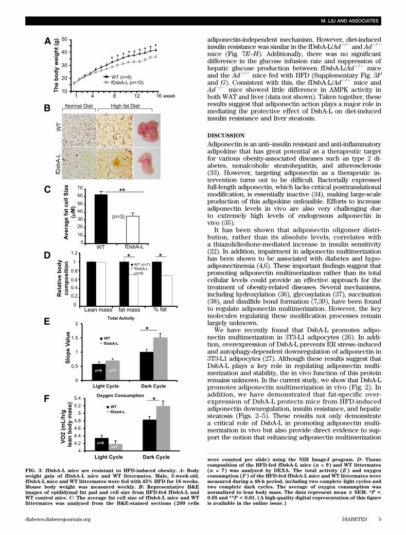
Generation of fat-specific DsbA-L transgenic mice.We have recently identified DsbA-L as a critical regulatorof adiponectin multimerization in 3T3-L1 adipocytes (26).To determine the functional role of DsbA-L in adiponectinmultimerization in vivo, we generated adipose-specificDsbA-L transgenic mice (fDsbA-L) using the murine FABP4/aP2 promoter (Fig. 1A). Western blot analysis revealed thatthe myc-tagged DsbA-L is specifically expressed in fat tis-sues (Fig. 1B), including white adipose tissue (WAT), brownadipose tissue (BAT), and macrophage, of the fDsbA-Ltransgenic mice with an approximately twofold higher ex-pression level compared with that of the endogenous protein(Fig. 1C–E). A similar expression level of DsbA-L was foundin an independent transgenic mouse line (data not shown).Overexpression of DsbA-L enhances adiponectinmultimerization in vivo. To determine whether over-expression of DsbA-L promotes adiponectin multimerizationand stability in vivo, we measured total adiponectin levelsand the ratio of each multimer to total adiponectin levels inboth adipose tissue and serum. Consistent with our previousfinding that DsbA-L promotes adiponectin assembly andstability in 3T3-L1 adipoctyes (26), the protein levels ofadiponectin and the ratio of the HMW form of this adipokineare increased in adipose tissue (Fig. 2A–D) and serum (Fig.2E–H) of the fDsbA-L mice compared with the control mice.Overexpression of DsbA-L greatly protected mice from HFD-induced downregulation of adiponectin levels (Fig. 2A and B)and its secretion (Fig. 2E and F). Gel filtration studiesrevealed a significant increase in the HMW form of adipo-nectin in WAT (Fig. 2C and D) and serum (Fig. 2G and H) ofthe fDsbA-L mice compared with WT littermates under HFD-feeding conditions. Similar results were also observed inanother DsbA-L overexpression transgenic mouse line (datanot shown). These results provide the first evidence thatDsbA-L promotes adiponectin multimerization in vivo.Overexpression of fDsbA-L in mice increased resistanceto diet-induced obesity. To determine whether over-expression of DsbA-L has an effect on energy homeostasis,we compared body weight and food intake between thefDsbA-L transgenic mice and WT littermates. On normalchow, the fDsbA-L mice showed little difference in foodintake and body weight compared with WT littermates(data not shown). There was also no significant differencein food intake between fDsbA-L and WT littermates fedwith an HFD (Supplementary Fig. 1B). However, the body
DsbA-L PREVENTS OBESITY AND INSULIN RESISTANCE
2 DIABETES diabetes.diabetesjournals.org
weight, epididymal fat pad, and fat cell size of the fDsbA-Lmice were notably reduced compared with WT littermates(Fig. 3A–C). Consistent with these findings, DEXA analysisrevealed that the fDsbA-L mice had a significantly lower fatcontent compared with the WT littermates (Fig. 3D). Therewas no significant difference in the activity between thefDsbA-L and WT mice during the light cycle, but the totalactivity of the fDsbA-L mice was significantly higher thanthat of the WT littermates during the dark cycle (Fig. 3E).Consistent with these findings, the overall metabolic rateof the fDsbA-L mice, expressed as a function of lean bodymass, was significantly greater than that of the WT controlmice in the dark cycle (Fig. 3F). The respiratory quotient(VCO2/VO2) was similar between fDsbA-L and WT control
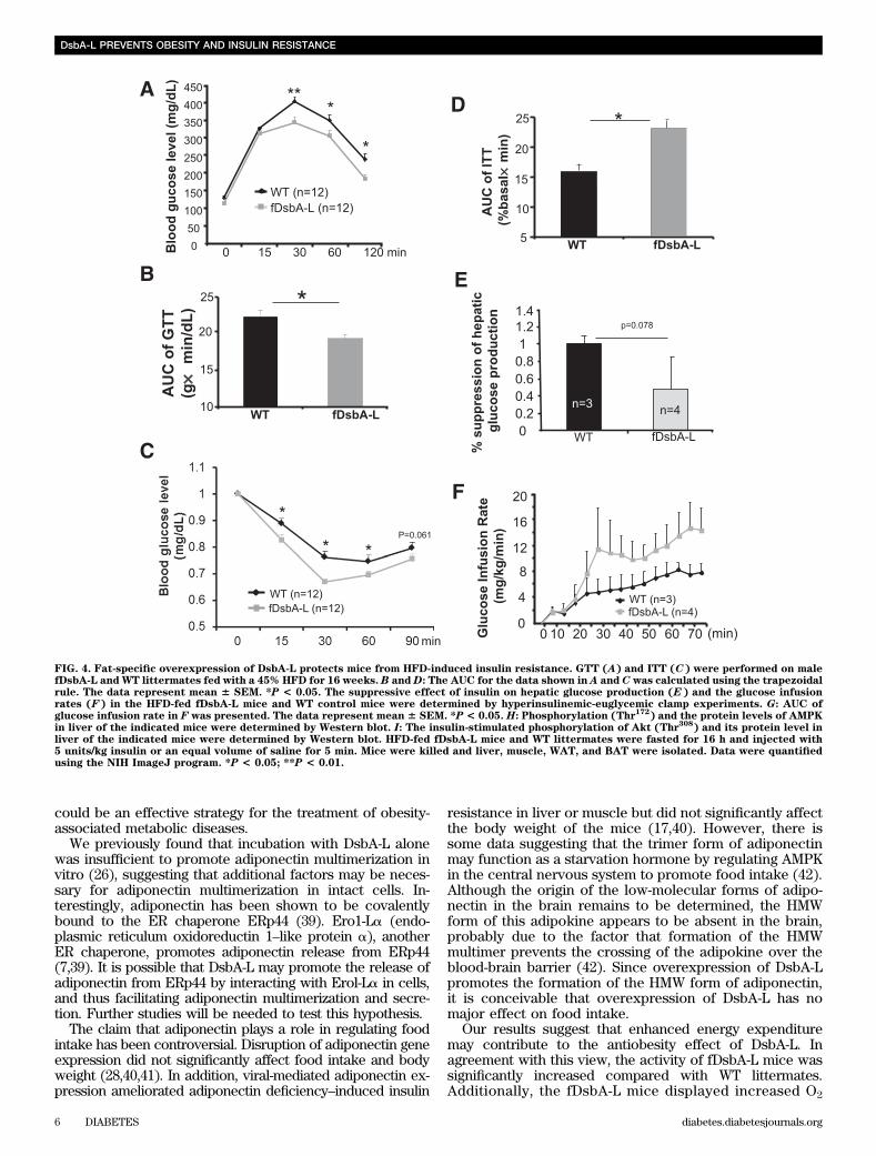
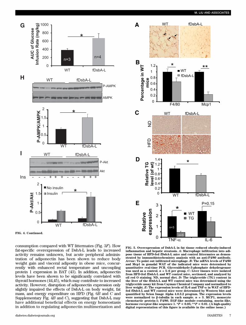
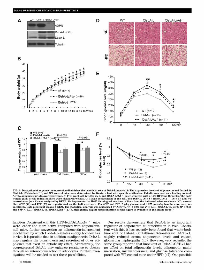
mice (data not shown), suggesting that there is no dif-ference in substrate utilization between these mice. Takentogether, these results suggest that increased metabo-lic rate may provide a mechanism by which over-expression of DsbA-L protects mice from diet-inducedobesity.The fDsbA-L mice are resistant to HFD-inducedinsulin resistance. The fDsbA-L mice displayed increasedglucose and insulin tolerance when compared with WT lit-termates under normal chow diet (Supplementary Fig. 1Cand D), but the difference between fDsbA-L and WT micedid not reach statistical significance. Under HFD, the fDsbA-Lmice showed a significantly enhanced glucose and insulintolerance compared with WT littermates (Fig. 4A–D). Hy-perglycemic clamp experiments revealed that insulin hada greater suppressive effect on hepatic glucose productionin fDsbA-L mice compared with WT mice under the HFDcondition (Fig. 4E). The fDsbA-L mice also exhibited ahigher insulin-mediated glucose infusion rate (Fig. 4F and G)and lower levels of fasting triglycerides compared withcontrol mice (Supplementary Fig. 2A). In agreement withprevious findings that the HMW form of adiponectin has amajor insulin-sensitizing effect in the liver (2,25,28,30),AMPK phosphorylation and insulin-stimulated Akt phos-phorylation were significantly enhanced in the liver (Fig.4H and I) and WAT (Supplementary Fig. 2B and C), but notin skeletal muscle (Supplementary Fig. 2D and E), of thefDsbA-L mice compared with WT littermates.Fat-specific overexpression of DsbA-L protects micefrom HFD-induced inflammation and hepatic steatosis.Since liver is the major organ for adiponectin action in vivo(2,25,30), we asked whether fat-specific overexpression ofDsbA-L protects mice from HFD-induced inflammation andliver dysfunction. In agreement with the findings of others(31,32), HFD feeding resulted in a large increase in mac-rophage infiltration into adipose tissue (Fig. 5A and B). TheHFD-induced fatty liver was significantly protected in thefDsbA-L mice (Fig. 5C). Overexpression of DsbA-L alsogreatly reduced HFD-induced macrosteatosis and accumu-lation of lipid droplets in the liver (Fig. 5D). Consistent withthese findings, the expression levels of TNF-a and IL-6 inWAT were significantly reduced in the fDsbA-L mice com-pared with WT littermates fed with an HFD (Fig. 5E).Targeted deletion of the adiponectin gene diminishesthe beneficial effects of DsbA-L on insulin resistanceand hepatic steatosis in mice. To determine whether thebeneficial effect of fat-specific overexpression of DsbA-L ismediated by adiponectin multimerization and action, wegenerated fat-specific DsbA-L transgenic mice in which theadiponectin gene targeted is disrupted (fDsbA-L/Ad2/2)(Fig. 6A). There was little difference in food intake (datanot shown), body weight (Fig. 6B and SupplementaryFig. 3A), and fat mass (Fig. 6C) between HFD-fed fDsbA-L/Ad2/2 and fDsbA-L mice. In addition, the promoting effectof DsbA-L on activity and energy expenditure was not sig-nificantly affected in the fDsbA-L/Ad2/2 mice comparedwith fDsbA-L mice (Supplementary Fig. 3B and C). How-ever, the protective effects of DsbA-L on diet-induced liversteatosis (Fig. 6D) and insulin resistance (Fig. 6E and Fand Supplementary Fig. 3D and E) were markedly reducedin fDsbA-L/Ad2/2 mice compared with fDsbA-L mice, sug-gesting that the protective effect of DsbA-L overexpressionon diet-induced insulin resistance and liver steatosis ismainly mediated by adiponectin action. These results alsosuggest that HFD feeding could have an effect on insulin
FIG. 1. Expression of the myc-tagged DsbA-L transgene in mouse tis-sues. A: The fDsbA-L transgene construct. An aP2 promoter was used todrive the expression of myc-tagged mouse DsbA-L gene. B: Western blotanalysis of tissue homogenates of the fDsbA-L transgenic mice using ananti-myc antibody. B, brain; F, fat; H, heart; K, kidney; L, liver; M,muscle; P, pancreas; S, spleen. The expression of the myc-tagged andendogenous DsbA-L in WAT (C), BAT (D), and macrophages (E) iso-lated from WT and fDsbA-L transgenic mice was analyzed by Westernblot using an anti–DsbA-L antibody. Tubulin was used as a loadingcontrol.
M. LIU AND ASSOCIATES
diabetes.diabetesjournals.org DIABETES 3
resistance and liver dysfunction independent of its causa-tive role in obesity.The protective effect of fat-specific overexpressionof DsbA-L on diet-induced obesity could be partiallymediated by an adiponectin-independent mechanism.The above results suggest that enhanced adiponectin levelsand multimerization play a major role in the insulin-sensitizingeffect of DsbA-L in vivo. However, fat-specific overex-pression of DsbA-L appears to have some additionalbeneficial effects in addition to promoting adiponectinmultimerization and function. To further test this possibility,we compared the physiological and metabolic properties of
the fDsbA-L/Ad2/2 and Ad2/2 littermates. There is no sig-nificant difference in food intake, body weight, and insulinsensitivity between fDsbA-L/Ad2/2 mice and Ad2/2 litter-mates under normal chow conditions (data not shown).However, the fDsbA-L/Ad2/2 mice displayed significantlyless body weight and smaller fat cell size compared withAd2/2 mice under HFD (Fig. 7A and B). In addition, thefDsbA-L/Ad2/2 mice were more active and displayed higherenergy expenditure throughout the light and dark cyclecompared with the Ad2/2 mice under HFD (Fig. 7C and D),suggesting that overexpression of DsbA-L had additionalbeneficial effects on diet-induced obesity through an
FIG. 2. Overexpression of DsbA-L enhanced adiponectin multimerization in vivo. A: The protein levels of adiponectin (ADPN) in WAT of fDsbA-Ltransgenic and WT control mice. Male fDsbA-L transgenic and WT littermates (5 weeks of age) were fed with normal chow diet (NC) or 45% HFDfor 16 weeks. Mice were killed and WAT was isolated. The expression levels of adiponectin in tissue homogenates were determined by Western blotwith an anti-adiponectin antibody. b-Tubulin was used as a loading control. B: The relative expression levels of adiponectin in A were quantified.Data represent mean 6 SEM. *P < 0.05. C: The relative expression levels of adiponectin multimers in WAT were determined by gel-filtrationchromatography, followed by Western blot.D: The ratio of HMW to total adiponectin in WAT as shown in C were quantified. Data represent mean6SEM. *P< 0.05. E: The serum adiponectin levels of fDsbA-L mice and WT littermates fed with NC or HFD were determined by Western blot with ananti-adiponectin antibody. IgG was used as a loading control. F: The relative expression levels of adiponectin in E were quantified. Data representmean 6 SEM. *P < 0.05. G: The relative expression levels of adiponectin multimers in serum of fDsbA-L transgenic and WT mice were determinedby gel-filtration chromatography, followed by Western blot using an anti-adiponectin antibody. H: The ratio of HMW to total adiponectin in serumas shown in G were quantified. Data represent mean 6 SEM. *P < 0.05.
DsbA-L PREVENTS OBESITY AND INSULIN RESISTANCE
4 DIABETES diabetes.diabetesjournals.org
adiponectin-independent mechanism. However, diet-inducedinsulin resistance was similar in the fDsbA-L/Ad2/2 and Ad2/2
mice (Fig. 7E–H). Additionally, there was no significantdifference in the glucose infusion rate and suppression ofhepatic glucose production between fDsbA-L/Ad2/2 miceand the Ad2/2 mice fed with HFD (Supplementary Fig. 3Fand G). Consistent with this, the fDsbA-L/Ad2/2 mice andAd2/2 mice showed little difference in AMPK activity inboth WAT and liver (data not shown). Taken together, theseresults suggest that adiponectin action plays a major role inmediating the protective effect of DsbA-L on diet-inducedinsulin resistance and liver steatosis.
DISCUSSION
Adiponectin is an anti–insulin resistant and anti-inflammatoryadipokine that has great potential as a therapeutic targetfor various obesity-associated diseases such as type 2 di-abetes, nonalcoholic steatohepatitis, and atherosclerosis(33). However, targeting adiponectin as a therapeutic in-tervention turns out to be difficult. Bacterially expressedfull-length adiponectin, which lacks critical posttranslationalmodification, is essentially inactive (34), making large-scaleproduction of this adipokine unfeasible. Efforts to increaseadiponectin levels in vivo are also very challenging dueto extremely high levels of endogenous adiponectin invivo (35).
It has been shown that adiponectin oligomer distri-bution, rather than its absolute levels, correlates witha thiazolidiedione-mediated increase in insulin sensitivity(22). In addition, impairment in adiponectin multimerizationhas been shown to be associated with diabetes and hypo-adiponectinemia (4,6). These important findings suggest thatpromoting adiponectin multimerization rather than its totalcellular levels could provide an effective approach for thetreatment of obesity-related diseases. Several mechanisms,including hydroxylation (36), glycosylation (37), succination(38), and disulfide bond formation (7,39), have been foundto regulate adiponectin multimerization. However, the keymolecules regulating these modification processes remainlargely unknown.
We have recently found that DsbA-L promotes adipo-nectin multimerization in 3T3-L1 adipocytes (26). In addi-tion, overexpression of DsbA-L prevents ER stress–inducedand autophagy-dependent downregulation of adiponectin in3T3-L1 adipocytes (27). Although these results suggest thatDsbA-L plays a key role in regulating adiponectin multi-merization and stability, the in vivo function of this proteinremains unknown. In the current study, we show that DsbA-Lpromotes adiponectin multimerization in vivo (Fig. 2). Inaddition, we have demonstrated that fat-specific over-expression of DsbA-L protects mice from HFD-inducedadiponectin downregulation, insulin resistance, and hepaticsteatosis (Figs. 2–5). These results not only demonstratea critical role of DsbA-L in promoting adiponectin multi-merization in vivo but also provide direct evidence to sup-port the notion that enhancing adiponectin multimerization
FIG. 3. fDsbA-L mice are resistant to HFD-induced obesity. A: Bodyweight gain of fDsbA-L mice and WT littermates. Male, 5-week-old,fDsbA-L mice and WT littermates were fed with 45% HFD for 16 weeks.Mouse body weight was measured weekly. B: Representative H&Eimages of epididymal fat pad and cell size from HFD-fed fDsbA-L andWT control mice. C: The average fat cell size of fDsbA-L mice and WTlittermates was analyzed from the H&E-stained sections (200 cells
were counted per slide) using the NIH ImageJ program. D: Tissuecomposition of the HFD-fed fDsbA-L mice (n = 9) and WT littermates(n = 7) was analyzed by DEXA. The total activity (E) and oxygenconsumption (F) of the HFD-fed fDsbA-L mice and WT littermates weremeasured during a 48-h period, including two complete light cycles andtwo complete dark cycles. The average of oxygen consumption wasnormalized to lean body mass. The data represent mean 6 SEM. *P <0.05 and **P< 0.01. (A high-quality digital representation of this figureis available in the online issue.)
M. LIU AND ASSOCIATES
diabetes.diabetesjournals.org DIABETES 5
could be an effective strategy for the treatment of obesity-associated metabolic diseases.
We previously found that incubation with DsbA-L alonewas insufficient to promote adiponectin multimerization invitro (26), suggesting that additional factors may be neces-sary for adiponectin multimerization in intact cells. In-terestingly, adiponectin has been shown to be covalentlybound to the ER chaperone ERp44 (39). Ero1-La (endo-plasmic reticulum oxidoreductin 1–like protein a), anotherER chaperone, promotes adiponectin release from ERp44(7,39). It is possible that DsbA-L may promote the release ofadiponectin from ERp44 by interacting with Erol-La in cells,and thus facilitating adiponectin multimerization and secre-tion. Further studies will be needed to test this hypothesis.
The claim that adiponectin plays a role in regulating foodintake has been controversial. Disruption of adiponectin geneexpression did not significantly affect food intake and bodyweight (28,40,41). In addition, viral-mediated adiponectin ex-pression ameliorated adiponectin deficiency–induced insulin
resistance in liver or muscle but did not significantly affectthe body weight of the mice (17,40). However, there issome data suggesting that the trimer form of adiponectinmay function as a starvation hormone by regulating AMPKin the central nervous system to promote food intake (42).Although the origin of the low-molecular forms of adipo-nectin in the brain remains to be determined, the HMWform of this adipokine appears to be absent in the brain,probably due to the factor that formation of the HMWmultimer prevents the crossing of the adipokine over theblood-brain barrier (42). Since overexpression of DsbA-Lpromotes the formation of the HMW form of adiponectin,it is conceivable that overexpression of DsbA-L has nomajor effect on food intake.
Our results suggest that enhanced energy expendituremay contribute to the antiobesity effect of DsbA-L. Inagreement with this view, the activity of fDsbA-L mice wassignificantly increased compared with WT littermates.Additionally, the fDsbA-L mice displayed increased O2
FIG. 4. Fat-specific overexpression of DsbA-L protects mice from HFD-induced insulin resistance. GTT (A) and ITT (C) were performed on malefDsbA-L and WT littermates fed with a 45% HFD for 16 weeks. B andD: The AUC for the data shown in A and C was calculated using the trapezoidalrule. The data represent mean 6 SEM. *P < 0.05. The suppressive effect of insulin on hepatic glucose production (E) and the glucose infusionrates (F) in the HFD-fed fDsbA-L mice and WT control mice were determined by hyperinsulinemic-euglycemic clamp experiments. G: AUC ofglucose infusion rate in F was presented. The data represent mean 6 SEM. *P < 0.05. H: Phosphorylation (Thr
172) and the protein levels of AMPK
in liver of the indicated mice were determined by Western blot. I: The insulin-stimulated phosphorylation of Akt (Thr308
) and its protein level inliver of the indicated mice were determined by Western blot. HFD-fed fDsbA-L mice and WT littermates were fasted for 16 h and injected with5 units/kg insulin or an equal volume of saline for 5 min. Mice were killed and liver, muscle, WAT, and BAT were isolated. Data were quantifiedusing the NIH ImageJ program. *P < 0.05; **P < 0.01.
DsbA-L PREVENTS OBESITY AND INSULIN RESISTANCE
6 DIABETES diabetes.diabetesjournals.org
consumption compared with WT littermates (Fig. 3F). Howfat-specific overexpression of DsbA-L leads to increasedactivity remains unknown, but acute peripheral adminis-tration of adiponectin has been shown to reduce bodyweight gain and visceral adiposity in obese mice, concur-rently with enhanced rectal temperature and uncouplingprotein 1 expression in BAT (43). In addition, adiponectinlevels have been shown to be significantly correlated withthyroid hormones (44,45), which may contribute to increasedactivity. However, disruption of adiponectin expression onlyslightly impaired the effects of DsbA-L on body weight, fatmass, and energy expenditure on HFD (Fig. 6B and C andSupplementary Fig. 4B and C), suggesting that DsbA-L mayhave additional beneficial effects on energy homeostasisin addition to regulating adiponectin multimerization and
FIG. 4. Continued.
FIG. 5. Overexpression of DsbA-L in fat tissue reduced obesity-inducedinflammation and hepatic steatosis. A: Macrophage infiltration into adi-pose tissue of HFD-fed fDsbA-L mice and control littermates as demon-strated by immunohistochemistry analysis with an anti-F4/80 antibody.Arrow: To point out infiltrated microphage. B: The mRNA levels of F4/80and Mcp1 in gonadal WAT of the indicated mice were determined byquantitative real-time PCR. Glyceraldehyde-3-phosphate dehydrogenasewas used as a control. n = 5–6 per group. C: Liver tissues were isolatedfrom HFD-fed fDsbA-L and WT control mice, sectioned, and analyzed byoil red O staining. ND, normal diet. D: The triglyceride (TG) content inthe liver of the fDsbA-L and WT control mice was determined using thetriglyceride assay kit from Cayman Chemical Company and normalized toliver weight. E: The expression levels of IL-6 and TNF-a in WAT of HFD-fed fDsbA-L and WT control mice were determined by Western blot andquantified by Scion Image Alpha 4.0.3.2 program. The expression levelswere normalized to b-tubulin in each sample. n = 3. MCP1, monocytechemotactic protein-1; F4/80, EGF-like module–containing, mucin–like,hormone receptor-like sequence 1. *P< 0.05; **P< 0.01. (A high-qualitydigital representation of this figure is available in the online issue.)
M. LIU AND ASSOCIATES
diabetes.diabetesjournals.org DIABETES 7
function. Consistent with this, HFD-fed fDsbA-L/Ad2/2 micewere leaner and more active compared with adiponectin-null mice, further suggesting an adiponectin-independentmechanism by which DsbA-L regulates energy homeostasisin vivo. It is possible that, in addition to adiponectin, DsbA-Lmay regulate the biosynthesis and secretion of other adi-pokines that exert an antiobesity effect. Alternatively, theoverexpressed DsbA-L may enhance resistance to obesitythrough an autonomous action in adipocytes. Further inves-tigations will be needed to test these possibilities.
Our results demonstrate that DsbA-L is an importantregulator of adiponectin multimerization in vivo. Consis-tent with this, it has recently been found that whole-bodyknockout of DsbA-L (glutathione S-transferase [GST]-k1)slightly reduced serum adiponectin levels and causedglomerular nephropathy (46). However, very recently, thesame group reported that knockout of DsbA-L/GST-k1 hadno effect on total adiponectin levels, adiponectin multi-merization, insulin tolerance, and glucose tolerance com-pared with WT control mice under HFD (47). One possible
FIG. 6. Disruption of adiponectin expression diminishes the beneficial role of DsbA-L in mice. A: The expression levels of adiponectin and DsbA-L infDsbA-L, fDsbA-L/Ad2/2
, and WT control mice were determined by Western blot with specific antibodies. Tubulin was used as a loading control.ADPN, adiponectin; O/E, overexpression. B: Five-week-old WT, fDsbA-L, and fDsbA-L/Ad2/2
mice were fed with a 45% HFD for 16 weeks. The bodyweight gains of the indicated mice were measured weekly. C: Tissue composition of the HFD-fed DsbA-L (n = 8), fDsbA-L/Ad2/2
(n = 4), and WTcontrol mice (n = 8) was analyzed by DEXA. D: Representative H&E histological sections of liver from the indicated mice are shown. ND, normaldiet. GTT (E) and ITT (F) were performed on the indicated mice. For GTT and ITT, 2 g/kg glucose and 0.075 units/kg insulin were used, re-spectively. Data represent means 6 SEM. The statistical analysis was performed by ANOVA. *P < 0.05 and P < 0.01 (fDsbA-L vs. WT); #P < 0.05and ##P < 0.01 (fDsbA-L vs. fDsbA-L/Ad2/2
). (A high-quality digital representation of this figure is available in the online issue.)
DsbA-L PREVENTS OBESITY AND INSULIN RESISTANCE
8 DIABETES diabetes.diabetesjournals.org
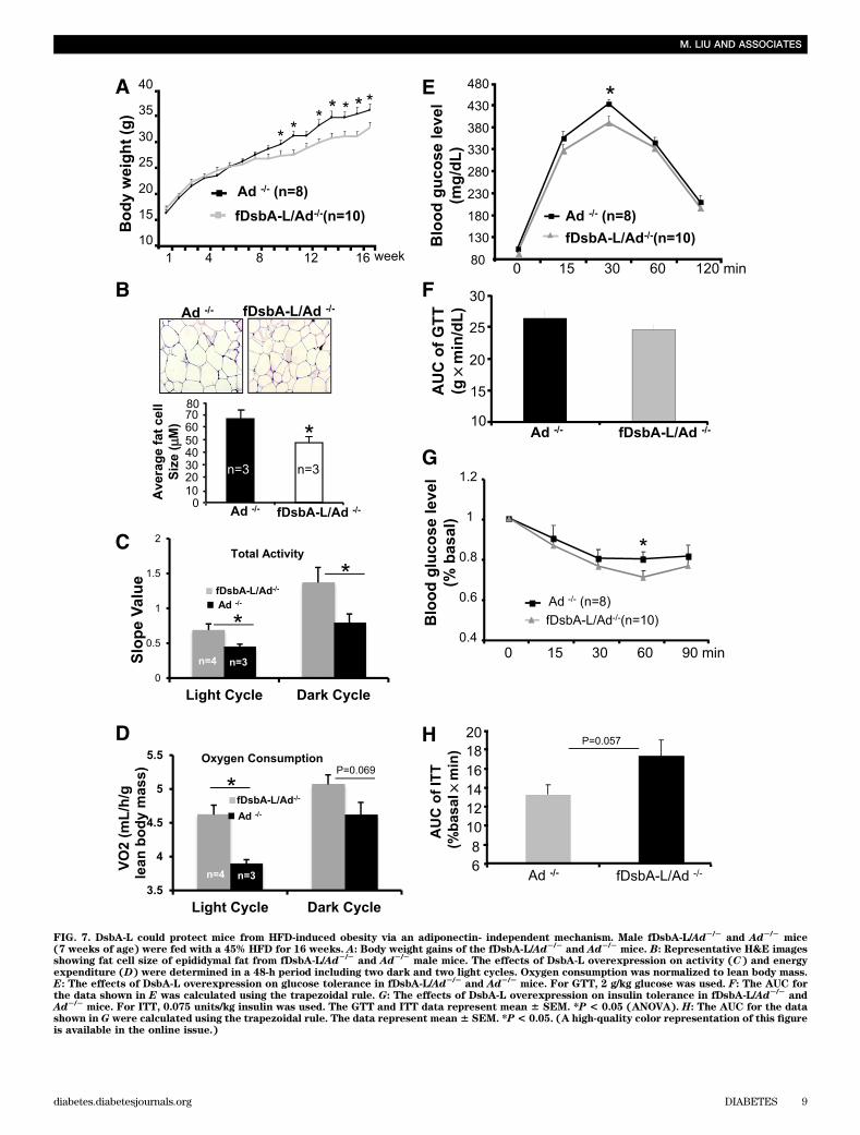
FIG. 7. DsbA-L could protect mice from HFD-induced obesity via an adiponectin- independent mechanism. Male fDsbA-L/Ad2/2and Ad2/2
mice(7 weeks of age) were fed with a 45% HFD for 16 weeks. A: Body weight gains of the fDsbA-L/Ad2/2
and Ad2/2mice. B: Representative H&E images
showing fat cell size of epididymal fat from fDsbA-L/Ad2/2and Ad2/2
male mice. The effects of DsbA-L overexpression on activity (C) and energyexpenditure (D) were determined in a 48-h period including two dark and two light cycles. Oxygen consumption was normalized to lean body mass.E: The effects of DsbA-L overexpression on glucose tolerance in fDsbA-L/Ad2/2
and Ad2/2mice. For GTT, 2 g/kg glucose was used. F: The AUC for
the data shown in E was calculated using the trapezoidal rule. G: The effects of DsbA-L overexpression on insulin tolerance in fDsbA-L/Ad2/2and
Ad2/2mice. For ITT, 0.075 units/kg insulin was used. The GTT and ITT data represent mean 6 SEM. *P < 0.05 (ANOVA). H: The AUC for the data
shown in G were calculated using the trapezoidal rule. The data represent mean 6 SEM. *P< 0.05. (A high-quality color representation of this figureis available in the online issue.)
M. LIU AND ASSOCIATES
diabetes.diabetesjournals.org DIABETES 9
explanation for these controversies is that knockout ofDsbA-L in vivo led to a compensatory increase in the ex-pression of molecules that promote adiponectin expres-sion and multimerization in adipose tissues. However, thefinding that HFD feeding increased the levels of HMWadiponectin in both WT and DsbA-L knockout mice, whichis contradictory to the findings of many others in the field(8,37,48–50), raises some concerns about the experimentalconditions under which the experiments were performed.Thus, it remains to be determined whether knockout ofDsbA-L under more physiologically relevant conditionsaffects adiponectin multimerization and function in vivo.
In summary, we have provided strong evidence for anin vivo role of DsbA-L in promoting adiponectin multi-merization and function. Our study also demonstrates thatenhanced adiponectin multimerization is sufficient to sup-press obesity-induced insulin resistance and liver damage,suggesting that upregulation of DsbA-L could be an effec-tive therapeutic approach for the treatment of obesity-induced insulin resistance and liver steatosis.
ACKNOWLEDGMENTS
This work was supported by an American Diabetes Associ-ation Research Award (1-12-BS-115 to F.L.) and by NationalInstitutes of Health RO1 grants DK-76902 (to F.L.), DK-69930(to L.Q.D.), P30-AG-13319-1SSI (to S.N.A.), DK-80157 and DK-89229 (to N.M.), HL-70963 (to R.A.), and R01-DK-55758, R01-CA-112023, RC1-DK-86629, and P01-DK-88761 (to P.E.S.).
No potential conflicts of interest relevant to this articlewere reported.
M.L. and F.L. designed the experiment; researched data;wrote, reviewed, and edited the manuscript; and contrib-uted to discussion. R.X. researched data and contributed todiscussion. S.A.W., N.Z., K.A., L.B.S., L.Z., H.C., and G.X.researched data. C.A.W., S.N.A., N.M., R.A.D., and R.A.reviewed and edited the manuscript. P.E.S. and L.Q.D.reviewed and edited the manuscript and contributed todiscussion. F.L. is the guarantor of this work and, as such,had full access to all the data in the study and takes respon-sibility for the integrity of the data and the accuracy of thedata analysis.
Parts of this work were presented in abstract form forpresentation at the 72nd Scientific Sessions of the AmericanDiabetes Association, Atlanta, Georgia, 8–12 June 2012.
The authors thank Derong Hu (UTHSCSA), Jessica Han(UTHSCSA), and Bing Zhu (UTHSCSA) for excellenttechnical assistance, Dr. Yihao Yu (Easton Hospital, Easton,PA) for the FABP4/aP2 promoter vector, and Anna Henicke(UTHSCSA) for editing the manuscript.
REFERENCES
1. Ahima RS. Adipose tissue as an endocrine organ. Obesity (Silver Spring)2006;14(Suppl. 5):242S–249S
2. Scherer PE. Adipose tissue: from lipid storage compartment to endocrineorgan. Diabetes 2006;55:1537–1545
3. Kadowaki T, Yamauchi T. Adiponectin and adiponectin receptors. EndocrRev 2005;26:439–451
4. Pajvani UB, Du X, Combs TP, et al. Structure-function studies of theadipocyte-secreted hormone Acrp30/adiponectin. Implications fpr meta-bolic regulation and bioactivity. J Biol Chem 2003;278:9073–9085
5. Tsao TS, Murrey HE, Hug C, Lee DH, Lodish HF. Oligomerization state-dependent activation of NF-kappa B signaling pathway by adipocytecomplement-related protein of 30 kDa (Acrp30). J Biol Chem 2002;277:29359–29362
6. Waki H, Yamauchi T, Kamon J, et al. Impaired multimerization of humanadiponectin mutants associated with diabetes. Molecular structure andmultimer formation of adiponectin. J Biol Chem 2003;278:40352–40363
7. Qiang L, Wang H, Farmer SR. Adiponectin secretion is regulated by SIRT1and the endoplasmic reticulum oxidoreductase Ero1-L alpha. Mol Cell Biol2007;27:4698–4707
8. Scherer PE, Williams S, Fogliano M, Baldini G, Lodish HF. A novel serumprotein similar to C1q, produced exclusively in adipocytes. J Biol Chem1995;270:26746–26749
9. Abbasi F, Chu JW, Lamendola C, et al. Discrimination between obesity andinsulin resistance in the relationship with adiponectin. Diabetes 2004;53:585–590
10. Hotta K, Funahashi T, Arita Y, et al. Plasma concentrations of a novel,adipose-specific protein, adiponectin, in type 2 diabetic patients. Arte-rioscler Thromb Vasc Biol 2000;20:1595–1599
11. Stefan N, Vozarova B, Funahashi T, et al. Plasma adiponectin concentrationis associated with skeletal muscle insulin receptor tyrosine phosphorylation,and low plasma concentration precedes a decrease in whole-body insulinsensitivity in humans. Diabetes 2002;51:1884–1888
12. Lindsay RS, Funahashi T, Hanson RL, et al. Adiponectin and developmentof type 2 diabetes in the Pima Indian population. Lancet 2002;360:57–58
13. Kumada M, Kihara S, Sumitsuji S, et al.; Osaka CAD Study Group. Coro-nary artery disease. Association of hypoadiponectinemia with coronaryartery disease in men. Arterioscler Thromb Vasc Biol 2003;23:85–89
14. Atzmon G, Pollin TI, Crandall J, et al. Adiponectin levels and genotype:a potential regulator of life span in humans. J Gerontol A Biol Sci Med Sci2008;63:447–453
15. Bik W, Baranowska-Bik A, Wolinska-Witort E, et al. The relationship be-tween adiponectin levels and metabolic status in centenarian, early el-derly, young and obese women. Neuroendocrinol Lett 2006;27:493–500
16. Fruebis J, Tsao TS, Javorschi S, et al. Proteolytic cleavage product of30-kDa adipocyte complement-related protein increases fatty acid oxi-dation in muscle and causes weight loss in mice. Proc Natl Acad SciUSA 2001;98:2005–2010
17. Yamauchi T, Kamon J, Waki H, et al. The fat-derived hormone adiponectinreverses insulin resistance associated with both lipoatrophy and obesity.Nat Med 2001;7:941–946
18. Bauche IB, El Mkadem SA, Pottier AM, et al. Overexpression of adipo-nectin targeted to adipose tissue in transgenic mice: impaired adipocytedifferentiation. Endocrinology 2007;148:1539–1549
19. Combs TP, Pajvani UB, Berg AH, et al. A transgenic mouse with a deletionin the collagenous domain of adiponectin displays elevated circulatingadiponectin and improved insulin sensitivity. Endocrinology 2004;145:367–383
20. Kim JY, van de Wall E, Laplante M, et al. Obesity-associated improvementsin metabolic profile through expansion of adipose tissue. J Clin Invest2007;117:2621–2637
21. Arita Y, Kihara S, Ouchi N, et al. Paradoxical decrease of an adipose-specific protein, adiponectin, in obesity. Biochem Biophys Res Commun1999;257:79–83
22. Pajvani UB, Hawkins M, Combs TP, et al. Complex distribution, not ab-solute amount of adiponectin, correlates with thiazolidinedione-mediatedimprovement in insulin sensitivity. J Biol Chem 2004;279:12152–12162
23. Tomas E, Tsao TS, Saha AK, et al. Enhanced muscle fat oxidation andglucose transport by ACRP30 globular domain: acetyl-CoA carboxylaseinhibition and AMP-activated protein kinase activation. Proc Natl Acad SciUSA 2002;99:16309–16313
24. Tsao TS, Tomas E, Murrey HE, et al. Role of disulfide bonds in Acrp30/adiponectin structure and signaling specificity. Different oligomers acti-vate different signal transduction pathways. J Biol Chem 2003;278:50810–50817
25. Yamauchi T, Kamon J, Minokoshi Y, et al. Adiponectin stimulates glucoseutilization and fatty-acid oxidation by activating AMP-activated proteinkinase. Nat Med 2002;8:1288–1295
26. Liu M, Zhou L, Xu A, et al. A disulfide-bond A oxidoreductase-like protein(DsbA-L) regulates adiponectin multimerization. Proc Natl Acad Sci USA2008;105:18302–18307
27. Zhou L, Liu M, Zhang J, Chen H, Dong LQ, Liu F. DsbA-L alleviates en-doplasmic reticulum stress-induced adiponectin downregulation. Diabetes2010;59:2809–2816
28. Nawrocki AR, Rajala MW, Tomas E, et al. Mice lacking adiponectin showdecreased hepatic insulin sensitivity and reduced responsiveness to per-oxisome proliferator-activated receptor gamma agonists. J Biol Chem2006;281:2654–2660
29. Wang L, Balas B, Christ-Roberts CY, et al. Peripheral disruption of theGrb10 gene enhances insulin signaling and sensitivity in vivo. Mol Cell Biol2007;27:6497–6505
30. Kadowaki T, Yamauchi T, Kubota N, Hara K, Ueki K, Tobe K. Adiponectinand adiponectin receptors in insulin resistance, diabetes, and the meta-bolic syndrome. J Clin Invest 2006;116:1784–1792
DsbA-L PREVENTS OBESITY AND INSULIN RESISTANCE
10 DIABETES diabetes.diabetesjournals.org
31. Saltiel AR, Kahn CR. Insulin signalling and the regulation of glucose andlipid metabolism. Nature 2001;414:799–806
32. Marceau P, Biron S, Hould FS, et al. Liver pathology and the metabolicsyndrome X in severe obesity. J Clin Endocrinol Metab 1999;84:1513–1517
33. Phillips SA, Kung JT. Mechanisms of adiponectin regulation and use asa pharmacological target. Curr Opin Pharmacol 2010;10:676–683
34. Wang Y, Xu A, Knight C, Xu LY, Cooper GJ. Hydroxylation and glycosyl-ation of the four conserved lysine residues in the collagenous domain ofadiponectin. Potential role in the modulation of its insulin-sensitizing ac-tivity. J Biol Chem 2002;277:19521–19529
35. Bełtowski J, Jamroz-Wiśniewska A, Widomska S. Adiponectin and its role incardiovascular diseases. Cardiovasc Hematol Disord Drug Targets 2008;8:7–46
36. Richards AA, Stephens T, Charlton HK, et al. Adiponectin multimerizationis dependent on conserved lysines in the collagenous domain: evidence forregulation of multimerization by alterations in posttranslational mod-ifications. Mol Endocrinol 2006;20:1673–1687
37. Wang Y, Lam KS, Yau MH, Xu A. Post-translational modifications of adi-ponectin: mechanisms and functional implications. Biochem J 2008;409:623–633
38. Frizzell N, Rajesh M, Jepson MJ, et al. Succination of thiol groups in adi-pose tissue proteins in diabetes: succination inhibits polymerization andsecretion of adiponectin. J Biol Chem 2009;284:25772–25781
39. Wang ZV, Schraw TD, Kim JY, et al. Secretion of the adipocyte-specificsecretory protein adiponectin critically depends on thiol-mediated proteinretention. Mol Cell Biol 2007;27:3716–3731
40. Maeda N, Shimomura I, Kishida K, et al. Diet-induced insulin resistance inmice lacking adiponectin/ACRP30. Nat Med 2002;8:731–737
41. Kubota N, Terauchi Y, Yamauchi T, et al. Disruption of adiponectin causesinsulin resistance and neointimal formation. J Biol Chem 2002;277:25863–25866
42. Kubota N, Yano W, Kubota T, et al. Adiponectin stimulates AMP-activatedprotein kinase in the hypothalamus and increases food intake. Cell Metab2007;6:55–68
43. Masaki T, Chiba S, Yasuda T, et al. Peripheral, but not central, adminis-tration of adiponectin reduces visceral adiposity and upregulates the ex-pression of uncoupling protein in agouti yellow (Ay/a) obese mice.Diabetes 2003;52:2266–2273
44. Lee JY, Takahashi N, Yasubuchi M, et al. Triiodothyronine induces UCP-1expression and mitochondrial biogenesis in human adipocytes. Am J PhysiolCell Physiol 2012;302:C463–C472
45. Malyszko J, Malyszko J, Wolczynski S, Mysliwiec M. Adiponectin, leptinand thyroid hormones in patients with chronic renal failure and on renalreplacement therapy: are they related? Nephrol Dial Transplant 2006;21:145–152
46. Blackburn AC, Coggan M, Shield AJ, et al. Glutathione transferase kappadeficiency causes glomerular nephropathy without overt oxidative stress.Lab Invest 2011;91:1572–1583
47. Theodoratos A, Blackburn AC, Coggan M, et al. The impact of glutathionetransferase kappa deficiency on adiponectin multimerisation in vivo. IntJ Obes (Lond). 17 January 2012 [Epub ahead of print]
48. Araki S, Dobashi K, Kubo K, Asayama K, Shirahata A. High molecularweight, rather than total, adiponectin levels better reflect metabolic ab-normalities associated with childhood obesity. J Clin Endocrinol Metab2006;91:5113–5116
49. Kaser S, Tatarczyk T, Stadlmayr A, et al. Effect of obesity and insulinsensitivity on adiponectin isoform distribution. Eur J Clin Invest 2008;38:827–834
50. Schraw T, Wang ZV, Halberg N, Hawkins M, Scherer PE. Plasma adipo-nectin complexes have distinct biochemical characteristics. Endocrinology2008;149:2270–2282
M. LIU AND ASSOCIATES
diabetes.diabetesjournals.org DIABETES 11