original article a study on in vitro cytotoxicity of … study on in vitro cytotoxicity of cortisol...
TRANSCRIPT
Int J Clin Exp Med 2016;9(2):1298-1305www.ijcem.com /ISSN:1940-5901/IJCEM0014152
Original ArticleA study on in vitro cytotoxicity of cortisol on madin-darby kidney cells
Ting-Xia Li1, Jian-Hua Mao1, Lei Huang1, Hai-Dong Fu1, Shuo-Hui Chen2, Ai-Min Liu1, Yu-Qin Liang1
1Department of Nephrology, The Children’s Hospital Zhejiang University School of Medicine, Hangzhou 310003, Zhejiang, China; 2Department of Nursing, The Children’s Hospital Zhejiang University School of Medicine, Hang-zhou 310003, Zhejiang, China
Received August 8, 2015; Accepted January 5, 2016; Epub February 15, 2016; Published February 29, 2016
Abstract: Stress hormones play a vital role in cancer development and tumor formation. However, this mechanism is still obscure. Cortisol plays at several important biochemical roles in disease progress and development. Evaluating the dose-dependent effect of cortisol on MDCK cells is contemplated as the primary task of this experiment. Cell viability did not change significantly following cortisol exposure. Morphologically, no changes were observed in the MDCK cells. However, few cells showed an oval profile. Fluorescent and confocal analysis showed the occurrence of apoptosis significantly at 0.1 mg/L of cortisol. However, the intensity of apoptosis was significantly reduced at higher concentration (0.2 mg/L) of cortisol. The method of qPCR quantitated the mRNA expression of caspase 3, matrix metalloproteinase-9 (MMP-9) and matrix metalloproteinase-2 (MMP-2). Caspase 3 mRNA expression was signifi-cantly altered at low concentration (0.1 mg/L) while it was tremendously reduced at the higher level of cortisol (0.2 mg/L). A remarkable increase in the MRNA expression of MMP-2 and MMP-9 was executed in a dose-dependent manner. Our experimental outcome implies that cortisol may induce the apoptosis in MDCK cells.
Keywords: Cortisol, MDCK cells, CLSM, SRB, caspase 3
Introduction
Stress and behavioral factors are known to pl- ay in disease progress and development [1]. Chronic stress and depression are believed to be associated with altered cellular immunity [2]. The psychological stress affects both im- mune system and the pathway of apoptosis. Immune system expresses receptors for cate-cholamine secreted by the sympathetic system. Hormone-receptor interaction leads to cellular action, cell trafficking and cytokine production [3]. However, the direct effects of cortisol on normal cells have not been investigated.
The adrenal cortex secretes cortisol. It is usu-ally increased in the acute and chronic stress condition [4]. High levels of pro-angiogenic fac-tors suggest that cortisol may probably expe-dite tumor metastasis. However, metastasis is contemplated to be an extremely complex pro-cess involving several sequential and interre-lated processes. Metastatic cells must com-plete all the necessary steps to produce clinical lesions [5]. In the pathogenesis of metastasis,
the process of invasion is contemplated to be a pivotal step [6]. However, there is no analysis of the effect of cortisol on the invasion potential of tumor cells. Norepinephrine may enhance the invasive potential because it affects tumor cell motility [7]. Circulating catecholamine has been associated the expression of matrix metalloproteinase (MMP) [8].
The present scrutiny was aimed to assess the effect of cortisol on the proliferation, viability, and morphology of MDCK cells. SRB assay determined cytotoxicity of cortisol on MDCK cells. Morphology of MDCK cells was observed by an inverted microscope, a fluorescence mi- croscope, and confocal laser scanning micro-scope (CLSM). Quantitative PCR (qPCR) was applied to determine caspase 3, MMP-9 and MMP-2 mRNA expression.
Materials and methods
Materials
Cortisol, dimethyl sulphoxide (DMSO), sulforho-damine B (SRB) was purchased from Sigma.
Cortisol effect
1299 Int J Clin Exp Med 2016;9(2):1298-1305
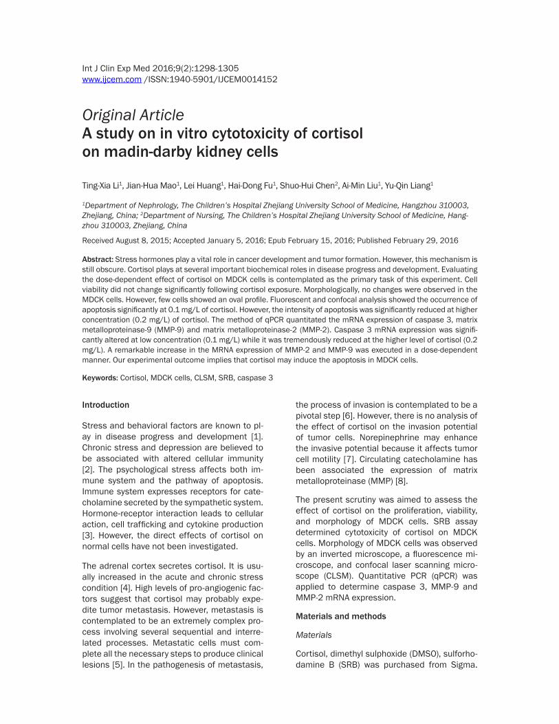
Figure 1. Cytotoxic effect of cortisol on MDCK cells by SRB assay at 48 h. Results are presented as a percentage of growth inhibition compared with the control. Values were expressed as means ± SEM.
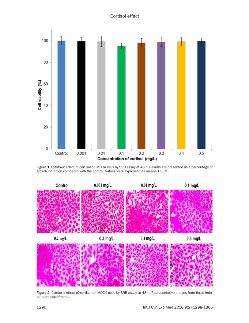
Figure 2. Cytotoxic effect of cortisol on MDCK cells by SRB assay at 48 h. Representative images from three inde-pendent experiments.
Cortisol effect
1300 Int J Clin Exp Med 2016;9(2):1298-1305
DMEM, FBS, penicillin-streptomycin, and tryp-sin-EDTA were acquired from Welgene (China). EB and Acridine Orange (AO) was obtained from Santa Cruz Biotechnology, Inc. (Delaware Av- enue, California, USA). Primers were purchased from Macrogen Inc. (China).
Cell culture
MDCK cells were acquired from the China Cell Line Bank (China). Cells were preserved in a growth medium and were supplemented with 10% FBS and 1% antibiotics (penicillin-strepto-mycin). The cells were developed in a CO2 incu-bator at 37°C with 5% CO2.
SRB assay
SRB assay [9] was used to measure the cyto-toxic effect of cortisol on MDCK cells. MDCK cells were cultured in 96 well plates at a densi-ty of 2.5 × 10⁴ cells and adhered for 24 h at 37°C. Cortisol was treated to the cells at differ-ent concentrations (0.001, 0.01, 0.1, 0.2, 0.3, 0.4 and 0.5 mg/L) for 48 h. Cells were fixed with acetone and air dried at the end of the treatment. After the cells had been fully dried, 100% of SRB solution (0.4% w/v) was added. 1% of acetic acid was used to wash the micro-plates that are then dried in drying oven. Stained MDCK cells were photographed using an inverted light microscope. Finally, 10 mM of Tris base was added and kept for overnight and measured at 540 nm.
Morphological observation
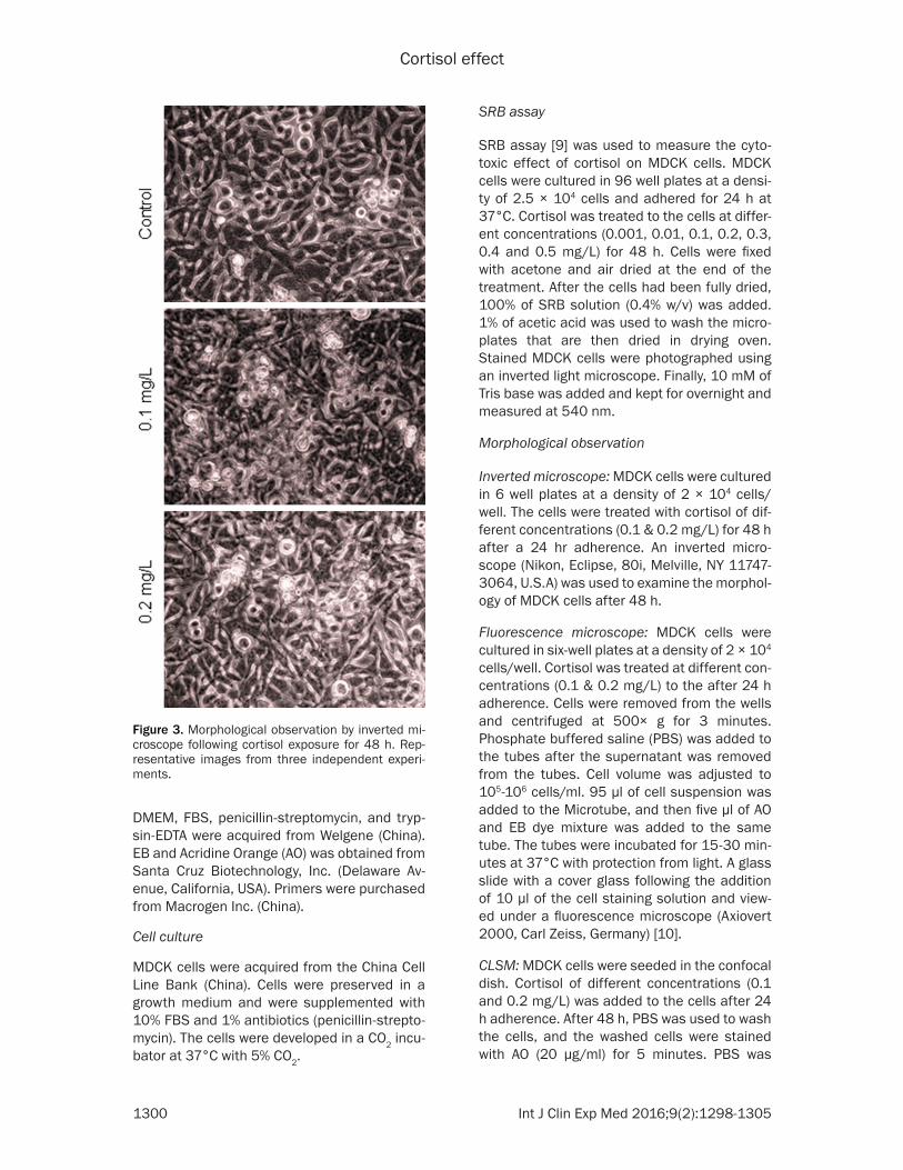
Inverted microscope: MDCK cells were cultured in 6 well plates at a density of 2 × 10⁴ cells/well. The cells were treated with cortisol of dif-ferent concentrations (0.1 & 0.2 mg/L) for 48 h after a 24 hr adherence. An inverted micro-scope (Nikon, Eclipse, 80i, Melville, NY 11747-3064, U.S.A) was used to examine the morphol-ogy of MDCK cells after 48 h.
Fluorescence microscope: MDCK cells were cultured in six-well plates at a density of 2 × 10⁴ cells/well. Cortisol was treated at different con-centrations (0.1 & 0.2 mg/L) to the after 24 h adherence. Cells were removed from the wells and centrifuged at 500× g for 3 minutes. Phosphate buffered saline (PBS) was added to the tubes after the supernatant was removed from the tubes. Cell volume was adjusted to 105-106 cells/ml. 95 µl of cell suspension was added to the Microtube, and then five µl of AO and EB dye mixture was added to the same tube. The tubes were incubated for 15-30 min-utes at 37°C with protection from light. A glass slide with a cover glass following the addition of 10 µl of the cell staining solution and view- ed under a fluorescence microscope (Axiovert 2000, Carl Zeiss, Germany) [10].
CLSM: MDCK cells were seeded in the confocal dish. Cortisol of different concentrations (0.1 and 0.2 mg/L) was added to the cells after 24 h adherence. After 48 h, PBS was used to wash the cells, and the washed cells were stained with AO (20 µg/ml) for 5 minutes. PBS was
Figure 3. Morphological observation by inverted mi-croscope following cortisol exposure for 48 h. Rep-resentative images from three independent experi-ments.
Cortisol effect
1301 Int J Clin Exp Med 2016;9(2):1298-1305
used to wash the cell twice, and the stained cells were viewed immediately under CLSM (1 × 81R motorized inverted microscope, Olympus [10].
Gene expression
MDCK cells were seeded T25 flask. Cortisol was treated at different concentrations (0.1 and 0.2 mg/L) to the cells after 24 h adher-ence. RNA was isolated from the contr- ol and cortisol treated cells [11]. qPCR was car-ried out using primers specific for caspase 3 (forward: 5’-TTAATAAAGGTATCCATGGAGAACA- CT-3’, reverse: 5’-TTAGTGATAAAAATAGAGTTCT- TTTGTGAG-3’), MMP-2 (forward: 5’-AGGATCAT-
TGGCTACACACC-3’, reverse: 5’-AGCTGTCATAGG- ATGTGCCC-3’, MMP-9 (forward: 5’-CGCAGACA- TCGTCATCCAGT-3’, reverse: 5’-GGATTGGCCT- TGGAAGATGA-3’ and GAPDH (forward: 5’-GG- TCACCAGGGCTGCTTTT-3’, reverse: 5-ATCTCG- CTCCTGGAAGATGGT-3’). According to the man-ufacturers instruction, the reaction was carried out in 10 μl using SYBR Green Master Mix (Bioneer). Based on the 2-ΔΔCT method [12] rela-tive ratios were calculated. CFX96TM Real-Time System (Bio-Rad) was used to monitor the PCR.
Statistical analysis
Values were expressed as mean ± SEM. ANOVA analysis has been carried out to compare the
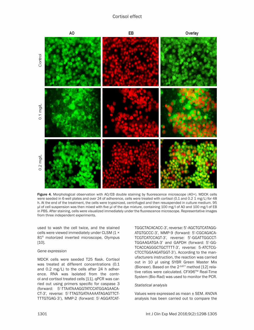
Figure 4. Morphological observation with AO/EB double staining by fluorescence microscope (40×). MDCK cells were seeded in 6-well plates and over 24 of adherence, cells were treated with cortisol (0.1 and 0.2 1 mg/L) for 48 h. At the end of the treatment, the cells were trypsinized, centrifuged and then resuspended in culture medium. 95 µl of cell suspension was then mixed with five µl of the dye mixture, containing 100 mg/l of AO and 100 mg/l of EB in PBS. After staining, cells were visualized immediately under the fluorescence microscope. Representative images from three independent experiments.
Cortisol effect
1302 Int J Clin Exp Med 2016;9(2):1298-1305
groups and control (SPSS 22, Statistical Pa- ckage) A P<0.05 was considered statistically significant.
Results
Cortisol on cell viability
The cytotoxic effect of cortisol on MDCK cells was observed (Figures 1 and 2). However, 0.3, 0.4 and 0.5 mg/L of cortisol are not related to physiological levels during normal and stress conditions. Therefore, 0.1 and 0.2 mg/L of cor-tisol was used for the further study. The invert-ed microscope could be used to observe the cell shape and its morphological changes. Control MDCK cells have a regular shape and morphology. Cortisol treated cells showed the sporadic distribution, loss of adhesion and oval profile at low concentration. However, these
effects were reversed marginally at a higher level (Figure 3).
Effect of cortisol on morphology
Fluorescence microscopy was carried out to resolve whether the cytotoxic effect of cortisol was related to the growth and proliferation of cancer cells. This method combines the dual uptake of the fluorescent DNA binding dyes EB and AO. The stained nucleus contains chroma-tin condensation that is used to differentiate viable, apoptotic, and necrotic cells. Control MDCK cells have typical morphological fea-tures. Cells treated with cortisol showed frag-mented chromatin in the nucleus, and the necrotic cells showed a uniform bright orange core. Significant morphological changes such as chromatin condensation, fragmented nuclei, and apoptotic bodies are observed at a low
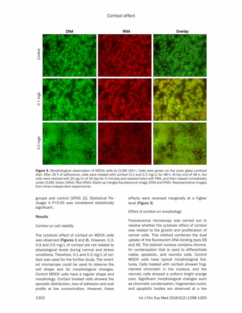
Figure 5. Morphological observation of MDCK cells by CLSM (40×). Cells were grown on the cover glass confocal dish. After 24 h of adherence, cells were treated with cortisol (0.1 and 0.2 mg/L) for 48 h. At the end of 48 h, the cells were stained with 20 µg/ml of AO dye for 5 minutes and washed twice with PBS, and then viewed immediately under CLSM. Green (DNA); Red (RNA); Stack-up merges fluorescence image (DNA and RNA). Representative images from three independent experiments.
Cortisol effect
1303 Int J Clin Exp Med 2016;9(2):1298-1305
concentration of cortisol (0.1 mg/L). However, the effect of cortisol was reduced at a higher level (Figure 4).
CLSM has been widely used to investigate in the morphological studies. AO is mainly used as a nucleic acid-selective fluorescent cationic dye that is applied to the determination of the cell cycle. AO interaction with DNA and RNA can occur by intercalation or electrostatic attrac-tions respectively, and spectrally fluorescein are bound to DNA with an excitation maximum at 502 nm and an emission maximum at 525 nm. Control cells have bigger and smoother nucleus than cortisol treated cells. Morpho- logical changes such as rounding, compact granular masses in the nucleus and reduced nuclear volume at a low concentration of corti-sol treatment. However, these effects were sig-nificantly reduced at the higher level of cortisol exposure (Figure 5).
Cortisol on gene expression
The effect of cortisol on selected gene mRNA expression and quantification was carried out by qPCR. MDCK cells were exposed to different concentration of cortisol for 48 h. No signifi-cant changes were observed at low concentra-
the beta component, which converts adenos-ine triphosphate to cyclic adenosine 3’, 5’-monophosphate (cyclic AMP). This, in turn, leads to phosphorylation of various proteins due to activation of several protein kinases. One of the proteins Myosin light chain regulates ATPase-actin activity and cell contraction [14].
The fluorescence microscopy study showed that morphological changes, including chroma-tin condensation, cell shrinkage and formati- on of apoptotic bodies. However, there was a reverse trend occurring at a higher level of cor-tisol exposure. CLSM has been widely used to study typical morphological changes [15] in cortisol treated cells, stained with AO dye. Our experimental results showed that the cortisol exerted the less cytotoxicity at a higher concen-tration compared to the low level. Morphological changes in cells are associated with general cellular functions that include mitosis, locomo-tion, phagocytosis, cytokinesis and secretion of macromolecules [16]. Actin filaments are used to mediate Cell-to-cell and cell-to-substrate attachments [17]. Enhanced levels of calcium ion, which are essential for the formation of the mitotic spindle, can be related to cortisol-in- duced cessation of mitotic activity [18]. Pro- longed exposure and repeated administration
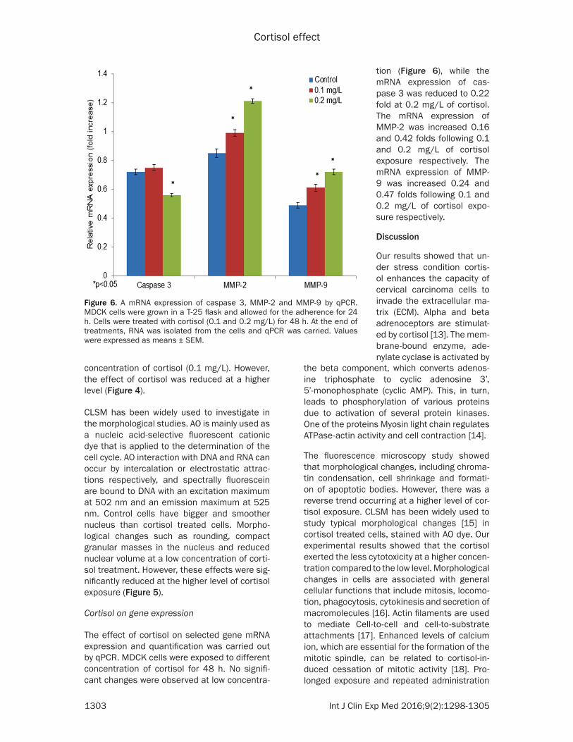
Figure 6. A mRNA expression of caspase 3, MMP-2 and MMP-9 by qPCR. MDCK cells were grown in a T-25 flask and allowed for the adherence for 24 h. Cells were treated with cortisol (0.1 and 0.2 mg/L) for 48 h. At the end of treatments, RNA was isolated from the cells and qPCR was carried. Values were expressed as means ± SEM.
tion (Figure 6), while the mRNA expression of cas-pase 3 was reduced to 0.22 fold at 0.2 mg/L of cortisol. The mRNA expression of MMP-2 was increased 0.16 and 0.42 folds following 0.1 and 0.2 mg/L of cortisol exposure respectively. The mRNA expression of MMP- 9 was increased 0.24 and 0.47 folds following 0.1 and 0.2 mg/L of cortisol expo-sure respectively.
Discussion
Our results showed that un- der stress condition cortis- ol enhances the capacity of cervical carcinoma cells to invade the extracellular ma- trix (ECM). Alpha and beta adrenoceptors are stimulat-ed by cortisol [13]. The mem-brane-bound enzyme, ade-nylate cyclase is activated by
Cortisol effect
1304 Int J Clin Exp Med 2016;9(2):1298-1305
of cortisol could produce oxidative free radi-cals. Due to an insufficient amount of protec-tive enzymes, especially at a higher concentra-tion of cortisol is one of the putative explanation for changing the cell shape and morphology.
Increased production of MMP-2 and MMP-9 occur when the invasive potential of cancer cells mediated by β-adrenergic receptors. Ce- rvical carcinoma penetration of extracellular matrix may be facilitated by altering MMPs expression. Β-Adrenergic receptors and MMP expression are believed to be key regulators in the pathogenesis of cervical carcinoma cells. The average level of cortisol ranges from 1 to 10 pmol/L and increases up to 10 nmol/L in the circulatory system under stress condition [19]. Stress can also increase the tissue corti-sol level through increased sympathetic activi-ty, which has been associated with metastasis [20]. The invasion to ECM is believed to be a critical factor in metastasis and blood flow in the tumor area. Cancer cell penetration in the host basement membrane contains an attach-ment, matrix dissolution, motility, and penetra-tion [21]. MMPs degrade components of the extracellular matrix and core protein of proteo-glycans [22]. Abnormal expression of MMPs leads to tumor cell invasion and metastasis [23]. MMP-9 play crucial role in the angiogenic switch that occurs during carcinogenesis [24]. Our data show that stress hormone can in- crease expression of MMPs under stress condi-tion. Our data indicate that MMPs might play a role in stress hormone-mediated changes in the cervical carcinoma cell metabolism. Yang et al. [25] have reported that the effect of stress on MMPs expression in a blister cham-ber wound model. Studies have indicated th- at the psychological stress can increase MMP expression in mice. The increased level of MMP-1, MMP-2, and MMP-9 mRNA expression occurred in colon tumors, and liver tissues of stress induced mice [8]. Cortisol reduced cas-pase 3 mRNA expression. Caspases act as cen-tral executioners of apoptosis [6]. The down-regulated caspase-3 reduces auto-catalysis [26].
Conclusion
Our experimental results show that cortisol can induce apoptosis in MDCK cells through up-regulation of MMP-2 and MMP-9. Cell viability, proliferation, and morphology of MDCK cells were significantly altered at a low concentration
while a reverse trend occurred at a higher level of cortisol. Stress and behavioral factors could contribute a significant role in cancer patho-genesis and prevention.
Acknowledgements
This work was suppoted by Public welfare Technology Application Research Project of Zhejiang province No. 2012C33G2010327.
Disclosure of conflict of interest
None.
Address correspondence to: Jian-Hua Mao, Depart- ment of Nephrology, The Children’s Hospital Zhe- jiang University School of Medicine, No. 57 Zhugan Lane, Hangzhou 310003, Zhejiang, China. Tel: 0086-0571-88873701; Fax: 0086-0571-888737- 01; E-mail: [email protected]
References
[1] Andersen BL, Farrar WB, Golden-Kreutz DM, Glaser R, Emery CF, Crespin TR, Shapiro CL, Carson WE. Psychological, behavioral, and im-mune changes after a psychological interven-tion: a clinical trial. J Clin Oncol 2004; 22: 3570-3580.
[2] Herbert TB, Cohen S. Depression, and immu-nity: a meta-analytic review. Psychol Bull 1993; 113: 472-486.
[3] Sanders VM, Straub RH. Norcortisol, the β- adrenergic receptor, and immunity. Brain Be-hav Immun 2002; 16: 290-332.
[4] Schmidt C, Kraft K. β-endorphin, and catechol-amine concentrations during chronic and ac- ute stress in intensive care patients. Eur J Med Res 1996; 1: 528-532.
[5] Fidler IJ. The pathogenesis of cancer metasta-sis: the “seed and soil” hypothesis revisited. Nat Rev Cancer 2003; 3: 453-458.
[6] Sood AK, Coffin JE, Schneider GB, Fletcher MS, DeYoung BR, Gruman LM, Gershenson DM, Schaller MD, Hendrix MJ. Biological signifi-cance of focal adhesion kinase in ovarian can-cer: role in migration and invasion. Am J Pathol 2004; 165: 1087-1095.
[7] Masur K, Niggemann B, Zanker KS, Entschlad-en F. Norcortisol-induced migration of SW 480 colon carcinoma cells is inhibited by β-blockers. Cancer Res 2001; 61: 2866-2869.
[8] Wu W, Yamaura T, Murakami K, Ogasawara M, Hayashi K, Murata J, Saiki I. Involvement of TNF-α in the enhancement of invasion and me-tastasis of colon 26-L5 carcinoma cells in mice
Cortisol effect
1305 Int J Clin Exp Med 2016;9(2):1298-1305
by social isolation stress. Oncol Res 1999; 11: 461-469.
[9] Vichai V, Kirtikara K. Sulforhodamine B colori-metric assay for cytotoxicity screening. Nature Protocs 2006; 1: 1112-1116.
[10] Xiao JX, Huang GQ, Zhu CP, Ren DD, Zhang SH. Morphological study on apoptosis MDCK cells induced by soyasaponins. Toxicol In Vitro 2007; 21: 820-826.
[11] Chomczynski P, Mackey K. A short technical report. Modification of the TRIZOL reagent pro-cedure for isolation of RNA from Polysaccha-ride-and proteoglycan-rich sources. Biotech-niques 1995; 19: 942-945.
[12] Pfaffl MW. A new mathematical model for rela-tive quantification in real-time RT-PCR. Nucleic Acids Res 2001; 29: e45.
[13] Potter DE, Rowland JM. Adrenergic drugs and intraocular pressure. Gen Pharmcol 1981; 12: 1-13.
[14] Rodger IW, Bowman WC. Adrenoceptors in skeletal muscle. In Adrenoceptors and Cate-cholamine Action, Vol. 1. Part B. In: Tunas G, editor. New York: John Wiley; 1983. pp. 123-55.
[15] Tattona NA, Rideout HJ. Confocal microscopy as a tool to examine DNA fragmentation, chro-matin condensation and other apoptotic ch- anges in Parkinson’s disease. Parkinsonism Relat Disord 1999; 5: 179-186.
[16] Adelstein RS, Scordilis SP, Trotter JA. The cyto-skeleton and cell movement: general consider-ations. Meth Achiev Exp Pathol 1979; 8: 1-41.
[17] Weihing RR. The cytoskeleton and plasma membrane. Meth Achiev Exp Pathol 1979; 8: 42-109.
[18] Sanger JW, Sanger JM. The cytoskeleton and cell division. Meth Achirv Exp Pathol 1979; 8: 110-142.
[19] Bierhaus A, Wolf J, Andrassy M, Rohleder N, Humpert PM, Petrov D, Ferstl R, von Eynatten M, Wendt T, Rudofsky G, Joswig M, Morcos M, Schwaninger M, McEwen B, Kirschbaum C, Nawroth PP. A mechanism converting psycho-social stress into mononuclear cell activation. Proc Natl Acad Sci U S A 2003; 100: 1920-1925.
[20] Paredes AH, Salvetti NR, Diaz AE, Dallard BE, Ortega HH, Lara HE. Sympathetic nerve activity in normal and cystic follicles from isolated bo-vine ovary: local effect of beta-adrenergic stim-ulation on steroid secretion. Reprod Biol Endo-crinol 2011; 9: 66.
[21] Liotta LA, Kohn EC. The microenvironment of the tumour host interface. Nature 2001; 411: 375-379.
[22] Kleiner DE, Stetler-Stevenson WG. Matrix me-talloproteinases and metastasis. Cancer Che-mother Pharmacol 1999; 43: S42-S51.
[23] Stack MS, Ellerbroek SM, Fishman DA. The role of proteolytic enzymes in the pathology of epithelial ovarian carcinoma. Int J Oncol 1998; 12: 569-576.
[24] Bergers G, Brekken R, McMahon G, Vu TH, Itoh T, Tamaki K, Tanzawa K, Thorpe P, Itohara S, Werb Z, Hanahan D. Matrix metalloprotein-ase-9 triggers the angiogenic switch during carcinogenesis. Nat Cell Biol 2000; 2: 737-744.
[25] Yang EV, Bane CM, MacCallum RC, Kiecolt-Glaser JK, Malarkey WB, Glaser R. Stress-relat-ed modulation of matrix metalloproteinase expression. J Neuroimmunol 2002; 133: 144-150.
[26] Chambers SK. In vitro invasion assays. In: Bartlett JS, editor. Ovarian cancer, methods, andprotocols. Totowa (New Jersey): Humana Press; 2000. pp. 179-185.