optimizing the cementation of femoral component in hip ... · pdf fileoptimizing the...
TRANSCRIPT
Optimizing the cementation of femoral component in hip arthroplasty
PhD dissertation
Juozas Petruskevicius
Faculty of Health SciencesAarhus University
2010

Optimizing the cementation of femoral component in hip arthroplasty
PhD dissertation
Juozas Petruskevicius
Orthopaedic DivisionNorth Denmark Region
Aalborg Hospital, University of Aarhus

to Ieva and Liepa
List of papers
This thesis is based on the following papers:
I. Assessment of the cementing quality after hip arthroplasty: comparison of Barrack’s grading with a new simplified cementation score.Juozas Petruskevicius, Mogens Berg Laursen, Poul Torben Nielsen, Kjeld SøballeSubmitted
II. No benefit of a proximal stem centralizer in cementing of femoral prosthesis in human cadaveric femora. Measures of intramedullary pressure, cement penetration, cement mantle thickness, and positioning of the stem. Juozas Petruskevicius, Thomas Lind-Hansen, Ramune Aleksyniene, Jens Randel Nyengaard, Poul Torben Nielsen, Kjeld SøballeBest poster Award, Annual Meeting of Danish Orthopaedic Society 2009Conditionally accepted for publication in Acta Orthopedica
III. Preheating of cemented femoral component in hip arthroplasty. Prospective randomized double-blinded study using radiostereometry, dual-energy X-ray absorptiometry and clinical scores. Juozas Petruskevicius, Mogens Brouw Jørgensen, Mogens Berg Laursen, Kjeld Søballe, Poul Torben NielsenManuscript preparation
IV. In-vivo temperature profile at cement-bone interface during total hip arthroplasty. Effect of stem preheating.Juozas Petruskevicius, Mogens Brouw Jørgensen, Mogens Berg Laursen, Kjeld Søballe, Poul Torben NielsenManuscript preparation
The papers will be referred in the text by their Roman numerals (I-IV)
List of papers
- 4 -
Preface
This PhD thesis was conducted during my employment as an orthopedic surgeon at the Department of Orthopedic Surgery, Aalborg Hospital, Orthopedic Division, North Denmark Region from April 2006 to May 2010. Simultaneously I was enrolled as a PhD student at the faculty of Health Sciences, Aarhus University. All studies were conducted in close collaboration between the orthopedic departments of Aalborg and Farsø Hospitals, as well as with Orthopaedic Research Unit in Aarhus. Many different departments and laboratories were involved in this scientific task. I am indeed grateful to my two principle supervisors, Kjeld Søballe and Poul Torben Nielsen. Kjeld introduced me to the orthopaedic research environment in Aarhus, provided me with contacts who helped finding the right answer to my questions. Poul Torben had a huge amount of ideas to be investigated. I admire his energy and enthusiasm and thank him holding the focus on the subject during the research period. This task would have been rather difficult to achieve without co-authors who did a great job helping me to manage this project. I would like to thank Mogens Berg Laursen for introducing me to RSA and DEXA methods, his constructive criticism and meticulous manuscript revisions. I thank Thomas Lind-Hansen and Ramune Aleksyniene for their friendship sharing their experience in PhD research and assisting me in performing biomechanical study. I am grateful to Mogens Brouw Jørgensen for introducing me to arthroplasty surgery, his support and encouragement managing clinical challenges.Ingrid Aaes from the Department of Earth Sciences, Jens Randel Nyengaard from Stereology and Electron Microscopy Laboratory and Niels Trolle from the Department of Biostatistics, University of Aarhus are acknowledged for helping me with bone sectioning, stereological evaluation and statistical data analysis. I am grateful to Professor Johan Kärrholm from the Department of Orthopaedics, Sählgrenska University Hospital, Sweden for his scientific advices evaluating RSA data. I also thank my friend Søren Kold who shared his expertise and knowledge on this topic, for his reviews and advices in manuscript preparation.I thank the staff at the Orthopaedic and Radiological Departments at Farsø Hospital for their tolerance and patience when surgery on cadaver bones was performed in operating theater and also for their managing the thermocouples during the hip arthroplasties. I owe particular thanks to Jess Riis who helped me with the development of in-vivo temperature measurements at cement-bone interface. I am grateful to Gitte Broholm and Ulla Hornum who spent a lot of time keeping the clinical trial on track, holding schedules of patients’ follow-up and investigating the missing data. I thank my colleagues and staff at the Orthopaedic Division, who supported me and showed great flexibility making this project possible. Finally, I thank my lovely family: my wife Irma for understanding and taking care of our daughters –Ieva and Liepa.
Preface
- 5 -
Acknowledgements and disclosures
I would like to thank “The program of Body Donation to Medical Science”, Institute of Anatomy, University of Aarhus for donation of human femora to biomechanical study. Biomet Denmark and NMS Inc. are acknowledged for supplying us with bone cement and cementing equipment.The study was financially supported by following institutions:
• Lundbeck Foundation• Sahva Fundation• Aase and Ejner Danielsens Foundation• The Research Unit of Northern Orthopedic Division• North Jutland Medical Association Research Foundation • The A.P. Møller and Chastine Mc-Kinney Møller Foundation for General Purposes • Dr. Heinrich Kopps grant (2006)
During the course of the PhD study I have benefited from institutional research support from the following company with commercial interests in this research area:
• Biomet Denmark
Contact address:
Juozas Petruskevicius, MD. Department of Orthopaedic SurgeryAalborg Hospital, University of AarhusNorthern Orthopaedic DivisionSdr. Skovvej 99000 AalborgDenmarkE-mail: [email protected]
Supervisors:
Kjeld Søballe, MD, PhD, DMSc, ProfessorDepartment of OrthopaedicsUniversity Hospital of Aarhus, Denmark
Poul Torben Nielsen, MD.Chief of the Arthroplasty Unit Department of Orthopaedic SurgeryAalborg Hospital, University of Aarhus, Denmark
Acknowledgments
- 6 -
Abbreviations
A anteriorAL anterior-lateralAM anterior-medialAP anterior-posteriorAUC area under the curveBMD bone mineral densityBMI body mass indexCBI cement-bone interfaceCoCrMo cobalt chromium molybdenumCT computer tomographyCV coefficient of varianceCg control groupDXA dual x-ray absorptiometryHHA hemi- hip arthroplastyHHS Harris hip scoreL lateralM medialP posteriorPCI prosthesis-cement interfacePL posterior-lateralPM posterior-medialPs preheated stemROI region of interestRSA radiostereometric analysisRT room temperatureTHA total hip arthroplastyTi titaniumVAS visual analog score
Abbreviations
- 7 -
Contents
Summary 9Introduction 11Aim of the PhD study 12Hypotheses 12Background 13Materials and methods 23
Femoral component 23Operating and cementation technique 24Radiological evaluation of cementing quality (Study I) 24Intramedullary pressures and cement penetration (Study II) 28Prospective randomized trial (Studies III and IV) 37
Statistics 47Considerations regarding Kappa method (Study I) 47Study II 48Study III 49Study IV 50
Summary of results 51Study I 51Study II 53Study III 55Study IV 59
Discussion 61Radiographic assessment of cementing quality (Study I) 61Cementation of stem with proximal stem centralizer (Study II) 62Migration of preheated femoral component in THA (Study III) 64Effect of preheating on heat generation at CBI (Study IV) 68
Conclusions 71References 74
Contents
- 8 -
Summary
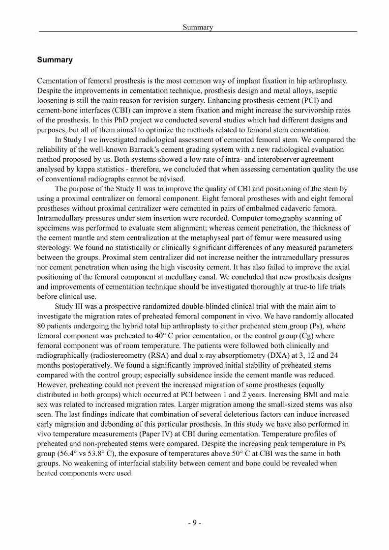
Cementation of femoral prosthesis is the most common way of implant fixation in hip arthroplasty. Despite the improvements in cementation technique, prosthesis design and metal alloys, aseptic loosening is still the main reason for revision surgery. Enhancing prosthesis-cement (PCI) and cement-bone interfaces (CBI) can improve a stem fixation and might increase the survivorship rates of the prosthesis. In this PhD project we conducted several studies which had different designs and purposes, but all of them aimed to optimize the methods related to femoral stem cementation.
In Study I we investigated radiological assessment of cemented femoral stem. We compared the reliability of the well-known Barrack’s cement grading system with a new radiological evaluation method proposed by us. Both systems showed a low rate of intra- and interobserver agreement analysed by kappa statistics - therefore, we concluded that when assessing cementation quality the use of conventional radiographs cannot be advised.
The purpose of the Study II was to improve the quality of CBI and positioning of the stem by using a proximal centralizer on femoral component. Eight femoral prostheses with and eight femoral prostheses without proximal centralizer were cemented in pairs of embalmed cadaveric femora. Intramedullary pressures under stem insertion were recorded. Computer tomography scanning of specimens was performed to evaluate stem alignment; whereas cement penetration, the thickness of the cement mantle and stem centralization at the metaphyseal part of femur were measured using stereology. We found no statistically or clinically significant differences of any measured parameters between the groups. Proximal stem centralizer did not increase neither the intramedullary pressures nor cement penetration when using the high viscosity cement. It has also failed to improve the axial positioning of the femoral component at medullary canal. We concluded that new prosthesis designs and improvements of cementation technique should be investigated thoroughly at true-to life trials before clinical use.
Study III was a prospective randomized double-blinded clinical trial with the main aim to investigate the migration rates of preheated femoral component in vivo. We have randomly allocated 80 patients undergoing the hybrid total hip arthroplasty to either preheated stem group (Ps), where femoral component was preheated to 40° C prior cementation, or the control group (Cg) where femoral component was of room temperature. The patients were followed both clinically and radiographically (radiostereometry (RSA) and dual x-ray absorptiometry (DXA) at 3, 12 and 24 months postoperatively. We found a significantly improved initial stability of preheated stems compared with the control group; especially subsidence inside the cement mantle was reduced. However, preheating could not prevent the increased migration of some prostheses (equally distributed in both groups) which occurred at PCI between 1 and 2 years. Increasing BMI and male sex was related to increased migration rates. Larger migration among the small-sized stems was also seen. The last findings indicate that combination of several deleterious factors can induce increased early migration and debonding of this particular prosthesis. In this study we have also performed in vivo temperature measurements (Paper IV) at CBI during cementation. Temperature profiles of preheated and non-preheated stems were compared. Despite the increasing peak temperature in Ps group (56.4° vs 53.8° C), the exposure of temperatures above 50° C at CBI was the same in both groups. No weakening of interfacial stability between cement and bone could be revealed when heated components were used.
Summary
- 9 -

In conclusion we recommend the RSA as a standard evaluation method of cemented femoral component. This method can show in vivo migrations which are most valuable predicting the long-term outcome of established prostheses or when evaluating the new concepts of femoral stem cementation.
Summary
- 10 -
Introduction
Since John Charnley introduced his concept of “low-friction” hip arthroplasty in the 1960’s many, not to say an unmanageable, number of papers have been published on this topic. Nevertheless, there are still sufficient numbers of non-fully answered questions. In primary total hip arthroplasty (THA) cemented fixation of femoral component takes place in 50 to 70% of cases as reported by Scandinavian national hip registries. Despite the improved survival rates, the main reason leading to revision is aseptic loosening with rates between 60 to 75%. Thus further research improving methods of cemented fixation are needed.
The purpose of this thesis is to draw attention to the factors which might improve prosthesis fixation and long-term survival.
It is commonly accepted that a simple radiological examination of hip replacement is the routine tool for both evaluating cementing quality and radiological follow-up. However, many papers have been published pointing to high intra- and inter-observer disagreement rates, making the radiological assessment unreliable. In Study I we investigated how to improve radiological assessment of cemented femoral component, where we also compared a new evaluation method with the well-known Barrack’s scoring system.
The development of modern cementing technique had increased THA outcomes by improving both cement mantle homogeneity and implant fixation at the cement-bone interface (CBI). To achieve sufficient cement mantle thickness and deep cement penetration high intramedullary pressures during cementation are required. Study II has focus on proximal centralizing device and its impact on cementation parameters influencing cementing quality.
Another important issue in cemented stem replacement is prosthesis-cement interface (PCI). Thorough research has confirmed that this is actually “a weak link” in stem-bone composite. Debonding of the rough-surfaced stem will inevitably lead to wear debris, osteolysis and aseptic loosening. Preheating of femoral components has been suggested to enhance the prosthesis-cement bond and maybe improve the long term fixation. In Study III we evaluated migration of the preheated stem in vivo using RSA. Moreover the influence on heat generation at CBI was also studied (Paper IV).
In all the studies the cemented Bi-Metric® (Biomet) stem was the main object of investigation. In Paper III we discuss the possible causes of considerable migration of this stem revealed by RSA, which might explain the contradictory survival rates for this stem reported by the Danish Hip Arthroplasty Registry.
Introduction
- 11 -
Aim of the PhD study
The aims of this thesis were:
1. To evaluate the reliability of the two grading systems in radiological assessment of cemented femoral component:
a. the five-scale Barrack’s grading system b. a new, simplified system proposed by authors (Paper I).
2. To measure cementing pressures, cement penetration, and stem positioning in cadaveric femora when cementing a straight femoral component with and without the use of a proximal stem centralizer (Paper II).
3. To compare in vivo migration rates, measured by radiostereometry when cementing matt-surfaced Ti femoral component preheated to 40° C with the same stem of room temperature (Paper III)
4. To investigate the temperature profile at cement-bone interface in vivo during cementation of preheated femoral component to 40° C (Paper IV).
Hypotheses
Study I:The quality of cementing technique can be evaluated with sufficiently high level of intra- and inter-observer agreement (kappa >60) using grading systems on postoperative radiographs.
Study II:Proximal stem centralizer increases:
pressure at CBI at metaphyseal part of the femurcement penetration into cancellous bone proximally
and improves axial positioning of femoral stem compared with the stem without proximal centralizer
Studies III and IV:Cementation of preheated femoral stem to 40° C
improves stem stability in vivohas no harmful effect on cement-bone interfacemight increase periprosthetic bone density (BMD) as a consequence of better fixation
increases cement curing temperature at CBI reduces cement polymerization time
Aim of the study
- 12 -
Background
Surgery using artificial hip implants have been performed since the 19th century. Materials such as
wood, glass, rubber, ivory were attempted as the components managing painful hip arthrosis. The
reviews of this interesting history of developing the modern total hip arthroplasty (THA) could be
found in orthopaedics textbooks or and historical articles [124, 148]. However, it should be
mentioned that bone cement has been known already in the beginning of the 20th century and the
first clinical use of it was in an attempt to close cranial defects in monkeys in 1938. Bone cement
called “dental acrylic” was also used by dentists [136, 148]. The Judet brothers (Dr. Jean Judet and
Dr. Robert Judet) were the first who attempted to use an acrylic material to replace arthritic hip
surfaces. They also developed the first short-stemmed prosthesis in 1946. This implant was made of
poly-methyl-methacrylate (PMMA) with a head that was 2/3 of a sphere attached to a short stem. In
1953 the first paper of well-documented use of bone cement for fixation of a THA was published by
Dr. Edward J. Haboush of The Hospital for Joint Diseases, New York City [52]. Several years later
John Charnley developed both a surgical and a cementing technique of THA known by the majority
of contemporary orthopaedic surgeons [16]. His invention was a milestone with regard to surgical
treatment of hip arthrosis, and these principles still dominate cemented hip replacement surgery –
the most successful procedure in orthopaedic surgery.
However, aseptic loosening of the hip prosthesis is the major complication leading to revision
[36, 60, 100-102]. A lot of research has been done both to reveal the loosening pathways and to
improve the fixation methods.
The subjects of this thesis are related to cemented femoral component. Therefore cementless
fixation will not be dealt with here.
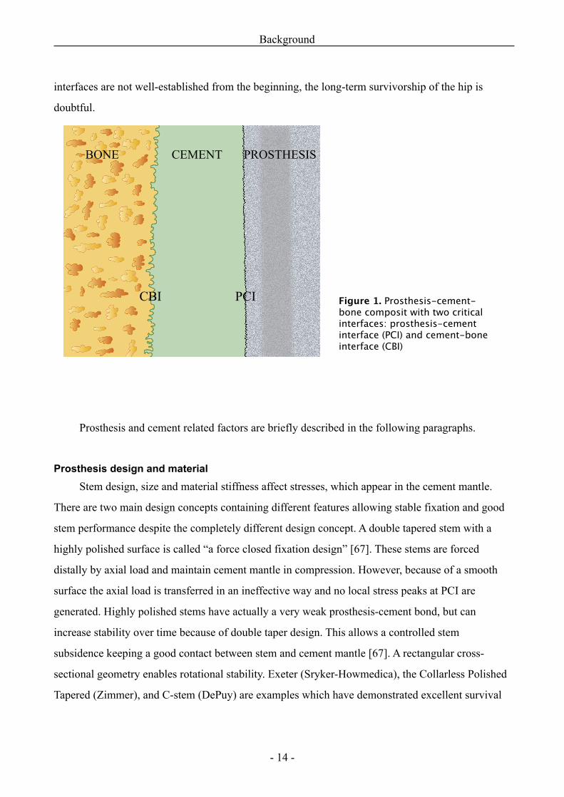
Cemented femoral stem is a composite consisting of metallic femoral stem, anchored by a
bone cement to a bone bed (Figure 1). Failure of one of these components can affect a stable
fixation and cause loosening. Initiation of failure is multifactorial, but the previous investigations
suggest that the most important factors influencing successful femoral stem fixation are related to:
1) patient selection [8, 38, 102], 2) stem geometry, design and surface finish, 3) surgical technique
[7, 60]. The surgeon is responsible for both a selection of materials and for surgical technique which
must establish the strong interfaces between both the prosthesis-cement and cement-bone. If these
Background
- 13 -
interfaces are not well-established from the beginning, the long-term survivorship of the hip is
doubtful.
Prosthesis and cement related factors are briefly described in the following paragraphs.
Prosthesis design and material Stem design, size and material stiffness affect stresses, which appear in the cement mantle.
There are two main design concepts containing different features allowing stable fixation and good
stem performance despite the completely different design concept. A double tapered stem with a
highly polished surface is called “a force closed fixation design” [67]. These stems are forced
distally by axial load and maintain cement mantle in compression. However, because of a smooth
surface the axial load is transferred in an ineffective way and no local stress peaks at PCI are
generated. Highly polished stems have actually a very weak prosthesis-cement bond, but can
increase stability over time because of double taper design. This allows a controlled stem
subsidence keeping a good contact between stem and cement mantle [67]. A rectangular cross-
sectional geometry enables rotational stability. Exeter (Sryker-Howmedica), the Collarless Polished
Tapered (Zimmer), and C-stem (DePuy) are examples which have demonstrated excellent survival
Background
- 14 -
Figure 1. Prosthesis-cement-bone composit with two critical interfaces: prosthesis-cement interface (PCI) and cement-bone interface (CBI)
BONE CEMENT PROSTHESIS
CBI PCI
rates[62, 96, 99, 102, 151, 155]. Another concept is known as “shape closed fixation design”.
Unlike previous ones, these stems are designed to be contained by a cement mantle. The rough
surface (matt-surfaced, grit-blasted stems) increases contact between stem and cement and transfers
a relatively large portion of axial load to the cement. Local stress peaks are generated around the
asperities of the surface, but strong interfacial bond reduces global cement stresses and prevents
both subsidence and micromotions occurring at the PCI [139, 146]. These stems have usually
anatomical form and other features such as more bulky shape, collar and ridges. Theses features are
designed to maintain stem stability and inhibit micromotions within the cement mantle [32, 56].
Reports of very good long-term survival have also been published for stems with such design, f.
exp.: Lubinus SP II, Spectron EF, Muller Straight stem [36, 102, 126, 129]. The disadvantage of this
design is that if the stems do debond, they may abrade the cement mantle, thereby producing
cement wear particles, initiating cement fractures and inducing osteolysis. The smooth stems, in
contrast, allows retention of debris on the stem surface, without significant damage to the cement
[63], though cement cracks usually appears in the corners between the stem and cement surface
[43].
Mixing design concepts to achieve better results can be detrimental for stem performance in
vivo. One design feature that suits well in one concept will not necessarily be advantageous in
another. An example of this mixed design concept was a double tapered Exeter stem with a rough
surface. Despite the improved initial interfacial strength, very bad clinical results was observed,
reporting catastrophically high revision rates [25, 64, 110, 131].
Materials used for cemented implants are cobalt-chromium alloy, titanium alloy and stainless steel.
Stems of different alloys vary in bending stiffness, where Ti-stems are most flexible. The rationale
of using titanium was to avoid the proximal medial bone resorption usually seen with rigid CoCr
and stainless steel stems. The stiffness of Ti–stems is closer to bone and cement and more load is
transferred to stem surroundings. It has been postulated that this flexibility should be beneficial for
bone biology avoiding stress shielding. Reports of stem failures have been published regarding all 3
materials [13, 112]. However, some studies of revised Ti implants caused wariness among the
surgeons [31, 149, 153]. Biomechanical experiments showed that Ti prostheses generate higher
stresses in the proximal region, whereas more rigid stems transfer load more distally. If the Ti-
implant has small dimensions (medial-lateral) proximally, the cement stresses may become too high
Background
- 15 -
resulting in stem micromotions and debonding between cement and implant. Moreover clinical
reports of crevice corrosion using Ti-stems suggest that titanium alloy is unfavourable for cemented
fixation. Nevertheless, the long-term results are influenced by multiple factors and the implant
stability not only depends on a single feature, but rather on a complexity of stem properties. Poor
survival rates have been demonstrated for certain stems (Capital hip, Centralign, Cenator, Iowa
stems) which was obviously caused by a combination of several inferior features [12, 106, 142].
A few clinical reports using cemented Bi-Metric® stems have previously been published. In a
study of 21 primary hybrid THA in young patients (mean age of 43 years), 18 hips were examined
at mean follow-up of 5.5 years. No hips were revised because of aseptic loosening, but a
radiolucency between stem and cement was observed in 2/18 cases and 1 patient had also had a
large bone osteolysis at cement-bone interface [88]. Other study with use of cemented Ti Bi-Metric
stem with collar reported 97% survival of 102 cemented THA. Only one stem was revised due to
aseptic loosening at mean follow-up of 5.7 years.
Contradictory outcomes are reported by the Danish National Hip Registry where both good
long-term survival and poor results are noticed. Eleven years survivorship with 1st. revision due to
aseptic loosening ranged from 92.5 to 100% in hybrid arthroplasties and from 91 to 95% in
cemented THA. However, the 11 years stem survival was only 88.6%, when revisions due to
loosening of femoral component in hybrid THA were calculated [120]. In study by Mette Ørskov
(2007) migration rates of Bi-Metric® stems made of different alloys (CoCr vs Ti) were compared. It
was not confirmed that Ti-stems should be inferior to stems made of CoCr [119].
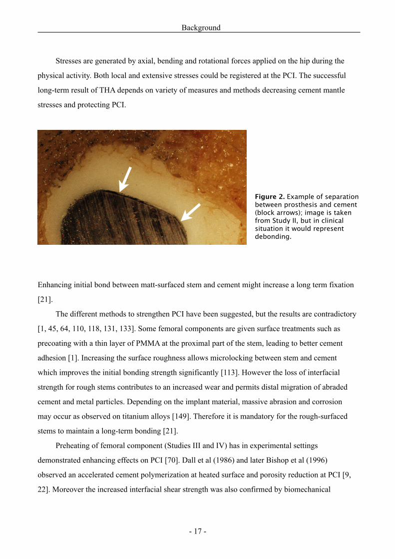
Prosthesis-cement interface (PCI)This is the most critical interface in the cemented stem-bone construct. Biomechanical, histological,
finite element and clinical studies showed that the initiation of loosening typically occurs between
stem and cement, so-called debonding [74, 103, 122] (Figure2). Subsequently, this induces cement
cracks, wear debris, fractures and fragmentation, which start a biological response at the cement-
bone interface too[2, 66].
Biomechanical experiments and finite element research revealed that stresses experienced at
PCI are the main causes leading to debonding when the endurance limit of both interfacial bond and
cement material is exceeded [67].
Background
- 16 -
Stresses are generated by axial, bending and rotational forces applied on the hip during the
physical activity. Both local and extensive stresses could be registered at the PCI. The successful
long-term result of THA depends on variety of measures and methods decreasing cement mantle
stresses and protecting PCI.
Enhancing initial bond between matt-surfaced stem and cement might increase a long term fixation
[21].
The different methods to strengthen PCI have been suggested, but the results are contradictory
[1, 45, 64, 110, 118, 131, 133]. Some femoral components are given surface treatments such as
precoating with a thin layer of PMMA at the proximal part of the stem, leading to better cement
adhesion [1]. Increasing the surface roughness allows microlocking between stem and cement
which improves the initial bonding strength significantly [113]. However the loss of interfacial
strength for rough stems contributes to an increased wear and permits distal migration of abraded
cement and metal particles. Depending on the implant material, massive abrasion and corrosion
may occur as observed on titanium alloys [149]. Therefore it is mandatory for the rough-surfaced
stems to maintain a long-term bonding [21].
Preheating of femoral component (Studies III and IV) has in experimental settings
demonstrated enhancing effects on PCI [70]. Dall et al (1986) and later Bishop et al (1996)
observed an accelerated cement polymerization at heated surface and porosity reduction at PCI [9,
22]. Moreover the increased interfacial shear strength was also confirmed by biomechanical
Background
- 17 -
Figure 2. Example of separation between prosthesis and cement (block arrows); image is taken from Study II, but in clinical situation it would represent debonding.
experiments [65, 68, 70, 71]. It has been suggested that heating of the component to 40° C could
produce an optimum effect without harm for bone tissue [42, 71, 95]. These advantages could be
beneficial especially for mat-surfaced stems, where strong initial fixation is required to avoid
micromotion and cement abrasion. Whether preheating can improve the long-term fixation in hip
arthroplasty is unknown. Increased fatigue strength of stem-cement construct was found by Iesaka
et al (2003), but these results were not confirmed by a recently published biomechanical and finite
model study performed by Damron et al (2006). No substantial improvement on the fatigue debond
response was registered, when a preheated stem was used [24]. So far, no clinical studies have been
published on this subject. There are no data about the in vivo migration of preheated stem.
Moreover, the knowledge about a possible biological response is sparse. Study III reveals the
relationship between the preheating and the stem migration.
Bone cement All bone cements used for arthroplasties are polymethilmetacrylates (PMMA) [86]. Cement is not a
glue, but filler, which both fills the space between the implant and bone and interdigitates into the
bone trabecullae. The primal function of bone cement is the implant fixation to the bone. As
mentioned previously cement is exposed by loading forces which transfer through the implant. In
order to provide durable stem fixation cement must resist these forces and ensure stable anchoring
of the implant[87]. The unique ability to distribute the load-induced stresses to the bone and “to
recover” when unloaded, makes bone cement an effective buffer [61, 92]. If the continuous stress
from outside exceeds the capability to transfer and absorb forces, a fatigue break will occur.
It has been shown that air voids and pores in the cement mantle act as stress risers. This
promotes the formation of micro-cracks and makes the cement susceptible to early fatigue failure
[14]. Furthermore the experiments performed by Mann et al (2006) demonstrated increasing stem
migration related to larger interfacial porosity [104, 125]. Vaccum mixing and cement
centrifugation have been adopted as methods to decrease cement porosity and enhance mechanical
strength of cement mantle [30, 73]. These measures have, however, a little impact on interfacial
porosity, but this can effectively be reduced by cementing of heated implants [9, 24, 42, 65, 69-71].
Whether preheating also influences mechanical properties of cement is unclear, because
contradictory results have been reported regarding compressive and bending strength of cement
mantle [42, 65, 71].
Background
- 18 -
Poor cement characteristics result in early failures as it was in case with Boneloc cement
[130]. But also cement handling properties and viscosity can affect THA outcome which has also
been shown by Swedish and Norwegian national hip registries. Low viscosity performed worse than
high viscosity cements which might be explained by the difficult handling characteristics of these
cements [37]. It has also been suggested that different prosthesis designs might require different
mechanical properties of the cement to achieve optimal performance [59].
Palacos R and Simplex P cements are associated with the lowest risk of revision. However,
new cement brands have been introduced on the marked recently. Refobacin® bone cement R with
gentamycin (Biomet UK Ltd, UK) is high-viscosity cement and is equivalent to Refobacin Palacos
R bone cement regarding chemical composition, molecular weight, particle size distribution and
mechanical properties. Some differences in handling and viscosity characteristics, however has
been noticed at in-vitro tests, but the clinical significance of this is unknown [23, 83]. Data from
RSA studies and national hip registries will be needed to confirm implications for the long-term
outcome.
Cement mantle and cement-bone interface (CBI)Establishment of a uniform and homogenuous cement mantle with a strong interlock at the CBI is
paramount for implant survival. The strength of this interface depends on sufficient cement
interdigitation in trabecular bone (macro-locking) and actually depends on operating technique. The
early experiments showed that the higher pressures produced both the deeper cement penetration
[105] and the stronger CBI [53, 85, 121]. Since the1980’s various methods improving both cement
and bone preparation were gradually introduced in hip replacement surgery [91]. This modern
cementing technique has significantly increased survivorship rates of femoral hip prostheses [10,
60, 101, 114, 123, 126, 132, 133, 137]. The so-called third generation technique consists of vacuum
mixing/or centrifugation of bone cement to reduce porosity, plugging of femoral canal distally,
thorough bone-bed preparation using pulsatile lavage, retrograde cement application, femoral
pressuriser to improve cement intrusion and the use of stem centralizers to secure neutral stem
alignment in the medullary canal.
The optimal thickness of cement mantle is a subject of debate. There is a common agreement
that a uniform cement mantle of 2 to 5 mm thickness is needed to ensure stable fixation of
cemented implant [33]. But the unusual method of stem cementation line-to-line has showed the
Background
- 19 -
same good long term results (“French paradox”) as those performed in the classical manner [54,
90]. However, the cadaver experiments revealed a considerable pressurization in the proximal part
of the femur and cement mantle for line-to-line stems was in fact thicker than anticipated. This
phenomenon confirms the significance of cement penetration into trabecular bone proximally, but
also demonstrates that cement mantle stresses are mostly affected by stem design, stiffness and
geometry which influence the long-term outcome.
Central positioning of the stem in the medullar cavity is preferable regardless of implant
geometry, surface finish and design. The use of distal stem centralizers helps to control alignment of
the stem avoiding direct contact between the bone and the tip of the prosthesis [5, 34, 55, 140].
However, this device alone cannot prevent cement mantle deficiencies, especially in the proximal
region of the femur [5, 11, 20]. Promising results using the proximal stem centralizer have been
reported in retrospective studies [47, 72]. Experimental trials have also shown that a proximal
centralizer can increase the intramedullary pressures in the proximal region of the femur, thereby
enhancing the cement-bone interlock [48, 49]. Nevertheless, no research has been published on the
relation between cementing pressures, cement penetration, cement mantle thickness and the use of a
proximal centralizer in a true-to-life study setup. This was the main subject of Study II.
An important physical feature of bone cement is heat generation during polymerization. In the
early years of replacement surgery, it was believed that one of the reasons of implant loosening
could be an endosteal bone necrosis due to high curing temperature of PMMA cement [111, 150].
The first well-documented in vivo measurement in 10 THA was performed by Meyer et al (1974)
using Simplex cement. The maximum temperature of 70° C was recorded at cement-bone interface
[109]. However, several years later the significant lower peak temperatures (max-48° C) were
obtained by Reckling and Dillon (1977), who performed measurements during 10 THA and 10 total
knee arthroplasties [128]. Since then several other investigations were performed reporting different
temperature ranges [144, 154]. Histological investigations of animal and human retrievals as well as
cadaver specimens demonstrated that CBI undergoes biological changes where primary bone
healing, remodelling and revascularization process are seen [28]. Clinical studies and experience
with modern cementing technique showed that low temperature cements have no benefits on
implant stability [130]. Recently histomorphological studies have also demonstrated that fibrous
tissue between the bone and the cement is a consequence of insufficient bone preservation and
Background
- 20 -
cement interdigitation due to poor operating technique, not because of heat-induced bone necrosis
as it was claimed previously [29].
New methods of cementation such as preheating of femoral stem have renewed the discussion
about eventual harmful effects of increased temperature at the CBI [95]. Conventionally, the
femoral stem of room temperature acts as the heat sinker during cement polymerization, thus
heating of the prosthesis above the room temperature could increase heat generation and might
cause bone necrosis. Several in vitro investigations demonstrated higher peak temperatures at CBI
during cementation of preheated stems compared with non-preheated [9, 42, 65, 68, 70]. These
results can not directly be extrapolated to the clinical situation. Recording cement curing
temperature at CBI in vivo is a technically demanding procedure. Moreover, the variation of
cancelous bone and cement mantle thickness, differences of blood circulation and amount of
cement penetration around thermocouple are some factors that influence temperature
measurements. Nevertheless, it is possible to achieve better understanding of heat fluctuations at
CBI when stems of different temperatures are cemented. RSA studies monitoring in vivo stem-
migration can reveal the effects of preheating on interfacial stability.
Radiological assessment of cemented implantThe main tool to evaluate cementation quality of hip implants is radiological evaluation of
postoperative radiographs, which are also usually used on follow-up examinations.
The radiological features of an adequate cementing technique, such as the thickness, shape, and
integrity of the cement mantle as well as prosthesis alignment, are important factors predicting the
longevity of cemented femoral implants [15, 31, 33, 123, 140]. But there are still high intra- and
inter-observer disagreement rates, which make the radiological assessment unreliable in predicting
the longevity of hip implants. The early radiological signs of loosening are often very sparse, and
the decision to perform a reoperation is often made in the late phase.
There are few published scoring systems that evaluate the cementation quality of femoral prostheses
[4, 50, 76]. The best known and most widely used scoring system is Barrack’s cement grading
published in 1992.
Some investigators [10, 15, 19] found a correlation between low-grade cement mantle and later
radiographic loosening of the femoral stem, whereas others could not show the same tendency [98].
Moreover, the accuracy of Barrack’s cement grading has been questioned [57, 80, 108]. Despite
Background
- 21 -
attempts to make this scoring system more “user-friendly,” intra- and interobserver disagreement is
still large, which makes reliable data comparison doubtful.
In Sweden the radiostereometric analysis (RSA) has been widely used for 2 decades as a
method to monitor the migration of the joint implants [134]. This technique has previously been
proved as a reliable method to predict THA fate. Important information regarding the risk of clinical
loosening and revision could be obtained after an observation period of 6 months to 2 years [77-79].
It has been demonstrated, that stem subsidence of 1 mm during the first postoperative years without
loosening is usually observed for highly polished tapered prostheses, but for matt-surfaced
components subsidence between 1 to 2 mm during the first 2 years is related to an increased risk of
failure. The risk of revision within 5 to 7 years after the operation exceeded 50%, if the subsidence
was more than 1.2 mm and 95% if it was more than 2.6 mm during the first 2 years. [78]. According
to the Swedish National Register the stems with a high survival rate showed mean subsidence
values below 0.2 mm (21). RSA’s high resolution means that migration rates of new prosthetic
design can be investigated using small sample sizes.
Background
- 22 -
Materials and methods
Femoral component
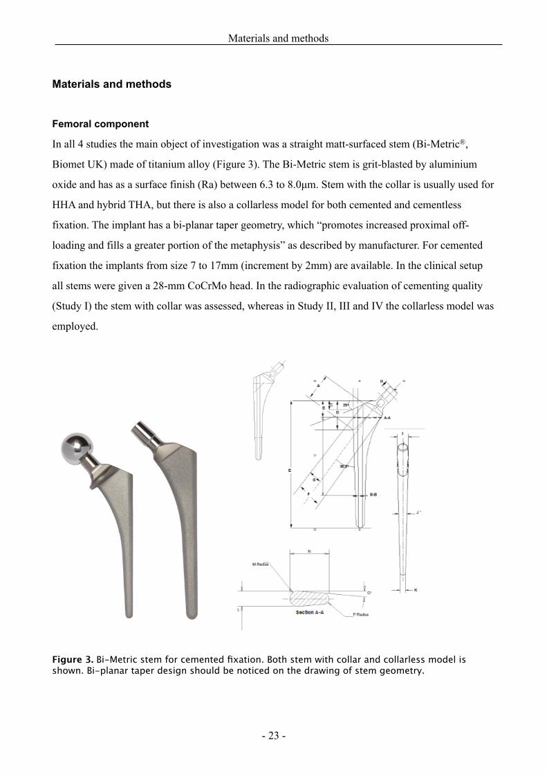
In all 4 studies the main object of investigation was a straight matt-surfaced stem (Bi-Metric®,
Biomet UK) made of titanium alloy (Figure 3). The Bi-Metric stem is grit-blasted by aluminium
oxide and has as a surface finish (Ra) between 6.3 to 8.0µm. Stem with the collar is usually used for
HHA and hybrid THA, but there is also a collarless model for both cemented and cementless
fixation. The implant has a bi-planar taper geometry, which “promotes increased proximal off-
loading and fills a greater portion of the metaphysis” as described by manufacturer. For cemented
fixation the implants from size 7 to 17mm (increment by 2mm) are available. In the clinical setup
all stems were given a 28-mm CoCrMo head. In the radiographic evaluation of cementing quality
(Study I) the stem with collar was assessed, whereas in Study II, III and IV the collarless model was
employed.
Figure 3. Bi-Metric stem for cemented fixation. Both stem with collar and collarless model is shown. Bi-planar taper design should be noticed on the drawing of stem geometry.
Materials and methods
- 23 -
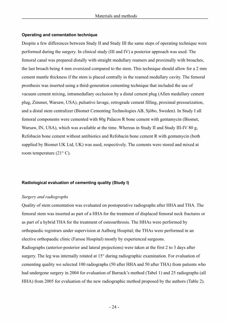
Operating and cementation technique
Despite a few differences between Study II and Study III the same steps of operating technique were
performed during the surgery. In clinical study (III and IV) a posterior approach was used. The
femoral canal was prepared distally with straight medullary reamers and proximally with broaches,
the last broach being 4 mm oversized compared to the stem. This technique should allow for a 2 mm
cement mantle thickness if the stem is placed centrally in the reamed medullary cavity. The femoral
prosthesis was inserted using a third-generation cementing technique that included the use of
vacuum cement mixing, intramedullary occlusion by a distal cement plug (Allen medullary cement
plug, Zimmer, Warsaw, USA), pulsative lavage, retrograde cement filling, proximal pressurization,
and a distal stem centralizer (Biomet Cementing Technologies AB, Sjöbo, Sweden). In Study I all
femoral components were cemented with 80g Palacos R bone cement with gentamycin (Biomet,
Warsaw, IN, USA), which was available at the time. Whereas in Study II and Study III-IV 80 g,
Refobacin bone cement without antibiotics and Refobacin bone cement R with gentamycin (both
supplied by Biomet UK Ltd, UK) was used, respectively. The cements were stored and mixed at
room temperature (21° C).
Radiological evaluation of cementing quality (Study I)
Surgery and radiographs
Quality of stem cementation was evaluated on postoperative radiographs after HHA and THA. The
femoral stem was inserted as part of a HHA for the treatment of displaced femoral neck fractures or
as part of a hybrid THA for the treatment of osteoarthrosis. The HHAs were performed by
orthopaedic registrars under supervision at Aalborg Hospital; the THAs were performed in an
elective orthopaedic clinic (Farsoe Hospital) mostly by experienced surgeons.
Radiographs (anterior-posterior and lateral projections) were taken at the first 2 to 3 days after
surgery. The leg was internally rotated at 15° during radiographic examination. For evaluation of
cementing quality we selected 100 radiographs (50 after HHA and 50 after THA) from patients who
had undergone surgery in 2004 for evaluation of Barrack’s method (Tabel 1) and 25 radiographs (all
HHA) from 2005 for evaluation of the new radiographic method proposed by the authors (Table 2).
Materials and methods
- 24 -
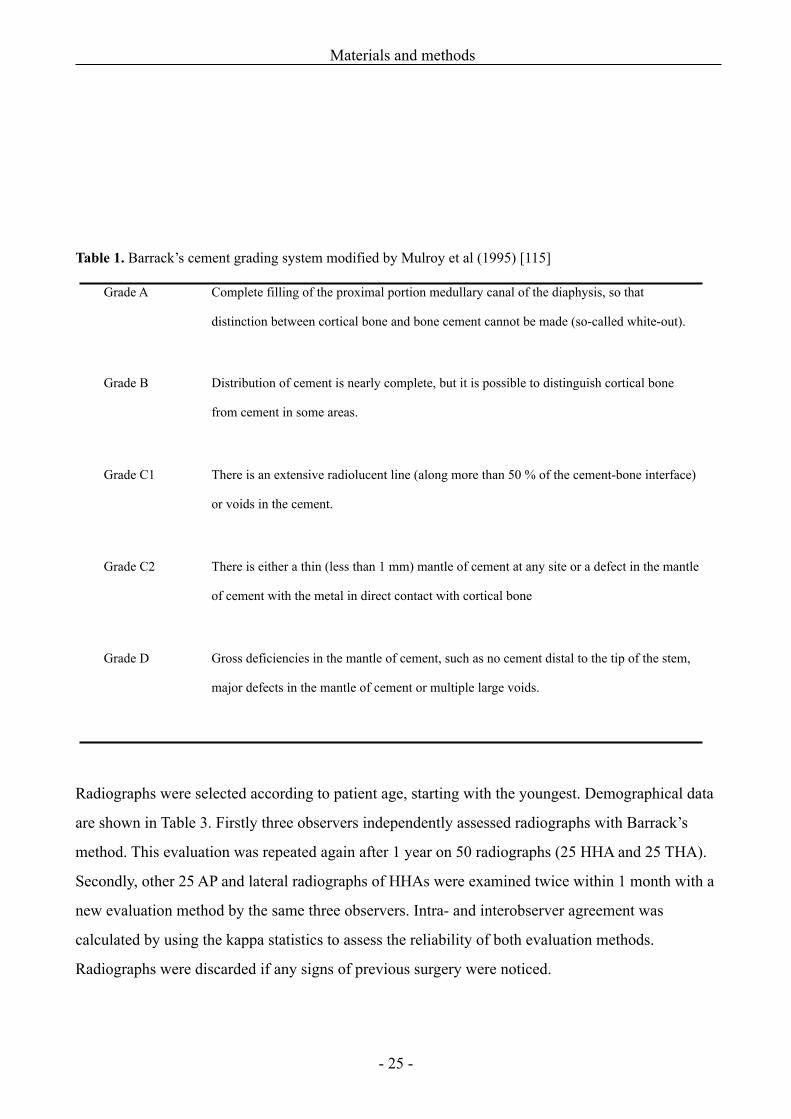
Table 1. Barrack’s cement grading system modified by Mulroy et al (1995) [115]
Grade A Complete filling of the proximal portion medullary canal of the diaphysis, so that
distinction between cortical bone and bone cement cannot be made (so-called white-out).
Grade B Distribution of cement is nearly complete, but it is possible to distinguish cortical bone
from cement in some areas.
Grade C1 There is an extensive radiolucent line (along more than 50 % of the cement-bone interface)
or voids in the cement.
Grade C2 There is either a thin (less than 1 mm) mantle of cement at any site or a defect in the mantle
of cement with the metal in direct contact with cortical bone
Grade D Gross deficiencies in the mantle of cement, such as no cement distal to the tip of the stem,
major defects in the mantle of cement or multiple large voids.
Radiographs were selected according to patient age, starting with the youngest. Demographical data
are shown in Table 3. Firstly three observers independently assessed radiographs with Barrack’s
method. This evaluation was repeated again after 1 year on 50 radiographs (25 HHA and 25 THA).
Secondly, other 25 AP and lateral radiographs of HHAs were examined twice within 1 month with a
new evaluation method by the same three observers. Intra- and interobserver agreement was
calculated by using the kappa statistics to assess the reliability of both evaluation methods.
Radiographs were discarded if any signs of previous surgery were noticed.
Materials and methods
- 25 -
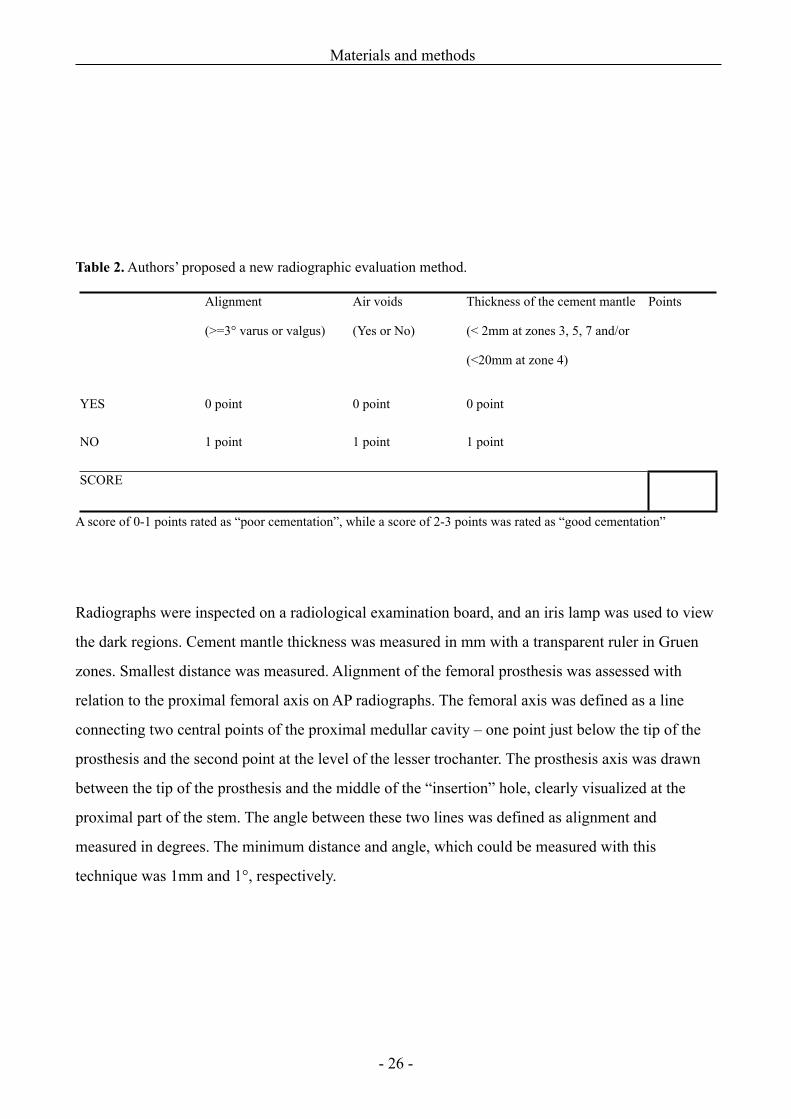
Table 2. Authors’ proposed a new radiographic evaluation method.
Alignment
(>=3° varus or valgus)
Air voids
(Yes or No)
Thickness of the cement mantle
(< 2mm at zones 3, 5, 7 and/or
(<20mm at zone 4)
Points
YES 0 point 0 point 0 point
NO 1 point 1 point 1 point
SCORESCORESCORE
A score of 0-1 points rated as “poor cementation”, while a score of 2-3 points was rated as “good cementation”
Radiographs were inspected on a radiological examination board, and an iris lamp was used to view
the dark regions. Cement mantle thickness was measured in mm with a transparent ruler in Gruen
zones. Smallest distance was measured. Alignment of the femoral prosthesis was assessed with
relation to the proximal femoral axis on AP radiographs. The femoral axis was defined as a line
connecting two central points of the proximal medullar cavity – one point just below the tip of the
prosthesis and the second point at the level of the lesser trochanter. The prosthesis axis was drawn
between the tip of the prosthesis and the middle of the “insertion” hole, clearly visualized at the
proximal part of the stem. The angle between these two lines was defined as alignment and
measured in degrees. The minimum distance and angle, which could be measured with this
technique was 1mm and 1°, respectively.
Materials and methods
- 26 -
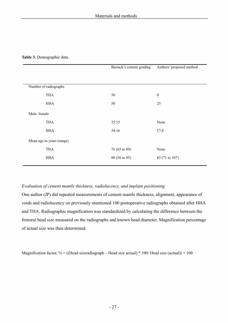
Table 3. Demographic data.
Barrack’s cement grading Authors’ proposed method
Number of radiographs
THA
HHA
50
50
0
25
Male: female
THA
HHA
35:15
34:16
None
17:8
Mean age in years (range)
THA
HHA
76 (65 to 89)
80 (56 to 95)
None
83 (71 to 107)
Evaluation of cement mantle thickness, radiolucency, and implant positioning
One author (JP) did repeated measurements of cement mantle thickness, alignment, appearance of
voids and radiolucency on previously mentioned 100 postoperative radiographs obtained after HHA
and THA. Radiographic magnification was standardized by calculating the difference between the
femoral head size measured on the radiographs and known head diameter. Magnification percentage
of actual size was then determined.
Magnification factor, % = ((Head sizeradiograph – Head size actual) * 100/ Head size (actual)) + 100
Materials and methods
- 27 -
Intramedullary pressures and cement penetration (Study II)
We prepared eight pairs of embalmed cadaveric femora. A mean donor age was 77 years
(range, 65 – 91). Cadavers were preserved using distilled water, glycerol, glutaraldehyd, glyoxal,
96% alcohol, and formaldehyde at Institute of Anatomy, University of Aarhus. Before the
experiments, the soft tissues were removed from the femora. Collarless Bi-Metric stem (Biomet
UK) was used for cementation. The majority of stems were size 9 (five pairs), while size 7 was used
in two pairs, and size 11 in one pair. Half of the eight left femora were randomly allocated to the
proximal centralizer group and the other half to the control group, providing an equal number of
right and left femora in both groups.
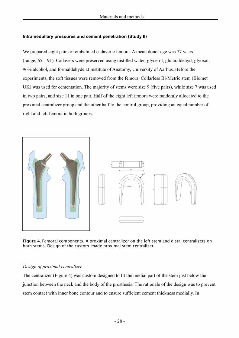
Figure 4. Femoral components. A proximal centralizer on the left stem and distal centralizers on both stems. Design of the custom-made proximal stem centralizer.
Design of proximal centralizer
The centralizer (Figure 4) was custom designed to fit the medial part of the stem just below the
junction between the neck and the body of the prosthesis. The rationale of the design was to prevent
stem contact with inner bone contour and to ensure sufficient cement thickness medially. In
Materials and methods
- 28 -
addition, we expected inhibition of cement outflow, thus increasing intramedullary pressures and
deeper penetration of cement into the proximomedial region of the femur. The centralizer was
hemispherical, 2 mm thick at its rounded part, and became evenly thinner at both the anterior and
posterior sides of the stem. The branches reached approximately two-thirds of the width of the stem.
The centralizer, 4 mm in height, was made of polyethylene powder using a high resolution 3D
printing machine (Danish Technological Institute, Aarhus, Denmark) and was firmly glued onto the
prosthesis before the cementation (Figure 4). Three different sizes with regard to the inner diameter
were made to fit the commonly used stem sizes 7, 9, and 11. All the prostheses were equipped with
a distal centralizer (Biomet Cementing Technologies AB, Sjöbo, Sweden) on the tip of the stem.
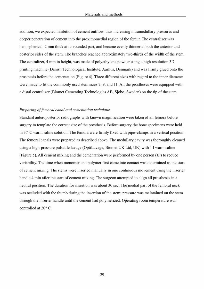
Preparing of femoral canal and cementation technique
Standard anteroposterior radiographs with known magnification were taken of all femora before
surgery to template the correct size of the prosthesis. Before surgery the bone specimens were held
in 37°C warm saline solution. The femora were firmly fixed with pipe–clamps in a vertical position.
The femoral canals were prepared as described above. The medullary cavity was thoroughly cleaned
using a high-pressure pulsatile lavage (OptiLavage, Biomet UK Ltd, UK) with 1 l warm saline
(Figure 5). All cement mixing and the cementation were performed by one person (JP) to reduce
variability. The time when monomer and polymer first came into contact was determined as the start
of cement mixing. The stems were inserted manually in one continuous movement using the inserter
handle 4 min after the start of cement mixing. The surgeon attempted to align all prostheses in a
neutral position. The duration for insertion was about 30 sec. The medial part of the femoral neck
was occluded with the thumb during the insertion of the stem; pressure was maintained on the stem
through the inserter handle until the cement had polymerized. Operating room temperature was
controlled at 20° C.
Materials and methods
- 29 -
Figure 5. Stepwise preparation of femoral canal and stem cementation, from top-left to right-bottom:Lavage of medullary cavity, retrograde cement application, pressurizing, stem insertion and finally implant in situ. Six pressure transducers on medial and lateral side could be seen.
Materials and methods
- 30 -
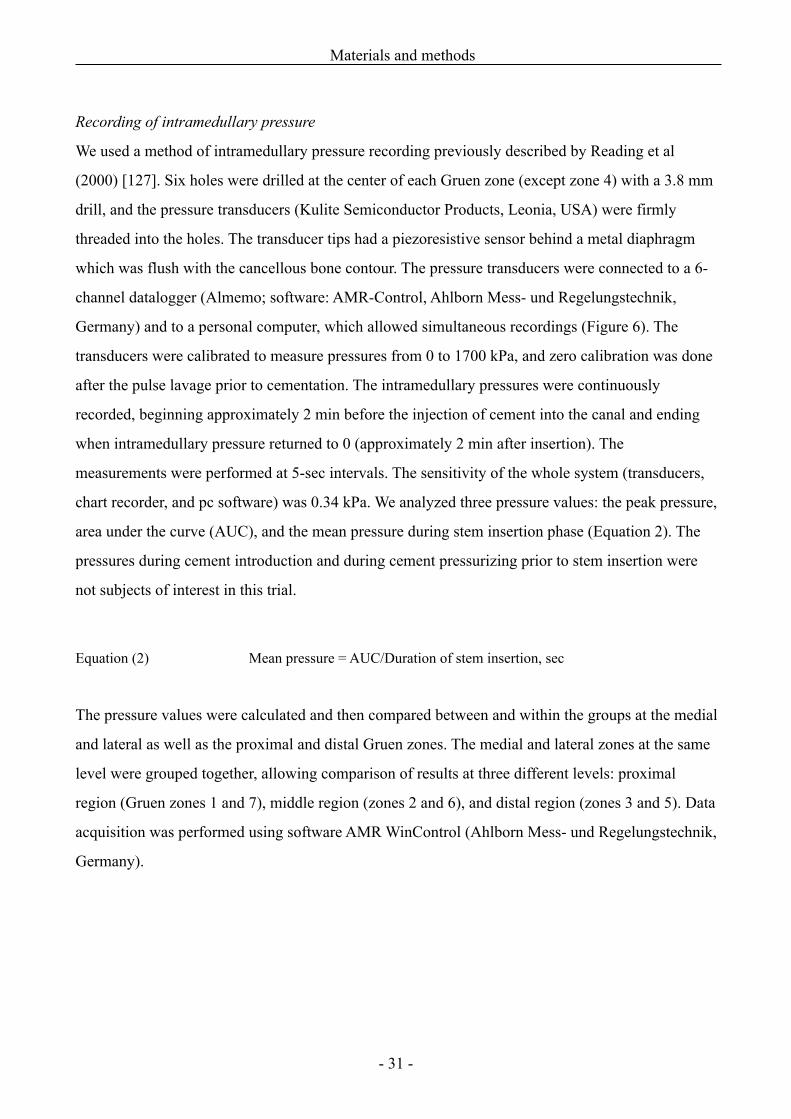
Recording of intramedullary pressure
We used a method of intramedullary pressure recording previously described by Reading et al
(2000) [127]. Six holes were drilled at the center of each Gruen zone (except zone 4) with a 3.8 mm
drill, and the pressure transducers (Kulite Semiconductor Products, Leonia, USA) were firmly
threaded into the holes. The transducer tips had a piezoresistive sensor behind a metal diaphragm
which was flush with the cancellous bone contour. The pressure transducers were connected to a 6-
channel datalogger (Almemo; software: AMR-Control, Ahlborn Mess- und Regelungstechnik,
Germany) and to a personal computer, which allowed simultaneous recordings (Figure 6). The
transducers were calibrated to measure pressures from 0 to 1700 kPa, and zero calibration was done
after the pulse lavage prior to cementation. The intramedullary pressures were continuously
recorded, beginning approximately 2 min before the injection of cement into the canal and ending
when intramedullary pressure returned to 0 (approximately 2 min after insertion). The
measurements were performed at 5-sec intervals. The sensitivity of the whole system (transducers,
chart recorder, and pc software) was 0.34 kPa. We analyzed three pressure values: the peak pressure,
area under the curve (AUC), and the mean pressure during stem insertion phase (Equation 2). The
pressures during cement introduction and during cement pressurizing prior to stem insertion were
not subjects of interest in this trial.
Equation (2) Mean pressure = AUC/Duration of stem insertion, sec
The pressure values were calculated and then compared between and within the groups at the medial
and lateral as well as the proximal and distal Gruen zones. The medial and lateral zones at the same
level were grouped together, allowing comparison of results at three different levels: proximal
region (Gruen zones 1 and 7), middle region (zones 2 and 6), and distal region (zones 3 and 5). Data
acquisition was performed using software AMR WinControl (Ahlborn Mess- und Regelungstechnik,
Germany).
Materials and methods
- 31 -

Figure 6. Schematic cartoon of the positions of the pressure transducers and pressure recorder. On the right a typical pressure profile during cementation is shown. Small pressure elevations in the beginning of curves correspond to cement application and pressurizing whereas large peaks are related to stem insertion.
Computer tomography scanning and sectioning of specimens
Each femur was scanned in a computer tomography (CT) scanner after the surgery. Three-
dimensional CT analysis was carried out using the medical data imaging software EasyViz TM
(Medical Insight A/S, Valby, Denmark), which allowed evaluation of stem alignment in relation to
the reamed medullary canal on the coronal and lateral projections. The femoral axis on the coronal
plan was defined as a line connecting two middle points of the medullar cavity – one point just
below the tip of the prosthesis and the other point at the level of the center of Gruen zone 2 (Figure
7). The femoral axis on the lateral projection was defined similarly, i.e., the middle point of the
femoral cavity at the tip of the prosthesis was connected to the middle point at the level of the
opening of the femoral canal proximally. The implant axis was drawn between the tip of the
prosthesis and the middle of the “insertion” hole, defined at the proximal part of the stem on both
projections. The angle between these two lines was measured with a digital angle ruler, and
alignment was defined in degrees. The least possible angle to measure was 0.1° degree. The
Materials and methods
- 32 -
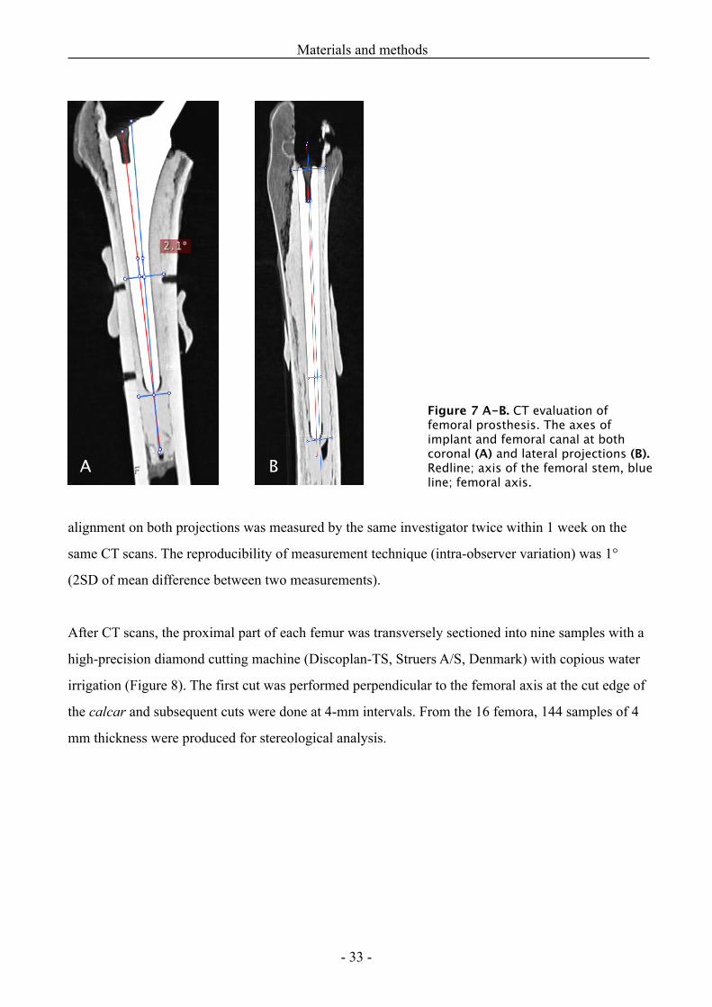
alignment on both projections was measured by the same investigator twice within 1 week on the
same CT scans. The reproducibility of measurement technique (intra-observer variation) was 1°
(2SD of mean difference between two measurements).
After CT scans, the proximal part of each femur was transversely sectioned into nine samples with a
high-precision diamond cutting machine (Discoplan-TS, Struers A/S, Denmark) with copious water
irrigation (Figure 8). The first cut was performed perpendicular to the femoral axis at the cut edge of
the calcar and subsequent cuts were done at 4-mm intervals. From the 16 femora, 144 samples of 4
mm thickness were produced for stereological analysis.
Materials and methods
- 33 -
Figure 7 A-B. CT evaluation of femoral prosthesis. The axes of implant and femoral canal at both coronal (A) and lateral projections (B). Redline; axis of the femoral stem, blue line; femoral axis.
A B
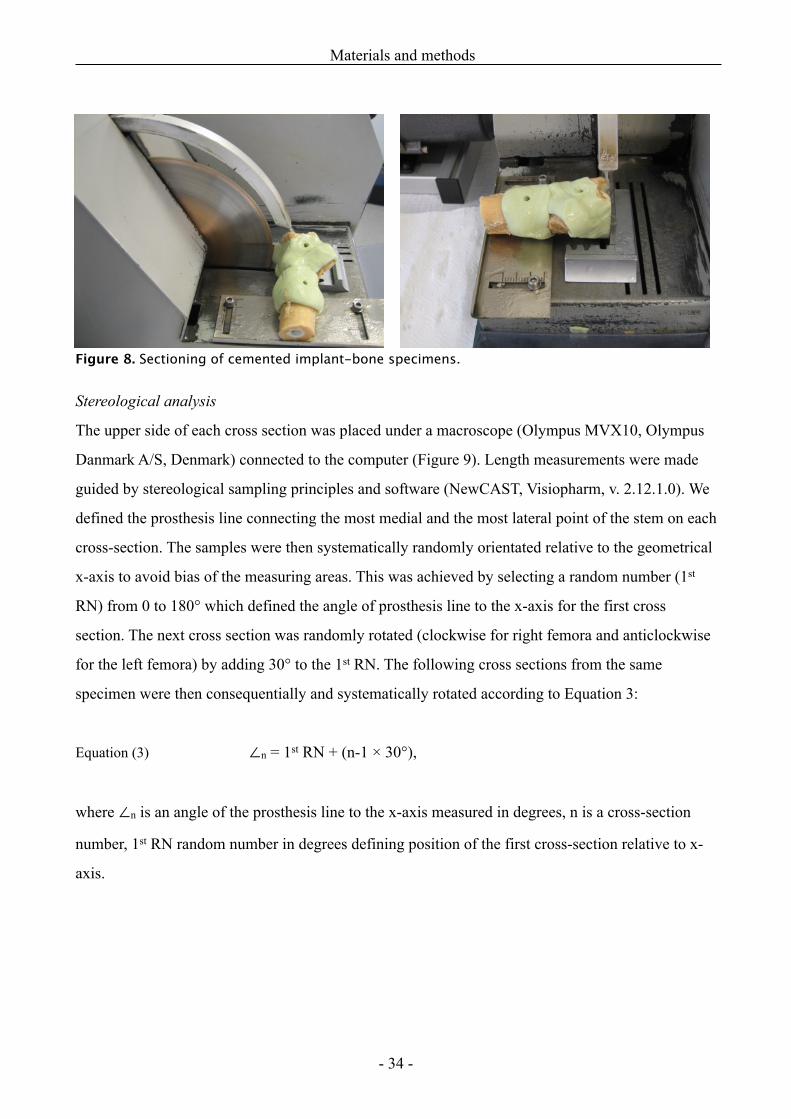
Figure 8. Sectioning of cemented implant-bone specimens.
Stereological analysis
The upper side of each cross section was placed under a macroscope (Olympus MVX10, Olympus
Danmark A/S, Denmark) connected to the computer (Figure 9). Length measurements were made
guided by stereological sampling principles and software (NewCAST, Visiopharm, v. 2.12.1.0). We
defined the prosthesis line connecting the most medial and the most lateral point of the stem on each
cross-section. The samples were then systematically randomly orientated relative to the geometrical
x-axis to avoid bias of the measuring areas. This was achieved by selecting a random number (1st
RN) from 0 to 180° which defined the angle of prosthesis line to the x-axis for the first cross
section. The next cross section was randomly rotated (clockwise for right femora and anticlockwise
for the left femora) by adding 30° to the 1st RN. The following cross sections from the same
specimen were then consequentially and systematically rotated according to Equation 3:
Equation (3) ∠n = 1st RN + (n-1 × 30°),
where ∠n is an angle of the prosthesis line to the x-axis measured in degrees, n is a cross-section
number, 1st RN random number in degrees defining position of the first cross-section relative to x-
axis.
Materials and methods
- 34 -
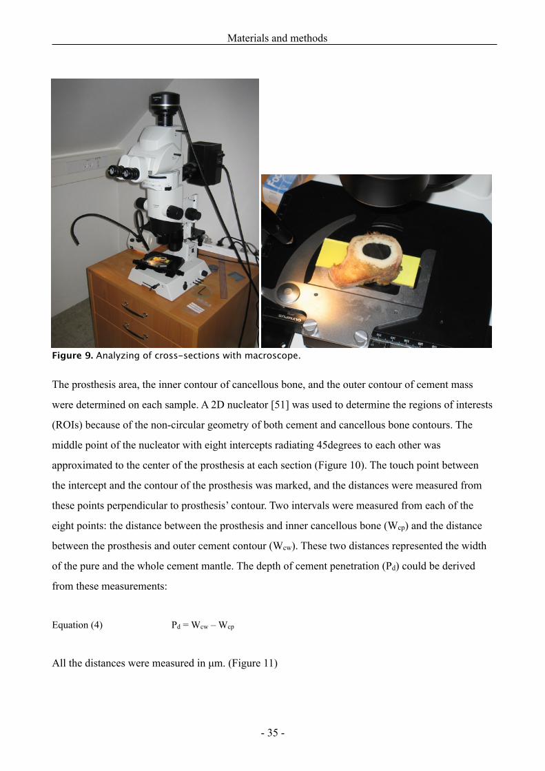
Figure 9. Analyzing of cross-sections with macroscope.
The prosthesis area, the inner contour of cancellous bone, and the outer contour of cement mass
were determined on each sample. A 2D nucleator [51] was used to determine the regions of interests
(ROIs) because of the non-circular geometry of both cement and cancellous bone contours. The
middle point of the nucleator with eight intercepts radiating 45degrees to each other was
approximated to the center of the prosthesis at each section (Figure 10). The touch point between
the intercept and the contour of the prosthesis was marked, and the distances were measured from
these points perpendicular to prosthesis’ contour. Two intervals were measured from each of the
eight points: the distance between the prosthesis and inner cancellous bone (Wcp) and the distance
between the prosthesis and outer cement contour (Wcw). These two distances represented the width
of the pure and the whole cement mantle. The depth of cement penetration (Pd) could be derived
from these measurements:
Equation (4) Pd = Wcw – Wcp
All the distances were measured in µm. (Figure 11)
Materials and methods
- 35 -
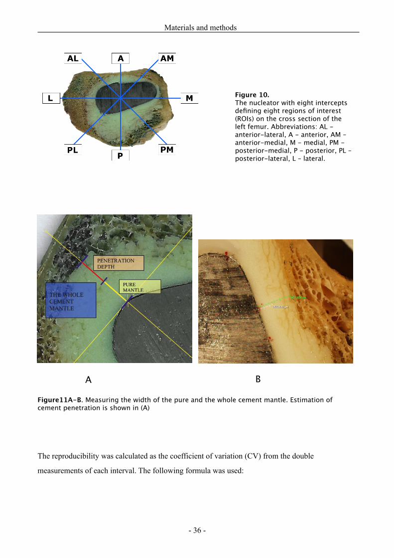
Figure11A-B. Measuring the width of the pure and the whole cement mantle. Estimation of cement penetration is shown in (A)
The reproducibility was calculated as the coefficient of variation (CV) from the double
measurements of each interval. The following formula was used:
Materials and methods
- 36 -
Figure 10. The nucleator with eight intercepts defining eight regions of interest (ROIs) on the cross section of the left femur. Abbreviations: AL - anterior-lateral, A - anterior, AM – anterior-medial, M - medial, PM - posterior-medial, P - posterior, PL – posterior-lateral, L – lateral.
A B
Equation (5) CV % =
where n is the number of measurements, d is the difference between two paired measurements (x1 and x2),
and their means ( and ). The coefficient of variation for Wcp and Wcw was correspondingly
0.93% and 0.54% .
Measuring centralization of femoral component
The centralization of the stem (∆Cp) was defined (in µm) by calculating the difference between the
largest (Maxcp) and the smallest (Mincp) distance of the pure cement mantle:
Equation (6) ∆Cp = Maxcp - Mincp, µm
If the thickness of the pure cement mantle is the same all the way around the prosthesis, the
difference (∆Cp) is 0, indicating a perfect centralization; in contrast a large difference will
express poor centralization. The mean value (from 9 cross sections per specimen) of the pure
cement mantle at each ROI was calculated according to the stereological method described above.
Two regions with the thickest and the thinnest pure mantle, were identified, and the difference
between these two regions was derived.
Prospective randomized trial (Studies III and IV)
Patients and follow-up
In the period from May 2006 to April 2008, 118 patients had a hybrid THA at arthroplasty unit,
Farsoe hospital (North Denmark Region). Of these, 88 who met the inclusion criteria (Table 4)
accepted participation.
Materials and methods
- 37 -
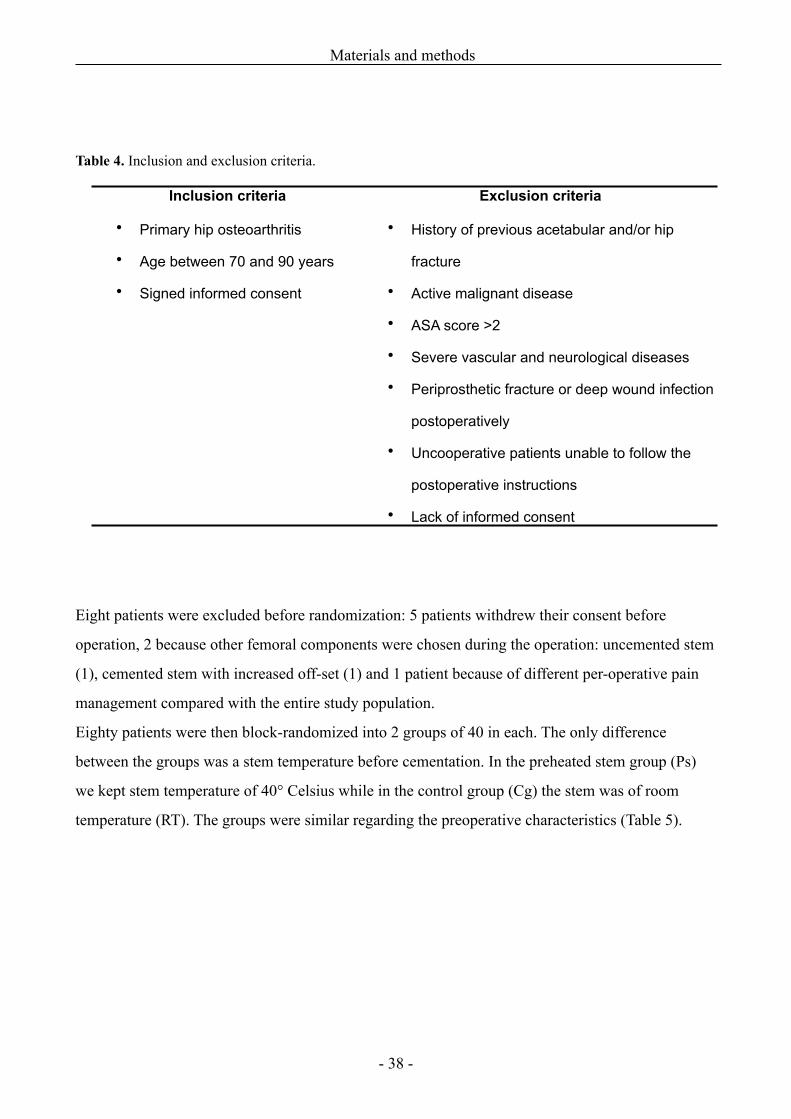
Table 4. Inclusion and exclusion criteria.
Inclusion criteria Exclusion criteria
• Primary hip osteoarthritis
• Age between 70 and 90 years
• Signed informed consent
• History of previous acetabular and/or hip
fracture
• Active malignant disease
• ASA score >2
• Severe vascular and neurological diseases
• Periprosthetic fracture or deep wound infection
postoperatively
• Uncooperative patients unable to follow the
postoperative instructions
• Lack of informed consent
Eight patients were excluded before randomization: 5 patients withdrew their consent before
operation, 2 because other femoral components were chosen during the operation: uncemented stem
(1), cemented stem with increased off-set (1) and 1 patient because of different per-operative pain
management compared with the entire study population.
Eighty patients were then block-randomized into 2 groups of 40 in each. The only difference
between the groups was a stem temperature before cementation. In the preheated stem group (Ps)
we kept stem temperature of 40° Celsius while in the control group (Cg) the stem was of room
temperature (RT). The groups were similar regarding the preoperative characteristics (Table 5).
Materials and methods
- 38 -
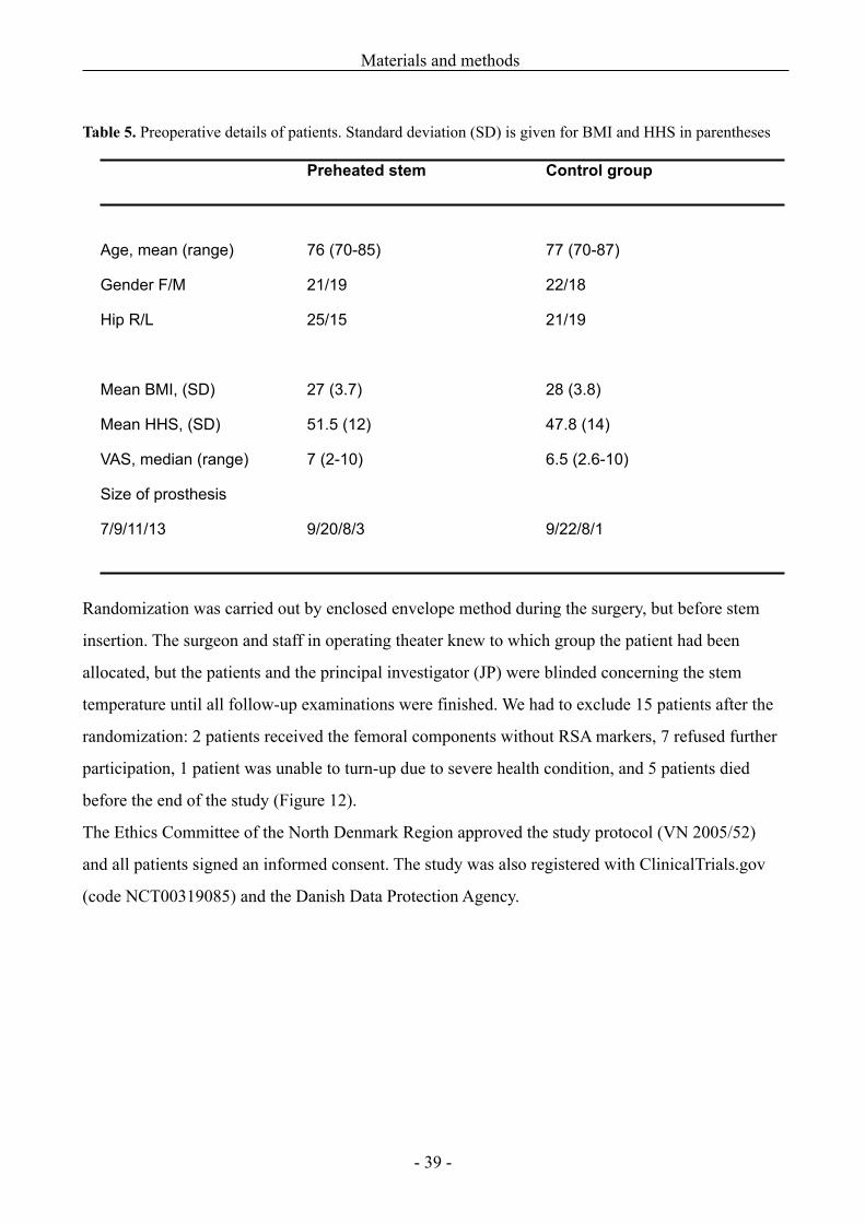
Table 5. Preoperative details of patients. Standard deviation (SD) is given for BMI and HHS in parentheses
Preheated stem Control group
Age, mean (range)
Gender F/M
Hip R/L
Mean BMI, (SD)
Mean HHS, (SD)
VAS, median (range)
Size of prosthesis
7/9/11/13
76 (70-85)
21/19
25/15
27 (3.7)
51.5 (12)
7 (2-10)
9/20/8/3
77 (70-87)
22/18
21/19
28 (3.8)
47.8 (14)
6.5 (2.6-10)
9/22/8/1
Randomization was carried out by enclosed envelope method during the surgery, but before stem
insertion. The surgeon and staff in operating theater knew to which group the patient had been
allocated, but the patients and the principal investigator (JP) were blinded concerning the stem
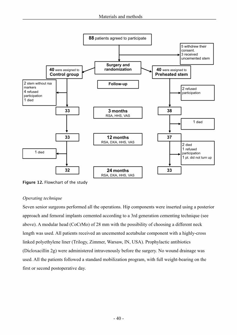
temperature until all follow-up examinations were finished. We had to exclude 15 patients after the
randomization: 2 patients received the femoral components without RSA markers, 7 refused further
participation, 1 patient was unable to turn-up due to severe health condition, and 5 patients died
before the end of the study (Figure 12).
The Ethics Committee of the North Denmark Region approved the study protocol (VN 2005/52)
and all patients signed an informed consent. The study was also registered with ClinicalTrials.gov
(code NCT00319085) and the Danish Data Protection Agency.
Materials and methods
- 39 -
2 refused participation
88 patients agreed to participate
40 were assigned to Control group
40 were assigned to Preheated stem
3 monthsRSA, HHS, VAS
33 38
37
33
33
32
2 stem without rsa markers4 refused participation1 died
Surgery and randomization
1 died
1 died
2 died1 refused participation1 pt. did not turn up
5 withdrew their consent.3 received uncemented stem
12 monthsRSA, DXA, HHS, VAS
24 monthsRSA, DXA, HHS, VAS
Follow-up
Figure 12. Flowchart of the study
Operating technique
Seven senior surgeons performed all the operations. Hip components were inserted using a posterior
approach and femoral implants cemented according to a 3rd generation cementing technique (see
above). A modular head (CoCrMo) of 28 mm with the possibility of choosing a different neck
length was used. All patients received an uncemented acetabular component with a highly-cross
linked polyethylene liner (Trilogy, Zimmer, Warsaw, IN, USA). Prophylactic antibiotics
(Dicloxacillin 2g) were administered intravenously before the surgery. No wound drainage was
used. All the patients followed a standard mobilization program, with full weight-bearing on the
first or second postoperative day.
Materials and methods
- 40 -
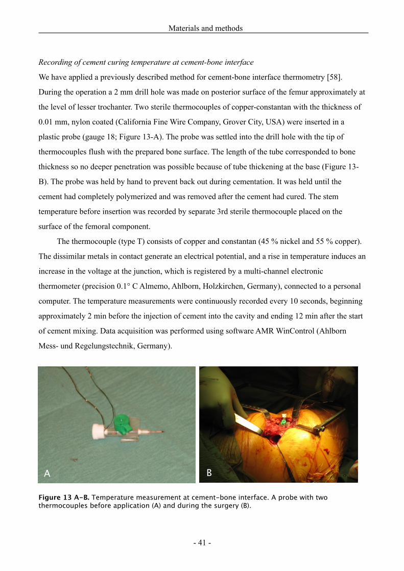
Recording of cement curing temperature at cement-bone interface
We have applied a previously described method for cement-bone interface thermometry [58].
During the operation a 2 mm drill hole was made on posterior surface of the femur approximately at
the level of lesser trochanter. Two sterile thermocouples of copper-constantan with the thickness of
0.01 mm, nylon coated (California Fine Wire Company, Grover City, USA) were inserted in a
plastic probe (gauge 18; Figure 13-A). The probe was settled into the drill hole with the tip of
thermocouples flush with the prepared bone surface. The length of the tube corresponded to bone
thickness so no deeper penetration was possible because of tube thickening at the base (Figure 13-
B). The probe was held by hand to prevent back out during cementation. It was held until the
cement had completely polymerized and was removed after the cement had cured. The stem
temperature before insertion was recorded by separate 3rd sterile thermocouple placed on the
surface of the femoral component.
The thermocouple (type T) consists of copper and constantan (45 % nickel and 55 % copper).
The dissimilar metals in contact generate an electrical potential, and a rise in temperature induces an
increase in the voltage at the junction, which is registered by a multi-channel electronic
thermometer (precision 0.1° C Almemo, Ahlborn, Holzkirchen, Germany), connected to a personal
computer. The temperature measurements were continuously recorded every 10 seconds, beginning
approximately 2 min before the injection of cement into the cavity and ending 12 min after the start
of cement mixing. Data acquisition was performed using software AMR WinControl (Ahlborn
Mess- und Regelungstechnik, Germany).
Figure 13 A-B. Temperature measurement at cement-bone interface. A probe with two thermocouples before application (A) and during the surgery (B).
Materials and methods
- 41 -
A B
Preheating of femoral components
Implants selected for preheating were stored for minimum 24 hours at 45° C in heating oven (with
the manufacturer’s permission). Because of rapid cool-down in laminar air flow, the sterile package
was unsealed just prior to cementation. The heated component was covered with warm sterile
dressings on assisting nurse table to inhibit heat dissipation. Stem temperature before insertion was
monitored by a sterile thermocouple. Femoral components to non-preheated group, cement,
acetabular shells, centralizers and intramedullary plugs were stored at room temperature (21° C).
Bone cement
Vacuum mixing and cement insertion was done with Cemvac cement delivery system (DePuy,
DePuy International Ltd, Engalnd).
All femoral components were cemented with 80 g. Refobacin® bone cement R with
gentamycin (Biomet UK Ltd, UK). This cement is known as high-viscosity cement and is
equivalent to Refobacin Palacos R bone cement regarding chemical composition, molecular weight,
particle size distribution and mechanical properties. Some differences in handling and viscosity
characteristics, however have been noticed at in-vitro tests, but the clinical significance of this is
unknown [23, 83]. Cement setting time is approximately 8 to10 min when mixing non-prechilled
cement at RT of 20° to 23° C. The shrinkage is about 2.67%.
Radiostereometric analysis (RSA)
The RSA procedure was performed as described previously [78, 134]. Prostheses were equipped
with three pegs (each one containing a 0.8 mm tantalum bead) located on the stem shoulder,
proximal-medial and tip of the prosthesis to enable RSA. We inserted tantalum balls both into the
bone and cement, which allowed evaluation of prosthesis-cement and prosthesis-bone micromotions
as well as cement-mantle migration relative to bone (CBI). Twenty cement markers of 1 mm were
poured in powder under cement mixing. Seven to nine tantalum balls of 0.8 mm were inserted with
RSA gun into the proximal femur.
The first RSA examination was performed at the second or third postoperative day and later at
3, 12 and 24 months postoperatively (Figure 14). All examinations were obtained with patient in
Materials and methods
- 42 -
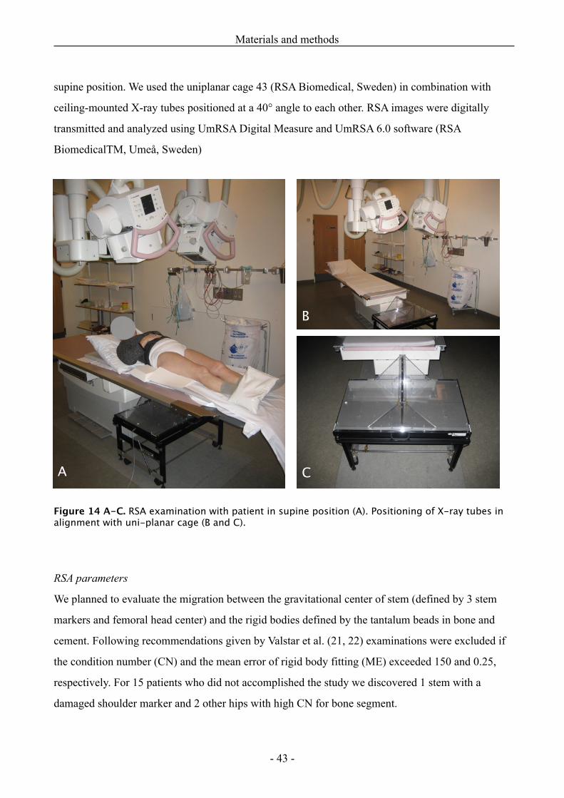
supine position. We used the uniplanar cage 43 (RSA Biomedical, Sweden) in combination with
ceiling-mounted X-ray tubes positioned at a 40° angle to each other. RSA images were digitally
transmitted and analyzed using UmRSA Digital Measure and UmRSA 6.0 software (RSA
BiomedicalTM, Umeå, Sweden)
Figure 14 A-C. RSA examination with patient in supine position (A). Positioning of X-ray tubes in alignment with uni-planar cage (B and C).
RSA parameters
We planned to evaluate the migration between the gravitational center of stem (defined by 3 stem
markers and femoral head center) and the rigid bodies defined by the tantalum beads in bone and
cement. Following recommendations given by Valstar et al. (21, 22) examinations were excluded if
the condition number (CN) and the mean error of rigid body fitting (ME) exceeded 150 and 0.25,
respectively. For 15 patients who did not accomplished the study we discovered 1 stem with a
damaged shoulder marker and 2 other hips with high CN for bone segment.
Materials and methods
- 43 -
A
B
C
In 65 patients who completed all RSA examinations we found 17 femoral stems (13 Control, 4
Preheated) with damaged or unstable stem markers and 5 hips (3 Control, 2 Preheated) with high
CN for bone (1 of those had also unstable stem marker). Valuable information of migration patterns
would be lost by excluding these 16 hips because the majority of unstable stem markers (12 hips)
became loose before 1 or 2 year’s follow-up. Therefore we evaluated the migration of femoral head
center (59 hips) and the tip of the prosthesis (60 hips) in relation to the bone and cement markers
(pb and pc). We measured the translations according to x-, y- and z-axes, which gives 6 degrees of
freedom. The maximum total point motion (MTPM) for both femoral head and femoral tip was also
calculated. Regarding the cement mantle migration, we evaluated only proximal-distal translation
(y-axis) between the rigid body of cement mantle and the rigid body of the bone.
The precision of RSA was determined by 15 double-examinations performed at the first
postoperative RSA examination. Complete repositioning of calibration cage, X-ray tubes and patient
were done between each exposure. Femoral component (center of femoral head, prosthesis tip) and
cement mantle displacements were calculated for these double examinations and the standard
deviations (SD) of the displacements were estimated. Migration exceeding 2.77SD of difference
was considered to be significant (P< 0.01). One hip had a high CN for bone (3774) due to poor
scattering of Ta balls, thus the precision values for pc-motions were derived from 15 examinations
whereas pb and CBI-motions were calculated in14 cases (Table 6).
Materials and methods
- 44 -
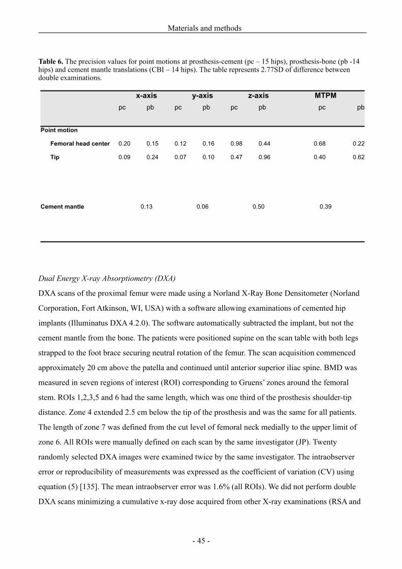
Table 6. The precision values for point motions at prosthesis-cement (pc – 15 hips), prosthesis-bone (pb -14 hips) and cement mantle translations (CBI – 14 hips). The table represents 2.77SD of difference between double examinations.
x-axisx-axis y-axisy-axis z-axisz-axis MTPMMTPMpc pb pc pb pc pb pc pb
Point motion
Femoral head center
Tip
0.20
0.09
0.15
0.24
0.12
0.07
0.16
0.10
0.98
0.47
0.44
0.96
0.68
0.40
0.22
0.62
Cement mantle 0.130.13 0.060.06 0.500.50 0.390.39
Dual Energy X-ray Absorptiometry (DXA)
DXA scans of the proximal femur were made using a Norland X-Ray Bone Densitometer (Norland
Corporation, Fort Atkinson, WI, USA) with a software allowing examinations of cemented hip
implants (Illuminatus DXA 4.2.0). The software automatically subtracted the implant, but not the
cement mantle from the bone. The patients were positioned supine on the scan table with both legs
strapped to the foot brace securing neutral rotation of the femur. The scan acquisition commenced
approximately 20 cm above the patella and continued until anterior superior iliac spine. BMD was
measured in seven regions of interest (ROI) corresponding to Gruens’ zones around the femoral
stem. ROIs 1,2,3,5 and 6 had the same length, which was one third of the prosthesis shoulder-tip
distance. Zone 4 extended 2.5 cm below the tip of the prosthesis and was the same for all patients.
The length of zone 7 was defined from the cut level of femoral neck medially to the upper limit of
zone 6. All ROIs were manually defined on each scan by the same investigator (JP). Twenty
randomly selected DXA images were examined twice by the same investigator. The intraobserver
error or reproducibility of measurements was expressed as the coefficient of variation (CV) using
equation (5) [135]. The mean intraobserver error was 1.6% (all ROIs). We did not perform double
DXA scans minimizing a cumulative x-ray dose acquired from other X-ray examinations (RSA and
Materials and methods
- 45 -
postoperative x-ray). Scanning was performed on the second or third postoperative day and again at
12 and 24 months postoperatively.
BMD (g/cm2) of each ROI was calculated at reference scan and the relative changes during the
follow-up period were expressed as the percentage difference between the consecutive
examinations.
Clinical examination
All patients were clinically examined preoperatively and after 3, 12 and 24 months. The clinical
results were assessed according to the Harris Hip Score (HHS) (24) and visual analog scale (VAS)
score.
Materials and methods
- 46 -
Statistics
Considerations regarding Kappa method (Study I)
Intra- and interobserver agreement was calculated by using the kappa statistic to assess the
reliability of both radiographic evaluation methods. Kappa values between the pairs and among all
three raters were presented. The Barrack’s scores were analyzed using the weighted kappa, whereas
a simple kappa statistics was used for the new radiographic evaluation method as there were only
two categories.
Equation (7) κ = Pobs – Pch/1 – Pch
where κ is kappa value, Pobs – proportion of observed agreement, Pch – proportion of agreement
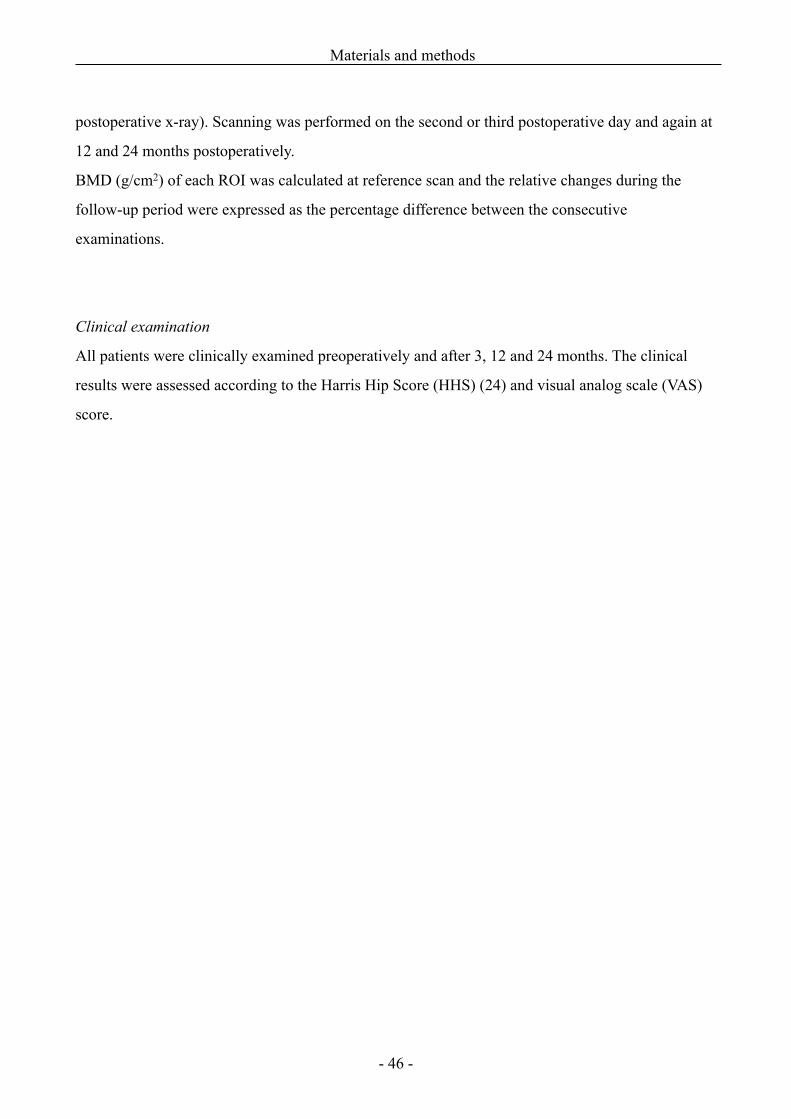
expected by chance. If there is complete agreement, then Pobs = 1 and thus κ = 1. The commonly
accepted level of kappa is >0.6 representing good reliability and reproducibility of classification or
grading system (Table 7).
Table 7. Interpretation of Kappa [89]
Kappa Agreement
<00.01-0.200.21-0.400.41-0.600.61-0.800.81-0.99
1.0
Less than chance agreementSlight agreementFair agreementModerate agreementSubstantial agreementAlmost perfect agreementPerfect agreement
A weighted kappa is a more reliable method to evaluate agreement between observers when the
analyzed issue has more than two different categories. The weighted kappa assigns less weight to
agreement as categories are further apart. In this study using Barrack’s classification we had five
possible categories (A, B, C1, C2, and D).
Statistics
- 47 -
Equation (8) κw = Σ (Pobs · w) – Σ (Pch · w) / 1 – Σ (Pch · w)
where κw is the weighted kappa value, Pobs – proportion of observed agreement, Pch – proportion of
agreement expected by chance, w – the weight which was calculated according to the number of
categories.
The kappa statistical method we used is the most commonly reported method to measure the
level of agreement [17, 57, 84, 108]. The advantage of this method is that it provides more
information than a simple calculation of the raw proportion of agreement; it takes into account the
disagreement between observations and allows calculation of the degree of chance agreement [81].
The disadvantage of the kappa statistic is that it is affected by the prevalence of the findings.
Disagreement on one category which has a low prevalence would result in low kappa in spite of
good agreement on other categories. That means that for rare categories, very low kappa values may
not necessarily reflect low rates of overall agreement [147]. An adjusted kappa statistics, which is
refinement of standard kappa method, allows analysis of categories of unequal sizes. This method,
however, was not applied in this study. We included a large number of radiographs after HHA for
both evaluation methods, as we expected to find a higher prevalence of low grades of cement
mantle making kappa statistic more reliable. The total number of observations has little influence on
kappa value which is rather affected of proportions of different categories. Therefore we did not
need to include the same large number of radiographs for authors’ proposed method as with the
Barrack’s method because we only had two categories there. We expected to find a balanced
number of both categories where conclusive statistical analysis could be done on 25 HHA
radiographs.
Non-parametric data analysis (Mann-Whitney rank sum test) for radiographic features, such as
presence of thin cement mantle and radiolucency, whereas the Pearson chi2 test was used to test the
distribution of air voids and stem alignment.
Study II
The differences of pressure, cement penetration, and cement mantle measurements between the
pairs are presented as mean ± SD and were analyzed using a multivariate analysis of
Statistics
- 48 -
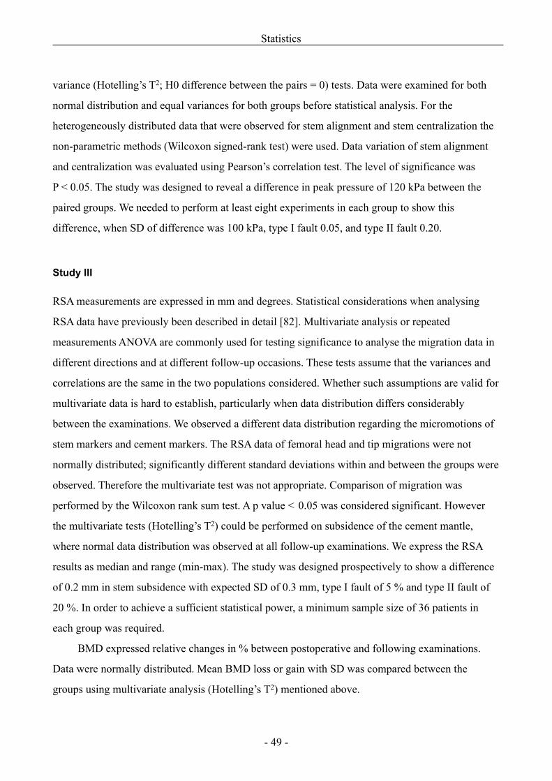
variance (Hotelling’s T2; H0 difference between the pairs = 0) tests. Data were examined for both
normal distribution and equal variances for both groups before statistical analysis. For the
heterogeneously distributed data that were observed for stem alignment and stem centralization the
non-parametric methods (Wilcoxon signed-rank test) were used. Data variation of stem alignment
and centralization was evaluated using Pearson’s correlation test. The level of significance was
P < 0.05. The study was designed to reveal a difference in peak pressure of 120 kPa between the
paired groups. We needed to perform at least eight experiments in each group to show this
difference, when SD of difference was 100 kPa, type I fault 0.05, and type II fault 0.20.
Study III
RSA measurements are expressed in mm and degrees. Statistical considerations when analysing
RSA data have previously been described in detail [82]. Multivariate analysis or repeated
measurements ANOVA are commonly used for testing significance to analyse the migration data in
different directions and at different follow-up occasions. These tests assume that the variances and
correlations are the same in the two populations considered. Whether such assumptions are valid for
multivariate data is hard to establish, particularly when data distribution differs considerably
between the examinations. We observed a different data distribution regarding the micromotions of
stem markers and cement markers. The RSA data of femoral head and tip migrations were not
normally distributed; significantly different standard deviations within and between the groups were
observed. Therefore the multivariate test was not appropriate. Comparison of migration was
performed by the Wilcoxon rank sum test. A p value < 0.05 was considered significant. However
the multivariate tests (Hotelling’s T2) could be performed on subsidence of the cement mantle,
where normal data distribution was observed at all follow-up examinations. We express the RSA
results as median and range (min-max). The study was designed prospectively to show a difference
of 0.2 mm in stem subsidence with expected SD of 0.3 mm, type I fault of 5 % and type II fault of
20 %. In order to achieve a sufficient statistical power, a minimum sample size of 36 patients in
each group was required.
BMD expressed relative changes in % between postoperative and following examinations.
Data were normally distributed. Mean BMD loss or gain with SD was compared between the
groups using multivariate analysis (Hotelling’s T2) mentioned above.
Statistics
- 49 -

Clinical results for HHS and VAS are presented as mean ±SD and median with range,
respectively. The differences between the groups and different follow-up intervals within the groups
were analyzed using multivariate analysis on repeated measurements (Hotelling’s T2).
Study IV
The following parameters from each temperature curve were registered:
1) the CBI temperature:
a. Tmix, at cement mixing, representing bone preparation,
b. Tstem, at stem insertion,
c. Tpeak, the peak temperature,
d. Tpoly, at the end of cement polymerization (1 min. after the peak temperature has
been reached).
2) the time intervals in seconds:
a. start of cement mixing as baseline
b. timepoint for peak temperature
c. polymerization time (the duration from cement mixing until 1 min following the
time of peak temperature)
d. exposure with temperature above 50° C.
3) The area under the temperature curve (AUC), from cement mixing to the end of cement
polymerization (1 min. after the peak temperature has been reached).
The temperature measurements are presented as means ± SD in degrees of Celsius. The differences
between means analyzed with (two-tailed) Student’s t-test as data were normally distributed. Time
intervals represented as medians with range and were analyzed using Wilcoxon rank sum test.
P values below <0.05 were considered significant.
The Intercooled STATA 9.0 (Stata Corporation, TX, USA) and SigmaStat 2.0 (Jandel
Corporation) statistical software were used for analyses.
Statistics
- 50 -
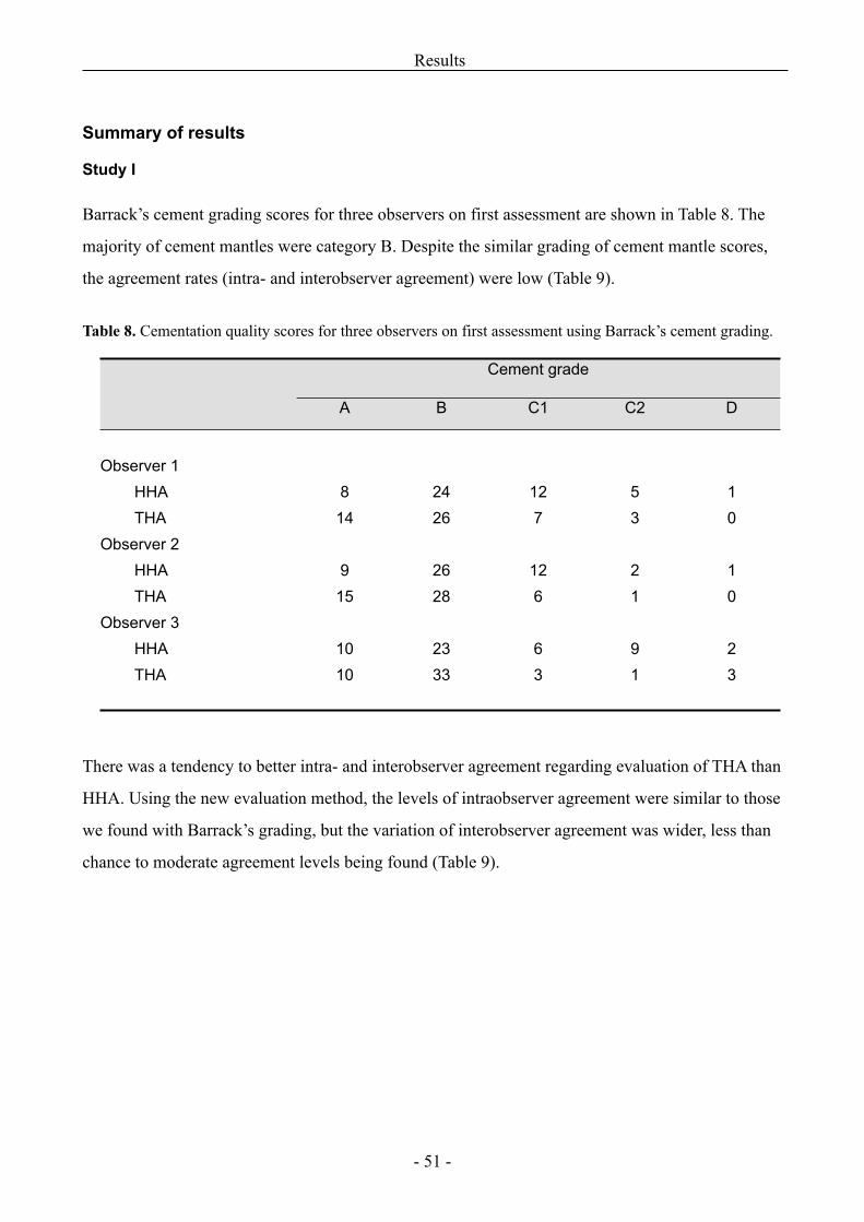
Summary of results
Study I
Barrack’s cement grading scores for three observers on first assessment are shown in Table 8. The
majority of cement mantles were category B. Despite the similar grading of cement mantle scores,
the agreement rates (intra- and interobserver agreement) were low (Table 9).
Table 8. Cementation quality scores for three observers on first assessment using Barrack’s cement grading.
Cement gradeCement gradeCement gradeCement gradeCement grade
A B C1 C2 D
Observer 1HHATHA
Observer 2HHATHA
Observer 3HHATHA
814
915
1010
2426
2628
2333
127
126
63
53
21
91
10
10
23
There was a tendency to better intra- and interobserver agreement regarding evaluation of THA than
HHA. Using the new evaluation method, the levels of intraobserver agreement were similar to those
we found with Barrack’s grading, but the variation of interobserver agreement was wider, less than
chance to moderate agreement levels being found (Table 9).
Results
- 51 -
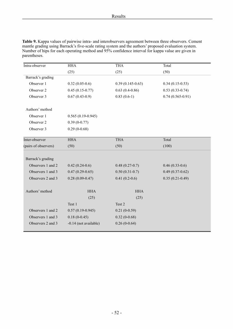
Table 9. Kappa values of pairwise intra- and interobservers agreement between three observers. Cement mantle grading using Barrack’s five-scale rating system and the authors’ proposed evaluation system. Number of hips for each operating method and 95% confidence interval for kappa value are given in parentheses.
Intra-observer HHA(25)
THA(25)
Total(50)
Barrack’s gradingObserver 1
Observer 2 Observer 3
Authors’ method
Observer 1 Observer 2
Observer 3
0.32 (0.05-0.6)
0.45 (0.15-0.77)0.67 (0.43-0.9)
0.565 (0.19-0.945)0.39 (0-0.77)
0.29 (0-0.68)
0.39 (0.145-0.63)
0.63 (0.4-0.86)0.83 (0.6-1)
0.34 (0.15-0.53)
0.53 (0.33-0.74)0.74 (0.565-0.91)
Inter-observer (pairs of observers)
HHA(50)
THA(50)
Total(100)
Barrack’s grading
Observers 1 and 2Observers 1 and 3
Observers 2 and 3
Authors’ method
Observers 1 and 2
Observers 1 and 3Observers 2 and 3
0.42 (0.24-0.6)0.47 (0.29-0.65)
0.28 (0.09-0.47)
HHA(25)
Test 10.57 (0.19-0.945)
0.18 (0-0.45)-0.14 (not available)
0.48 (0.27-0.7)0.50 (0.31-0.7)
0.41 (0.2-0.6)
HHA(25)
Test 20.21 (0-0.59)
0.32 (0-0.68)0.26 (0-0.64)
0.46 (0.33-0.6)0.49 (0.37-0.62)
0.35 (0.21-0.49)
Results
- 52 -
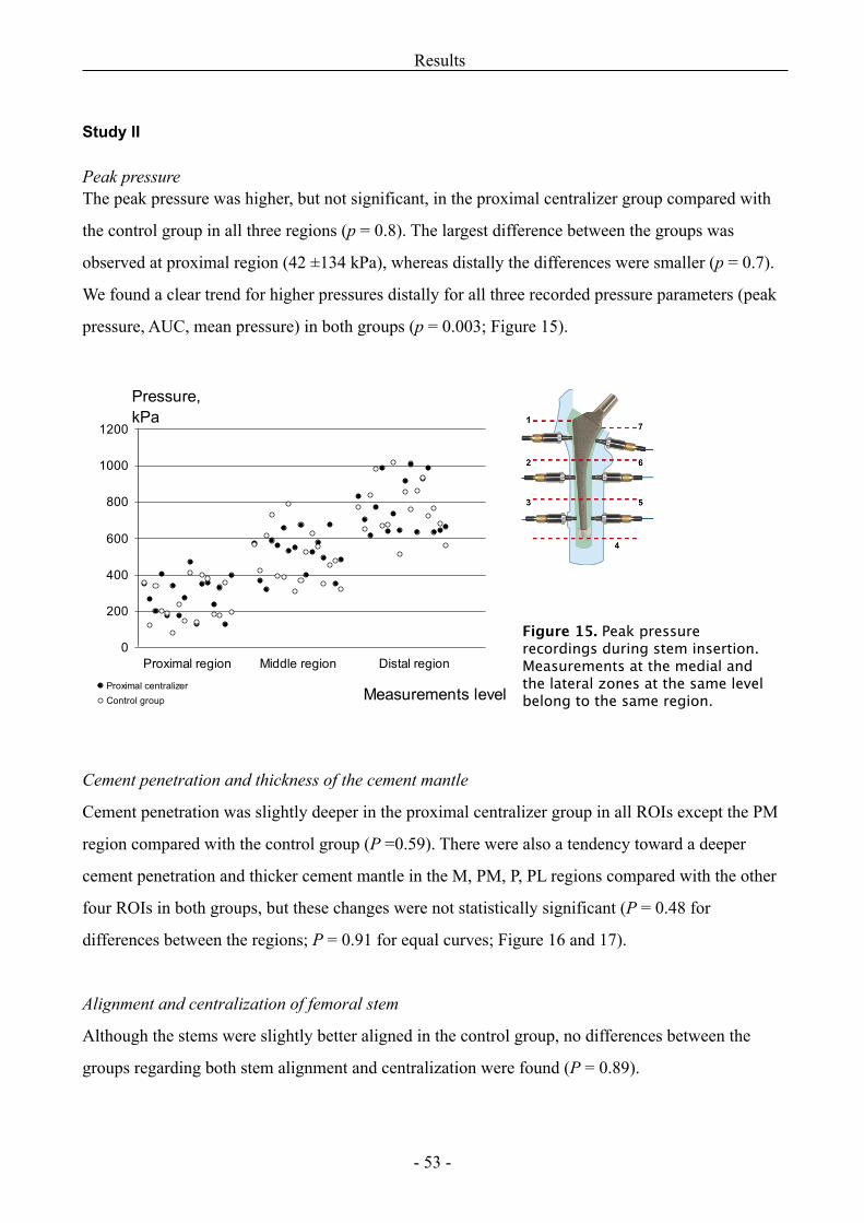
Study II
Peak pressureThe peak pressure was higher, but not significant, in the proximal centralizer group compared with
the control group in all three regions (p = 0.8). The largest difference between the groups was
observed at proximal region (42 ±134 kPa), whereas distally the differences were smaller (p = 0.7).
We found a clear trend for higher pressures distally for all three recorded pressure parameters (peak
pressure, AUC, mean pressure) in both groups (p = 0.003; Figure 15).
Cement penetration and thickness of the cement mantle
Cement penetration was slightly deeper in the proximal centralizer group in all ROIs except the PM
region compared with the control group (P =0.59). There were also a tendency toward a deeper
cement penetration and thicker cement mantle in the M, PM, P, PL regions compared with the other
four ROIs in both groups, but these changes were not statistically significant (P = 0.48 for
differences between the regions; P = 0.91 for equal curves; Figure 16 and 17).
Alignment and centralization of femoral stem
Although the stems were slightly better aligned in the control group, no differences between the
groups regarding both stem alignment and centralization were found (P = 0.89).
Results
- 53 -
Figure 15. Peak pressure recordings during stem insertion. Measurements at the medial and the lateral zones at the same level belong to the same region.
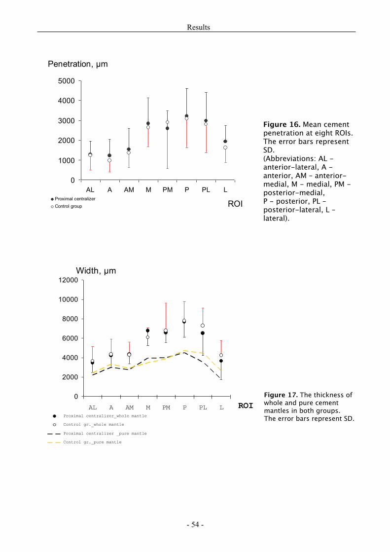
Results
- 54 -
Figure 17. The thickness of whole and pure cement mantles in both groups. The error bars represent SD.
Figure 16. Mean cement penetration at eight ROIs. The error bars represent SD. (Abbreviations: AL - anterior-lateral, A - anterior, AM – anterior-medial, M - medial, PM - posterior-medial, P - posterior, PL – posterior-lateral, L – lateral).
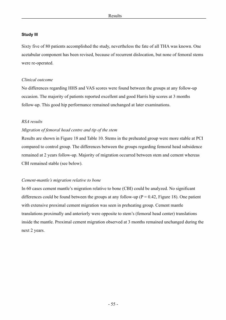
Study III
Sixty five of 80 patients accomplished the study, nevertheless the fate of all THA was known. One
acetabular component has been revised, because of recurrent dislocation, but none of femoral stems
were re-operated.
Clinical outcome
No differences regarding HHS and VAS scores were found between the groups at any follow-up
occasion. The majority of patients reported excellent and good Harris hip scores at 3 months
follow-up. This good hip performance remained unchanged at later examinations.
RSA results
Migration of femoral head centre and tip of the stem
Results are shown in Figure 18 and Table 10. Stems in the preheated group were more stable at PCI
compared to control group. The differences between the groups regarding femoral head subsidence
remained at 2 years follow-up. Majority of migration occurred between stem and cement whereas
CBI remained stable (see below).
Cement-mantle’s migration relative to bone
In 60 cases cement mantle’s migration relative to bone (CBI) could be analyzed. No significant
differences could be found between the groups at any follow-up (P = 0.42, Figure 18). One patient
with extensive proximal cement migration was seen in preheating group. Cement mantle
translations proximally and anteriorly were opposite to stem’s (femoral head center) translations
inside the mantle. Proximal cement migration observed at 3 months remained unchanged during the
next 2 years.
Results
- 55 -
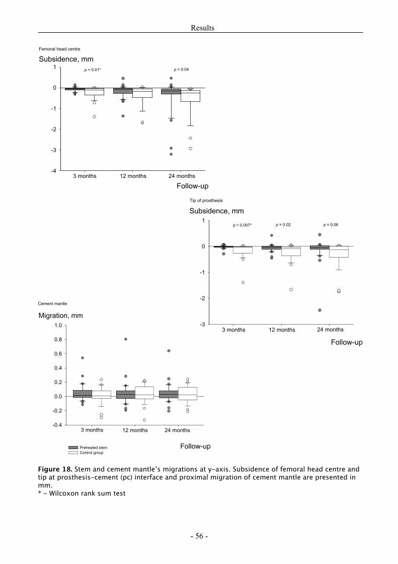
Figure 18. Stem and cement mantle’s migrations at y-axis. Subsidence of femoral head centre and tip at prosthesis-cement (pc) interface and proximal migration of cement mantle are presented in mm.* - Wilcoxon rank sum test
Results
- 56 -
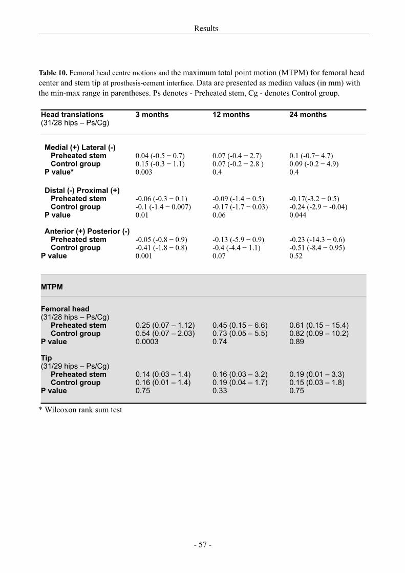
Table 10. Femoral head centre motions and the maximum total point motion (MTPM) for femoral head center and stem tip at prosthesis-cement interface. Data are presented as median values (in mm) with the min-max range in parentheses. Ps denotes - Preheated stem, Cg - denotes Control group.
Head translations (31/28 hips – Ps/Cg)
3 months 12 months 24 months
Medial (+) Lateral (-)Preheated stemControl group
P value*
0.04 (-0.5 − 0.7)0.15 (-0.3 − 1.1)0.003
0.07 (-0.4 − 2.7)0.07 (-0.2 − 2.8 )0.4
0.1 (-0.7− 4.7)0.09 (-0.2 − 4.9)0.4
Distal (-) Proximal (+)Preheated stemControl group
P value
-0.06 (-0.3 − 0.1)-0.1 (-1.4 − 0.007)0.01
-0.09 (-1.4 − 0.5)-0.17 (-1.7 − 0.03)0.06
-0.17(-3.2 − 0.5)-0.24 (-2.9 − -0.04)0.044
Anterior (+) Posterior (-)Preheated stemControl group
P value
-0.05 (-0.8 − 0.9)-0.41 (-1.8 − 0.8)0.001
-0.13 (-5.9 − 0.9)-0.4 (-4.4 − 1.1)0.07
-0.23 (-14.3 − 0.6)-0.51 (-8.4 − 0.95)0.52
MTPM
Femoral head(31/28 hips – Ps/Cg)
Preheated stem Control group
P value
0.25 (0.07 – 1.12)0.54 (0.07 – 2.03)0.0003
0.45 (0.15 – 6.6)0.73 (0.05 – 5.5)0.74
0.61 (0.15 – 15.4)0.82 (0.09 – 10.2)0.89
Tip(31/29 hips – Ps/Cg)
Preheated stemControl group
P value
0.14 (0.03 – 1.4)0.16 (0.01 – 1.4)0.75
0.16 (0.03 – 3.2)0.19 (0.04 – 1.7)0.33
0.19 (0.01 – 3.3)0.15 (0.03 – 1.8)0.75
* Wilcoxon rank sum test
Results
- 57 -
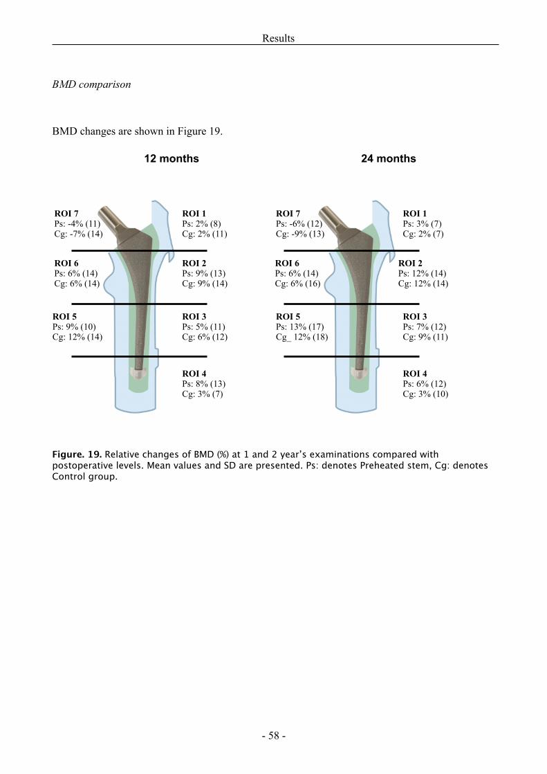
BMD comparison
BMD changes are shown in Figure 19.
12 months 24 months
Figure. 19. Relative changes of BMD (%) at 1 and 2 year’s examinations compared with postoperative levels. Mean values and SD are presented. Ps: denotes Preheated stem, Cg: denotes Control group.
Results
- 58 -
ROI 1Ps: 2% (8)Cg: 2% (11)
ROI 2Ps: 9% (13)Cg: 9% (14)
ROI 3Ps: 5% (11)Cg: 6% (12)
ROI 4Ps: 8% (13)Cg: 3% (7)
ROI 5Ps: 9% (10)Cg: 12% (14)
ROI 6Ps: 6% (14)Cg: 6% (14)
ROI 7Ps: -4% (11)Cg: -7% (14)
ROI 1Ps: 3% (7)Cg: 2% (7)
ROI 2Ps: 12% (14)Cg: 12% (14)
ROI 3Ps: 7% (12)Cg: 9% (11)
ROI 4Ps: 6% (12)Cg: 3% (10)
ROI 5Ps: 13% (17)Cg_ 12% (18)
ROI 6Ps: 6% (14)Cg: 6% (16)
ROI 7Ps: -6% (12)Cg: -9% (13)
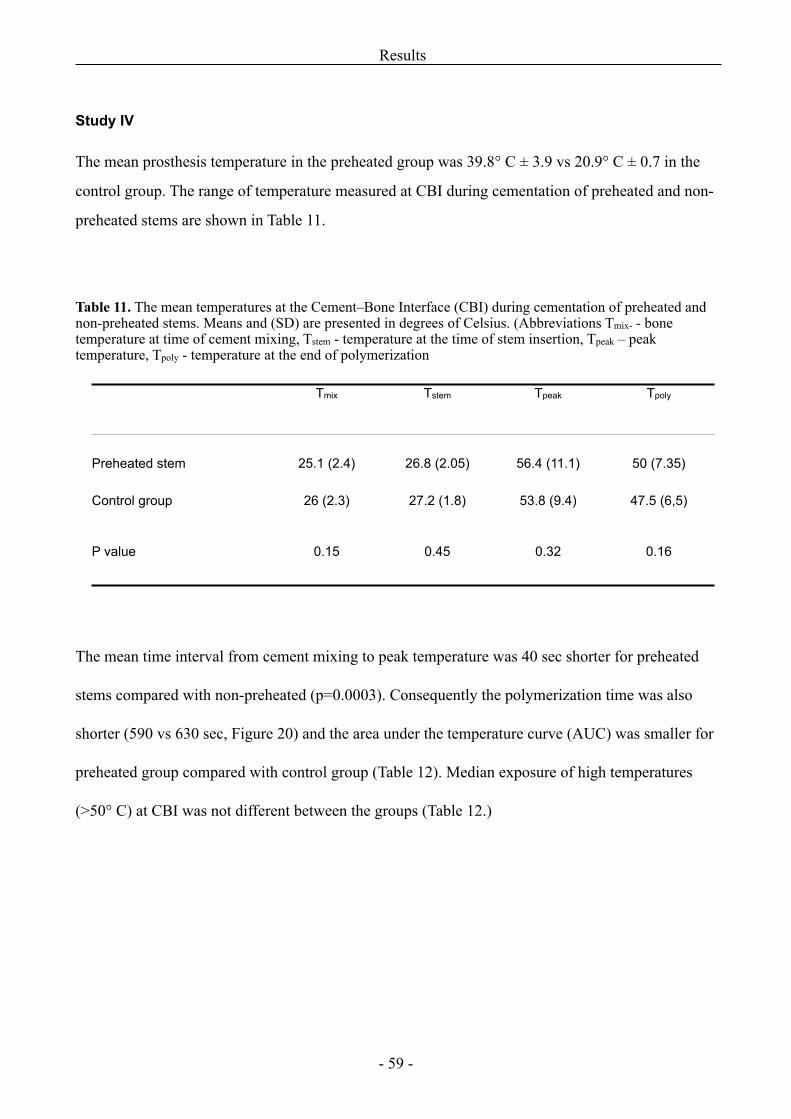
Study IV
The mean prosthesis temperature in the preheated group was 39.8° C ± 3.9 vs 20.9° C ± 0.7 in the
control group. The range of temperature measured at CBI during cementation of preheated and non-
preheated stems are shown in Table 11.
Table 11. The mean temperatures at the Cement–Bone Interface (CBI) during cementation of preheated and non-preheated stems. Means and (SD) are presented in degrees of Celsius. (Abbreviations Tmix- - bone temperature at time of cement mixing, Tstem - temperature at the time of stem insertion, Tpeak – peak temperature, Tpoly - temperature at the end of polymerization
Tmix Tstem Tpeak Tpoly
Preheated stem 25.1 (2.4) 26.8 (2.05) 56.4 (11.1) 50 (7.35)
Control group 26 (2.3) 27.2 (1.8) 53.8 (9.4) 47.5 (6,5)
P value 0.15 0.45 0.32 0.16
The mean time interval from cement mixing to peak temperature was 40 sec shorter for preheated
stems compared with non-preheated (p=0.0003). Consequently the polymerization time was also
shorter (590 vs 630 sec, Figure 20) and the area under the temperature curve (AUC) was smaller for
preheated group compared with control group (Table 12). Median exposure of high temperatures
(>50° C) at CBI was not different between the groups (Table 12.)
Results
- 59 -
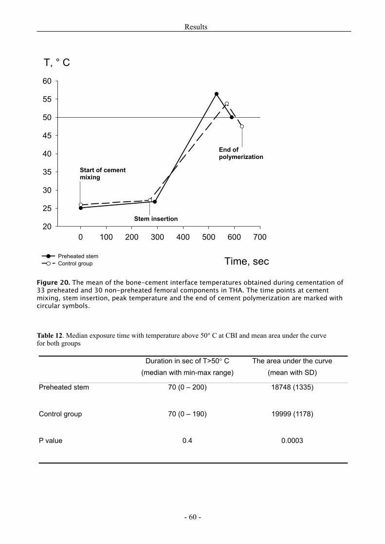
Figure 20. The mean of the bone-cement interface temperatures obtained during cementation of 33 preheated and 30 non-preheated femoral components in THA. The time points at cement mixing, stem insertion, peak temperature and the end of cement polymerization are marked with circular symbols.
Table 12. Median exposure time with temperature above 50° C at CBI and mean area under the curvefor both groups
Duration in sec of T>50° C (median with min-max range)
The area under the curve(mean with SD)
Preheated stem 70 (0 – 200) 18748 (1335)
Control group 70 (0 – 190) 19999 (1178)
P value 0.4 0.0003
Results
- 60 -
Discussion
Radiographic assessment of cementing quality (Study I)
Reliable methods of radiographic evaluation of cementing quality should be used for assessment of
the new prosthesis designs and improvements in cementing technique. Barrack’s cement grading
system was proposed to be such a method and our intention was to apply this classification in the
other studies (II and III) for evaluation of cementing quality after changes in prosthesis design and
cementing technique were introduced.
A thorough literature review revealed that the reliability of Barrack’s system has been
questioned previously [108]. McCaskie found a very large variation of intra- and interobserver
agreement levels (unweighted κ values 0.42 and -0.04, respectively) using this system. Especially, no
better agreement was achieved between more experienced surgeons (κ = 0.18) compared with
agreement between trainees (κ = 0.052) and consultants (κ = - 0.48), indicating that the system is
difficult to apply correctly. Similar outcome was observed by Kelly et al. (1996), who reported fair to
moderate intra- and interobserver agreement (weighted κ ranged from 0.38 to 0.53) between two
experienced surgeons. They did not recommend the use of this system due to poor reproducibility
[80]. Better interobserver agreement (adjusted κ values range: 0.56 to 0.73) was found by Harvey et
al. in 1998 [57] evaluating 100 hips on both AP and lateral radiographs by three observers. An
explanation for this discrepancy between the studies may be that different kappa statistical methods
were applied. But even using the best results, there will still be 20% to 43% of radiographs which
would be scored differently by different observers. The fact is that Barrack’s cement grades are not
clearly described, as complete definitions of all terms are also missing.
Our results were very similar to those of Kelly et al. (1996), confirming that Barrack’s cement
grading is associated with a large intra- and interobserver variability, as a consequence a new method
of radiographic evaluation was proposed by us. The idea was to make a simple scoring system with
radiological features which could be easily recognized and familiar to most orthopaedic surgeons. The
parameters we chose in our cementing score have been reported to have prognostic values on
longevity of cemented femoral stem, as confirmed by previous researchers [15, 33, 103, 123, 140].
Discussion
- 61 -
The short time interval of 1 month between the two assessments should enhance the reproducibility of
this new method. However, this method was not superior to Barack’s system and surely be even less
reliable when used by others.
The examples above and our poor results confirm that radiographic evaluation is not particularly
suitable for evaluation of the cementing quality of the femoral stem on postoperative radiographs.
Even though a reliable classification should appear one day it will hardly have a prognostic value,
because cement grading systems usually evaluate the quality of cement mantle and CBI, but initiation
of failure usually starts at PCI [74, 103]. This interface between prosthesis and cement is very difficult
to assess both on postoperative and follow-up radiographs.
We propose that early postoperative radiographs should only be used to detect gross
postoperative complications, such as periprosthetic fracture, cement interposition between the femoral
head and polyethylene, hip dislocation, as well as to document that an operation has been performed.
We support previous investigators in their recommendation not to use grading systems in follow-up
papers as reliable comparisons between the different centres cannot be made.
Cementation of stem with proximal stem centralizer (Study II)
Despite the intensive research regarding cement penetration into cancellous bone, only a few
studies have investigated measures influencing cementation pressures and cement intrusion at
the most proximal femoral region when cadaver femora are used [27, 107, 127, 138]. It has been
suggested that a proximal stem centralizer could increase the cementing pressure by preventing
cement outflow proximally.
The findings in our study showed a smaller pressure increase and a larger variation than
expected, ie. no significant effect of proximal centralizer on cementing pressure was revealed.
We obtained typical intramedullary pressure recordings, with the highest pressures during stem
insertion. Despite the presence of a proximal centralizer, the greatest values were still achieved at the
distal end of the femoral canal. This was in agreement with most other studies [18, 107, 116, 117, 127,
138, 156]. However, we could not confirm the results published by Gozzard et al (2003 and 2005),
who found significantly increased pressure in the proximal regions during cementation of the CPS-
Plus stem with a proximal centralizer [48, 49]. The reason for this discrepancy could be explained by
the use of different laboratory models. Gozzard et al. used a round proximal centralizer, which may
Discussion
- 62 -
have yielded a better occlusion of the femoral cavity than our centralizer. Prostheses were cemented in
femoral moulds, which had no cancellous bone interstices. Therefore cementation pressures recorded
in studies of Gozzard et al cannot be directly compared with our results where normal bone anatomy
was present. This limitation of in-vitro experiments, have been pointed out previously [107, 116].
It should be mentioned that larger standard deviations of pressure values were found in our
study compared with studies where simulated femur canals were used and stem insertion was fully
automated. We think the anatomical variation of the femurs, different size of the prosthesis, manual
femur preparation and stem insertion could partially explain this difference. But also the rheological
properties of the cement could have a great influence on intramedullary pressures, as previously
reported by others [18]. We did not know the exact cement viscosity at the time of stem insertion. This
is perhaps the most important limitation of our study. The time after cement mixing was used as an
indicator of cure stage. However, some batch-to-batch variability in cement cure time is always
expected [23]. If cement viscosity was completely the same at the time of stem insertion in both
groups the different outcome might be found. Nevertheless, we think that our results can better
represent the conditions existing in the operating theater.
The stereological technique we used for image analysis allowed design-based evaluation of both
the depth of cement penetration and cement thickness. Only the proximal 5 cm of each femur was
used because most of the cancellous bone is preserved here. No significant differences regarding the
depth of cement penetration and cement mantle thickness were found between the groups. The
average penetration depth in both groups was less than 4 mm, which has previously been claimed to
be the optimal for the interfacial strength between the cement and bone [3]. Previous investigators,
using low viscosity cement, needed only 76 kPa to achieve this penetration depth, while significantly
higher pressure values recorded in our study could not yield the same penetration. These discrepancies
can be explained by the use of high viscosity cement in our trial and the differences between the
canellous bone porosity.
We found a tendency toward thicker cement in the antero-medial and medial regions in the
proximal centralizer group compared with control group. This indicates that the proximal centralizer
works in accordance with its design, i.e. creating a thicker cement mantle medially, but at the expense
of the lateral regions of the femur. It seemed that the proximal centralizer pushes the stem more
laterally, resulting in an asymmetrical cement mantle around the prosthesis. This can also explain
Discussion
- 63 -
more valgus deviation in the AP plan and a significantly larger variation of lateral alignment when
stems with proximal centralizer were inserted.
Both cement penetration and the cement mantle were thinnest anteriorly. This supports the
previously reported observations that sufficient cement mantle is difficult to achieve in this region
when a straight stem design is used [20].We found no significant difference between the groups
regarding stem centralization in the metaphyseal part of the femur. However, the larger variation in
the proximal centralizer group expresses the lower precision of stem positioning in the reamed canal.
The reason might be an anatomical variation of the femoral neck influencing the position of the stem.
The possibility to manipulate stem into the right position was inhibited because proximal centralizer
reduced the space of medullary canal.
All these findings may indicate difficulties related to stem positioning when a proximal
centralizer was used. Similar observations were made by Goldberg et al. despite the circumferential
centralizer design. They concluded that the proximal centralizer not always maintains the alignment
of the stem, but can lead to suboptimal cement thickness between the lateral side of the prosthesis and
the medullary cavity [47]. Our findings support these results because the risk of inferior stem
positioning was increased when proximal centralizing device was used on a straight stem.
Migration of preheated femoral component in THA (Study III)
Several important findings were discovered by this study: the better initial stability of preheated stem
compared with stem of RT; no signs of weakening of cement-bone interface in preheated stem group;
a considerable migration of many femoral components in both groups at 2 years.
Reduced in vivo migration of heated stems has finally confirmed earlier experiments that
preheating improves fixation of cemented prostheses. These biomechanical studies showed a stronger
cement bond to heated component and have supposed use of preheating clinically [9, 65, 68, 70, 71].
The stem subsidence was reduced and significant differences between the groups were still observed
at 2 years. However, the prolonged effect on stability for this stem design in particular, could still be
debated because overall migration (MTPM) rates of preheated stems did not differ significantly
compared with non-preheated at 2 years of follow-up. This indicates that the strong interfacial bond
alone (seen at 3 months) cannot ensure a good long-term fixation. The last postulation could be
indirectly confirmed by Damron et al (2006), who found no marked effect on the fatigue debond
Discussion
- 64 -
response when preheated stem was used. The prostheses used by Damron et al were of CrCo alloy, but
had a similar surface finish (5.1 µm) as in our study [24].
Our observation of 20 components, which subsided >0.4 mm at PCI signifies a potential risk of
later debonding. This finding is of great importance for this femoral component, where initial stability
during the first 2 years is required for long-term survival. Stem subsidence of 1 mm without loosening
may be allowed for highly polished tapered prostheses, but for matt-surfaced components subsidence
between 1 to 2 mm during the first 2 years is a cause of concern, because the migration can be
expected to continue. According to Swedish National Register the stems with a high survival rate
showed mean subsidence values below 0.2 mm, the mean femoral head subsidence in our study was
-0.51 and -0.39 mm (Cg/Ps) indicating inferior results for this stem design. Our RSA findings can
explain the poor performance of this prosthesis in hybrid hip arthroplasty observed by Danish Hip
Register (DHR 2007), where a survivorship rate of only 88.6 % after 11 years was noticed [120].
The previously reported RSA study of cemented Ti Bi-Metric stem showed similar migration
patterns as in our study, though subsidence was smaller (median: -0.1 mm at 2 years) [119].
Interestingly a very small migration for this stem was found in a study by Ström et al [141]. Only 0.04
mm proximal translation of femoral head centre was observed in a sample of 23 cemented THA in
young patients. This contrast is hard to explain as we could expect a larger migration due to higher
activity in the young population. Ström et al used Palacos R cement, anterolateral approach and
cemented acetabular cup in their hip replacements. The surgical approach might have the influence on
stem migration as it was previously shown for Exeter stems. The posterior approach was related to
greater internal rotation of the stem measured by RSA [46]. Combined with other circumstances
(BMI, gender) influencing stem stability, better results might be achieved.
Several factors affecting the stresses within cement mantle could be responsible for unexpected
large stem migration in our study. The correlation between male sex, increasing BMI and increased
femoral head migration was observed by us. This observation is supported by the Norwegian and the
Swedish national hip registries, which reported that males are revised more often and earlier than
females [41]. Increasing weight was also a risk factor among male patients [38]. We think that stem
size also matters [75, 142] even no obvious correlation between stem migration and the size of the
implant was found. The collarless stems we used in the study could also increase the cement stresses
because a collar has a positive effect on stability for grit-blasted stems as it was claimed previously
Discussion
- 65 -
[32, 93]. Biomechanical experiments showed that Ti prostheses generate higher stresses in the
proximal region, whereas more rigid stems transfer load more distally [39, 93]. Ti implants with small
dimensions are more flexible and cement stresses may become too high. These forces together with
stress peaks which are generated around the asperities of rough surface [146] could exceed a fatigue
endurance limit of stem-cement interface resulting in stem micromotions and debonding [40, 44, 145].
High BMI and male sex, the last might correspond to increased physical activity, magnify the loading
forces in the hip. In this manner the initiation of debonding and micromotion can occur and later on
the migration detectable by RSA.
Our explanation could be argued as the cement mantle’s stresses can be significantly affected
also by cement mantle thickness, uniformity and homogeneity. Nevertheless our operation technique
should allow a minimum cement thickness of 2 mm for all stem sizes if the stem aligned in the middle
of femoral canal. Larger migration of other stem sizes at CBI might also be observed if poor
cementation technique was performed, but this was not a case in our study. We do think that the
sufficient and symmetric cement mantle inhibits debonding and partially resists the loosening even
though micromotion has occurred. Many elderly patients would probably decrease their physical
activity and might ignore the symptoms or may not seek medical attention despite obvious
radiographic loosening. Good cement mantle’s quality and low activity level of the patient explains
those satisfactory mid- and long-term survivorship rates for this stem reported by others [143].
The cement mantle’s migration patterns recorded by us signify that the cement mantle acts as a
buffer, moving in opposite direction compared with motions of femoral head centre. The stem
subsidence forced by axial load might push the cement mantle proximally which was noticed both in
our study and by other researchers [78]. Whether these proximal migrations are caused by real
micromotions at CBI or by plastic cement deformation known as creeping is difficult to distinguish.
Although the stable rigid body of cement mantle during the follow-up period denies a larger cement
deformation. We have also noticed cement mantle rotations, especially at y-axis. However, we
interpreted these findings with caution. Different degree of scattering of Ta-markers at longitudinal
(good scattering) and other axes (usually poor for x- and z-axes) represent cement mantle as a thin
long segment in a RSA coordinate system. This can affect the RSA measurements resulting in large
rotation values, which might not necessarily be a true rotation (personal communication with
Discussion
- 66 -
J.Kärrholm). Significance of cement mantle’s retroversion on longevity of femoral stems is not clearly
discussed in the literature, and the knowledge regarding cement rotations at x and z-axes is sparse.
Results of both the RSA and the BMD measurements did not show any signs of harmful effects
on cement-bone interface caused by stem preheating. Only one patient with extensive proximal
cement migration and a low HHS was seen in preheating group. We believe that rather high BMI (30)
and male sex than preheating were the main reasons of this migration. Otherwise the magnitude of
cement mantle micromotions was equally distributed between the groups. Generally the CBI was
stable during the follow-up period, while the stem migrations were increasing inside a cement mantle.
Previous investigators were cautious about the risk of thermal necrosis when cementing the preheated
stem [95]. The heat generation during cement curing has previously been considered as the main
reason of initiation of aseptic loosening [111, 152]. Our findings in Study III can obviously reject this
theory.
The bone mineral density was unaffected by preheating as we found the same changes in both
groups. Increased BMD values in zones from 2 to 6 during the 2 years follow-up are a contradictory
outcome, because it could be affected by a presence of cement and cortical bone. Nevertheless, the
BMD changes at proximal-medial region (zone 7) and at major trochanter (zone 1) are more reliable,
as the most of the trabecular bone is preserved here. Significant BMD decrease in zone 7 recorded in
our study has also been reported previously [26, 94, 119]. The postulation that the stems made of Ti-
alloy could prevent stress-shielding and the calcar resorption could not be verified here for this
particular stem design.
Study III has some limitations. The RSA measurements were based on migration of 2 stem
markers (femoral head centre and tip), i.e. the 3D migration of stem gravity centre could not be
analysed. A model-based RSA system could solve this problem, but the system was not equipped in
our research centre. We performed no double examinations of DEXA scanning, although the precision
of BMD analysis is based on intra-observer measuring error. Moreover, we were unable to exclude the
cement mantle when analysing BMD, which might affect the results.
Discussion
- 67 -
Effect of preheating on heat generation at CBI (Study IV)
This study is the largest study known to date regarding temperature measurements at CBI during
THA. Moreover the live temperature profiles during cementation of stems with 2 different
temperatures could be analyzed. In this randomized study the groups were comparable regarding
different individual and technical parameters influencing heat generation and dissipation. Thus the
stem temperature was the only relevant variable affecting temperature at CBI.
Most important finding was that the mean peak temperature reached >50° C in both groups
when measured at proximal part of the femur. Our measurements were lower than the temperature of
70° C demonstrated by Meyer et al (1973) [109], but higher than 48° C and 40° C found in other in-
vivo studies [128, 144]. Actually our results were very close to those measured in-vitro experiments
or predicted by finite element studies (Table 13) [68, 70, 95].
The temperature measurements at endosteal surface confirmed that medullary canal has
considerably lower in-vivo temperature after bone preparation than 37° C. We think that this is due to
vertical laminar air flow in the operating theatre and thorough pulsalative lavage performed with 1l
saline of RT. Many in–vitro studies assumes that bone surface at medullary canal has a body core
temperature of 37° C, but both our recordings and previous reports show that the femoral canal has
temperatures between 27° to 33° C during bone preparation [71, 144, 154]. Nevertheless, these low
temperatures at the beginning of cementation could not prevent the high temperatures at interface
during cement polymerization as it was also showed by Toksvig-Larsen even when the precooled
femoral prostheses were used [144]. In contrast to previous studies, we were able to show a prolonged
heat exposure above 50° C in median 70 seconds. Both exposure and heat levels could be even higher
below the tip of the prosthesis where cement mantle is thicker.
Discussion
- 68 -
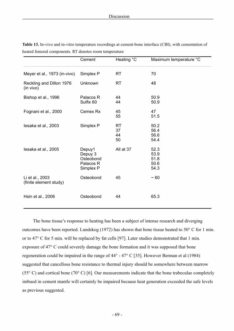
Table 13. In-vivo and in-vitro temperature recordings at cement-bone interface (CBI), with cementation of heated femoral components. RT denotes room temperature
Cement Heating °C Maximum temperature °C
Meyer et al., 1973 (in-vivo)
Reckling and Dillon 1976 (in vivo)
Bishop et al., 1996
Fognani et al., 2000
Iesaka et al., 2003
Iesaka et al., 2005
Li et al., 2003 (finite element study)
Hsin et al., 2006
Simplex P
Unknown
Palacos R Sulfix 60
Cemex Rx
Simplex P
Depuy1Depuy 3OsteobondPalacos RSimplex P
Osteobond
Osteobond
RT
RT
4444
4555
RT374450
All at 37
45
44
70
48
50.9 50.9
4751.5
50.256.456.654.4
52.353.951.850.654.3
~ 60
65.3
The bone tissue’s response to heating has been a subject of intense research and diverging
outcomes have been reported. Lundskog (1972) has shown that bone tissue heated to 50° C for 1 min.
or to 47° C for 5 min. will be replaced by fat cells [97]. Later studies demonstrated that 1 min.
exposure of 47° C could severely damage the bone formation and it was supposed that bone
regeneration could be impaired in the range of 44° - 47° C [35]. However Berman et al (1984)
suggested that cancellous bone resistance to thermal injury should be somewhere between marrow
(55° C) and cortical bone (70° C) [6]. Our measurements indicate that the bone trabeculae completely
imbued in cement mantle will certainly be impaired because heat generation exceeded the safe levels
as previous suggested.
Discussion
- 69 -
In study IV preheating had a limited effect on temperature increase at CBI as the difference
between the groups was 2.6° C. This is in accordance with 3.1° C reported by Iesaka et al (2005),
where stems of 37° C were cemented with Palacos R cement (identical to Refobacin bone cement R,
used by us) in vitro [68]. However the maximum temperatures were approx. 6° C higher in our study
than those measured by Iesaka et al. We had expected that presence of blood, tissue fluid, or irrigation
fluid at the interface, as well as blood circulation would keep heat generation at lower levels than
those measured by Iesaka et al [68, 70]. The main reason might be different temperature profile of
cements, but also an increasing cement penetration at the site of temperature recordings affected the
measurements in our study.
It should be pointed out that cement and bone contact in-vivo is not uniform between these two
surfaces; therefore a deeper cement penetration around the thermocouple will result in higher
temperatures. Thus, recordings made at one point on the interface may not indicate the temperature at
all areas of the interface. Moreover, the maximum temperature may not represent the temperature at
the interface, because of penetrated mass of cement. This is the limitation of our and other studies
where temperatures are recorded only at one site in the bone wall.
Experience and science regarding aseptic loosening of femoral component has shown that
despite the high curing temperatures, the well-cemented stems had a stable interlock at CBI. The
modern cementing technique had increased the survival rates of hip prostheses even more. This
proves that optimal cement penetration but not a cement temperature at interface influences stem
stability.
Our study revealed that a minor temperature increase from 53.8° C to 56.4° C when cementing
the heated femoral components (40° C) had no harmful effects on CBI in comparison with non-
preheated stems. This conclusion is also supported by our calculation of the total heat transfer to CBI,
expressed as AUC. The smaller AUC for preheated stems was recorded as the consequence of shorter
polymerization time. The last outcome could be also instrumental in shortening the operating time of
THA.
Discussion
- 70 -
Conclusions
Study I
A new radiographic evaluation method to assess cementing quality of femoral stem was compared
with the well-known Barrack’s cement grading. Both methods demonstrated a low rate of intra- and
interobserver agreement when evaluated by 3 experienced hip surgeons. We conclude that simple
radiographic evaluation is not suitable for the evaluation of cementation quality on postoperative
radiographs and support previous investigators in their recommendation not to use radiographic
grading systems in follow-up papers. Evaluation methods with documented prognostic value (e.g.
radiostereometry) should be applied in preference for simple visual radiographic assessment, when
introducing new methods in cementing technique and new prosthetic designs.
Study II
The results from cadaveric study did not reveal any effect of proximal stem centralizer on cementation
pressures and cement penetration during cementation of Bi-Metric stem. The hemispherical design
failed to increase the cement flow into bone at metaphyseal part of the femur. Moreover, a larger
variation of stem alignment when a proximal centralizer was used indicates lower precision of stem
placement proximally in the reamed cavity. Proximal centralizer with the present design did not
improve stem positioning when a straight stem was used. A circumferential proximal centralizer might
yield a better proximal seal and thus could increase the cementing pressures and cement penetration in
the metaphyseal part of the femur, but further investigations are needed.
Study III and IV
From our RSA analysis we concluded that preheating improved the initial stability of stem, especially
subsidence inside the cement mantle was reduced. This particular stem mostly migrated at stem-
cement interface in spite of matt-surfaced finish; meanwhile the micromotions between the cement
and bone were limited. No detrimental effects by heating components to 40°C on stability between
cement and bone or periprosthetic mineral bone density could be detected. The loss of initial stability
after 1 year among several stems strongly indicates that this particular stem risk debonding and
developing of aseptic loosening especially in overweight male patients. Preheating of femoral
components reduces the cement polymerization time and thus will shorten the total operation time.
Conclusions
- 71 -
Aspects for future research
The studies investigating improvements of both PCI and CBI are needed to increase long-term
fixation of cemented implants. Biomechanical research using cadaveric femora should be a main
experimental setup when new implant designs are intended to clinical use. This experimental model
should be used to test different physical conditions influencing the interfacial relationship between
implant, cement and bone. Outcome from these experiments are close to that expected in a clinical
situation and surely have a good prognostic value.
On other hand the well-designed randomized clinical trials are the golden standard to measure
an effect of any new management, drug or implant clinically. Implant migration studies based on RSA
can reveal changes in small experimental samples, thus avoiding the unnecessary costs of both patient
health and medical care in case of failure.
The effect of preheating revealed in our study should be further investigated on femoral components
of different designs and surface finish in all major joint replacements. A longer follow-up period of 5
to 10 years using RSA will clarify whether preheating has a prolonged effect on the fixation of
implants.
Aspects for future research
- 72 -
Summary in Danish
Cementering af lårbensproteser er den mest almindelige fikseringsmetode i total hoftealloplastik. På trods af forbedringer i både cementeringsteknik, protesedesign og metallegeringer, er aseptisk proteseløsning stadig den vigtigste årsag til revisionskirurgi. Forbedringen af bindingsstyrken mellem protese-cement og cement-knogle kan øge lårbensprotesens fikseringsstyrke og kan forbedre protesens holdbarhed på lang sigt.
I dette ph.d.-projekt, blev der udført 4 studier, hvor radiologisk vurdering af cementeringsteknik samt metoder, der kan forbedre protesefiksation, blev undersøgt.
I Studie I, blev den velkendte Barrack's cement klassifikationssystem sammenlignet med en ny radiologisk evalueringsmetode udarbejdet af studiets medforfattere. Cementeringskvaliteten af lårbensproteser blev vurderet på røntgenbilleder efter total og hemi-hoftealloplastik. Reproducerbarhed af begge metoder blev analyseret. Begge systemer viste høj intra- og inter-observatør variation, målt med kappastatistik, hvorfor kan vurderingen af cementeringskvalitet ved brug af konventionelle røntgenbilleder ikke anbefales.
Formålet med Studie II var at forbedre kvaliteten af cement-knogle interfasen samt placeringen af lårbensprotese ved hjælp af en proximal centralizer. Otte lårbensproteser med og otte proteser uden proksimal centralizer blev cementeret i parrede kadaver femora. Det intramedullære tryk blev registreret under cementeringen. CT skanning af de cementerede implantater blev udført for at bedømme protesealignment i forhold til femuraksen. Cement-penetration, tykkelsen af cement kappen og protesecentraliseringen i den metafyseale del af lårbenet blev målt ved hjælp af stereologi. Vi fandt ingen signifikant forskel i de målte parametre mellem grupperne. Den proksimale centralizer øgede hverken det intramedullære tryk eller cement penetrationen. Protesealignment og centralisering blev heller ikke forbedret. Man konkluderede, at nye protesedesigns og forbedringer af cementerings teknik bør undersøges grundigt på virkelighedstro modeller før klinisk brug.
Undersøgelse III var et prospektivt randomiseret dobbelt-blindet klinisk forsøg med hovedformålet at sammenligne protesemigration in vivo efter cementeringen af lårbensproteser, der blev varmet op til 40° C og samme protese ved stue-temperatur. 80 patienter, der fik udført hybrid hoftealloplastik blev randomiseret i to grupper. Patienterne blev fulgt både klinisk og med røntgenstereofotometri (RSA) samt DEXA skanninger. To år efter operationen blev der fundet en betydeligt forbedret stabilitet af de opvarmede proteser sammenlignet med de ikke-opvarmede. Især nedsynkning i forhold til cementkappe blev hæmmet. Ingen svækkelse af stabilitet mellem cement og knogle kunne observeres. Men opvarmningen kunne ikke forhindre betydelig migration af nogen proteser (ligeligt fordelt i begge grupper), der fandt sted mellem protesen og cementen efter 1 og 2 år. Stigende BMI og mandligt køn var relateret til større migration. Øget migrationsvariation blandt de små protesestørrelser blev også set. Disse observationer indikerer, at kombinationen af flere faktorer kan fremkalde tidlig migration og debonding af denne protese. I dette forsøg har vi også foretaget in vivo temperaturmålinger (Studie IV) ved cement-knogle interfasen. Trods den stigende maksimale temperatur i den opvarmede gruppe (56,4° vs 53,8° C) var eksponeringen af temperaturer over 50° C den samme i begge grupper (median 70 sek). Da ingen skadelige virkninger forårsaget af opvarmningen kunne observeres (målt både med RSA og DEXA) kan opvarmning af lårbensprotese til 40° C før cementering anbefales.
Summary in Danish
- 73 -
References1. Ahmed AM, Raab S, Miller JE. Metal/cement interface strength in cemented stem fixation. J Orthop Res 1984;2(2):105-18. 2. Anthony PP, Gie GA, Howie CR, Ling RS. Localised endosteal bone lysis in relation to the femoral components of cemented total hip arthroplasties. J Bone Joint Surg Br 1990;72(6):971-9. 3. Askew MJ, Steege JW, Lewis JL, Ranieri JR, Wixson RL. Effect of cement pressure and bone strength on polymethylmethacrylate fixation. J Orthop Res 1984;1(4):412-20. 4. Barrack RL, Mulroy RD,Jr., Harris WH. Improved cementing techniques and femoral component loosening in young patients with hip arthroplasty. A 12-year radiographic review. J Bone Joint Surg Br 1992;74-B(3):385-9. 5. Berger RA, Seel MJ, Wood K, Evans R, D'Antonio J, Rubash HE. Effect of a centralizing device on cement mantle deficiencies and initial prosthetic alignment in total hip arthroplasty. J Arthroplasty 1997;12(4):434-43. 6. Berman AT, Reid JS, Yanicko DR,Jr, Sih GC, Zimmerman MR. Thermally induced bone necrosis in rabbits. Relation to implant failure in humans. Clin Orthop Relat Res 1984;(186):284-92. 7. Berry DJ. Cemented femoral stems: what matters most. J Arthroplasty 2004;19(4):83-4. 8. Berry DJ, Harmsen WS, Cabanela ME, Morrey BF. Twenty-five-year survivorship of two thousand consecutive primary Charnley total hip replacements: factors affecting survivorship of acetabular and femoral components. J Bone Joint Surg Am 2002;84-A(2):171-7. 9. Bishop NE, Ferguson S, Tepic S. Porosity reduction in bone cement at the cement-stem interface. J Bone Joint Surg Br 1996;78(3):349-56. 10. Bourne RB, Rorabeck CH, Skutek M, Mikkelsen S, Winemaker M, Robertson D. The Harris Design-2 total hip replacement fixed with so-called second-generation cementing techniques. A ten to fifteen-year follow-up. J Bone Joint Surg Am 1998;80(12):1775-80. 11. Breusch SJ, Lukoschek M, Kreutzer J, Brocai D, Gruen TA. Dependency of cement mantle thickness on femoral stem design and centralizer. J Arthroplasty 2001;16(5):648-57. 12. Callaghan JJ, Forest EE, Sporer SM, Goetz DD, Johnston RC. Total hip arthroplasty in the young adult. Clin Orthop Relat Res 1997;(344):257-62. 13. Callaghan JJ, Tooma GS, Olejniczak JP, Goetz DD, Johnston RC. Primary hybrid total hip arthroplasty: an interim followup. Clin Orthop Relat Res 1996;(333):118-25. 14. Carter DR, Gates EI, Harris WH. Strain-controlled fatigue of acrylic bone cement. J Biomed Mater Res 1982;16(5):647-57. 15. Chambers IR, Fender D, McCaskie AW, Reeves BC, Gregg PJ. Radiological features predictive of aseptic loosening in cemented Charnley femoral stems. J Bone Joint Surg Br 2001;83(6):838-42. 16. Charnley J. Acrylic cement in orthopaedic surgery. Baltimore: Williams and Wilkins. 1970. 17. Christensen F, Soballe K, Ejsted R, Luxhoj T. The Catterall classification of Perthes' disease: an assessment of reliability. J Bone Joint Surg Br 1986;68(4):614-5. 18. Churchill DL, Incavo SJ, Uroskie JA, Beynnon BD. Femoral stem insertion generates high bone cement pressurization. Clin Orthop Relat Res 2001 (393):335-44. 19. Clohisy JC, Harris WH. Primary hybrid total hip replacement, performed with insertion of the acetabular component without cement and a precoat femoral component with cement. An average ten-year follow-up study. J Bone Joint Surg Am 1999;81(2):247-55. 20. Crawford RW, Psychoyios V, Gie G, Ling R, Murray D. Incomplete cement mantles in the sagittal femoral plane: an anatomical explanation. Acta Orthop Scand 1999;70(6):596-8. 21. Crowninshield RD, Jennings JD, Laurent ML, Maloney WJ. Cemented femoral component surface finish mechanics. Clin Orthop Relat Res 1998;(355):90-102.
References
- 74 -
22. Dall DM, Miles AW, Juby G. Accelerated polymerization of acrylic bone cement using preheated implants. Clin Orthop 1986 (211):148-50. 23. Dall GF, Simpson PM, Breusch SJ. In vitro comparison of Refobacin-Palacos R with Refobacin Bone Cement and Palacos R + G. Acta Orthop 2007;78(3):404-11. 24. Damron LA, Kim DG, Mann KA. Fatigue debonding of the roughened stem-cement interface: effects of surface roughness and stem heating conditions. J Biomed Mater Res B Appl Biomater 2006;78(1):181-8. 25. Della Valle AG, Zoppi A, Peterson MG, Salvati EA. A rough surface finish adversely affects the survivorship of a cemented femoral stem. Clin Orthop Relat Res 2005;(436):158-63. 26. Digas G, Thanner J, Anderberg C, Karrholm J. Fluoride-containing acrylic bone cement in total hip arthroplasty. Randomized evaluation of 97 stems using radiostereometry and dual-energy x-ray absorptiometry. J Arthroplasty 2005;20(6):784-92. 27. Dozier JK, Harrigan T, Kurtz WH, Hawkins C, Hill R. Does increased cement pressure produce superior femoral component fixation? J Arthroplasty 2000;15(4):488-95. 28. Draenert K. The John Charnley Award Paper. Histomorphology of the bone-to-cement interface: remodeling of the cortex and revascularization of the medullary canal in animal experiments. Hip 1981:71-110. 29. Draenert KD, Draenert YI, Krauspe R, Bettin D. Strain adaptive bone remodelling in total joint replacement. Clin Orthop Relat Res 2005;(430):12-27. 30. Dunne NJ, Orr JF, Mushipe MT, Eveleigh RJ. The relationship between porosity and fatigue characteristics of bone cements. Biomaterials 2003;24(2):239-45. 31. Ebramzadeh E, Normand PL, Sangiorgio SN, Llinas A, Gruen TA, McKellop HA, et al. Long-term radiographic changes in cemented total hip arthroplasty with six designs of femoral components. Biomaterials 2003;24(19):3351-63. 32. Ebramzadeh E, Sangiorgio SN, Longjohn DB, Buhari CF, Dorr LD. Initial stability of cemented femoral stems as a function of surface finish, collar, and stem size. J Bone Joint Surg Am 2004;86-A(1):106-15. 33. Ebramzadeh E, Sarmiento A, McKellop HA, Llinas A, Gogan W. The cement mantle in total hip arthroplasty. Analysis of long-term radiographic results. J Bone Joint Surg Am 1994;76(1):77-87. 34. Egund N, Lidgren L, Onnerfalt R. Improved positioning of the femoral stem with a centralizing device. Acta Orthop Scand 1990;61(3):236-9. 35. Eriksson AR, Albrektsson T. Temperature threshold levels for heat-induced bone tissue injury: a vital-microscopic study in the rabbit. J Prosthet Dent 1983;50(1):101-7. 36. Espehaug B, Furnes O, Engesaeter LB, Havelin LI. 18 years of results with cemented primary hip prostheses in the Norwegian Arthroplasty Register: concerns about some newer implants. Acta Orthop 2009;80(4):402-12. 37. Espehaug B, Furnes O, Havelin LI, Engesaeter LB, Vollset SE. The type of cement and failure of total hip replacements. J Bone Joint Surg Br 2002;84(6):832-8. 38. Espehaug B, Havelin LI, Engesaeter LB, Langeland N, Vollset SE. Patient-related risk factors for early revision of total hip replacements. A population register-based case-control study of 674 revised hips. Acta Orthop Scand 1997;68(3):207-15. 39. Estok DM,2nd, Orr TE, Harris WH. Factors affecting cement strains near the tip of a cemented femoral component. J Arthroplasty 1997;12(1):40-8. 40. Fisher DA, Tsang AC, Paydar N, Milionis S, Turner CH. Cement-mantle thickness affects cement strains in total hip replacement. J Biomech 1997;30(11-12):1173-7. 41. Flugsrud GB, Nordsletten L, Espehaug B, Havelin LI, Meyer HE. The effect of middle-age body weight and physical activity on the risk of early revision hip arthroplasty: a cohort study of 1,535 individuals. Acta Orthop 2007;78(1):99-107.
References
- 75 -
42. Fognani R, Baleani M, Toni A. Experimental study on the cement mantle in hip arthroplasty: effect of defects on the property of the materials used. Chir Organi Mov 2000;85(4):403-8. 43. Fowler JL, Gie GA, Lee AJ, Ling RS. Experience with the Exeter total hip replacement since 1970. Orthop Clin North Am 1988;19(3):477-89. 44. Friedman RJ, Black J, Galante JO, Jacobs JJ, Skinner HB. Current concepts in orthopaedic biomaterials and implant fixation. Instr Course Lect 1994;43:233-55. 45. Gardiner RC, Hozack WJ. Failure of the cement-bone interface. A consequence of strengthening the cement-prosthesis interface? J Bone Joint Surg Br 1994;76(1):49-52. 46. Glyn-Jones S, Alfaro-Adrian J, Murray DW, Gill HS. The influence of surgical approach on cemented stem stability: an RSA study. Clin Orthop Relat Res 2006l;448:87-91. 47. Goldberg BA, al Habbal G, Noble PC, Paravic M, Liebs TR, Tullos HS. Proximal and distal femoral centralizers in modern cemented hip arthroplasty. Clin Orthop Relat Res 1998(349):163-73. 48. Gozzard C, Gheduzzi S, Miles AW, Learmonth ID. An in-vitro investigation into the cement pressurization achieved during insertion of four different femoral stems. Proc Inst Mech Eng [H ] 2005;219(6):407-13. 49. Gozzard C, Gheduzzi S, Miles AW, Learmonth ID. An in-vitro investigation of the CPS-Plus femoral stem: influence of the proximal centraliser on cement pressurisation during stem insertion. Acta Orthop Scand 2003;74(2):154-8. 50. Gruen TA, McNeice GM, Amstutz HC. "Modes of failure" of cemented stem-type femoral components: a radiographic analysis of loosening. Clin Orthop Relat Res 1979(141):17-27. 51. Gundersen HJ. The nucleator. J Microsc 1988;151(Pt 1):3-21. 52. Haboush EJ. A new operation for arthroplasty of the hip based on biomechanics, photoelasticity, fast-setting dental acrylic, and other considerations. 1953 [classicle article. Bull Hosp Jt Dis 1996;55(2):95-111. 53. Halawa M, Lee AJ, Ling RS, Vangala SS. The shear strength of trabecular bone from the femur, and some factors affecting the shear strength of the cement-bone interface. Arch Orthop Trauma Surg 1978;92(1):19-30. 54. Hamadouche M, Baque F, Lefevre N, Kerboull M. Minimum 10-year survival of Kerboull cemented stems according to surface finish. Clin Orthop Relat Res 2008;466(2):332-9. 55. Hanson PB, Walker RH. Total hip arthroplasty cemented femoral component distal stem centralizer. Effect on stem centralization and cement mantle. J Arthroplasty 1995;10(5):683-8. 56. Harrington MA,Jr, O'Connor DO, Lozynsky AJ, Kovach I, Harris WH. Effects of femoral neck length, stem size, and body weight on strains in the proximal cement mantle. J Bone Joint Surg Am 2002;84-A(4):573-9. 57. Harvey EJ, Tanzer M, Bobyn JD. Femoral cement grading in total hip arthroplasty. J Arthroplasty 1998;13(4):396-401. 58. Harving S, Soballe K, Bunger C. A method for bone-cement interface thermometry. An in vitro comparison between low temperature curing cement Palavit, and Surgical Simplex P. Acta Orthop Scand 1991;62(6):546-8. 59. Havelin LI, Espehaug B, Vollset SE, Engesaeter LB. The effect of the type of cement on early revision of Charnley total hip prostheses. A review of eight thousand five hundred and seventy-nine primary arthroplasties from the Norwegian Arthroplasty Register. J Bone Joint Surg Am 1995t;77(10):1543-50. 60. Herberts P, Malchau H. Long-term registration has improved the quality of hip replacement: a review of the Swedish THR Register comparing 160,000 cases. Acta Orthop Scand 2000;71(2):111-21. 61. Holm NJ. The relaxation of some acrylic bone cements. Acta Orthop Scand 1980;51(5):727-31. 62. Hook S, Moulder E, Yates PJ, Burston BJ, Whitley E, Bannister GC. The Exeter Universal stem: a minimum ten-year review from an independent centre. J Bone Joint Surg Br 2006;88(12):1584-90.
References
- 76 -
63. Howell JR,Jr, Blunt LA, Doyle C, Hooper RM, Lee AJ, Ling RS. In vivo surface wear mechanisms of femoral components of cemented total hip arthroplasties: the influence of wear mechanism on clinical outcome. J Arthroplasty 2004;19(1):88-101. 64. Howie DW, Middleton RG, Costi K. Loosening of matt and polished cemented femoral stems. J Bone Joint Surg Br 1998;80(4):573-6. 65. Hsieh PH, Tai CL, Chang YH, Lee MS, Shih HN, Shih CH. Precooling of the femoral canal enhances shear strength at the cement-prosthesis interface and reduces the polymerization temperature. J Orthop Res 2006;24(9):1809-14. 66. Huddleston HD. Femoral lysis after cemented hip arthroplasty. J Arthroplasty 1988;3(4):285-97. 67. Huiskes R, Verdonschot N, Nivbrant B. Migration, stem shape, and surface finish in cemented total hip arthroplasty. Clin Orthop Relat Res 1998;(355):103-12. 68. Iesaka K, Jaffe WL, Jones CM, Kummer FJ. The effects of fluid penetration and interfacial porosity on the fixation of cemented femoral components. J Bone Joint Surg Br 2005;87(9):1298-302. 69. Iesaka K, Jaffe WL, Kummer FJ. Effects of the initial temperature of acrylic bone cement liquid monomer on the properties of the stem-cement interface and cement polymerization. J Biomed Mater Res B Appl Biomater 200415;68(2):186-90. 70. Iesaka K, Jaffe WL, Kummer FJ. Effects of preheating of hip prostheses on the stem-cement interface. J Bone Joint Surg Am 2003;85-A(3):421-7. 71. Jafri AA, Green SM, Partington PF, McCaskie AW, Muller SD. Pre-heating of components in cemented total hip arthroplasty. J Bone Joint Surg Br 2004;86(8):1214-9. 72. Jarrett SD, Lachiewicz PF. Precoated femoral component with proximal and distal centralizers: results at 5 to 12 years. J Arthroplasty 2005;20(3):309-15. 73. Jasty M, Davies JP, O'Connor DO, Burke DW, Harrigan TP, Harris WH. Porosity of various preparations of acrylic bone cements. Clin Orthop 1990;(259):122-9. 74. Jasty M, Maloney WJ, Bragdon CR, O'Connor DO, Haire T, Harris WH. The initiation of failure in cemented femoral components of hip arthroplasties. J Bone Joint Surg Br 1991;73(4):551-8. 75. Jergesen HE, Karlen JW. Clinical outcome in total hip arthroplasty using a cemented titanium femoral prosthesis. J Arthroplasty 2002;17(5):592-9. 76. Johnston RC, Fitzgerald RH,Jr., Harris WH, Poss R, Muller ME, Sledge CB. Clinical and radiographic evaluation of total hip replacement. A standard system of terminology for reporting results. J Bone Joint Surg Am 1990;72(2):161-8. 77. Karrholm J, Borssen B, Lowenhielm G, Snorrason F. Does early micromotion of femoral stem prostheses matter? 4-7-year stereoradiographic follow-up of 84 cemented prostheses. J Bone Joint Surg Br 1994;76(6):912-7. 78. Karrholm J, Herberts P, Hultmark P, Malchau H, Nivbrant B, Thanner J. Radiostereometry of hip prostheses. Review of methodology and clinical results. Clin Orthop Relat Res 1997;(344):94-110. 79. Karrholm J, Nivbrant B, Thanner J, Anderberg C, Borlin N, Malchau H. Radiostereometric Evaluation of Hip Implant, Design and Surface Finish. Micromotion of Cemented Femoral Stems. 67th Annual Meeting of the American Academy of Orthopaedic Surgeons. March 15-19, Orlando, USA; 2000.80. Kelly AJ, Lee MB, Wong NS, Smith EJ, Learmonth ID. Poor reproducibility in radiographic grading of femoral cementing technique in total hip arthroplasty. J Arthroplasty 1996;11(5):525-8. 81. Kirkwood BR, Sterne JAC. Essential Medical Statistics. 2003;433-7. 82. Kiss J, Murray DW, Turner-Smith AR, Bithell J, Bulstrode CJ. Migration of cemented femoral components after THR. Roentgen stereophotogrammetric analysis. J Bone Joint Surg Br 1996;78(5):796-801.
References
- 77 -
83. Kock HJ, Huber FX, Hillmeier J, Jager R, Volkmann R, Handschin AE, et al. In vitro studies on various PMMA bone cements: a first comparison of new materials for arthroplasty. Z Orthop Unfall 2008;146(1):108-13. 84. Kramhoft M, Gehrchen PM, Bodtker S, Wagner A, Jensen F. Inter- and intraobserver study of radiographic assessment of cemented total hip arthroplasties. J Arthroplasty 1996;11(3):272-6. 85. Krause WR, Krug W, Miller J. Strength of the cement-bone interface. Clin Orthop Relat Res 1982;(163):290-9. 86. Kuehn KD. Bone cements. Up-to-date comparison of Physical and Chemical Properties of Commercial Materials. Berlin Heidelberg 2000: Springer-Verlag; 2000. 87. Kuehn KD, Ege W, Gopp U. Acrylic bone cements: mechanical and physical properties. Orthop Clin North Am 2005;36(1):29-39. 88. Kurtinaitis J, Porvaneckas N, Kocius M, Broga R. [Primary hybrid and cementless total hip arthroplasty. 5 to 10 years follow-up results]. Lietuvos Chirurgija 2003;1(2):155-63. 89. Landis JR, Koch GG. The measurement of observer agreement for categorical data. Biometrics 1977 Mar;33(1):159-74. 90. Langlais F, Kerboull M, Sedel L, Ling RS. The 'French paradox.'. J Bone Joint Surg Br 2003 Jan;85(1):17-20. 91. Lee AJ, Ling RS. Improved cementing techniques. Instr Course Lect 1981;30:407-13. 92. Lee AJ, Ling RS, Gheduzzi S, Simon JP, Renfro RJ. Factors affecting the mechanical and viscoelastic properties of acrylic bone cement. J Mater Sci Mater Med 2002 Aug;13(8):723-33. 93. Lewis JL, Askew MJ, Wixson RL, Kramer GM, Tarr RR. The influence of prosthetic stem stiffness and of a calcar collar on stresses in the proximal end of the femur with a cemented femoral component. J Bone Joint Surg Am 1984 Feb;66(2):280-6. 94. Li C, Mason J, Yakimicki D. Thermal characterization of PMMA-based bone cement curing. J Mater Sci Mater Med 2004 Jan;15(1):85-9. 95. Li C, Schmid S, Mason J. Effects of pre-cooling and pre-heating procedures on cement polymerization and thermal osteonecrosis in cemented hip replacements. Med Eng Phys 2003 09;25(7):559-64. 96. Ling RS, Charity J, Lee AJ, Whitehouse SL, Timperley AJ, Gie GA. The long-term results of the original Exeter polished cemented femoral component: a follow-up report. J Arthroplasty 2009 Jun;24(4):511-7. 97. Lundskog J. Heat and bone tissue. An experimental investigation of the thermal properties of bone and threshold levels for thermal injury. Scand J Plast Reconstr Surg 1972;9:1-80. 98. Madey SM, Callaghan JJ, Olejniczak JP, Goetz DD, Johnston RC. Charnley total hip arthroplasty with use of improved techniques of cementing. The results after a minimum of fifteen years of follow-up. J Bone Joint Surg Am 1997;79(1):53-64. 99. Makela K, Eskelinen A, Pulkkinen P, Paavolainen P, Remes V. Cemented total hip replacement for primary osteoarthritis in patients aged 55 years or older: results of the 12 most common cemented implants followed for 25 years in the Finnish Arthroplasty Register. J Bone Joint Surg Br 2008;90(12):1562-9. 100. Makela KT, Eskelinen A, Pulkkinen P, Paavolainen P, Remes V. Total hip arthroplasty for primary osteoarthritis in patients fifty-five years of age or older. An analysis of the Finnish arthroplasty registry. J Bone Joint Surg Am 2008;90(10):2160-70. 101. Malchau H, Herberts P, Ahnfelt L. Prognosis of total hip replacement in Sweden. Follow-up of 92,675 operations performed 1978-1990. Acta Orthop Scand 1993 Oct;64(5):497-506. 102. Malchau H, Herberts P, Eisler T, Garellick G, Soderman P. The Swedish Total Hip Replacement Register. J Bone Joint Surg Am 2002;84-A Suppl 2:2-20. 103. Maloney WJ, Schmalzried T, Harris WH. Analysis of long-term cemented total hip arthroplasty retrievals. Clin Orthop Relat Res 2002;(12):70-8.
References
- 78 -
104. Mann KA, Damron LA, Miller MA, Race A, Clarke MT, Cleary RJ. Stem-cement porosity may explain early loosening of cemented femoral hip components: experimental-computational in vitro study. J Orthop Res 2007;25(3):340-50. 105. Markolf KL, Amstutz HC. Penetration and flow of acrylic bone cement. Clin Orthop Relat Res 1976;(121):99-102. 106. Massoud SN, Hunter JB, Holdsworth BJ, Wallace WA, Juliusson R. Early femoral loosening in one design of cemented hip replacement. J Bone Joint Surg Br 1997;79(4):603-8. 107. McCaskie AW, Barnes MR, Lin E, Harper WM, Gregg PJ. Cement pressurisation during hip replacement. J Bone Joint Surg Br 1997;79(3):379-84. 108. McCaskie AW, Brown AR, Thompson JR, Gregg PJ. Radiological evaluation of the interfaces after cemented total hip replacement. Interobserver and intraobserver agreement. J Bone Joint Surg Br 1996;78(2):191-4. 109. Meyer PR,Jr, Lautenschlager EP, Moore BK. On the setting properties of acrylic bone cement. J Bone Joint Surg Am 1973;55(1):149-56. 110. Middleton RG, Howie DW, Costi K, Sharpe P. Effects of design changes on cemented tapered femoral stem fixation. Clin Orthop Relat Res 1998;(355):47-56. 111. Mjoberg B. Loosening of the cemented hip prosthesis. The importance of heat injury. Acta Orthop Scand Suppl 1986;221:1-40. 112. Mohler CG, Callaghan JJ, Collis DK, Johnston RC. Early loosening of the femoral component at the cement-prosthesis interface after total hip replacement. J Bone Joint Surg Am 1995;77(9):1315-22. 113. Muller RT, Schurmann N. Shear strength of the cement metal interface--an experimental study. Arch Orthop Trauma Surg 1999;119(3-4):133-8. 114. Mulroy RD,Jr., Harris WH. The effect of improved cementing techniques on component loosening in total hip replacement. An 11-year radiographic review. J Bone Joint Surg Br 1990;72(5):757-60. 115. Mulroy WF, Estok DM, Harris WH. Total hip arthroplasty with use of so-called second-generation cementing techniques. A fifteen-year-average follow-up study. J Bone Joint Surg Am 1995;77(12):1845-52. 116. Munro NA, Nicol M, Selvaraj S, Hussain SM, Finlayson DF. Femoral cement pressurization in hip arthroplasty: a comparison of 3 systems. J Arthroplasty 2007;22(6):893-901. 117. Oh I, Carlson CE, Tomford WW, Harris WH. Improved fixation of the femoral component after total hip replacement using a methacrylate intramedullary plug. J Bone Joint Surg Am 1978;60(5):608-13. 118. Ong A, Wong KL, Lai M, Garino JP, Steinberg ME. Early failure of precoated femoral components in primary total hip arthroplasty. J Bone Joint Surg Am 2002;84-A(5):786-92. 119. Ørskov M. On the longevity of cemented hip prostheses and the influence on implant design [PhD-dissertation]. University of Aarhus: Faculty of Health Science, University of Aarhus, Denmark; 2007. 120. Overgaard S, Lucht U, Nielsen PT, Varmarken J-, Pedersen AB, Bartels P. The Danish Hip Arthroplasty Register. Report 2007 [Dansk Hoftealloplastik Register. Årsrapport 2007]. 2008;58,62-67. 121. Panjabi MM, Goel VK, Drinker H, Wong J, Kamire G, Walter SD. Effect of pressurization on methylmethacrylate-bone interdigitation: an in vitro study of canine femora. J Biomech 1983;16(7):473-80. 122. Peters CL, Bachus KN, Craig MA, Higginbotham TO. The effect of femoral prosthesis design on cement strain in cemented total hip arthroplasty. J Arthroplasty 2001;16(2):216-24. 123. Poss R, Brick GW, Wright RJ, Roberts DW, Sledge CB. The effects of modern cementing techniques on the longevity of total hip arthroplasty. Orthop Clin North Am 1988;19(3):591-8. 124. Pramanik S, Agarwal A, Rai KN. Chronology of Total Hip Joint Replacement and Materials Development. Trends Biomater Artif Organs 2005;19(1):15-26.
References
- 79 -
125. Race A, Miller MA, Clarke MT, Mann KA. Cement-implant interface gaps explain the poor results of CMW3 for femoral stem fixation: A cadaver study of migration, fatigue and mantle morphology. Acta Orthop 2005;76(5):679-87. 126. Rasquinha VJ, Dua V, Rodriguez JA, Ranawat CS. Fifteen-year survivorship of a collarless, cemented, normalized femoral stem in primary hybrid total hip arthroplasty with a modified third-generation cement technique. J Arthroplasty 2003;18 Suppl 1:86-94. 127. Reading AD, McCaskie AW, Barnes MR, Gregg PJ. A comparison of 2 modern femoral cementing techniques: analysis by cement-bone interface pressure measurements, computerized image analysis, and static mechanical testing. J Arthroplasty 2000;15(4):479-87. 128. Reckling FW, Dillon WL. The bone-cement interface temperature during total joint replacement. J Bone Joint Surg Am 1977;59(1):80-2. 129. Riede U, Luem M, Ilchmann T, Eucker M, Ochsner PE. The M.E Muller straight stem prosthesis: 15 year follow-up. Survivorship and clinical results. Arch Orthop Trauma Surg 2007;127(7):587-92. 130. Riegels-Nielsen P, Sorensen L, Andersen HM, Lindequist S. Boneloc cemented total hip prostheses. Loosening in 28/43 cases after 3-38 months. Acta Orthop Scand 1995;66(3):215-7. 131. Rockborn P, Olsson SS. Loosening and bone resorption in exeter hip arthroplasties. Review at a minimum of five years. J Bone Joint Surg Br 1993;75(6):865-8. 132. Russotti GM, Coventry MB, Stauffer RN. Cemented total hip arthroplasty with contemporary techniques. A five-year minimum follow-up study. Clin Orthop Relat Res 1988;(235):141-7. 133. Schmalzried TP, Harris WH. Hybrid total hip replacement. A 6.5-year follow-up study. J Bone Joint Surg Br 1993;75(4):608-15. 134. Selvik G. Roentgen stereophotogrammetry. A method for the study of the kinematics of the skeletal system. Acta Orthop Scand Suppl 1989;232:1-51. 135. Smart RC, Barbagallo S, Slater GL, Kuo RS, Butler SP, Drummond RP, et al. Measurement of periprosthetic bone density in hip arthroplasty using dual-energy x-ray absorptiometry. Reproducibility of measurements. J Arthroplasty 1996;11(4):445-52. 136. Smith DC. The genesis and evolution of acrylic bone cement. Orthop Clin North Am 2005;36(1):1-10. 137. Smith SE, Harris WH. Total hip arthroplasty performed with insertion of the femoral component with cement and the acetabular component without cement. Ten to thirteen-year results. J Bone Joint Surg Am 1997;79(12):1827-33. 138. Song Y, Goodman SB, Jaffe RA. An in vitro study of femoral intramedullary pressures during hip replacement using modern cement technique. Clin Orthop Relat Res 1994;(302):297-304. 139. Speirs AD, Slomczykowski MA, Orr TE, Siebenrock K, Nolte LP. Three-dimensional measurement of cemented femoral stem stability: an in vitro cadaver study. Clin Biomech 2000;15(4):248-55. 140. Star MJ, Colwell CW,Jr., Kelman GJ, Ballock RT, Walker RH. Suboptimal (thin) distal cement mantle thickness as a contributory factor in total hip arthroplasty femoral component failure. A retrospective radiographic analysis favoring distal stem centralization. J Arthroplasty 1994;9(2):143-9. 141. Strom H, Kolstad K, Mallmin H, Sahlstedt B, Milbrink J. Comparison of the uncemented Cone and the cemented Bimetric hip prosthesis in young patients with osteoarthritis: an RSA, clinical and radiographic study. Acta Orthop 2006;77(1):71-8. 142. Sylvain GM, Kassab S, Coutts R, Santore R. Early failure of a roughened surface, precoated femoral component in total hip arthroplasty. J Arthroplasty 2001;16(2):141-8. 143. Tarasevicius S, Tarasevicius R, Zegunis V, Smailys A, Kalesinskas RJ. Metal type of the femoral stem in total hip arthroplasty. Medicina (Kaunas) 2005;41(11):932-5. 144. Toksvig-Larsen S, Franzen H, Ryd L. Cement interface temperature in hip arthroplasty. Acta Orthop Scand 1991;62(2):102-5.
References
- 80 -
145. Verdonschot N. Implant Choice:stem design philosophies. In: Breusch SJ, Malchau H, editors. The well-cemented total hip arthroplasty. Theory and practise. Heidelberg, Germany: Springer Medizin Veralg; 2005; 168-179. 146. Verdonschot N, Tanck E, Huiskes R. Effects of prosthesis surface roughness on the failure process of cemented hip implants after stem-cement debonding. J Biomed Mater Res 1998;42(4):554-9. 147. Viera AJ, Garrett JM. Understanding interobserver agreement: the kappa statistic. Fam Med 2005;37(5):360-3. 148. Webb JC, Spencer RF. The role of polymethylmethacrylate bone cement in modern orthopaedic surgery. J Bone Joint Surg Br 2007;89(7):851-7. 149. Willert HG, Broback LG, Buchhorn GH, Jensen PH, Koster G, Lang I, et al. Crevice corrosion of cemented titanium alloy stems in total hip replacements. Clin Orthop Relat Res 1996;(333):51-75. 150. Willert HG, Ludwig J, Semlitsch M. Reaction of bone to methacrylate after hip arthroplasty: a long-term gross, light microscopic, and scanning electron microscopic study. J Bone Joint Surg Am 1974;56(7):1368-82. 151. Williams HD, Browne G, Gie GA, Ling RS, Timperley AJ, Wendover NA. The Exeter universal cemented femoral component at 8 to 12 years. A study of the first 325 hips. J Bone Joint Surg Br 2002;84(3):324-34. 152. Wiltse LL, Hall RH, Stenehjem JC. Experimental studies regarding the possible use of self-curing acrylic in orthopaedic surgery. J Bone Joint Surg Am 1957;39-A(4):961-72. 153. Witt JD, Swann M. Metal wear and tissue response in failed titanium alloy total hip replacements. J Bone Joint Surg Br 1991;73(4):559-63. 154. Wykman AG. Acetabular cement temperature in arthroplasty. Effect of water cooling in 19 cases. Acta Orthop Scand 1992;63(5):543-4. 155. Yates PJ, Burston BJ, Whitley E, Bannister GC. Collarless polished tapered stem: clinical and radiological results at a minimum of ten years' follow-up. J Bone Joint Surg Br 2008;90(1):16-22. 156. Yee AJ, Binnington AG, Hearn T, Protzner K, Fornasier VL, Davey JR. Use of a polyglycolide lactide cement plug restrictor in total hip arthroplasty. Clin Orthop Relat Res 1999;(364):254-66.
References
- 81 -