open access original article metabolic bone disease...
TRANSCRIPT
* Corresponding author: Kobra Shiasi Arani, Research Center for Biochemistry and Nutrition in Metabolic Disorders, Kashan
University of Medical Sciences, Kashan, Iran. Tel: 03155574430; E-mail: [email protected]
Original Article Open Access Metabolic Bone Disease in Very Low-Birth-Weight Neonates Kobra Shiasi Arani1*, Asghar Lotfi2, Mohammad Jahangiri3, Hamid Reza Talari4, Kamran Hami4, Hossein Akbari5 1. Research Center for Biochemistry and Nutrition in Metabolic Diseases, Kashan University of Medical Sciences, Kashan, Iran
2. Department of Nursing, Faculty of Nursing & Midwifery, Najafabad Branch, Islamic Azad University, Najafabad, Isfahan, Iran
3. Kashan University of Medical Sciences, Kashan, Iran
4. Department of Radiology, Kashan University of Medical Sciences, Kashan, Iran
5. Department of Biostatistics and Public Health, Kashan University of Medical Sciences, Kashan, Iran
ABSTRACT
Background: Metabolic bone diseases (MBD), including rickets and osteopenia, are important neonatal complications among preterm infants. This study aimed to determine the prevalence and risk factors of MBD in neonates with very low birth weight (VLBW). Methods: This prospective study was conducted on VLBW infants from January 2012 to July 2013. Inclusion criteria were birth weight of ≤1500 g and age of ≤7 days, and the exclusion criteria were cholestatic disorders, skeletal anomalies and genetic syndromes. Serum calcium, phosphorus and alkaline phosphatase (ALP) concentrations were measured regularly until the 12th week of birth. In addition, wrist and chest radiographs were obtained from the neonates at 8-12 weeks of age. Results: In total, 58 neonates with the mean gestational age of 30.6±2.65 weeks, weight of 1265±262 g and height of 38.06±2.49 cm were enrolled in this study. The correlation between biochemical parameters in multiple analysis and radiological findings of rickets was examined, and a significant association was observed between serum phosphorus level at the first week of age and the incidence of rickets. Moreover, 14 infants had only one radiologic sign of rickets (e.g. fraying, cupping, widening or lack of provisional zone of calcification (PZC), and 8 subjects (13.7%) showed at least two radiologic signs. The prevalence of osteopenia and rickets among infants with birth weight of <1200 g was 32.7% and 81.8%, respectively. In addition, 72.2% of the neonates with birth weight of >1200 g had normal X-rays (P=0.036). Conclusion: Despite the remarkable advances in the management of VLBW infants, MBD is still a prevalent complication during the neonatal period. According to the results of this study, birth weight and gestational age are the most significant risk factors for MBD.
Keywords: Infant; Premature; Rickets; Very low birth weight
Introduction Metabolic bone diseases (MBD) are considered
as important medical complications in the management of preterm infants (1, 2). MBD could vary from mild impairment of bone growth to severe defects of bone mineralization, and may lead to prolonged ventilator dependence, growth retardation and spontaneous fractures in severe cases (3).
MBD includes complications such as rickets and osteopenia; rickets are defined on the basis of radiographic evidence in the growing ends of long bones, while osteopenia implies low bone mineralization (4). MBD normally occurs within 1-4 months after birth. Patients tend to be asymptomatic, and the diagnosis of the diseases is
based on laboratory and radiographic findings of spontaneous, non-traumatic fractures, especially in legs, arms and ribs.
Fractures may occur within 36-40 weeks of pregnancy in 10% of neonates weighing less than 1000 g. Softening of the ribs and fractures lead to the decreased compliance of the chest, and infants may experience respiratory distress due to poor ventilation (5, 6). This rachitic respiratory distress often manifests after the 5th week of birth, and it needs to be differentiated from early-onset respiratory disease in premature infants (5). Among other long-term morbidities of MBD are enamel hypoplasia, dolichocephaly, frontal bossing due to the poor mineralization of the skull
Shiasi Arani K et al. Metabolic Bone Disease in VLBW Neonates
Iranian Journal of Neonatology 2015; 6(2): 8
and classical rickets (5, 7). The major causes of MBD among preterm
infants are deficiency of calcium, phosphorus and magnesium (8-11). During pregnancy, the total amount of calcium increases from 5 g at the end of the second trimester to 30-35 g at term, and 80% of calcium and phosphorus transmission from the mother to fetus occurs during the third trimester of pregnancy; preterm birth disrupt this transfer (5).
On the other hand, breast milk and usual formula are not able to provide the infant with the essential calcium and phosphorus independently. Therefore, early provision of highly bioavailable minerals, as well as the correction of vitamin D deficiency and phosphorus concentrations, are recommended as helpful methods of MBD prevention (2). In a study in this regard, Siddhartha et al. (2014) reported the prevalence of radiological rickets to be 7% at the end of 3 months, which increased to 17% by the end of the 6th month of birth in LBW neonates (1500-2000 g) who were exclusively breastfed (12).
Among other risk factors of MBD are delayed breastfeeding, soy formula, obstructive jaundice, prolonged use of total parenteral nutrition (TPN) and medications such as diuretics, Methylxanthines and corticosteroids (1, 13). In another study, Viswanathan, et al. (2013) reported a prevalence of 30.9% of radiological evidence of MBD in 230 ELBW infants of which 33.8% developed spontaneous fractures (14). In addition, the prevalence of rickets in LBW infants and neonates with normal weight was reported to be 13.4% and 4.9%, respectively between 12-14 weeks of corrected age (15).
In another research, out of 32 ELBW infants, 18 cases showed radiologic signs of rickets, and 14 had osteopenia without rickets. Therefore, early screening for MBD is recommended in preterm infants, specialty those with birth weight of <600 g, or alkaline phosphatase (ALP) of >800 IU/L (16).
Evidence suggests that ALP concentrations of >750 IU/L may cause osteopenia of prematurity (8). As a result, weekly measurement of biochemical bone profile (i.e. Ca, P and ALP) have been recommended for early detection of MBD in high-risk neonates (17). This study aimed to determine the prevalence of MBD in VLBW neonates.
Method Participants
This prospective observational study was conducted from January 2012 to July 2013 on
VLBW neonates (≤1500 g) who were born or admitted during the first week of birth in Shahid Beheshti Hospital of Kashan, Iran. The study was approved by the Ethical Committee of Kashan University of Medical Sciences, and written informed consent was obtained from the parents of neonates.
Inclusion criteria of this study were birth weight of ≤1500 g and age of ≤7 days. The exclusion criteria were as follows: 1) age and weight above the aforementioned criteria; 2) failure to perform the required tests and graphs; 3) death of the neonate during the study; 4) skeletal anomalies and 5) associated genetic syndromes. Furthermore, the medical birth records of the subjects were reviewed prior to the study.
Admission data of the neonates including weight, height, gestational age, multiple fetuses, mode of delivery and cause of preterm labor were recorded as well. Furthermore, possible risk factors of MBD such as hyaline membrane disease (HMD), surfactants therapy, necrotizing enter colitis, Broncho pulmonary dysplasia, TPN, interventricular hemorrhage (IVH) and Cholestasis (i.e. conjugated bilirubin of >2 mg/dL) were determined.
In this study, method of feeding was selected based on the condition of the mother and neonate; infants of mothers with the ability and desire for breastfeeding were fed with breast milk, alternate with special formula for preterm infants (PRE-NAN), while other infants were only fed with special formula for preterm infants (PRE-NAN). Each 100 g of PRE-NAN formula powder contained calcium (620 mg), phosphorus (340 mg), Mg (52 mg), vitamin D (500 iu) and energy (501 kcal).
Laboratory tests including serum calcium, phosphorus and ALP were performed weekly on all the neonates until discharge. After discharge, the tests were repeated every 2-4 weeks.
All the neonates were followed-up for 12 weeks, and samples were provided at least 3-6 times during this period. Moreover, wrist and chest radiographs were obtained from all the infants between 8-12 weeks of age. Radiographs were conducted by two independent radiologists who were unaware of the laboratory results. Radiological findings were as follows: Early rickets, which were diagnosed in the
presence of only one of the following radiologic signs of rickets in each report: widening, fraying, cupping or loss of provisional zone of calcification (PZC) at the distal ends of the radius and ulna.
Metabolic Bone Disease in VLBW Neonates Shiasi Arani K et al.
Iranian Journal of Neonatology 2015; 6(2): 9
Established rickets, which were diagnosed in the presence of at least two of the aforementioned radiologic signs of rickets in each report.
Osteopenia, which was diagnosed in the presence of reduced bone mineral density.
Pathological fractures, which were diagnosed in the presence of fractures in X-ray graphics without any obvious trauma.
Statistical Analysis
Statistical analysis of this study was performed using SPSS version 16.0 (SPSS software Inc, Chicago, IL, USA), and the obtained values were presented as Mean ± Standard Deviation (SD); a P value of ≥0.05 (two-sided) was considered statistically significant.
In addition, changes in the biochemical parameters in multiple measuring were examined by Friedman test and Kruskal-Wallis test was used to determine the relationship between biochemical parameters in multiple measures and radiologic evidence of rickets.
Results General and Anthropometric Data
In total, 58 VLBW neonates with the mean gestational age of 30.6±2.65 weeks were enrolled in this study, out of which 19 cases (32.7%) were small for gestational age and others were appropriate for gestational age. The mean weight, height and head circumference of the studied neonates were 1265±262 g, 38.06 ±2.49 cm and 28.02±2.06 cm, respectively.
Age of infants at the onset of nutrition varied from 1 to 90 days (mean: 14.48 ± 12.3 days), and 28 neonates (48%) were born from twin or triplet gestations. Mode of delivery was natural vaginal in 5 cases and cesarean section in others, and 35 subjects (60%) had been diagnosed with HMD. In addition, 45 cases (77.6%) had received TPN, and 13 patients (22.4%) were found to have IVH via head ultrasound. Biochemical Parameters
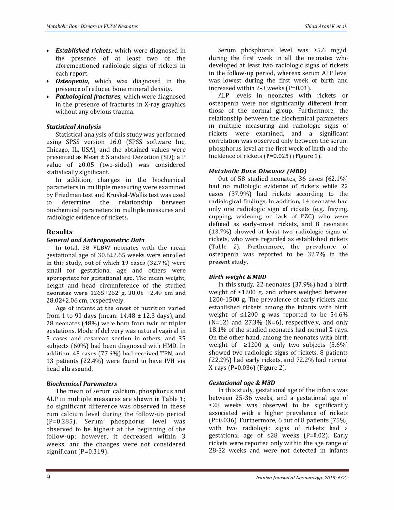
The mean of serum calcium, phosphorus and ALP in multiple measures are shown in Table 1; no significant difference was observed in these rum calcium level during the follow-up period (P=0.285). Serum phosphorus level was observed to be highest at the beginning of the follow-up; however, it decreased within 3 weeks, and the changes were not considered significant (P=0.319).
Serum phosphorus level was ≥5.6 mg/dl during the first week in all the neonates who developed at least two radiologic signs of rickets in the follow-up period, whereas serum ALP level was lowest during the first week of birth and increased within 2-3 weeks (P=0.01).
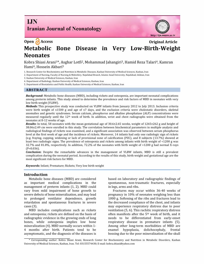
ALP levels in neonates with rickets or osteopenia were not significantly different from those of the normal group. Furthermore, the relationship between the biochemical parameters in multiple measuring and radiologic signs of rickets were examined, and a significant correlation was observed only between the serum phosphorus level at the first week of birth and the incidence of rickets (P=0.025) (Figure 1).
Metabolic Bone Diseases (MBD)
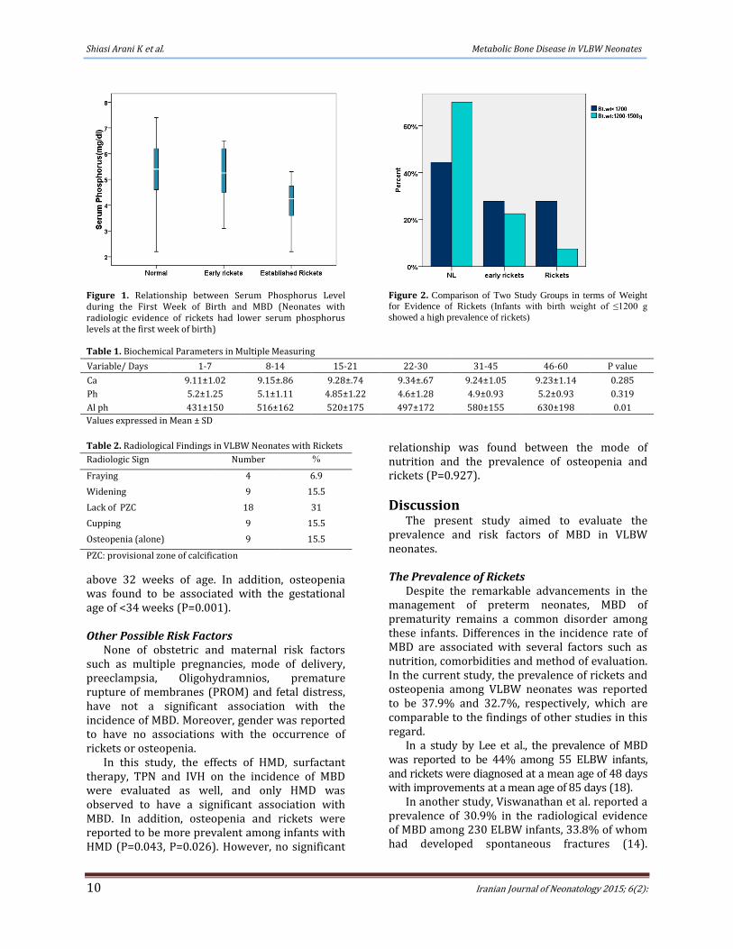
Out of 58 studied neonates, 36 cases (62.1%) had no radiologic evidence of rickets while 22 cases (37.9%) had rickets according to the radiological findings. In addition, 14 neonates had only one radiologic sign of rickets (e.g. fraying, cupping, widening or lack of PZC) who were defined as early-onset rickets, and 8 neonates (13.7%) showed at least two radiologic signs of rickets, who were regarded as established rickets (Table 2). Furthermore, the prevalence of osteopenia was reported to be 32.7% in the present study. Birth weight & MBD
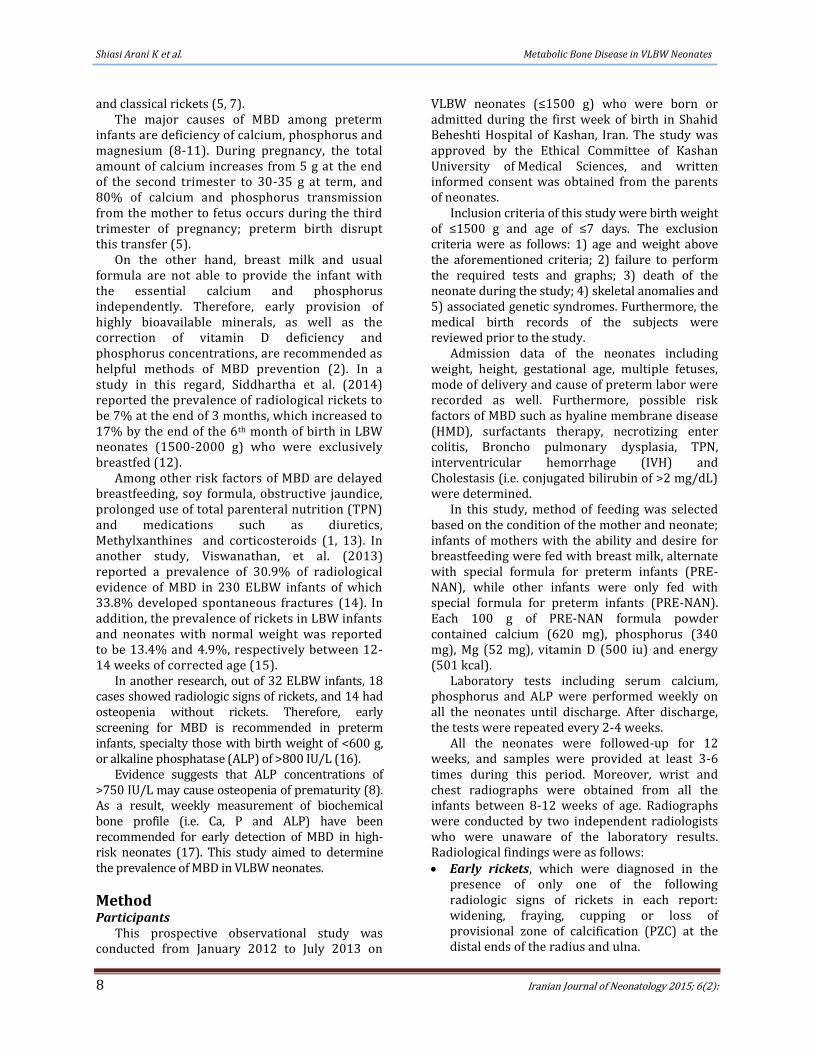
In this study, 22 neonates (37.9%) had a birth weight of ≤1200 g, and others weighed between 1200-1500 g. The prevalence of early rickets and established rickets among the infants with birth weight of ≤1200 g was reported to be 54.6% (N=12) and 27.3% (N=6), respectively, and only 18.1% of the studied neonates had normal X-rays. On the other hand, among the neonates with birth weight of ≥1200 g, only two subjects (5.6%) showed two radiologic signs of rickets, 8 patients (22.2%) had early rickets, and 72.2% had normal X-rays (P=0.036) (Figure 2). Gestational age & MBD
In this study, gestational age of the infants was between 25-36 weeks, and a gestational age of ≤28 weeks was observed to be significantly associated with a higher prevalence of rickets (P=0.036). Furthermore, 6 out of 8 patients (75%) with two radiologic signs of rickets had a gestational age of ≤28 weeks (P=0.02). Early rickets were reported only within the age range of 28-32 weeks and were not detected in infants
Shiasi Arani K et al. Metabolic Bone Disease in VLBW Neonates
Iranian Journal of Neonatology 2015; 6(2): 10
Figure 1. Relationship between Serum Phosphorus Level during the First Week of Birth and MBD (Neonates with radiologic evidence of rickets had lower serum phosphorus levels at the first week of birth)
Figure 2. Comparison of Two Study Groups in terms of Weight
for Evidence of Rickets (Infants with birth weight of ≤1200 g
showed a high prevalence of rickets)
Table 1. Biochemical Parameters in Multiple Measuring
P value 46-60 31-45 22-30 15-21 8-14 1-7 Variable/ Days
0.285 9.23±1.14 9.24±1.05 9.34±.67 9.28±.74 9.15±.86 9.11±1.02 Ca
0.319 5.2±0.93 4.9±0.93 4.6±1.28 4.85±1.22 5.1±1.11 5.2±1.25 Ph
0.01 630±198 580±155 497±172 520±175 516±162 431±150 Al ph
Values expressed in Mean ± SD
Table 2. Radiological Findings in VLBW Neonates with Rickets
% Number Radiologic Sign
6.9 4 Fraying
15.5 9 Widening
31 18 Lack of PZC
15.5 9 Cupping
15.5 9 Osteopenia (alone)
PZC: provisional zone of calcification
above 32 weeks of age. In addition, osteopenia was found to be associated with the gestational age of <34 weeks (P=0.001). Other Possible Risk Factors
None of obstetric and maternal risk factors such as multiple pregnancies, mode of delivery, preeclampsia, Oligohydramnios, premature rupture of membranes (PROM) and fetal distress, have not a significant association with the incidence of MBD. Moreover, gender was reported to have no associations with the occurrence of rickets or osteopenia.
In this study, the effects of HMD, surfactant therapy, TPN and IVH on the incidence of MBD were evaluated as well, and only HMD was observed to have a significant association with MBD. In addition, osteopenia and rickets were reported to be more prevalent among infants with HMD (P=0.043, P=0.026). However, no significant
relationship was found between the mode of nutrition and the prevalence of osteopenia and rickets (P=0.927).
Discussion The present study aimed to evaluate the
prevalence and risk factors of MBD in VLBW neonates. The Prevalence of Rickets
Despite the remarkable advancements in the management of preterm neonates, MBD of prematurity remains a common disorder among these infants. Differences in the incidence rate of MBD are associated with several factors such as nutrition, comorbidities and method of evaluation. In the current study, the prevalence of rickets and osteopenia among VLBW neonates was reported to be 37.9% and 32.7%, respectively, which are comparable to the findings of other studies in this regard.
In a study by Lee et al., the prevalence of MBD was reported to be 44% among 55 ELBW infants, and rickets were diagnosed at a mean age of 48 days with improvements at a mean age of 85 days (18).
In another study, Viswanathan et al. reported a prevalence of 30.9% in the radiological evidence of MBD among 230 ELBW infants, 33.8% of whom had developed spontaneous fractures (14).
Metabolic Bone Disease in VLBW Neonates Shiasi Arani K et al.
Iranian Journal of Neonatology 2015; 6(2): 11
Furthermore, the prevalence of rickets among LBW infants was reported to be 13.4% while it was 4.9% among neonates with normal weight within 12-14 weeks of corrected age (15).
In another research, rickets were diagnosed in 32 ELBW neonates with radiographic evidence of rickets and the associated risk factors (e.g. ALP >800 IU/L, TPN >3-4 weeks, or clinical suspicion). Of these neonates, 18 cases had radiologic rickets and 14 had osteopenia without rickets.
According to other studies, the prevalence of radiologic rickets is approximately 50% among infants with birth weight of <600 g, while it is 14% in neonates with birth weight of 600-800 g and 4% in infants with birth weight of 800-1000 g (P<0.01) (16). The lower prevalence of rickets reported in the present study might be due to the selective assessment of newborns; however, rickets may be diagnosed incidentally (19).
Birth weight and Gestational age
In the present study, birth weight and gestational age were important predictors of MBD; accordingly, gestational age of <28 weeks was observed to be significantly associated with a higher prevalence of rickets.
Moreover, early rickets were detected only within the gestational age of 28-32 weeks, and infants ageing over 32 weeks were not reported to have early rickets. On the other hand, osteopenia was associated with the gestational age of <28.5 weeks, and infants weighing less than 1200 g were reported to be more susceptible to early rickets (P=0.036).
Similar to our study, Viswanathan et al. (2013) evaluated the frequency of the radiological evidence of MBD and spontaneous fractures in 230 ELBW infants. According to their findings, neonates with MBD had a lower gestational age and birth weight compared to normal infants (14). In addition, Mitchell et al. indicated that infants born with a birth weight of ≤600 grams were at a higher risk of osteopenia and rickets (16).
Maternal and Obstetric Risk Factors
According to the results of this study, possible obstetric and maternal risk factors associated with rickets include multiple pregnancies, mode of delivery, preeclampsia, oligohydramnios, PROM and fetal distress, have not a significant association with the incidence of MBD. Other studies have variable results on this subject.
On the other hand, the findings of the present study were indicative of a similar prevalence of MBD between the two genders. In a study
conducted on 41 VLBW infants, Funke et al. reported a higher risk of bone disorders among male preterm neonates (20); however, no significant difference was observed between the bone mineral content, which was measured by Dual-energy X-ray absorptiometry, and the gender of the studied neonates (21).
Postpartum Complications
In the current study, we investigated the role of HMD, surfactant therapy, TPN and IVH in the incidence of MBD, and only HMD was found to have a significant association with MBD. This finding is inconsistent with other studies in this regard.
Some of the important risk factors for MBD include long-term TPN (more than 4 weeks) and use of medications such as diuretics and corticosteroids (22). In a research, the presence of Cholestasis, necrotizing enter colitis, Broncho-pulmonary dysplasia, and gastrointestinal perforation were not significantly associated with radiological rickets despite the fact that cholestatic patients were observed to have a higher ALP (16).
Biochemical Parameters
In a study by Chuhan et al. (2011), serum calcium and phosphorus levels were reported to be significantly lower in preterm infants (28-32 weeks), and serum ALP, creatinine and electrolytes were significantly higher in these infants compared to the control group (34-38 weeks) (23).
In preterm infants, serum calcium may not be a useful screening measure for MBD since these neonates are able to maintain normal plasma calcium at the expense of removal from the bone (22). Therefore, no significant correlation could be found between serum calcium and the diagnosis of rickets (16).
Another important predictor of MBD is the level of serum phosphorus. In the present study, serum phosphorus levels declined over time, and the lowest level was observed during the 4th week of birth; however, this level increased along with the improvement of nutrition.
In this study, all the neonates with rickets had serum phosphorus levels of <5.6 mg/dl (1.8 mmol/l), which is consistent with the findings of previous studies in this regard (8, 24).
Preterm infants with serum phosphorus levels of <2 mmol/l are at the possible risk of osteopenia, and serum phosphorus levels of <1.8 mmol/l are strongly associated with radiographic evidence of rickets. Unfortunately, this parameter has a low sensitivity despite its high specificity (8).
Shiasi Arani K et al. Metabolic Bone Disease in VLBW Neonates
Iranian Journal of Neonatology 2015; 6(2): 12
In a study by Mitchell et al. (2009), a direct correlation was observed between serum phosphorus levels and the birth weight of neonates. Furthermore, increased ALP and decreased serum phosphorus were found to be associated with a higher risk of osteopenia and rickets in preterm infants (16).
In another study, Lapillonne et al. (2004) demonstrated that higher intake of calcium and phosphorus by 25% and 40% were likely to increase bone mineral content in preterm infants with gestational age of 28-32 weeks (25).
On the other hand, supplementary calcium and phosphorus are known to reduce the radiographic evidence of rickets in VLBW infants; nevertheless, monitoring for urinary calcium and phosphorus concentrations has been recommended by several researchers (13).
Bone is the origin of 90% of plasma alkaline phosphatase (P-ALP) in infants, which is an indicator of high bone metabolism during this period (26). In all neonates, P-ALP rises within the first 2-3 weeks of birth and increases further in case of mineral deficiency.
However, appropriate mineral supplementation in preterm infants may not be able to reduce P-ALP (27); some researchers have suggested that P-ALP of >500 might be an indicator of possible MBD (22). In the present study, P-ALP levels in neonates with rickets or osteopenia were not significantly different from the control group, which is inconsistent with the findings of other studies in this regard.
In a cohort study conducted on 113 ELBW neonates, P-ALP was observed to have a significant inverse correlation with birth weight, gestational age and serum phosphorus levels; however, it was not significantly higher in infants with radiologic rickets compared to those without rickets (16).
According to some data, provision of minerals in preterm infants could prevent the increase of ALP (1). In a study by Backstorm et al. (2000), it was demonstrated that at the corrected age of 3 months, total ALP activities higher than 900 IU/l have a sensitivity of 88% and specificity of 71% for low bone mineral density (measured by Dual- energy X-ray absorptiometry).
Furthermore, measurements of bone isoenzyme activity were not associated with
higher diagnostic value. In the same study, serum phosphate levels of <1.8 mmol/l (5.6 mg/dl) were associated with low bone density (96% of specificity and 50% of sensitivity), and a
combination of these two criteria yielded a sensitivity of 100% at a specificity of 70% (24).
One of the limitations of the present study, similar to other researches, was the unavailability of DXA device appropriate for the determination of bone density in infants. In addition, wrist X-ray is essential for the accurate diagnosis of rickets. Another limitation of this study was the measurement of total ALP activity without evaluating bone isoenzyme activity although it may not have improved the diagnostic value (24).
Due to financial limitations, we not measured parathyroid hormone and 25-hydroxy vitamin D level. Therefore, it is recommended that future studies be conducted with large sample sizes on the evaluation of the relationship between MBD and maternal and neonatal vitamin D deficiency.
Conclusion Despite the remarkable advances in the
management of VLBW infants, MBD remains a prevalent and important medical issue in the field of neonatology.
Acknowledgement Hereby, we extend our gratitude to Kashan
University of Medical Sciences for their support of this research project. This study was extracted from a research proposal (No. 9121) in the Department of Pediatric Endocrinology.
Conflict of Interest
The authors declare no conflict of interest, and no funding was received for this research project.
References 1. Lothe A, Sinn J, Stone M. Metabolic bone disease of
prematurity and secondary hyperparathyroidism. J Paediatr Child Health. 2011; 47:550-3.
2. Pieltain C, de Halleux V, Senterre T, Rigo J. Prematurity and bone health. World Rev Nutr Diet. 2013; 106:181-8.
3. Sharma M, Sohi I. Linear and skull growth in extremely low birth weight babies with rickets of prematurity. Indian J Pediatr. 2012; 79:655-8.
4. Chen HY, Chiu LC, Yek YL, Chen YL. Detecting rickets in premature infants and treating them with calcitriol: experience from two cases. Kaohsiung J Med Sci. 2012; 28:452-6.
5. Greenbaum LA. Rickets and Hypervitaminosis D.. Kliegman RM, editor. 19th ed. Philadelphia: ELSEVIER; 2011. P:208 .
6. Tomlinson C, McDevitt H, Ahmed SF, White MP. Longitudinal changes in bone health as assessed by
Metabolic Bone Disease in VLBW Neonates Shiasi Arani K et al.
Iranian Journal of Neonatology 2015; 6(2): 13
the speed of sound in very low birth weight preterm infants. J Pediatr. 2006; 148:450-5.
7. Rustico SE, Calabria AC, Garber SJ. Metabolic bone disease of prematurity.Journal of Clinical & Translational Endocrinology. 2014; 1:85-91.
8. Grataroli E. Newborn Ostheopenia; 2014. Available from: http://flipper.diff.org/app/items/6486.
9. Mihatsch W, Trotter A, Pohlandt F. Calcium and phosphor intake in preterm infants: sensitivity and specifity of 6-hour urine samples to detect deficiency. Klin Padiatr. 2012; 224:61-5.
10. Pereira-da-Silva L, Costa A, Pereira L, Filipe A, Virella D, Leal E, et al. Early high calcium and phosphorus intake by parenteral nutrition prevents short-term bone strength decline in preterm infants. J Pediatr Gastroenterol Nutr. 2011; 52:203-9.
11. Wesol-Kucharska D, Laskowska J, Sibilska M, Friedman-Gruszczynska J, Blonska M, Gawecka A, et al. [Prevention of osteopenia in premature infants]. Med Wieku Rozwo. 2008; 12:924-32.
12. Siddhartha S, Sharma S, Arya S, Chellani H, Roy N. Metabolic bone disease in low birth weight babies between 1500 and 2000 g on exclusive breast feeding. Astrocyte. 2014; 1:75-9.
13. Maas C, Pohlandt F, Mihatsch WA, Franz AR. [Prevention of bone mineral deficiency in premature infants: review of the literature with focus on monitoring of urinary calcium and phosphate]. Klin Padiatr. 2012; 224:80-7.
14. Viswanathan S, Khasawneh W, McNelis K, Dykstra C, Amstadt R, Super DM, et al. Metabolic bone disease: a continued challenge in extremely low birth weight infants. JPEN J Parenter Enteral Nutr. 2013; 38:982-90.
15. Agarwal R, Virmani D, Jaipal ML, Gupta S, Gupta N, Sankar MJ, et al. Vitamin D status of low birth weight infants in Delhi: a comparative study. J Trop Pediatr. 2012; 58:446-50.
16. Mitchell SM, Rogers SP, Hicks PD, Hawthorne KM, Parker BR, Abrams SA. High frequencies of elevated alkaline phosphatase activity and rickets exist in extremely low birth weight infants despite current nutritional support. BMC pediatr. 2009; 9:47.
17. Dokos C, Tsakalidis C, Tragiannidis A, Rallis D. Inside the “fragile” infant: pathophysiology, molecular
background, risk factors and investigation of neonatal osteopenia. Clin Cases Miner Bone Metab. 2013; 10(2):86-90.
18. Lee SM, Namgung R, Park MS, Eun HS, Park KI, Lee C. High incidence of rickets in extremely low birth weight infants with severe parenteral nutrition-associated cholestasis and bronchopulmonary dysplasia. J Korean Med Sci. 2012; 27:1552-5.
19. Rennie LM, Beattie TF, Wilkinson AG, Crofton P, Bath LE. Incidental radiological diagnosis of rickets. Emerg Med J. 2005; 22:534-7.
20. Funke S, Morava E, Czako M, Vida G, Ertl T, Kosztolanyi G. Influence of genetic polymorphisms on bone disease of preterm infants. Pediatr Res. 2006; 60:607-12.
21. Rohana J, Hasmawati J, Zulkifli SZ. Risk factors associated with low bone mineral content in very low birth weight infants. Singapore Med J. 2007; 48:191-4.
22. Harrison CM, Johnson K, McKechnie E. Osteopenia of prematurity: a national survey and review of practice. Acta Paediatr. 2008; 97:407-13.
23. Chauhan SS, Sarkar PD, Bhimte B. Prematurity and related biochemical outcomes: study of bone mineralization and renal function parameters in preterm infants. Biochemistry Res Int. 2011; 2011.
24. Backstrom MC, Kouri T, Kuusela AL, Sievanen H, Koivisto AM, Ikonen RS, et al. Bone isoenzyme of serum alkaline phosphatase and serum inorganic phosphate in metabolic bone disease of prematurity. Acta Paediatr. 2000; 89:867-73.
25. Lapillonne A, Salle BL, Glorieux FH, Claris O. Bone mineralization and growth are enhanced in preterm infants fed an isocaloric, nutrient-enriched preterm formula through term. Am J Clin Nutr. 2004; 80:1595-603.
26. Boceanu E, Ilie C, Enatescu I, Iacob D. Metabolic bone disease of prematurity. Revista Societăţii Române de Chirurgie Pediatrică. 2013:42.
27. Torabi Z, Moemeni N, Ahmadiafshar A, Mazloomzadeh S. The effect of calcium and phosphorus supplemtation on metabolic bone disorders in premature infants. J Pak Med Assoc. 2014; 64:635-9.