ontogenetic changes in the skull of the european wildcat ... · pdf fileontogenetic changes in...
TRANSCRIPT

281© Museum für Tierkunde Dresden, ISSN 1864-5755, 18.07.2012
62 (2) 2012281 – 294
Ver tebrate Zoology
Ontogenetic changes in the skull of the European wildcat (Felis silvestris Schreber, 1777)
Clara Stefen 1 & DietriCh heiDeCke ✝ 2
1 Senckenberg Naturhistorische Sammlungen Dresden, Museum für Tierkunde, Königsbrücker Landstraße 159 clara.stefen(at)senckenberg.de
2 Institut für Zoologie der Martin Luther Universität Halle-Wittenberg, Zoologische Sammlungen, Domplatz 4, 06099 Halle/Saale
Accepted on January 20, 2012.
Published online at www.vertebrate-zoology.de on July 06, 2012.
> Abstract The postnatal changes of the skull of the male and the female European wildcat are described in detail using linear measure-ments and geometric morphometrics. Overall, the analysis of the linear measurements and of the geometric morphometrics of the landmark data indicate the same postnatal changes in the skull morphology, which are, however, described better by geometric morphometrics. The changes mainly affect the caudal part of the skull and, in the mandible, the coronoid and the angular process as well as the ventral rim. Three growth phases can be distinguished particularly on the basis of the linear measurements with the fastest growth and the most obvious changes in the skull occurring from birth to about 7 months, slowed down growth from about 7 to 14 months, and only little growth from 14 to about 24 months of age, when the growth curves for most linear measurements level off. None of the easy and non-destructive means of the linear measurements em-ployed was found to clearly determine the age of a skull.
> ZusammenfassungDie postnatalen Veränderungen des Schädels von weiblichen und männlichen Europäischen Wildkatzen werden anhand von linearen Messungen und mit Hilfe der Geometrischen Morphometrie im Detail beschrieben. Insgesamt lassen sich die postnatalen Veränderungen des Schädels mit beiden Methoden darstellen, doch werden sie mit der Geometrischen Morphometrie besser illustriert. Drei Wachstumsphasen werden anhand der linearen Maße deutlich unterschieden: 1. Von Geburt bis etwa 7 Monate mit dem schnellsten Wachstum und den deutlichsten Veränderungen der Form des Schädels. 2. Von etwa 7 bis 14 Monaten mit langsameren Wachstum. 3. Von 14 bis etwa 24 Monaten mit nur geringem Wachstum; die adulten Werte sind für die meisten Parameter erreicht. Im Schädel betreffen Veränderungen vor allem den caudalen Bereich, in der Mandibel den Coronoid und Angular Fortsatz sowie den ventralen Rand. Keine der genutzten linearen Messstrecken eignete sich als einfache Methode zur individuellen Altersbestimmung des Schädels.
> Key wordsPostnatal changes, geometric morphometrics.
Introduction
The outer and cranial morphology of the European wildcat (Felis silvestris Schreber, 1777) has been dealt with for centuries (e.g. summarized by Piechocki, 1990; Stefen & Görner, 2009). One of the major concerns has often been the differentiation of wild and domestic cats (e.g. SchauenberG, 1969, 1977; kratochvíl & kratochvíl, 1970; DanielS et al.,
1998; beaumont et al., 2001; reiG et al., 2001). The variability within populations has been less focussed on (e.g. kratochvíl, 1973; SláDek et al., 1971, 1972; Stefen & heiDecke, 2011). Ontogenetic changes in the skull morphology of wild and domestic cats have, in particular, been indicated by kratochvíl (1973) and krüGer et al. (2009). So far, no direct correlations be-

C. Stefen & D. HeiDecke: Ontogenetic changes in the skull of Felis silvestris 282
tween the morphological changes and the age of an individual specimen have been documented in detail for wildcats. The aim of this study was to describe in more detail the postnatal changes in the skull morphology of wild-cats by considering the age of both sexes and, further-more, to see which areas of the skull change the most, how fast these changes occur, and whether they dif-fer between the sexes. Classical linear measurements and correlations to age were used as well as the meth-ods of geometric morphometrics as the latter permit a good quantification and visualization of shape change (Drake & klinGenbrG, 2008). One aspect of studying the linear measurements was to find a non-destructive and easy-to-use tool to determine the unknown age of wildcats in museum and institute collections. Skull de-velopment will be discussed in relation to life history events.
Materials and methods
Materials
The 67 wildcats, 40 males (m) and 27 females (f), in the collection of the Zoological Institute of the Mar-tin-Luther-Universität Halle-Wittenberg in Halle were used in this study. Furthermore, several cats from other collections were included: 5 (3 f, 2 m) from the Sen-ckenberg Forschungsinstitut and Naturmuseum Frank-furt and 3 from the Museum für Naturkunde Berlin. The specimens ranged from 3 to 78 months in age (Fig. 1). All specimens originated from the Harz region or from Thuringia. The wildcats were distinguished from domestic cats by cranial volume, cranial index, and, when avail-able, the intestine length learned from the museums’ records. According to Piechocki (1990), the cranial volume of wildcats ranges from 32.5 – 50 cm3 and for domestic cats from 20 – 35 cm3, hence, a cranial vol-ume of > 35 cm3 can be used to identify wildcats. For cats with a cranial volume of 32 – 35 cm3 the cranial index (= greatest total skull length : cranial volume) must be calculated to clearly identify the type of cat. SchauenberG (1969) used the cranial index to dif-ferentiate wild and domestic cats: a cranial index of < 2.75 is indicative of wildcats whereas one of > 2.75 is indicative of domestic cats (for skulls with fully developed adult dentition). In cases where neither the cranial volume nor the intestine length were available,
a combination of the glabella, the location of the pala-tine foramen, the nasal length, and the angular process were used for the determination. Prior to the present study the determination of the individual age had been performed by counting the cement layers at the roots of the maxillary canines (Piechocki & Stiefel, 1988). The age of the specimens were taken from this publication. For the geometric morphometric analysis, photo-graphs were taken of the skulls and mandibles from the collection in Halle by placing them on a cushion to have the respective aspect of interest parallel to the camera. The sample that could be used consisted of 37 mandibles (m 23, f 14), 39 skulls in the dorsal view (m 23, f 16), 42 skulls in the ventral view (m 26, f 16) and 44 skulls in the lateral view (m 30, f 14). As far as pos-sible, all the individual specimens were photographed from the same side. In a few cases, mirror images of the other side had to be used for the digitization of the landmarks.
Methods
Linear measurements were taken in partial accordance with french et al. (1988), kratochvíl (1973), and YamaGuchi et al. (2004). The measurements are listed in Table 1 and illustrated in Figure 2. These data were used in Pearson correlation analyses against age per-formed with SPSS 16.
Fig. 1. Frequency of the known age of the studied individuals separated according to sex.

283Vertebrate Zoology n 62 (2) 2012
ing variables could no statistically significant correla-tion to age be observed in either sex: bn, afor, schind, and for the tooth measurements LP4, BP4, CH, Cl, pml, lp3, lp4, lm1, cl, and ch. From the scatter diagrams of the variables opposite age it can be derived that the increase in size for near-ly all the variables with growth takes place the fastest during the first few months. The curves indicate three basic growth phases: the fastest growth from birth to about 7 months, slowed down growth from about 7 to about 14 months, and only little growth from 14 months to about 24 months of age, when the curves level off. Particularly ZW, ZwM1, bCa, mandl_1, mandl_2, corh, and y17 continue to grow with even a slight increase in CranV. For a few variables the lowest level of the approxi-mate adult size range is reached at about 7 months: cond, bullh, which is, however, very variable, and shbull.
Shape
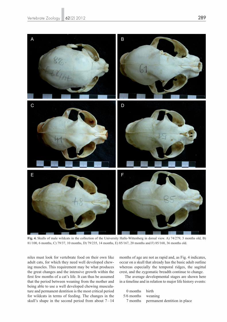
The overall shape of the skull changes markedly par-ticularly within the first 5 – 7 months. The skull of ju-veniles, particularly newborns, but also of up to about three-month-old wildcats, is well rounded and oval in shape (including the zygomatic arches) in the dorsal view (Fig. 4). It appears compact and well inflated in the lateral view (Fig. 5). With growth, the skull elongates, particularly in the caudal cranial region, the orbits appear to tilt more, and the frontal flattens whereas the anteriodorsal naso-frontal-maxillar region becomes a bit rounder. In the dorsal view of grown wildcats the overall shape is more complex than in newborns, the cranium appears rounded whereas the frontal with the postor-bital processes, the nasals, and the developing sagit-tal crest at the corners forms a squared rhombic roof. The zygomatic arches extend far, laterally encasing the large orbitae and the temporal fossae. Whereas in very young skulls the largest zygomatic width is at the orbita, it is at the distal part of the zygomatic arc at the part that encloses the temporal fossa in skulls from about 6 months of age. The development of the nuchal and the sagittal crests starts in male wildcats at about 6 – 10 months of age (Fig. 3), and the temporal ridges are developed at about 5 to 6 months. The temporal fossa increases markedly in size from birth to about 6 months of age and continues to increase as the zygomatic width con-tinues to widen, yet more slowly.
For the geometric morphometric analyses, 18 land-marks (LM) were chosen on the dorsal and the ventral aspect of the skull and the lateral view of the mandible and 20 on the lateral aspect of the skull as illustrated in Figure 3 and listed in Table 2. Overall landmarks were chosen to cover the entire structure of the skull and the mandible. However, concerning the mandible, the coronoid, the condyle, and the angular process as well as the course of the mandible joining them were focused on as relations between them are assumed to be particularly important for making the distinction between wild and domestic cats. The aim was to look for easily identifiable landmarks in each specimen. For this, junctions of sutures in the skull, extreme points, and maximum or minimal curvatures were selected as landmarks. The digitalization of the landmarks was carried out using the TPSdig2 software (SUNY Stony Brook, http://life.bio.sunysb.edu/morph/index.html). These co - or di nate data were imported into the MorphoJ pro-gram (klinGenberG, 2010; http://www.flywings.org.uk/MorphoJ_page.htm). This program was then used for all further shape analyses. First, the Procrustes su-perimposition was used to rescale the landmark con-figurations to a standard size, position, and orientation. In this way the centroid size of the landmark configu-ration was computed as the square root of the sum of the squared distances of all the landmarks from their centroid. For the analysis of the changes during the ontogeny, multivariate regressions of the Procrustes coordinates were performed on the respective vari-ables. To test for digitalization errors, the landmarks on six mandibles were digitized four times with none of them indicating digitization errors.
Results
Linear measurements
The postnatal changes of male wildcats are illustrated by several photographs (Fig. 4, 5) as well as in scatter diagrams for some of the cranial parameters (Fig. 6). The Pearson Correlations between the cranial varia-bles and the individual age show a clear correlation of most variables and age, with most of them increasing, but with dtempr decreasing. A marked sexual dimor-phism can also be seen, with the males showing more correlations to age and a stronger statistical signifi-cance, than the females (Table 3). Only for the follow-

C. Stefen & D. HeiDecke: Ontogenetic changes in the skull of Felis silvestris 284
the nasals. The distal end of the nasals, the maxilla-frontal-nasal meeting, and the anterior orbital rim stay in place relative to each other, but the distance of the orbits to the nasals increases slightly. In the ventral view (Fig. 7B) the shape changes are more the same all over the skull but smaller in the anterior-most re-gion. They include the widening of the skull at the zygomatic arch, the relative movement of the choana towards the anterior and thus the relative shortening of the palate. As LM 10 and LM 11 move towards each other with age, a decrease in the height of the fora-men magnum could be assumed since formh is very variable and shows a correlation to age only in males. Postnatal changes are also apparent in the lateral view of the skull (Fig. 76C) and affect the caudal part of the
Geometric morphometrics
The sexual dimorphism was clearly supported by the results of discriminant analyses with the Procrustes superimposition of the landmark data for all the views of the skull and the mandible. The dorsal aspect of the skull (Fig. 7A) shows that most of the postnatal shape changes occur in the cau-dal part of the skull, including the development of the nuchal and the sagittal crest, the change of the rela-tive position of the fronto-parietal sutures (LM 3), the broadening of the skull at the postorbital process and in the zygomatic width, and a few changes in the fa-cial part of the skull, particularly in the elongation of
Fig. 2. Schematic illustration of linear measurements taken as explained in table 1.

285Vertebrate Zoology n 62 (2) 2012
Table 1. List of abbreviations and explanation of measurements taken.
gsl greatest skull length, from inion (point where the two superior nuchal crest meet in sagittal plane) to prosthion (maxillary bone at alveoles of incisors in sagittal plane), nearest waycbl condylobasal length (condylion, furthest extension of condyles to inion) zw zygomatic width, maximumzwM1 zygomatic width at M1, measured from ventral nucr maximum width across nuchal cresthsb maximum cranial width (maximum across squamosum)dtempr distance between frontoparietal ridges at their intersection with frontoparietal suturesiob interorbital width measured between grooves of angularis occuli weinsbn width of nasal bones at premaxilla/maxilla/nasal suturesnasapw maximum internal width of nasal aperturenasaph maximum internal height of nasal aperture (might be oblique)cranh cranial height from porion (dorsal most point of auditory meatus) to bergma (intersection of frontoparietal sutures at sagittal plane)lsagcr potential length of sagittal crest, from intersection of frontoparietal sutures to inionbop width across orbital processespob width across postorbital constrictionskullh skull height from condyles to inionln_1 length of nasal bones at midlineln_2 maximal length of nasal bonesfacl facial length, from prosthion to nasion (caudal end of nasals) vertorb maximal vertical diameter of orbithororb horizontal diameter across orbithzvorb vertical distance from orbital to maxillary bone between P4 and M1bCa width of maxillary bone at caninesbCi distance between caninesrostb rostral breadth, maximal width of snoutpw palate width measured between M1 (tips of callipers tugged in between palate and M1 from distal in ventral view)acbull width across bullae from porion to porionformw maximal width of foramen magnumformh height of foramen magnum (occasionally occurring high notches of the foramen are not included in the measurement)cond maximal width across occipital condylesnpalno distance from internal nares to anterior palatal notchpalnobull distance from anterior palatal notch to depression of the tympanic bulla at base of styloid processbulll length of tympanic bullabullw width of tympanic bullabullh height of tympanic bulla, from top of auditory meatus to maximal ventral extension of bullaozrl length of maxillary tooth row from distal of canine to distal end of P4lP4 length of P4 crownbP4 width of P4 crownCh height of crown of maxillar caninus from alveole to tipCl length of crown of maxillar caninus afor distance between foramen lacerum and foramen ovaleshbull height of skull measured vertically above bullae tympanicacranV cranium volume, measured to the nearest 0.5 cm3 using glass beads of 1 mm diameter mandls length of mandible measured from the alveoles of the incisors at sagittal plane (pogonion) to the caudal end of angular processmandl length of mandible measured from pogonion to condyle parallel to ventral rim of mandiblecorh height of mandibular ramus at coronoid process
angfd from angular process to an imaginary line extended from condyle and coronoid process; positive values indicate an angular process not extending as far caudally as this line, negative values indicate coronoid process extending not as far caudally as angular process
mand height of mandibular ramus between p4 and m1uzrl length of mandibular tooth row from distal end of caninus to distal end of m1pm1 length of p-m1 (at alveoles)lp3 length of p3 at alveolelp4 length of p4 at alveolelm1 length of crown of m1 (measured from above)cl length of mandibular caninusch height of mandibular caninus from alveole to tipy4 distance from prosthion to middle of infraorbital forameny17 greatest width across both P4 y6 length of tooth row from P2-P4kliob interorbital breadth measured at shortest distance between orbits

C. Stefen & D. HeiDecke: Ontogenetic changes in the skull of Felis silvestris 286
krüGer et al. (2009), and GarcíaPerea et al. (1996). They are documented here in more detail (Fig. 4, 5) as well as being discussed in relation to life history events. The postnatal changes in the expression, the form, and the course of the temporal ridges and the sagittal crest, including the development of a lyrate form as well as the development of a nuchal crest are e.g. also known from lynxes (GarciaPerea, 1996), whose development is very similar to that of wildcats. They are further known also from other mammals such as beavers (hinze, 1950), where the postnatal changes of the temporal ridges and the sagittal crest help deter-mine the age of specimens (Stefen, 2009). The nearly constant values of the tooth measure-ments result from the fact that only permanent teeth were included in the analyses as their size does not change with age; only the height of the canines chang-es due to wear. In the ventral view of the skull, the changes of the rim of the foramen magnum are indicated with geo-metric morphometrics (Fig. 7B), which is not support-ed in the correlations to age of the linear measurements of formh and formw (Tab. 3). It is therefore assumed that the apparent age-related decrease in size of the fo-
skull more than the anterior part. The changes associ-ated with the development of the nuchal and the sagit-tal crest as well as the overall flattening of the roof of the skull (LM 5, 6) are prominent. The elongation of the zygomatic arch and the cranium (LM 11 – 16) and some changes associated with the dental arch (LM 18, 19, 1) are also indicated. The shape changes of the mandible (Fig. 7D) in-clude nearly all the landmarks but differ in direction and magnitude. The coronoid process increases in height, the angular process in length, and the condyle shifts slightly towards the posterior. The ascending ra-mus of the mandible also broadens (LM 5, 11).
Discussion
The changes in the shape and the size of the skull of wildcats, from neonate to older age, were indicated particularly in the dorsal view by kratochvíl (1973),
Fig. 3. Schematic illustration of the landmarks used in dorsal, ventral and lateral view of the skull as well as on lateral side of the mandible. Explanations of landmarks in table 2.

287Vertebrate Zoology n 62 (2) 2012
9 – 10 months for males (Piechocki, 1990). The last period with the slowest growth lasts until the age of about 20 – 24 months. For domestic cats, fusion of the ossification centres of bones occurs between 14 and 20 months of age, with a possible delay of the upper limit (Smith, 1969). For wildcats, SchauenberG (1980) stat-ed that “physical maturity, which is the stage reached when the process of ossification of the epiphyses is completed, arrives between 18 and 19 months.” The findings of the reduced growth of the skull with its growth apparently coming to an end at about two years of age which, in the present study, was observed for wildcats correspond well to Schauenberg’s observa-tions concerning domestic cats and wildcats. Only
ramen magnum reflects a slight shift in the angle of the occipital plane to the basiocranium. In the present study, this angle was not measured on the skulls. Three periods of growth are suggested of which the first one, marked by very fast growth, is completed at about 7 months of age. A few variables seem to reach the lowest level of the approximate adult range at this age at which permanent dentition is completed (conDé & SchauenberG, 1978). From this age onwards there is no constraint on the mandible and the skull to increase in size in accordance with the teeth, or alveoles. The second period of growth lasts up to an age of about 14 months, which is shortly after sexual maturity has been reached – at about 12 months for females and
Table 2. List of landmarks chosen for geometric morphometric analysis.
landmarks on dorsal aspect of skull
01 anterior point of midline between nasals02 posterior most point of midline between nasals03 meeting of frontoparietal sutures 04 anterior most end of sagital crest if developed (otherwise identical to next landmark)05 distal most point of midline of skull06 prominent bulging of nuchal crest07 end of nuchal crest08 lateral most extension of skull 09 posterior end of zygomatic arch10 laterodistal most extension of zygomatic arch11 point anterior of orbital process of zygomatic arch12 anteriolateral rmost extension of zygomatic arch13 ‘depression’ anterior to zygomatic arch14 extension of alveole of Caninus15 suture of maxillary, nasal and frontal bone16 where maxillary intersects orbital rim17 tip of orbital process18 point of postorbital constriction
landmarks on ventral aspect of skull
01 between first incisors in midline02 at apparently ‚deepest‘ point between incisor and canines03 at posterior end of alveole of Caninus04 at anterior onset of zygomatic arch 05 at slight depression of zygomatic arch06 at lateral most extension of zygomatic arch07 at postriolateral most extension of zygomatic arch 08 at postorbital constriction09 at condyle at foramen magnum 10 distal most extension of foramen magnum in midline 11 anterior most extension of foramen magnum in midline12 at anterior end of choane or posterior end of palatine bone in midline13 premaxillary-maxillary suture in midline14 caudal end of incisive foramen15 anterior most notch of palate16 anterior end of bulla17 extension of bulla towards midline18 caudal end of bulla
landmarks on lateral aspect of skull
01 at anterir rim of alveole of incisor02 at lateral tip of nasal03 at midline of nasal bones 04 at distal most extension of nasals05 at ‚knickungspunkt‘ of skull (more or less in extension of rim of orbital process)06 at parietal-frontal suture at midline07 anteriormost end of sagital crest if there is any08 most distal point of skull09 extension of occipital10 condyle11 distal point of bulla12 vental point of bulla13 anterioventral point of external auditory meatus14 dorsoposterior most point of external auditory meatus15 extension of squamosal rim16 jugal-maxillary suture at dorsal rim of zygomatic arch17 dorsal rim of maxillary below zygomatic arch18 posterior end of alveole of P419 greatest extension of curvature anterior to P320 anterioventral rim of orbita
landmarks on lateral side of mandible
01 midline between incisors of both mandibles02 tip of incisor03 distal rim of alveole of incisor04 anterior rim of alveole of premolar teeth (p3)05 depression just distal of carnassial (m1)06 most dorsally extended tip of coronoid process07 most distally extended tip of coronoid process08 border of visible „thickening“ of coronoid process09 maximal anterior curvature of ramus between coronoid and condyle10 anterior point of condyle11 distal most point of condyle12 maximal anterior curvature of ramus between condyle and angular process13 distal most tip of angular process14 ventral most curvature of angular process15 ventral most curvature below anterior part of masseteric fossa16 ventral rim below between p4 and m117 curvature below diastema, below point 418 position of distal most foramen mentale
C. Stefen & D. HeiDecke: Ontogenetic changes in the skull of Felis silvestris 288
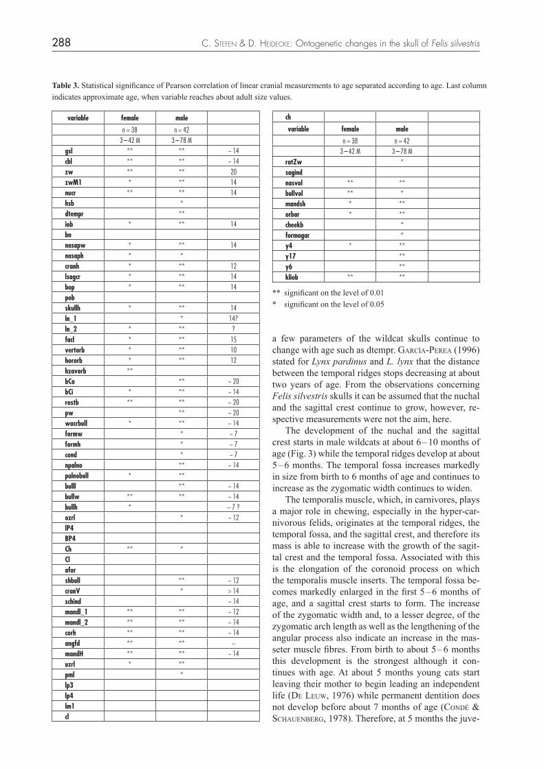
Table 3. Statistical significance of Pearson correlation of linear cranial measurements to age separated according to age. Last column indicates approximate age, when variable reaches about adult size values.
variable female male
n = 38 n = 423 – 42 M 3 – 78 M
gsl ** ** ~ 14cbl ** ** ~ 14zw ** ** 20zwM1 * ** 14nucr ** ** 14hsb *dtempr **iob * ** 14bnnasapw * ** 14nasaph * *cranh * ** 12lsagcr * ** 14bop * ** 14pobskullh * ** 14ln_1 * 14?ln_2 * ** ?facl * ** 15vertorb * ** 10hororb * ** 12hzavorb **bCa ** ~ 20bCi * ** ~ 14rostb ** ** ~ 20pw ** ~ 20wacrbull * ** ~ 14formw * ~ 7formh * ~ 7cond * ~ 7npalno ** ~ 14palnobull * **bulll ** ~ 14bullw ** ** ~ 14bullh * ~ 7 ?ozrl * ~ 12lP4BP4Ch ** *Claforshbull ** ~ 12cranV * > 14schind ~ 14mandl_1 ** ** ~ 12mandl_2 ** ** ~ 14corh ** ** ~ 14angfd ** ** ~mandH ** ** ~ 14uzrl * **pml *lp3lp4lm1cl
ch
variable female male
n = 38 n = 423 – 42 M 3 – 78 M
ratZw *sagindnasvol ** **bullvol ** *mandsh * **orbar * **cheekb *formagar *y4 * **y17 **y6 **kliob ** **
** significant on the level of 0.01 * significant on the level of 0.05
a few parameters of the wildcat skulls continue to change with age such as dtempr. GarcíaPerea (1996) stated for Lynx pardinus and L. lynx that the distance between the temporal ridges stops decreasing at about two years of age. From the observations concerning Felis silvestris skulls it can be assumed that the nuchal and the sagittal crest continue to grow, however, re-spective measurements were not the aim, here. The development of the nuchal and the sagittal crest starts in male wildcats at about 6 – 10 months of age (Fig. 3) while the temporal ridges develop at about 5 – 6 months. The temporal fossa increases markedly in size from birth to 6 months of age and continues to increase as the zygomatic width continues to widen. The temporalis muscle, which, in carnivores, plays a major role in chewing, especially in the hyper-car-nivorous felids, originates at the temporal ridges, the temporal fossa, and the sagittal crest, and therefore its mass is able to increase with the growth of the sagit-tal crest and the temporal fossa. Associated with this is the elongation of the coronoid process on which the temporalis muscle inserts. The temporal fossa be-comes markedly enlarged in the first 5 – 6 months of age, and a sagittal crest starts to form. The increase of the zygomatic width and, to a lesser degree, of the zygomatic arch length as well as the lengthening of the angular process also indicate an increase in the mas-seter muscle fibres. From birth to about 5 – 6 months this development is the strongest although it con-tinues with age. At about 5 months young cats start leaving their mother to begin leading an independent life (De leuw, 1976) while permanent dentition does not develop before about 7 months of age (conDé & SchauenberG, 1978). Therefore, at 5 months the juve-
289Vertebrate Zoology n 62 (2) 2012
months of age are not as rapid and, as Fig. 4 indicates, occur on a skull that already has the basic adult outline whereas especially the temporal ridges, the sagittal crest, and the zygomatic breadth continue to change. The average developmental stages are shown here in a timeline and in relation to major life history events:
0 months birth 5/6 months weaning 7 months permanent dentition in place
niles must look for vertebrate food on their own like adult cats, for which they need well developed chew-ing muscles. This requirement may be what produces the great changes and the intensive growth within the first few months of a cat’s life. It can thus be assumed that the period between weaning from the mother and being able to use a well developed chewing muscula-ture and permanent dentition is the most critical period for wildcats in terms of feeding. The changes in the skull’s shape in the second period from about 7 – 14
Fig. 4. Skulls of male wildcats in the collection of the University Halle-Wittenberg in dorsal view. A) 74/279, 3 months old, B) 81/100, 6 months, C) 79/37, 10 months, D) 79/235, 14 months, E) 85/167, 20 months and F) 85/168, 36 months old.
A B
C D
E F

C. Stefen & D. HeiDecke: Ontogenetic changes in the skull of Felis silvestris 290
since the estimators on the mandible, as given by raDinSki (1981), were not measured. It can however be deduced from Fig. 7C that the moment arm or in-lever of the superficial masseter (distance from the tip of the coronoid process to the condyle) and of one portion of the temporalis muscle (distance from the condyle to the ventral rim of the angular process, raDinSki, 1981) increase but cannot be put in rela-tion to the out-lever, the carnassial (distance from the mandibular condyle to the bite point). “Carnassial function in young lynxes is assumed by dP3 and dp4, but this function shifts to P4 and m1 in adults, the morphology of pP3 and dp4 being similar to that of P4 and m1” (tumilSon & macDaniel, 1984). This shift occurs in other carnivores such as domestic cats (Gaunt, 1959) and Felis silvestris in a similar way (García-Perea et al., 1996). The sample size, the high overall variability – pro-bably at all age stages – and the rapid development in the first 5 – 6 months most likely render it difficult to differentiate between the shape changes of the skull in wildcats prior to and after weaning. Such dif-ferences were noted in e.g. spotted hyenas, Crocuta crocuta (tanner et al., 2010). The authors argue for “protracted skull development, with adult morphology not achieved until well after reproductive maturity” (p. 359). The sample of aged specimens with intact skulls available for this study was small, therefore, a larg-er sample should be aimed at in future. There were hardly any specimens older than 40 months (3 years) although the maximum possible age of wildcats was once estimated at up to 16 years (Piechocki & möller, 1983), however currently it is assumed to be about 6 years (büttner, 1994). For the initial growth, the ma-jor growth spurt, and the shape changes the unavail-ability of aged specimens may not be problematic, but for the changes in the development of the nuchal and the sagittal crest as well as dtempr it would be desirable to be able to follow them up to old age. Furthermore, using a larger sample and including the indicators of the lever arms of the masseter and the temporalis muscles according to raDinSki (1981) might help to analyse the development of these muscles and their relative mechanical advantage as well as and the more detailed pre- and post-weaning differences in the skull development also for wildcats.
Aging of individuals
One aim of this study was to find linear measurements that could be used for the non-destructive age deter-mination of skulls. Only a few variables were of po-
9 – 12 months reproductive maturity 20 months adult skull shape
In lynxes, the development of the sagittal crest begins at the end of the first year, when [the young] adopt an independent way of life (GarcíaPerea, 1996). This is clearly later than in wildcats, but cor-responds to the adoption of an independent life in a similar way and supports the importance of the tem-poralis muscle. During the ontogeny, the in-levers as well as the out-levers of the masseter and the temporalis muscle increase with skull size. Whether there is a mechani-cal advantage to these two chewing muscles, and if, to what extent, cannot n be clearly determined here
Fig. 5. Skulls of male wildcats in the collection of the University Halle-Wittenberg in lateral view. A) 87/125, first weeks, but not exactly aged, B) 86/47 7 months, can C) 86/25, 2 years old.
A
B
C

291Vertebrate Zoology n 62 (2) 2012
published measurements for comparability were fol-lowed, would be the length of the developed sagittal crest. Given the overall variability for all the measure-ments taken, it can be assumed that the development and the size of the sagittal crest would also prove to be too variable for a good age determination. Another non-destructive but also time-consuming method is the radiography of the mandible and the
tential interest in this respect, particularly dtempr was considered as it was obvious to narrow with age when studying the skulls (also indicated in García-Perea et al., 1996). However, as in all variables, the strong overall variability (Stefen & heiDecke, 2011) renders it unsuitable for the direct age-determination of speci-mens. Another possible measurement for age deter-mination, nevertheless not considered here as other
age months50403020100
110
100
90
80
70malefemale
age months50403020100
27.5
25.0
22.5
20.0
17.5
age months50403020100
36
34
32
30
age months50403020100
35
30
25
20
15
10
5
gsl bCa
pob dtempr
A
mm
mm
mmmm
B
C Dage months50403020100
110
100
90
80
70malefemale
age months50403020100
27.5
25.0
22.5
20.0
17.5
age months50403020100
36
34
32
30
age months50403020100
35
30
25
20
15
10
5
gsl bCa
pob dtempr
A
mm
mm
mmmm
B
C D
Fig. 6. Scatter diagrams of some cranial variables and age (in months) of the specimens separated according to sex. A) Greatest skull length (gsl), B) width across Canines (BCa), C) width of postorbital constriction (pob), D) distance between frontoparietal crests at their intersection with coronal suture (dtemp).

C. Stefen & D. HeiDecke: Ontogenetic changes in the skull of Felis silvestris 292
5 4
74 9
18
1710 11
12
2
10
16
7
65
15
12
4
32
14
131
8
11
9
7
6 5
20
17
18
19 1
2
34
168
1514
13
12
1110
9
7
8
6
9
5
15 161413
12
2
3 14
18 17
1011
18
17
15
14
1
13
16
6
3
Fig. 7. Regressions of shape changes with age and illustrated shape change for wildcat skull and mandible. A) skull in dorsal view, B) skull in ventral aspect, C) skull in lateral aspect, and D) mandible. Size factor for all shape changes 100, only for lateral view 1000.
A
B
C
D

293Vertebrate Zoology n 62 (2) 2012
Skulls of Lynxes, Genus Lynx (Mammalia: Carnivora). – J. Morph., 229: 241 – 254.
GarcíaPerea, R., baquero, R.A., fernánDez-SalvaDor, & GiSbert, J. (1996): Desarrollo Juvenil de Craneo en las Pobla-ciones Ibericas de Gato Montes, Felis silvestris Schreber, 1777. – Donana, Acta Vertebrata, 23(3): 153 – 164.
GuStafSon, G. (1950): Age determinations on teeth. – J. Am. Dent. Ass. 41: 45 – 54.
klinGenberG, C.P. (2010): MorphoJ: an integrated software package for geometric morphometrics. – Molecular Ecology Resources, doi: 10.1111/j.1755-0998.2010.02924.x
kratochvíl, J. & kratochvíl, Z. (1970): Die Unterscheidung von Individuen der Population Felis s. silvestris aus den West karpaten von Felis s. f. catus. – Zool. Listy, 19(4): 293 – 302.
kratochvíl, Z. (1973): Schädelkriterien der Wild- und Haus-katze (Felis silvestris silvestris Schreber 1777 und Felis s. f. catus L. 1758). – Acta Sci. Nat. Brno, 7: 1 – 50.
krüGer, M., hertwiG, S.T., JetSchke, G. & fiScher, M.S. (2009): Evaluation of anatomical characters and the question of hybridization with domestic cats in the wildcat popula-tion of Thuringia, Germany. – J. Zool. Syst. Evol. Res., 47: 268 – 282.
kvam, T. (1984): Age determination in European lynx Lynx lynx by incremental lines in canine tooth cementum. – Acta Zool. Fenn., 171: 221 – 223.
meachen-SamuelS, J.A. & binDer, W.J. (2010): Sexual dimor-phism and ontogenetic growth in the American lion and sabertoothed cat from Rancho La Brea. – J. Zool., 280: 271 – 279.
Piechocki, R. (1990): Die Wildkatze Felis silvestris. – Neue Brehm Bücherei A. Ziemsen Verlag, Wittenberg, 232 S.
Piechocki, R. & möller, H. (1983): Über das Ansprechen, den Schutz und die Lebensweise der Wildkatze I, II, Schluß. – Unsere Jagd, 33: 14 – 15, 52 – 53, 82 – 83.
Piechocki, R. & Stiefel, a. (1988): Über die Altersstruktur der Verluste der Wildkatze (Felis silvestris Schreber 1777) – Hercynia N. F., 25: 235 – 258.
raDinSki, L.B. (1981): Evolution of skull shape in carnivores. I. Representative modern carnivores. – Biol. J. Linn. Soc., 15: 369 – 388.
reiG, S., DanielS, M.J. & macDonalD, D.W. (2001): Cranio me-tric differentiation within wild-living cats in Scotland using 3D morphometrics. – J. Zool. Lond., 253: 121 – 132.
SchauenberG, P. (1969): L’identification du Chat forestier d’ Eu rope, Felis s. silvestris Schreber 1777, par une méthode ostéo métrique. – Rev. Suisse Zool., 76: 433 – 441.
SchauenberG, P. (1977): Longueur de l’intestin du chat fores-tier Felis silvestris Schreber. – Mammalia, 41(3): 357 – 30.
SchauenberG, P. (1980): Note sur le squelette et la maturité physique du chat forestier Felis silvestris Schreb. – Revue Suisse Zool., 87: 549 – 556.
SláDek, J., mošanSký, A. & PalášthY, J. (1971): Die Varia bi-li tät der Schädelkapazität bei der Westkarpaten-Population der Wildkatze, Felis silvestris Schreber, 1777. – Zool. Listy, 20(2): 153 – 160.
lower canine and the measurement of the pulp cavity opening. The older the specimen, the more filled is the pulp cavity (GuStavSon, 1950). binDer et al. (2002) showed that this method yields similar results as the methodology of counting cementum annuli (e.g. kvam 1984; Piechocki & Stiefel, 1988) which was used for lions and fossil saber-toothed cats (meachen-SamuelS & binDer, 2010).
Acknowledgement
I would very much like to thank Mr. T. Datzmann, Dresden, for taking most of the digital pictures, Dr. C. klinGenberG, Man-chester, for his help with the geometric morphometrics ana lysis, and the reviewers for their helpful comments. Mrs. or riSon, Dresden, checked the English.
References
beaumont, M., barrat, E.M., Gottelli, D., kitchener, A.C., DanielS, M.J., PritcharDS, J.K. & bruforD, M.W. (2001): Genetic diversity and introgression in the Scottish wild-cat. – Molecular Ecology, 10: 319 – 336.
binDer, W.J., thomPSon, W.N., & van valkenburGh, B. (2002): Temporal variation in tooth fracture among Rancho La Brea dire wolves. – J. Vertebrate Paleontology, 22: 423 – 428.
büttner, K. (1994): Katzenjammer vorbei? Bilanz des Wie der-ansiedlungsprojektes Wildkatze in Nordbayern. – National-park, 85(4): 17 – 21.
conDé, B. & P. SchauenberG (1978): Remplacement des cani-nes chez le Chat forestier Felis silvestris Schreb. – Revue Suisse de Zoologie, 85(2): 241 – 24.
DanielS, M.J., balharrY, D., hirSt, D., kitchener, A.C. & aSPinall, R.J. (1998): Morphological and pelage characteris-tics of wild living cats in Scotland: implications for defin-ing the ‘wildcat’. – J. Zool. Lond., 244: 231 – 247.
Drake, A.G. & klinGenberG, C.P. (2008): The pace of morpho-logical change: historical transformation of skull shape in St Bernard dogs. – Proc. R. Soc. B, 275: 71 – 76.
fernánDez, E. & loPe, F. de (1990): Sobre el dimorfismo sex-ual en el cráneo de gato montés Felis silvestris Schreber, 1777, en el suroeste ibérico. – Donana, Acta Vertebrata, 17: 213 – 219.
french, D.D., corbett, L.K. & eaSterbee, N. (1988): Mor pho-logical discriminants of Scottish wildcats Felis silvestris, domestic cats F. catus and their hybrids. – J. Zool. Soc. Lond., 214: 235 – 259.
Gaunt, W.A. (1959): The development of the deciduous cheek teeth of the cat. – Acta Anat., 38: 187 – 212.
GarcíaPerea, R. (1996): Patterns of Postnatal Develpment in
C. Stefen & D. HeiDecke: Ontogenetic changes in the skull of Felis silvestris 294
Mechanical Advantage in the Spotted Hyena (Crocuta crocuta). – J. Morph., 271: 353 – 365.
tumilSon, R. & mcDaniel, R. (1984): Morphology, replace-ment mechanisms, and functional conservation in dental re-placement patterns of the bobcat (Felis rufus). – J. Mamm., 65:111 – 117.
YamaGuchi, N., DriScoll, C.A., kitchener A.C., warD, J.M., & macDonalD, D.W. (2004): Craniological differentia-tion between European wildcats (Felis silvestris silvestris), African wildcats (F. s. lybica) and Asian wildcats (F. s. ornata): implications for their evolution and conservation. – Biol. J. Linn. Soc., 83: 47 – 63.
SláDek, J., mošanSký, A. & PalášthY, J. (1972): Variabilität der linearen kraniologischen Merkmale bei der westkarpa-tischen Population der Wildkatze, Felis silvestris Schreber, 1777. – Zool. Listy, 21(1): 23 – 37.
Smith, R.N. (1969): Fusion of Ossification Centres in the Cat. – J. Small Anim. Pract., 10: 523 – 530.
Stefen, C. (2009): Intraspecific Variability of beaver teeth (Ca-storidae: Rodentia). – Zool. J. Linn. Soc., 155: 926 – 936.
Stefen, C. & heiDecke, D. (2011): Kraniometrische Variabilität der Wildkatze (Felis silvestris Schreber, 1777) im Harz-gebiet. – Hercynia N. F., 44: 253 – 285.
Stefen, C. & Görner, M. (2009): Wildkatze in Deutschland und Mitteleuropa – zum Stand der Forschung und Konse-quen zen für den Schutz. – Säugetierkd. Inf., 7(38): 1 – 216.
tanner, J.B., zelDitch, M.L., lunDriGan, B. L. & holekamP, K.E. (2010): Ontogenetic Change in Skull Morphology and