online lupus
TRANSCRIPT
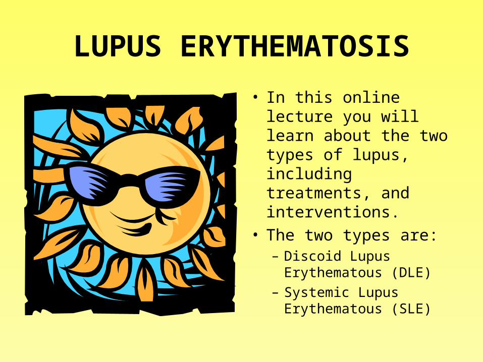
LUPUS ERYTHEMATOSIS
• In this online lecture you will learn about the two types of lupus, including treatments, and interventions.
• The two types are:– Discoid Lupus
Erythematous (DLE)– Systemic Lupus
Erythematous (SLE)

The readings that accompany this lecture are:
• Reading Assignment
• Iggy- pp 429-436


What is Discoid Lupus Erythematous?
• A chronic skin condition of sores with inflammation and scarring favoring the face, ears, and scalp and at times on other body areas.
• These lesions develop as a red, inflamed patch with a scaling and crusty appearance. The center areas may appear lighter in color with a rim darker than the normal skin.
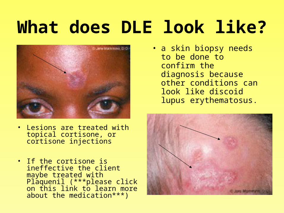
What does DLE look like?
• Lesions are treated with topical cortisone, or cortisone injections
• If the cortisone is ineffective the client maybe treated with Plaquenil (***please click on this link to learn more about the medication***)
• a skin biopsy needs to be done to confirm the diagnosis because other conditions can look like discoid lupus erythematosus.
What causes DLE?
• The exact cause is unknown, but it is thought to be autoimmune with the body's immune system incorrectly attacking normal skin.
• This condition tends to run in families. Females out number males with this condition 3 to 1.
• In some patients with discoid lupus erythematosus, sunlight and cigarette smoking may make the lesions come out.

What interventions will help with DLE?
• Patients whose condition is sensitive to sunlight need to wear a UVA blocking sunscreen daily and a hat while out doors.
• Follow-up with the doctor is important and necessary every six months to once a year to make sure the disease is not spreading to the internal organs and to minimize scarring
• If the client is taking Plaquenil yearly eye
exams are a must.


What is Systemic Lupus Erythematosus (SLE)?
• SLE is a complex chronic connective-tissue disease.
• It affects almost all body systems.• The manifestations are widely variable but they
are thought to be the result of cell and tissue damage caused by the deposition of antigen-antibody complexes in connective tissues.
• SLE can range from a mild, episodic disorder to a rapidly fatal disease process.
What is the epidemiology of SLE?
• Females are affected more than males in a ratio of 9:1
• The disease usually affects women of childbearing age, but can occur at any age.
• It is more common in African Americans, Hispanics, and Asians than it is in Caucasians.
What is the etiology of SLE?• The exact etiology is unknown.• Genetic, environmental and hormonal factors play a role in its
development.• The above statements are idiopathic forms of SLE. It can also be
drug induced by the following medications:• Procainamide • Isoniazid • Hydralazine • Minocycline • Phenytoin • Ethosuximide • D-Penicillamine
– Manifestations of drug induced lupus usually resolve when the medication is discontinued.

What are the initial symptoms of SLE?
• The initial manifestations of SLE are: fatigue, fever, malaise, weight loss, musculoskeletal manifestations similar to arthritis.
• SLE can affect multiple systems. Let’s take a look at each of those systems.
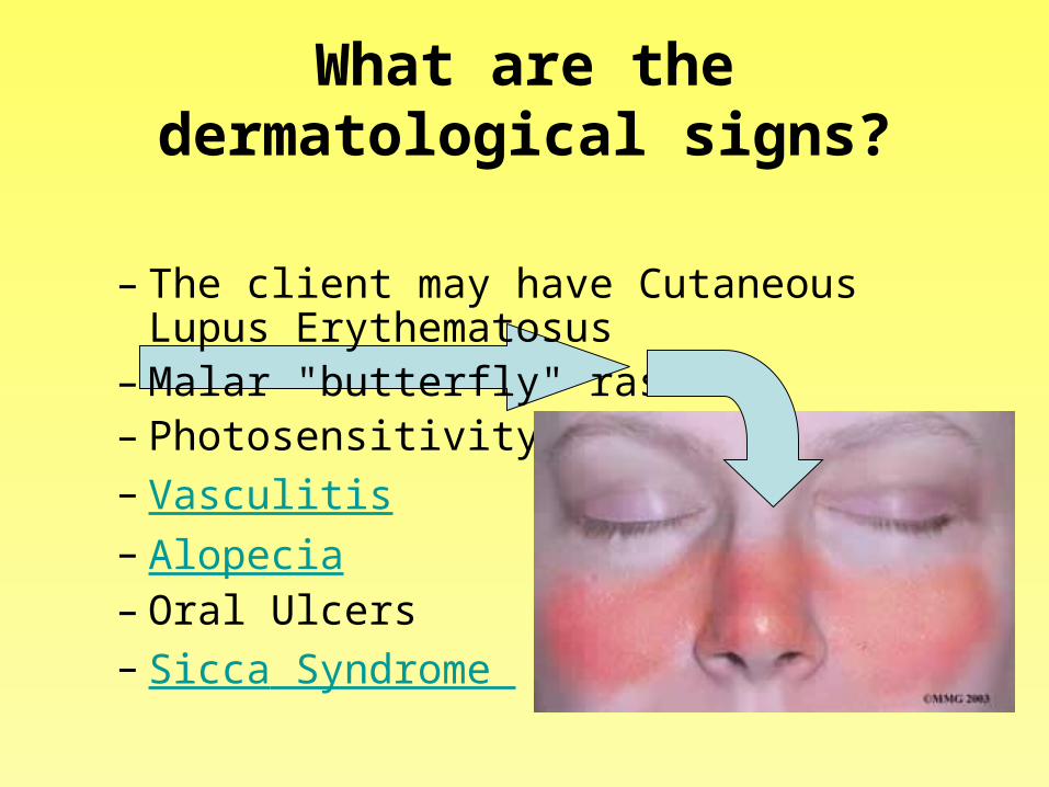
What are the dermatological signs?
– The client may have Cutaneous Lupus Erythematosus
– Malar "butterfly" rash – Photosensitivity – Vasculitis – Alopecia – Oral Ulcers– Sicca Syndrome
What are the neurological symptoms?
A client may have the following symptoms:
• Neuropathies (peripheral and central)
• Seizures• Depression• Psychosis
A client may have the following complications from an exacerbation of SLE:
• CVA• Organic Brain Syndrome
– Intellectual impairment– Memory Loss– Personality Changes– Disorientation
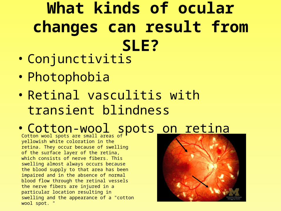
What kinds of ocular changes can result from SLE?
• Conjunctivitis
• Photophobia
• Retinal vasculitis with transient blindness
• Cotton-wool spots on retina
Cotton wool spots are small areas of yellowish white coloration in the retina. They occur because of swelling of the surface layer of the retina, which consists of nerve fibers. This swelling almost always occurs because the blood supply to that area has been impaired and in the absence of normal blood flow through the retinal vessels the nerve fibers are injured in a particular location resulting in swelling and the appearance of a "cotton wool spot. "

What are the musculoskeletal changes with SLE?
• Morning Stiffness
• Arthralgias
• Symmetric Polyarthritis
• Joint Swelling and Effusion

How can SLE effect the renal system?
• Proteinuria• Cellular casts
Potential complications resulting from SLE are:
• Nephrotic syndrome• Renal failure

How can SLE effect the GI system?
• Hepatomegaly
• Anorexia
• Nausea
• Abdominal Pain
• Diarrhea

How can SLE effect the Cardiovascular system?
• Pericarditis• Myocarditis• Endocarditis• Vasculitis• Venous or arterial
thrombosis (anywhere in the body)

How does SLE effect the hematologic system?
• Anemia
• Leukopenia
• Thrombocytopenia
• Splenomegaly

How can SLE effect the Respiratory System?
• Pleurisy
• Pleural effusion
• Pneumonitis
• Interstitial fibrosis
What Lab Assessments help in the diagnosis?
• Initially and ANA titer is completed. *Please remember an ANA titer alone cannot be used to diagnose a disease, it must be used in combination with an evaluation of symptoms and other tests.
• Secondary testing if the ANA titer positive – Complete Blood Count *** pay particular attention to the
WBC, Hgb & Plt counts**
– Coagulation factors– Urinalysis – Serum Creatinine – Antiphospholipid Antibody – Double Stranded DNA Antibody (Anti-dsDNA) – Smith Antibody (Anti-Smith or Anti-Sm)
How is a diagnosis of SLE made?
• A diagnosis of SLE is made when a client has 4 of 11 following criteria: – Malar Rash – Discoid rash – Photosensitivity – Oral Ulcers – Polyarthritis involving more than 2 joints – Pleuritis or Pericarditis – Antinuclear Antibody positive titer– Renal disease – Neurologic disorder (e.g. Seizures, Psychosis) – Anemia, Neutropenia or Thrombocytopenia – Anti-dsDNA, Anti-Sm positive
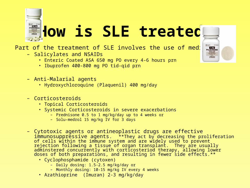
How is SLE treated?Part of the treatment of SLE involves the use of medications
– Salicylates and NSAIDs • Enteric Coated ASA 650 mg PO every 4-6 hours prn • Ibuprofen 400-800 mg PO tid-qid prn
– Anti-Malarial agents • Hydroxychloroquine (Plaquenil) 400 mg/day
– Corticosteroids • Topical Corticosteroids • Systemic Corticosteroids in severe exacerbations
– Prednisone 0.5 to 1 mg/kg/day up to 4 weeks or – Solu-medrol 15 mg/kg IV for 3 days
– Cytotoxic agents or antineoplastic drugs are effective immunosuppressive agents. **They act by decreasing the proliferation of cells within the immune system and are widely used to prevent rejection following a tissue of organ transplant. They are usually adminstered concurrently with corticosteriod therapy, allowing lower doses of both preparations, and resulting in fewer side effects.**
• Cyclophosphamide (cytoxen)– Daily dosing: 1.5-2.5 mg/kg/day or – Monthly dosing: 10-15 mg/kg IV every 4 weeks
• Azathioprine (Imuran) 2-3 mg/kg/day
What else does a client with SLE need?
• A client with SLE also needs Opthamology exams with dilation upon starting steriods or plaquenil and yearly there after.
• Interventions to reduce fatigue.• Sunscreen and other protection against
photosensitivity.• Interventions to prevent infection.• Birth control is critical during exacerbations.
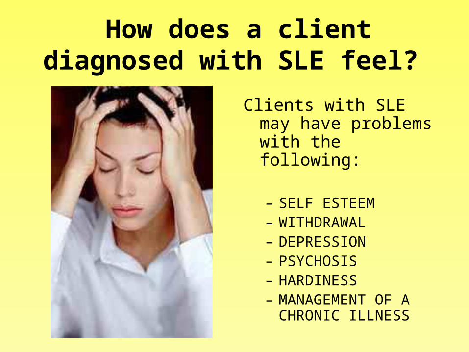
How does a client diagnosed with SLE feel?
Clients with SLE may have problems with the following:
– SELF ESTEEM– WITHDRAWAL– DEPRESSION– PSYCHOSIS– HARDINESS– MANAGEMENT OF A
CHRONIC ILLNESS

What are the outcomes?
Although there is no cure for SLE, the 10 year survival rate is greater than 70% among clients with this disease, which
once was considered fatal in most cases.
• Click on the following link to watch a video about Lupus and initiatives regarding the disease http://www.lupusresearchinstitute.org/video.php

A Case Study
• D.W. is a 23 year old married woman with 3 children under the age of 5. She presented to her physician 2 years ago with vague complaints of intermittent fatigue, joint pain, and low-grade fever. Her physician noted a scaly rash across her nose cheeks, back and chest at that time.
Laboratory Results Revealed
• Positive antinuclear antibody titer• Positive Lupus Erythematosus cell prep• Elevated C-reactive protein• Elevated ESR• Hgb 10 g/dL
Joint X-rays revealed joint swelling in bilateral knees without joint erosion.

What is the diagnosis: systemic lupus erythematosus or
cutaneous lupus erythematosus?
• D.W. was diagnosed with systemic lupus erythematosus.– The malar rash– Polyarthritis– Anemia– Positive ANA titer
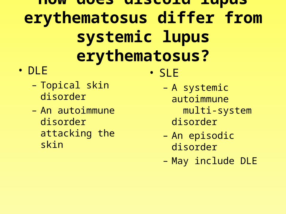
How does discoid lupus erythematosus differ from
systemic lupus erythematosus?• DLE
– Topical skin disorder– An autoimmune
disorder attacking the skin
• SLE– A systemic
autoimmune multi-system disorder
– An episodic disorder– May include DLE
• D.W. was subsequently diagnosed with systemic lupus erythematosus. She was initially treated with Cyclophosphamide (cytoxen) 150mg PO every day and prednisone (Deltasone) 20 mg po every day, bedrest, ice packs, and aspirin to control discomfort.

What priorities need to be addressed with D.W.?
• Monitor blood count with particular attention to WBC and platelet counts. Notify the physician if the WBC’s fall below 4000, or platelets below 75,000.
• Monitor renal & liver function studies• Cytoxen should be administered with food to
minimize gastrointestinal effects• Monitor for signs of abnormal bleeding• Use meticulous hand washing and assess for
signs of infection• Ensure adequate nutrient intake
• D.W. responded well to treatment and resumed her job in environmental services at a large geriatric facility. Eighteen months after diagnosis, D.W. developed puffy hands and feet and increased fatigue. D.W. reported that she had been working longer hours because of the absence of 2 of her fellow workers.
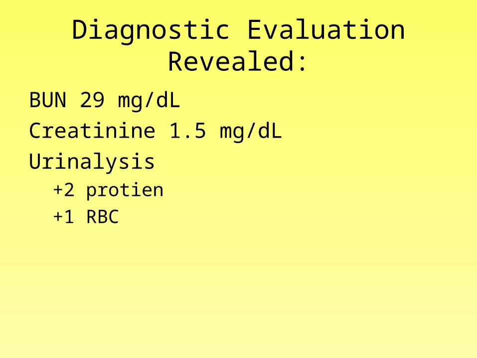
Diagnostic Evaluation Revealed:
BUN 29 mg/dL
Creatinine 1.5 mg/dL
Urinalysis+2 protien
+1 RBC

• What are the significance of these findings?
• What is the relationship of such findings to D.W.’s diagnosis of SLE?
• How will D.W.’s treatment and nursing plan likely to change?
(**Hint – This is a great place to apply your Renal Content****)

• There will be a pass session on Lupus to discuss and questions and talk about the case study. Hope to see you there.
• For any other questions please see Jean or email her at [email protected]