on the optical measurement of corneal thickness
TRANSCRIPT

A C T A O P H T H A L M O L O G I C A V O L . 4 9 1 9 7 1
The Department of Oplithalmology (Head: Pro{. Viggo A. Jensen) Kommunehospitalet, Universi ty of Arlzz~s. DK-8000 Denmark
ON THE OPTICAL MEASUREMENT OF CORNEAL THICKNESS
BY
NlELS EHLERS and FINN KRUSE HANSEN
According to ancient literature the thickness of the central cornea is about one millimeter. This value originates from post mortem anatomical measurements, and corresponds fairly well to the thickness of the maximal swollen human cornea (Elders 1966).
Blix (1880) was the first to perform a direct optical measurement of the cor- neal thickness in a living eye. From measurements on ten eyes he concluded that the central thickness of the cornea is about 0.5 mm in young men, in good ac- cordance with most later authors. In recent years the study of the corneal thick- ness has been taken up, and more simple instruments have been deviced ( V O ~ Z
Bahr 1948, Muurice S- Giardini 1951, Jaeger 1952). The purpose of the present paper is to review the different optical principles
for the measurement of the corneal thickness, and to discuss the use and the errors of the Haag-Streit pachometer, which will be used in some following cli- nical studies.
I Optical principles
1. Successive focusing on specular reflexes. ‘7. Simultaneous observation of specular reflexes. 3. The use of an astigmatic light bundle. 4. Simultaneous observation of doubled specular reflexes. 3. Measurement of the apparent thickness of the optical section.
Received August 5th 1970
65 Act2 Ophthnlmol . -1’3. I 5

I1 The Haag-Streit pachometer
1. The apparatus and its use. 2. The significance of the angle kappa. 3. Measuring accuracy.
I Optical principles
1. Successive focusing on specular reflexes
By successive focusing on the anterior and the posterior surface of the cornea the distance moved by the apparatus is the apparent thickness. From this the real thickness of the cornea can be calculated when the radius of curvature of the anterior surface and the refractive index of the cornea are known. Blix (1880) used two microscope tubes with optical systems of equal power, converging at an angle of 39' towards a point in front of the tubes. The appa- ratus was movable along the line bisecting the angle between the tubes. An illuminated cross in the one tube gave an image at the point of intersection of the microscope axes. The apparatus was at first so adjusted, that this image by reflection in the anterior corneal surface, was observed in the other microscope. By moving the apparatus forwards along the line bisecting the angle between the two microscopes until the image was seen by reflection in the posterior sur- face, the moved distance is the apparent thickness of the cornea. From this the real thickness may be calculated exactly.
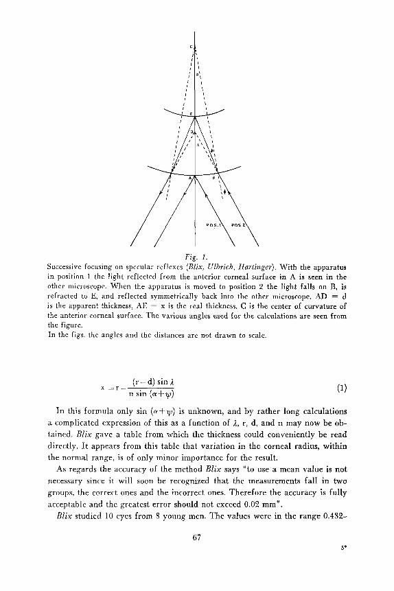
Fig. 1 shows the principle of the method. With the apparatus in position 1 the light reflected from the anterior corneal surface in A is seen in the other microscope. In position 2 the light falls on B, is refracted to E, and reflected symmetrically back into the other microscope. The various angles are seen from the figure. AD = d is the apparent thickness, AE = x is the real thickness.
To perform the calculation we know besides the apparent thickness d, the radius of the anterior corneal surface r, the refactive index n (BZix used 1.375) and the angle A (= 19.5'). From CBE the sine relations give:
sin y r r - x
sin (n-a-y) - - -
from which
r sin y x = r - -- sin ( a + ~ )
sin p, (r-d) sin1 n r
Inserting in this equation sin y = - , and sin p, = the latter
obtained by the sine relations applied to CBD, we obtain:
66
C
, I I
,
I
I
Fig. 1. Successive focusing on specular reflexes (Blix, Ulbrich, Hnrtinger). With the apparatus in position 1 the light reflected from the anterior corneal surface in A is seen in the other microscope. When the apparatus is moved to position 2 the light falls on B, is refracted to E, and reflected symmetrically back into the other microscope. AD = d is the apparent thickness, AE = x is the real thickness. C is the center of curvature of the anterior corneal surface. The various angles used for the calculations are seen from the figure. In the figs. the angles and the distances are not drawn to scale.
(r - d) sin n sin (a+y)
x = r -
I n this formula only sin (a+y) is unknown, and by rather long calculations a complicated expression of this as a function of 2, r, d, and n may now be ob- tained. BZix gave a table from which the thickness could conveniently be read directly. I t appears from this table that variation in the corneal radius, within the normal range, is of only minor importance for the result.
As regards the accuracy of the method BZix says “to use a mean value is not necessary since it will soon be recognized that the measurements fall in two groups. the correct ones and the incorrect ones. Therefore the accuracy is fully acceptable and the greatest error should not exceed 0.02 mm”.
BZix studied 10 eyes from 8 young men. T h e values were in the range 0.482-
67
0.668 mm and if one of his persons who showed both the upper and the lower extreme is excluded, the range in 8 eyes is 0.506-0.576 mm.
The so-called microscopic method of Doaders consists in successive focusing a microscope on the anterior and the posterior surface of the cornea. Considering paraxial rays the real thickness may easily be calculated from the movement of the apparatus (fig. 1). The microscope is in position 1 focused on the anterior surface of the cornea. By moving to position 2 the posterior corneal surface is brought into focus. The apparent thickness (= the displacement of the micro- scope) is d, the real thickness x. At the anterior corneal surface the difference in vergence between the incident and the refracted rays equals the vergence power of the surface. One may therefore write:
1 n n - 1 d x r _ _ _ = -
from which by multiplication with rxd,
ndr r + d (n- 1)
X = (Hartinger 192 I )
This method is simple in use. The readings can be made with an accuracy of 0.1 mm (Hartinger 1921).
The greatest obstacle to the use of the method of successive focusing is the possibility of a movement of the cornea between the two adjustments, which will of course invalidate the result.
Schiwtz (1 913) used this principle for thickness measurements on excised cor- neae. With the cornea placed on a glass hemisphere the focusing on the posterior surface was facilitated, and the possibility of a displacement of the cornea bet- ween the two successive adjustments was avoided. Ulbrich (1914) introduced the use of a slit lamp and a corneal microscope provided with a micrometer drum to measure the displacement of the microscope. Fincham (quoted by Koby 1930) used this principle and in 12 eyes found values from 0.48 to 0.59 mm (mean 0.53 mm), and Sobanski (1934) also using this principle found a value of 0.407 to 0.671 mm for the central cornea.
2. Simultaneous observation of specular reflexes
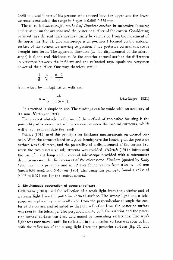
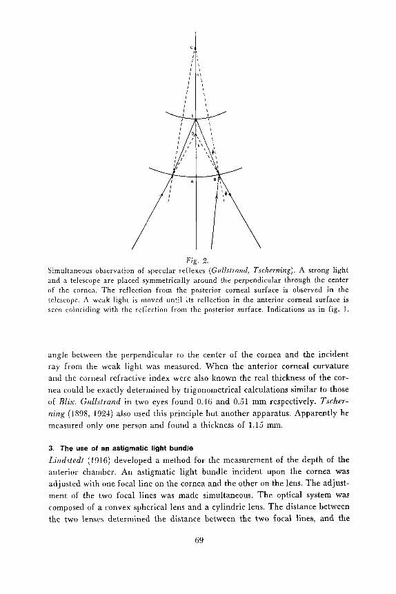
Gz~11~fruizd (1909) used the reflection of a weak light from the anterior and of a strong light from the posterior corneal surface. The strong light and a tele- scope were placed symmetrically 25' from the perpendicular through the cen- ter of the cornea and adjusted so that the reflection from the posterior surface was seen in the telescope. The perpendicular to both the anterior and the poste- rior corneal surface was first determined by coinciding reflections. The weak light was now moved until its reflection in the anterior surface was seen in line with the reflection of the strong light froin the posterior surface (fig. 2). The
68
C
I I I I I
I I I
I
I ,
F i g . 2. Simultaneous observation of specular reflexes (Gullstrmd, Tscherning). A strong light and n telescope are placed symmetrically around the perpendicular through the center of the cornea. The reflection from the posterior corneal surface is observed in the telescope. A weak light is moved until its reflection in the anterior corneal surface is seen coinciding with the reflection froin the posterior surface. Indications as in fig. 1.
angle between the perpendicular to the center of the cornea and the incident ray from the weak light was measured. When the anterior corneal curvature and the corneal refractive index were also known the real thickness of the cor- nea could be exactly determined by trigonometrical calculations similar to those of Blix. GuZZsl,and in two eyes found 0.46 and 0.51 mm respectively. Tscher- ?zing (1898, 1924) also used this principle but another apparatus. Apparently he measured only one person and found a thickness of 1.15 mm.
3. The use of an astigmatic light bundle
Lindstedt (1916) developed a method for the measurement of the depth of the anterior chamber. An astigmatic light bundle incident upon the cornea was adjusted with one focal line on the cornea and the other on the lens. The adjust- ment of the two focal lines was made simultaneous. The optical system was composed of a convex spherical lens and a cylindric lens. The distance between the two lenses determined the distance between the two focal lines, and the
6 9

chamber depth could be calculated. Rosengren (1930) used this apparatus for studies on the chamber depth. A commercially available modification has been described by Steizstrom (1953).
The principle, although it seems accurate apparently has not been used in the study of corneal thickness.
4. Simultaneous observation of doubled specular reflexes
won Bahr (1948) designed an apparatus, in optical principle similar to that of Blix, but permitting simultaneous observation of the anterior and the posterior corneal surface. Plane parallel glass plates were placed in the lower half of the incident and the reflected light (fig. 3 ) . The glass plates were symmetrically movable around vertical axes. With the glass plates perpendicular to the light no refraction takes place, and in this position the apparatus is so adjusted that
I Fig. 3.
Simultaneous observation of doubled specular reflexes. The principle of von Bohr. Plane parallel glass plates in the lower half of the incident and the reflected light bring the reflection from the posterior corneal surface to coincide with that from the anterior. The displacement of the light by the plane parallel glass plate is calculated from FGH. Indications as in fig. 1.
70

the reflection from the anterior corneal surface is seen in the microscope. If the axis of the apparatus (= the line bisecting the right angle between incident and reflected light) coincides with the perpendicular common to both corneal sur- faces, the glass plates may be turned and the reflection from the posterior cor- neal surface seen to coincide with that from the anterior surface.
The displacement a of the light by passing a plane glass plate of thickness b is seen from fig. 3. From A FGH we may write:
a a cos F sin ( 8 - t ) = - -___
F G - b
b sin ( a - E ) a =
cos E
For the calculation of the real thickness of the cornea we may proceed as in the method of Blix . Equation (1) is valid also in this case and by inserting d =
alsin?., the only unknown is sin ( a + y ) , which may again be expressed by a rather complicated formula. uon Bahr gives a graph showing angle of rotation of the glass plate versus corneal thickness for an average corneal radius of 7.8 mm. By way of examples uon Bahr has shown how variation in corneal cur- vature only insignificantly affects the calculated thickness of the cornea.
The accuracy of this method was studied by double determinations on 36 eyes from 18 persons. From this the standard deviation for a single reading was found to be 0.013 mm. In a measurement of 224 eyes from 125 persons a mean value of 0.565k 0.002 mm (mean 4 standard error of mean) was found.
Mazirice &- Giardini (1951) designed an attachment to the Haag-Streit slit lamp model 360. A glass plate was placed on the slit lamp arm between the slit and the focusing lens, and covering the half of the light beam. The arm of the slit lamp and the microscope were fixed at an angle of 50'. The slit was focused on the cornea with the reflection from the anterior surface seen in the microscope. The glass plate was now rotated until the reflection from the posterior surface was seen aligned with that from the anterior (fig. 4). As a theoretical calcula- tion of the real thickness has not been possible the apparatus was empirically calibrated on glass tubes of known wall thickness. This apparatus, which is commercially available (Sterks-Martin, London) has been used extensively in studies on corneal physiology. The theoretical background of the apparatus has recently been treated by Kalberer, W a l z & Griin (1965) and Walz , Gautschi & Griin (1968).
The scale of the apparatus can be read to 0.25', corresponding to a thickness of about 0.005 111171. The accuracy of the method was estimated by taking 25 readings in one position on one eye, resulting in a standard deviation of 0.011 mm. Maurice & Giardini measured the corneae of 44 subjects and gave a value of 0.507 ? 0.0042 mm (mean & standard error of mean, N = 44).
7 1

Fig. 4. Simultaneous observation of doubled specular reflexes. The principle o f Maurice & Gicudini. A plane parallel glass plate (not shown) is placed in the incident light in front of the focusing lens (K). When the glass plate is rotated the reflection from the posterior surface is aligned with that from the anterior surface. Indications as in fig. 1.
Hedbys &- Mishima (1962) designed a similar apparatus for experimental use. The plane glass plate was placed in the reflected light behind a condensing lens. The apparatus was calibrated against the apparatus of Maurice & Giardini.
5. Measurement of the apparent thickness of the optical section
The principle of the following methods is a measurement of the apparent thick- ness of the optical section, which is seen by diffuse reflection from the colloids of the cornea (the Tyndall phenomenon). Dependent upon the angle of inci- dence of the light and the observation angle the real thickness may be calcu- lated in different ways from the measured apparent thickness (juillerat & Koby 1928). The measurement of the apparent thickness is improved and simplified by simultaneous observation of the anterior and the posterior surface as intro- duced by Goldmaizn (1932).
The main optical principles of calculation were treated by ]uillerat & Koby (1928), and two appeared particular simple (figs. 5 & 6). In the first, the optical
72

Fig . 5. Measurement of the apparent thickness of the optical section. The principle of Juillerat E Koby. Payclia, DonaZdson. The optical axis of the microscope coincides with that of thc obscrved cornea, and the apparent thickness of the optical section is measured. Indi- cations as in fig. 1.
axis of the microscope coincides with that of the eye. The angle of incidence of the light upon the cornea may be 45’ (Koby 1928), 71’ (Paycha 1953) or 51.5’ (Donaldson 1968). When the angle of incidence is around 71’ the apparent thickness equals the real thickness as emphasized already by Juillerat & Koby (1928).
Koby (1928) measured 20 subjects and found a value of 0.590 & 0.014 mm (mean & standard error of mean). This high mean value is partly due to the as- sumption of a refractive index of 1.4, and the high standard error probably to the use of an eye-piece micrometer requiring two successive readings to obtain the apparent thickness. Donaldson (1968) measured 268 eyes and gave a value of 0.522 & 0.041 mm (mean k standard deviation, number of statistically inde- pendent values not given). As to the accuracy Donaldson gives “an average de- viation in consecutive readings of 1.20/0”. A part of this material was more thoroughly reported by Martola 8~ Baum (1968).
Jueger (1952) developed the principle illustrated in fig. 6. The apparatus is available as an attachment to the Zeiss slit lamp (Zeiss, Oberkochen) and a simi-
73

c i
Fig. 6 . Measurement of the apparent thickness of the optical section. The principle of juillerat & Koby, Ineger. The light falls perpendicular onto the cornea. The apparent optical section is measured from the side. Indications as in fig. 1.
I I
l a'
I I I I I I
lar, technically improved apparatus is now available for use with the Haag- Streit slit lamp model 900 (Haag-Streit, Berne).
The optical section of the light falling perpendicular onto the corneal surface is observed at an angle of 40'. In the microscope the image of the optical sec- tion is now seen. The thickness of this is measured by aligning the anterior and the posterior corneal surfaces by means of a rotating plane parallel glass plate, covering the lower half of the reflected light. The calculation of the real thick- ness proceeds as in the method of Blix and uon Bahr. Substituting in equation ( 1 ) d = a/sin 1, we have
a r sin I - sin I n sin (c.+y)
x = r - (3)
a is the displacement of the light caused by the glass plate, and known from the angle of rotation (cf. eq. 2). The only unknown factor is therefore again sin (a + y) , which can be exactly calculated. Iaeger provided for different va- lues of r graphs showing the real thickness as a function of the displacement a. Variation in r gave only minor variations in thickness. In the attachment to the
74

Haag-Streit slit lamp the scale is linearly calibrated in mm thickness, and cor- rection for variations in r and in non-linearity is made by a table. Hedbys & Mishima (1968) using the Haag-Streit apparatus found a value of 0.518 t- 0.003 mm in 40 eyes (mean L- standard error of mean, number of subjects not stated). Lowe (1969) found 0.517 k 0.003 mm (mean t- standard error of mean) in 157 eyes from 80 subjects.
The same principle was used by Lavergne & Kelecom (1962), who found a value of 0.51 L- 0.003 mm (mean L- standard error of mean) in 198 eyes, the only difference being an angle of incidence of 60' and the use of the Goldmann "messokular" (1932). The method is discussed by Weekers, Grieten & Lavergne (1961).
Jaeger stated that the angle of rotation of the glass plate could be read with an accuracy of 0.5', corresponding to 0.02 mm in apparent thickness. Honegger Rc Genie (1968) gave for a slightly modified attachment to the Zeiss slit lamp a 95 o / o probability of being within ? 0.01 mm of the real value when taking the mean of 5 readings. By the attachment to the Haag-Streit slit lamp the same accuracy was obtained by 4 readings.
An objection to this principle is that the measurement is made along the line of sight and not along the optical axis. Mishima & Hedbys (1968) proposed the use of two small additional lights, the reflections of which made it possible to perform the measurement with the light falling perpendicular to the anterior corneal surface. In this situation the patient had not to fix the incident light but another movable mark.
I I The Haag-Streit Pachometer
Honcgger S- Genie (1968) made a direct comparison between the instruments at present commercially available, viz, the Maurice-Giardini pachometer, the attachment to the Zeiss slit lamp (in a slightly modified form), and the attach- ment I to the Haag-Streit slit lamp model 900. The latter showed the smallest measuring error, and appears to be the most simple in use.
During the last years we have worked with the Haag-Streit pachometer in clinical and experimental studies. In connection with the preceeding review of the optical principles for the measurement of corneal thickness we have found occasion to present some comments on the use of this pachometer.
1. The apparatus and its use
The principle was given by Juillerat & Koby (1928), but it was particularly and independently developed by jaeger (1952). (see section I, 5 and fig. 6). The apparatus consists of an attachment to the Haag-Streit slit lamp containing two glass plates in front of the right microscope, a lower fixed and an upper, rotat-
75

able around a vertical axis. The incident light comes through a vertical aperture in a diaphragm extending from the attachment, and securing an angle of 40" between the incident light beam and the axis of the right microscope. The right ocular is replaced by a special slit image ocular, dividing the visual field into lower and upper halves. The light passing through the upper rotatable and the lower fixed glass plates is seen in the upper and the lower visual field, re- spectively.
In the microscope the optical section through the cornea is seen. When the upper glass plate is rotated, the upper part of the image of the optical section is displaced (fig. 7). The angle of rotation of the glass plate is a measure of the corneal thickness (cf. eqs. 2 & 3), and is read on a scale directly calibrated in mm. As mentioned in section I, 5 a correction for variation in corneal curvature and non-linearity is necessary. This is obtained from a table supplied by Haag- Streit. This correction is numerically of minor importance except in cases of megalocornea, keratoconus, etc.
An advantage of this apparatus over those using specular reflection from the corneal surfaces is the possibility of measuring total thickness, stromal thickness (Ehlers 1970), or the depth of a stromal clouding or a vessel (fig. 7c). This last will be of importance in planning lamellar keratoplasty. With this principle the microscopic magnification does not influence the measurement (jaeger 1952). and the greater objective magnification (x 1.6) may be recommended. Likewise the refraction of the observer does not influence the measurement (Goldmmn 1968).
2. The significance of the angle kappa
According to the optical principle the incident light should fall perpendicular to the cornea. This is obtained by having the patient fix the incident light. Be- sides introducing the limitation that only central thickness can be measured this gives the possibility of certain measuring errors. T o the trained observer it is to some degree possible from the Tyndall reflection in the optical section to know
Fig. 7 . Diagrammatic illustration of the image seen in the ocular of the Haag-Streit pacho- meter. a. measurement o f total corneal thickness, b. measurement o f stromal thickness,
c. mcasurement of the depth of an opacity.
76

if the patient looks into the light, and this together with the position of the light in the pupil generally gives a sufficient localization.
When the patient fixes the incident light the measurement is made along the line of sight and not along the perpendicular to the cornea. This introduces a measuring error and a systematic difference between right and left eye.
Some optical definitions. As regards the different axis in the eye and the angles between them nearly every author uses his own definitions, and in case of equal defi- nitions often uses different symbols. In the present connection we need to define the line of sight by which we understand the line in the optical system followed by the light ray to the fovea. The localization of this in the optical system is unknown in the individual case, and there is ol course no reason to assume that it goes perpendicular through the center of the cornea. The other line of interest is this line perpendicular through the center of the cornea, along which we want to measure the corneal thickness
Clinically we can measure an angle, here called kappa, between the line of sight and a line perpendicular to the anterior corneal surface and going through the center of the apparent pupil. Where these two lines intersect, if they do so at all, we do not know.
During the use of the pachometer it was noted that often the corneal thick- ness was the greater in the left eye, and in a material of normal eyes (Kruse HnnscJn 197 1) a statistically significant difference could be demonstrated (P< 0.001). In fig. 8 corneal thickness difference between the left and the right eye is plotted against the sum of the angles kappa of the two eyes. A linear regres- sion analysis showed significant correlation (P<O.OOl, R = 0.84).
On the assumption that an angle kappa equivalates a decentered fixation, the
1 I I 0 5 10 15 E r
Fig. 8. Corneal thickness difference between left and right eye plotted against the sum of the angles kappa of the two eyes. Equation of regression ahscissa = degrees, ordinate = m-B.
= 1.62K - 2.3.
77

effect of this was studied by use of a small movable fixation light on the dia- phragm of the pachometer. Fig. 9 shows that a systematic variation occurs. If the subject fixes to the left of the incident light too high values are obtained, and if he fixes to the right too low values are found. This systematic variation, also observed by uon Bahr (1948) probably corresponds qualitatively to the dif- ference observed between right and left eye, and as seen from the figures it is of a considerable magnitude. The thickness difference caused by a given angle is not equal in the two diagrams. This could be because in fig. 9 the decentering occurs around the center of rotation of the eye, whereas in fig. 8 the decentering is caused by different optical errors in the eye. A geometrical calculation of this error is for the same reason rather speculative and any attempt to make a cor- rection for a measured angle kappa has not been performed.
uon Bolzr (1948) also noted a difference between the right and the left eye, caused by a systematic error of the method. With his apparatus the right eye was found to be the thicker. The two principles are, however, so different that no direct comparison is possible, and no controversy exists.
MiJhirna K. Hedbys (1968) have introduced a modification of the pachometer with two small lights placed 40' to the left of the incident light (for the obser- ver). If the light beam falls perpendicular onto the anterior corneal surface the reflection of these small lights will be seen aligned with the corneal epithelium. This modification is theoretically correct, and in addition makes a measure- ment of peripheral thickness possible.
Fig. 9. Systematic variation in the measured thickness when the incident light is not fixed exactly. Right eye of subject with angle kappa 0. As the corneal area studied is within the approximately spherical optical portion of the cornea (Berg 1929, Tscherning 1924) no correction for change in corneal curvature has been performed. abscissa = degrees. ordinate = m-6.
78

On a clinical material, however, a reduction of the standard deviation of the value for the central corneal thickness in a group of subjects is not evident (com- pare values of Mishima & Hedbys and Kruse Hansen 1971). An explanation of this may be the difficulty in fixing steadiIy a small target near to the incident light, which seems necessary in the Mishima-Hedbys modification.
3. Measuring accuracy
When the accuracy of the measurement of the cornea is discussed it must be realized that the thickness varies with the thickness of the precorneal film. This means that the cornea changes its thickness with 1 or 2 p in the period between two blinkings. This obviously rises the question what should be understood by the thickness. This problem is in fact even greater, as there is a priori no reason to assume that methods using specular reflections and methods based on obser- vation of the optical section measure exactly the same distance of a given cornea.
In the methods using measurement of the apparent thickness of the optical section the width of the slit lamp light and the focusing of the microscope will set a theoretical limit for the accuracy. With the relatively low magnification used in the Haag-Streit pachometer this is however no practical problem.
Krirte H a n ~ e n (1971) made 5 determinations of the thickness in 37 subjects. From this the statistical variance of the method may be calculated as the mean of the variances of the 5 readings. Of course, the variation in thickness during the period between two blinkings probably reduces the use of Gaussian statistics to a matter of convention. The values obtained are s,lxt = 0.0085 and s,in =
0.0079. Apparently, there is no difference in the accuracy of the determination of the thickness of the right and the left cornea. The mean value sds = 0.008 mm may be given as the error of a single measurement. This value is similar to that of Honegger & Genie (1968) and a little smaller than the values given by von Balzr (1948) and Maurice & Giardini (1951) for other instruments.
Summary
The different principles used or proposed to be used in the optical measurement of the corneal thickness are reviewed and illustrated in diagrammatic form.
The measurement of the thickness of the apparent optical section of the cor- nea, in the technical elaboration of the attachment to the Haag-Streit slit lamp, is simple in practice, and appears to be the most accurate. The error of the measurement (the standard deviation) is found to be 0.008 mm. With this appa- ratus, however, a small but systematic difference between right and left eye may be demonstrated, caused by the angle kappa between the optical axis of the eye and the line of sight.
79

References
vo/7 Bahr, G. (1948): Measurements of the thickness of the cornea. Acta ophthal., Kbh.
Berg. F. (1929): Vergleichende Messungen der Form der vorderen Hornhautflache mit ophthalmometer und mit photographischer Methode. Acta ophthal., Kbh. 7: 386-423.
Blix, M. (1879-80): Oftalmometriska studier. Uppsala Lakareforenings Forhandlingar
Donddson, D. D. (1966): A new instrument for the measurement of corneal thickness.
Ehlers, N . (1966): Variations in hydration properties of the cornea. Acta ophthal., Kbh.
Ehlers, N . (1970): On corneal thickness and intraocular pressure 11. Acta ophthal., Kbh.
Goldmai~~r , H . (1932): Ein neues Messokular fur die Spaltlampe. Ber. deutsche Ophthal.
Goldniaiz~i, H . (1968): Biomicroscopy of the eye. Amer. J. Ophthal. 66: 789-804. GuIISti-and, A. (1909): Die Dioptrik des Auges. In Handh. der Physiol. Optik (H. von
Helmholtz) 3. Aufl., 1. Bd. Voss, Hamburg. p. 279-282. Hortiriger, H. (1921): Zur Messung der Kammertiefe und des Irisdurchmessers. Zschr.
ophthal. Optik 9: 135-143. Herfbys. B. 0. 8L Mishinzn, S . (1962): Flow of water in the corneal stroma. Exp. Eye
Res. 1 : 262-215. Hoizegger, H . & Genie, E. (1968) : Hornhautdickenmessung. Ein Vergleich verschiedener
Gerate. Graefes Arch. klin. exp. Ophthal. 174: 262-270. Jneger, W. (1 952) : Tiefenmessung der menschlichen Vorderkammer mit planparallelen
Platten (Zusatzgerat zur Spaltlampe). Graefes Arch. Ophthal. 153: 120-131. ]itillo.at & Koby, F . E. (1928): Determination de I’kpaisseur de l a corn& sur le vivant
a u moyen de la lampe B fente. Rev. gen. d’0phtal. 42: 203-227. Kalberer, M., W a l z , D. k Griin, F. (1965): Studien uber optische Methoden zur Be-
stimmung von Hornhautdicken I. Theorie der Messungen mit dem Pachometer. Graefes Arch. Ophthal. 168: 17-32.
Koby, F . E. (1928): De l’epaisseur, mesurCe sur le vivant, des parties centrales de la cornte. Rev. gen. d’Ophta1. 42: 293-296.
Koby , F . E. (1930): A propos de l’epaissseur de la c o d e vivante. Rev. gen. d’0phtal. -14: 222-225.
Krrrse Hrrnsen, F . (1971): A clinical study of the normal human central corneal thick- ness. Acta ophthal., Kbh. 49: 82-89.
Lavrrgnc,, G. k Kelccom, /. (1962): Applications cliniques de la mesure de l’epaisseur de la cornte. Bull. SOC. belge. Ophtal. 131: 323-333.
Liitdsterlt, F. (1916): Ober die Messung der Tiefe der vorderen Augenkammer mittels eines neuen: liir klinischen Gebrauch bestimmten Instruments. Arch. Augenheilk. 80: 104-1 67.
Lowc, X. F . (1969): Central corneal thickness. Ocular corrrelations in normal eyes and those with primary angle-closure glaucoma. Brit. J. Ophthal. 53: 824-826.
Murtuln, E.-L. & Bauni, /. L. (1968): Central and peripheral corneal thickness. Arch. Ophthal., Chicago 79: 28-30.
Mnrtricc. D. M. 8L Giardini, A . A. (1951): A simple optical apparatus for measuring the corneal thickness, and the average thickness of the human cornea. Brit. J. Ophthal. 33: 169-177.
26: 247-265.
15: 349-420.
Arch. Ophthal., Chicago 76: 25-31.
-14: 461471.
48: 1107-1112.
Ges. 49: 435-437 .
80

M i s h i m , S. (1968): Corneal thickness. Survey Ophthal. 13: 57-96. Mishimn. S. k Hedbys, B. 0. (1968): Measurement of corneal thickness with the Haag-
Streit pachometer. Arch. Ophthal., Chicago 80: 710-7 13. Paychn, F . C. (1953): Methode de mesure de I’epaisseur de la cornke. Arch. Ophtal.,
Paris 13: 156-158. Rosengren, B. (1930): Studien iiber die Tiefe der vorderen Augenkammer mit besonderer
Hinsicht auf ihr Verhalten beim primaren Glaukom. Acta ophthal., Kbh. 8: 99-136. Schieti, If. (1913): Optische Mitteilungen. Arch. Augenheilk. 75: 351-352. Sobahski. /. (1934): Die Hornhautdicke in vivo und ihre Bestimmung. Klin. oczna 12:
Stenslriim, S. (1953): An apparatus for the measurement of the depth of the anterior
Tscherning, M . (1898): Optique physiologique. Masson, Paris. Tscherni7zg. M . (1924): Physiologic optics. The Keystone Press, Philadelphia. Ulbricli. H . (1914) : Methode der successiven Scharfeneinstellung. Klin. Mbl. Augen-
heilk. 53: 241. Wrrlz, D.. Gnutsclri, /. k Griiiz, F . (1968): Studien iiber optische Methoden zur Bestim-
mung von Hornhautdicken 11. Messungen an Corneamodellen. Graefes Arch. klin. exp. Ophthal. 176: 13-29.
Weekers, R., Grict~n, /. k Laverg7ae, G. (1961): Etude des dimensions de la chambre anterieurs de I ’ d humain. Premiere partie: Considerations biometriques. Ophthalmo- logica 142: 6.50-662.
317-323. Cited by Zbl. ges. Ophthal. 32: 354-355.
chamber, based on the principle of Lindstedt. Acta ophthal., Kbh. 31: 265-270.
81 Acta Ophthalrnol. 49. 1 ti