olfactory nerve
TRANSCRIPT

Olfactory NerveOlfactory Nerve
Dr Arun OommenDr Arun Oommen

• Anatomy and physiology

The olfactory nerve is a sensory nerve with only one function- smell
First order neurons of olfactory system are bipolar sensory cells
The olfactory receptors are located in the superior posterior nasal septum and lateral wall of the nasal cavity

Olfactory receptors have the unique property to regenerate
Specific odorants stimulate specific receptor cells and specific cells respond to particular odorants
Around 20 central processes are given off from these ciliated cells (filaments of the 1st nerve)


These filaments (olfactory nerve) penetrate the cribriform plate of ethmoid to enter the olfactory bulb. They acquire a sheath of myelin
. In the bulb the olfactory afferent fiber
synapse with the dendrites of the 2nd order neurons called the mitral and tufted cells
At the point of synapse conglomerate of fibres called the olfactory glomeruli are formed


• The axons of the mitral and tufted cells leave the bulb and course posteriorly as the olfactory tract in the olfactory sulcus on the orbital surface of the frontal lobe
• The olfactory tract divide into medial and lateral olfactory stria on either side of the anterior perforating substance


Some of these fibres decussate in the anterior commissure and join fibres from the opposite olfactory pathway. Some go to the olfactory trigone and tuberculum olfactorium (In the APS)

Some of the medial olfactory stria terminate in paraolfactory area, inf part of cingulate gyrus, subcallosal gyrus
•

Other fibres esp the lateral stria supply the ipsilateral piriform lobe of the temporal cortex (primary olfactory cortex) and terminate in the
• uncus,• anterior hippocampal gyrus,•pyriform cortex,• entorhinal cortex,• amygdaloid nucleus,


• The parahippocampal gyrus sent impulse to the hippocampus
• The amygdaloid and hippocampal nuclei (connected on each side thru the ant commissure) sent projecting fibres to the ant hypothalamic nuclei, mamillary body ,tuber cinerum and habenular nucleus

• This in turn project to the thalamus, cingulate gyrus,striatum and mesencephalic reticular formation]
• Olfaction is the only sensation not directly processed in the thalamus
• Connection with the superior and inferior salivatory nucleus is important in reflex salivation


Clinical examination

2 types of deficits
Conductive deficits
Sensorineural/Neurogenic

• Proper history Past head injury Smoking Recent UTI• Systemic illness• Toxins medications and illicit drugs
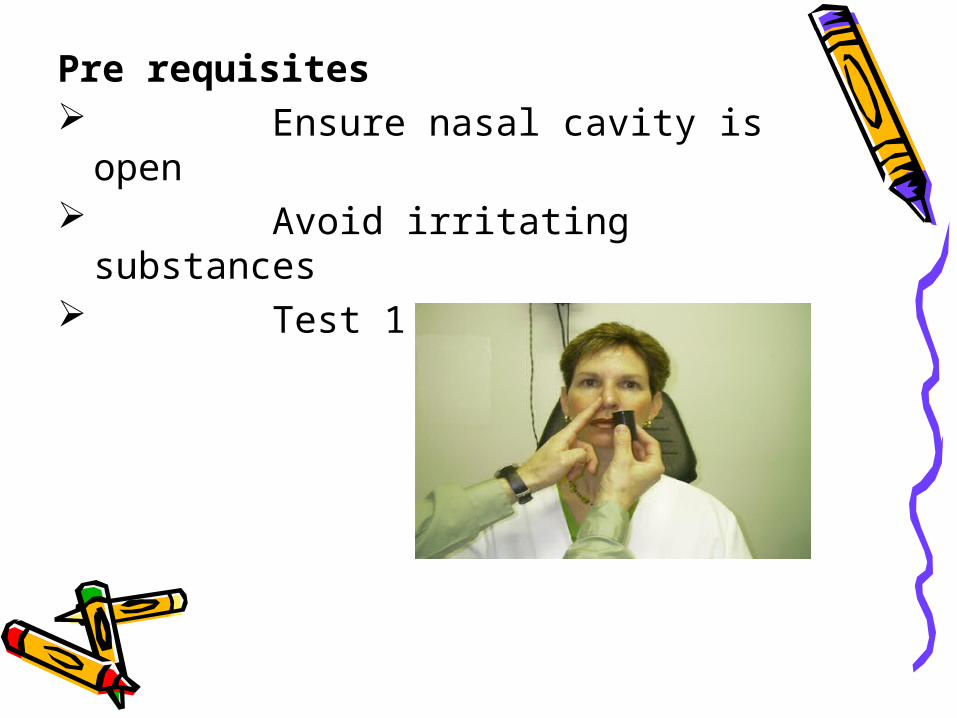
Pre requisites Ensure nasal cavity is open Avoid irritating substances Test 1 nostril at a time

Substances used Cloves, Coffee ,Cinnamon
Commercially available substance like UPSIT (University of Pennsylvania smell identification test)

Unilateral loss of smell is more significant than bilateral
Perception of odor is more important than accurate identification
Perceiving the presence of an odor indicate continuity of the
olfactory pathway
Key points

Identification of odor indicate intact
cortical function
Since there is bilateral innervations, lesion central to decussation does not cause loss of smell and lesion in olfactory cortex does not produce anosmia
The appreciation of presence of smell even without recognition excludes anosmia

Dissorders of olfactory function and Dissorders of olfactory function and localisationlocalisation

Terminologies• Anosmia -Decreased sense of smell• Hyperosmia -Increased sense of smell• Dysosmia -Defective sense of smell• Parosmia -Pervertion of smell• Phantosmia -Perception of smell that
is no real• Presbyosmia -Decresed smell due to
aging• Cacosmia -Inappropriately
disagreeable odor• Coprosmia -Faecal scent• Olfactory agnosia - Inability to identify
detected odors

Conditions causing Conditions causing disturbed olfactiondisturbed olfaction

Congenital-Cleft palate, Downs syn, Turners, Kallmans , Familial dysautonomia
Endocrine/metabolic -adrenal insufficiency ,Diabetes, Hypothyroidism
Iatrogenic-Ethmoidectomy, Hypertelorism, orbitofrontal lobectomy, Radiotherapy, Rhinoplasty, temporal lobectomy, Repair of ACA anuerysm
Infections-HIV, Herpes simplex, UTI Liver diseases -Cirrhosis, hepatitis
Local processes -Hansens disease ,Polyps, Rhinitis, Adenoids, tumors
Neurogenic-Alzheimers disease, Head trauma, Huntingtons disease Migraines, meningiomas,(Foster kennedy syndrome) ,parkinsonism, temporal lobe disease
Psychiatric-Schizophrenia, Hypochondriasis
Uremia
Miscellaneous-Cystic fibrosis ,sarcoidosis ,Occupational exposure ,Refsums disease

Most common causes of anosmia
• Upper resp tract infection
• Head injury (15-30%) 1. Local injury to olfactory nerves at
cribriform plate due to coup or contrecoup forces
2.Temporal/orbito frontal injury
• Nasal and sinus disease
• Idiopathic

• Lesions involving the orbital surface of brain may cause unilateral anosmia
• In meningiomas of olfactory groove or cribriform plate areas unilateral anosmias occur followed by bilateral anosmias
• Parosmias and cacosmias are often due to Psychiatric diseases or may follow head injuries
•

• Olfactory hallucinations are often due to Psychosis but can result from neoplastic or vascular lesions of the central olfactory system or following seizures
• In seizure focus involving medial temporal lobe structures (uncinate or complex partial seizures) often preceded by disagreeable olfactory aura

• Following temporal lobectomy olfactory discrimination is confined to ipsilateral nostrils.
• Following right fronto orbital lobectomy impairment seen in both nostrils
• In olfactory epileptic auras tumors are the most common cause of seizures and the amygdyla is the most likely symptomatic zone

Foster kennedy syndrome
Seen in olfactory groove or sphenoidal ridge meningiomas or frontal lobe ICSOL
3 signs-Ipsilateral anosmia -Ipsilateral optic atrophy -Contralateral papilledema

Pseudo Foster Kennedy syndrome
Seen when increased IC pressure of any cause occur in patients who have previous unilateral optic atrophy
Most commonly seen due to sequential anterior ischaemic optic neuropathy or optic neuritis (optic disc oedema on one side associated with optic disc atrophy on other side)

Kallmann’s syndrome
X linked dissorder
Familial syndrome of permanent anosmia with hypogonadotropic hypogonadism.
Hypoplasia or aplasia of olfactory bulbs and tract
Can be associated with cerebellar ataxia and mirror movements of hands