occlusion in natural dentition
TRANSCRIPT

OCCLUSION IN Natural Dentition
Dr. M. Ahsen Saeed
BDS, BSc
FMH
According to GPT/8 : Occlusion may be defined as :
- Act or process of closure or being closed or shut off.
-Static relationship between incisal and masticatory surfaces of maxillary or mandibular teeth / tooth analogues.
In latin Occ = Up Clusion =Closing
Ahsen Saeed, 1010
Occlusion --> Oc + Claudre
" To Close Up"
The term "occlusion' used in dentistry means contact relationship of teeth in function or parafunction.
However, term refers not only to contact of arches at an occlusal interface but also to all those factors concerned with development and stability of masticatory system and with use of teeth in oral motor behaviour. The primary components of human dental occlusion are :
(a) The dentition
(b) Neuro Muscular System
(c) Cranio facial structures.
The development and maturation of these components are inter-related, so that growth, adaptations and change actively participate in development of adult occlusion.
INTRODUCTION
JABLONSKI (1982) It is relationship between all components of masticatory system in normal function, parafunction and dysfunction including morphology, and functional features of contacting surfaces of opposing teeth and restorations, occlusal trauma, neuromuscular physiology, psychophysiological state and the diagnosis, prevention and treatment of functional disorders.
DYNAMIC OCCLUSION (Davies and Gray)Refers to the occlusal contacts that are made whilst
mandible is moving relative to the maxilla, the mandible being guided by muscles of mastication and anterior and posterior guidance mechanism of mandible, Anterior guidance of teeth which may touch during eccentric movement of mandible, posterior guidance, TMJ.
As defined by Dorland's Medical Dictionary
Occlusion is Act of closure or state of being closed.
According to "WHEELERS occlusion refers not only to contact of arches at an occlusal interface but also to all those factors concerned with development and stability of masticatory system and with use of teeth in oral motor behaviour.
According to Bishara, Occlusion is way maxillary and mandibular teeth articulates.
Ash and Ramfjord defines occlusion as contact relationship of teeth in function and parafunction.

In reality, Dental Occlusion is much more complex relationship because it involves study of
Teeth : Morphology
: Angulation
Muscles of Mastication
Skeletal Structures
TMJ
Functional Jaw Movements.
Classification of Occlusion: Type I Maximal intercuspation occurs in harmony with the
verifiable centric relation
Type I (A) Maximal intercuspation occurs in harmony with adaptive centric posture.
Type II Condyles must displace from verified centric relation for maximal intercuspation to occur.
Type II (A) Condyles must displace from adaptive centric posture for maximal intercuspation to occur
Type III Centric relation or adaptive centric posture can not be verified. The TMJs accepts loading without causing discomfort, so the relationship of maximal intercuspation to correct condyle position can not be immediately determined.The condition is diagnosed as transitory and treatable to achieve centric relation or adaptive centric posture
Type IV occlusal relationship is in a stage of progressive disorder because of pathologically unstable and actively progressive deformity of TMJs.
Type IV occlusion may be described specifically as (1) progressive open occlusion (2) Progressive asymmetry (3) progressive mandibular retrusion.
CONCEPTS OF OCCLUSION GNATHOLOGICAL APPROACH
PHILOSOPHY OF ARNE G. LAURITZEN
NILES GUICHET AND GNATHOLOGY
VISION OF TRANSOGRAPHIC CONCEPT
FREEDOM IN CENTRIC CONCEPT
OCCLUSAL CONCEPTS OF SCHUYLER
BEYRON’S OCCLUSAL CONCEPTS
THE PANKEY – MANN PHILOSOPHY
DAWSON’S CONCEPT OF COMPLETE DENTISTRY
GERBER’S CONDYLAR DISPLACEMENT THEORY
DEVELOPMENT OF OCCLUSION
MOUTH OF NEONATE
(1) Gum Pads :
Alveolar processes at birth are called Gum Pads
- There are horse shoe shaped
- Consist of 2 parts : labiobuccal and lingual.
- The 2 portions of gum pads are separated from arch other by groove called Dental Groove. The gum pads are divided into 10 segments by certain grooves called Transverse Grooves. Each of these segments consist of one developing deciduous tooth sac.
Gingival Groove Separates gum pad from palate and floor of mouth.
Transverse groove between canine of first deciduous molar segment is called lateral sulcus. The lateral sulcii are useful in judging inter-arch relationship in early stage. The lateral sulcus of mandibular arch is normally more distal to that of maxillary arch.
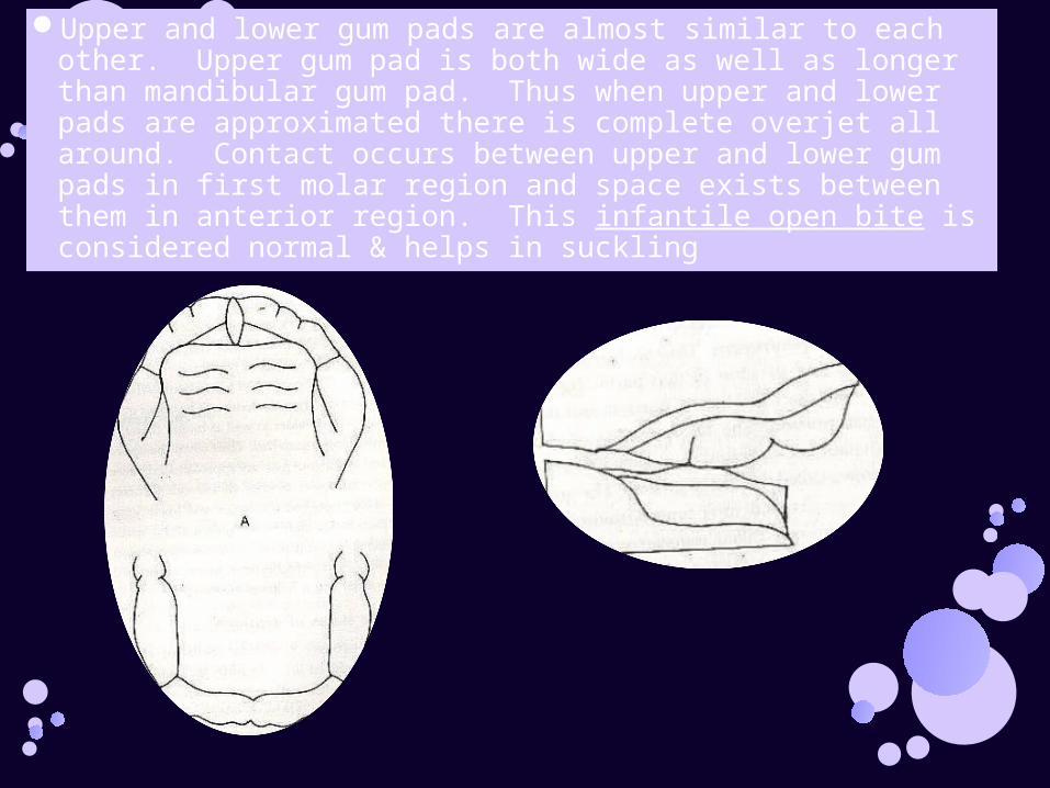
Upper and lower gum pads are almost similar to each other. Upper gum pad is both wide as well as longer than mandibular gum pad. Thus when upper and lower pads are approximated there is complete overjet all around. Contact occurs between upper and lower gum pads in first molar region and space exists between them in anterior region. This infantile open bite is considered normal & helps in suckling
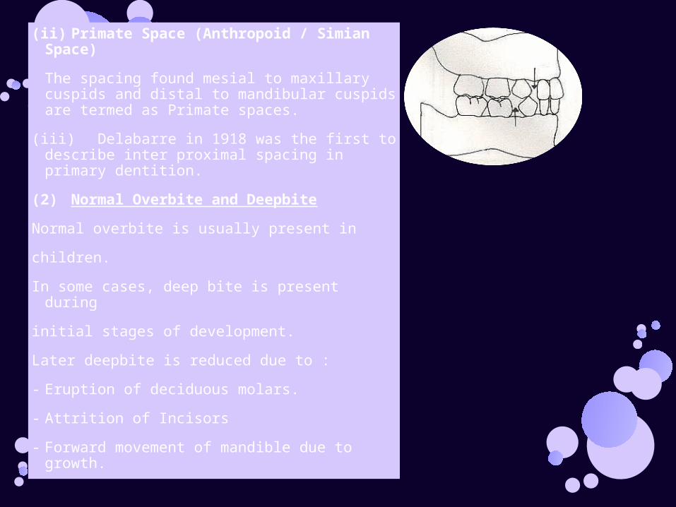
(ii) Primate Space (Anthropoid / Simian Space)
The spacing found mesial to maxillary cuspids and distal to mandibular cuspids are termed as Primate spaces.
(iii) Delabarre in 1918 was the first to describe inter proximal spacing in primary dentition.
(2) Normal Overbite and Deepbite
Normal overbite is usually present in
children.
In some cases, deep bite is present during
initial stages of development.
Later deepbite is reduced due to :
- Eruption of deciduous molars.
- Attrition of Incisors
- Forward movement of mandible due to growth.
(3) Overbite and OverjetDuring Primary dentition- Overbite decreases by slight amount. - Overjet reduced to zero. From the early mixed dentition to completion of permanent
occlusion. average overbite increases slightly and then decreases. (4)Inter Incisal Angle : It is angle formed between the intersection of long axis of
upper incisors and lower incisors. In Primary dentition, as incisor are more upright, there is an
increase in Inter incisal angle. (5)Occlusal Relations
Baumes Classification(a) Flush terminal plane - 76%(b) Mesial Step Terminal Plane - 14%(c) Distal Step Terminal Plane - 10%
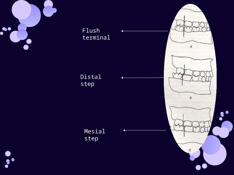
Flush terminal
Distal step
Mesial step
Mixed Dentition Period
- Begins at approximately 6 years of age.
- Classified into 3 phases :
(a) First Transitional Period :
Characterized by :
(i) Emergence of 1st permanent molars.
• Mandibular 1st molar - 1st permanent tooth to erupt at 6 years of age.
• Location and relationship depends upon
distal surface relationship upper and lower
2nd deciduous molars.
Mesial Step Terminal Plane (14%) : In this type of relationship distal surface of lower 2nd
deciduous molar is more mesial than that of upper. This permanent molars erupt directly in
Angles Class - I occlusion. This type of mesial step terminal plane most commonly occur due to early forward growth of mandible. If differential growth of mandible in forward direction persists, it can lead to Angles Class-III malocclusion
Distal Step Terminal Plane (10%)
This is characterized by distal surface of lower
2nd deciduous surface of lower 2nd deciduous
molar being more distal to that of upper. Thus
erupting molars may be in Angle's class II
malocclusion.
Alignment and Occlusion of Dentition :
Dental Arch Form• The teeth are positioned on maxilla and mandible in
such a way as to produce curved arch when seen from occlusal surface. The arch form is in large part determined by shape of underlying basal bone.
• On basis of qualitative observations, antheropologists have described general shape of palatal arch as being paraploid, U-shaped, ellipsoid, round and horse shoe shape
• Discrepancies in arch between the maxillary and mandibular arches generally result in poor occlusal relationships.
INTRA ARCH ALIGNMENT
Intra arch tooth alignment refers to relationship of teeth to each other within dental arch.
The occlusal planes of dental arches are curved in manner that permits maximum use of tooth contact during function as flat occlusal plane will not permit simultaneous functional contact in more than one area of dental arch.
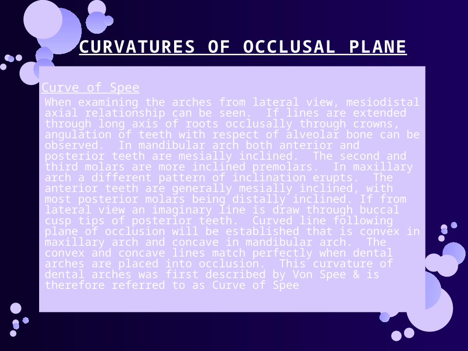
CURVATURES OF OCCLUSAL PLANE
Curve of SpeeWhen examining the arches from lateral view, mesiodistal axial relationship can be seen. If lines are extended through long axis of roots occlusally through crowns, angulation of teeth with respect of alveolar bone can be observed. In mandibular arch both anterior and posterior teeth are mesially inclined. The second and third molars are more inclined premolars. In maxillary arch a different pattern of inclination erupts. The anterior teeth are generally mesially inclined, with most posterior molars being distally inclined. If from lateral view an imaginary line is draw through buccal cusp tips of posterior teeth. Curved line following plane of occlusion will be established that is convex in maxillary arch and concave in mandibular arch. The convex and concave lines match perfectly when dental arches are placed into occlusion. This curvature of dental arches was first described by Von Spee & is therefore referred to as Curve of Spee
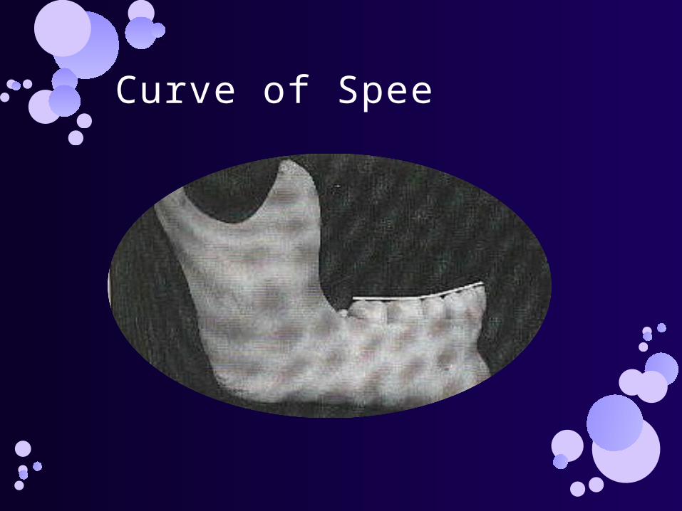
Curve of Spee
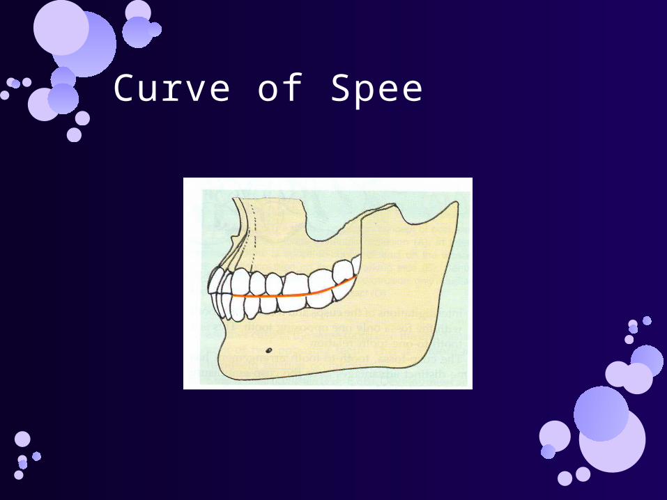
Curve of Spee
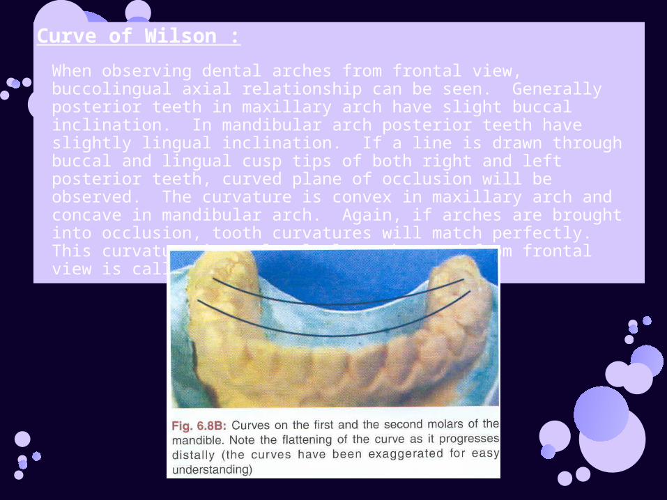
Curve of Wilson :
When observing dental arches from frontal view, buccolingual axial relationship can be seen. Generally posterior teeth in maxillary arch have slight buccal inclination. In mandibular arch posterior teeth have slightly lingual inclination. If a line is drawn through buccal and lingual cusp tips of both right and left posterior teeth, curved plane of occlusion will be observed. The curvature is convex in maxillary arch and concave in mandibular arch. Again, if arches are brought into occlusion, tooth curvatures will match perfectly. This curvature in occlusal plane observed from frontal view is called CURVE OF WILSON

Curve of Monsoon
Bonwill, one of first to describe dental arches, noted that an equilateral triangle existed between centres of condyles and mesial contact areas of mandibular central incisors. He depicted this as having 4-inch sides. In other words, the distance from mesial contact area of mandibular central incisor to centre of either condyle was 4 inches and distance between centres of condyles was 4 inch. In 1932, Monson, used Bonwill's triangle and proposed a theory that sphere erusted with radius of 4 inches, with centre that was an equal distance from occlusal surfaces of posterior teeth and from centres of condyles. The curve formed was known as CURVE OF MONSON
DENTITION
Relative positions of the maxillary and mandibular teeth influence mandibular movement.
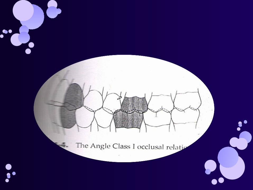
Angle class I considered to be the ideal occlusion in which mesiobuccal cusp of the maxillary first molar is aligned with the buccal groove of the mandibular first molar and overjet and overbite of anterior teeth is about 2 mm.
CENTRIC RELATIONThe maxillomandibular relation in which the condyles articulate
with the thinnest avascular portion of their respective discs with the complex in the anterior superior position against the shapes of the articular eminences.
This position is independent of tooth contact.
This position is clinically discernable when the mandible is directed superiorly and anteriorly.
It is restricted to a purely rotary movement about the transverse horizontal axis.
MAXIMUM INTERCUSPATION
This is a position in which the maxillary and mandibular teeth make maximum surface contact with each other.
The mandible is elevated as superiorly as possible in the sagittal plane.
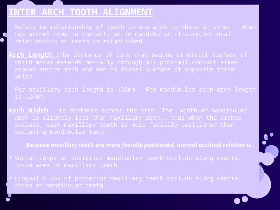
INTER ARCH TOOTH ALIGNMENT
Refers to relationship of teeth in one arch to those in other. When two arches come in contact, as in mandibular closure,occlusal relationship of teeth is established.
Arch Length :The distance of line that begins at distal surface of third molar extends mesially through all proximal contact areas around entire arch and end at distal surface of opposite third molar.
For maxillary arch length is 128mm. For mandibular arch arch length is 126mm.
Arch Width : is distance across the arch. The width of mandibular arch is slightly less than maxillary arch., thus when the arches occlude, each maxillary tooth is more facially positioned than occluding mandibular tooth.
Because maxillary teeth are more facially positioned, normal occlusal relation is :
Buccal cusps of posterior mandibular teeth occlude along central fossa area of maxillary teeth.
Lingual cusps of posterior maxillary teeth occlude along central fossa of mandibular teeth.
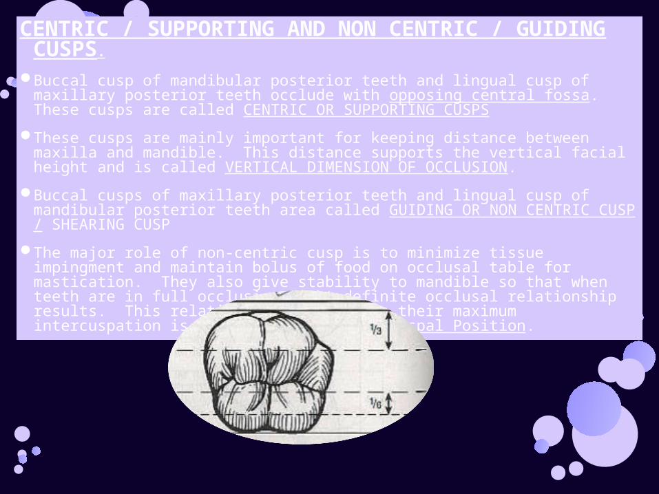
CENTRIC / SUPPORTING AND NON CENTRIC / GUIDING CUSPS.
Buccal cusp of mandibular posterior teeth and lingual cusp of maxillary posterior teeth occlude with opposing central fossa. These cusps are called CENTRIC OR SUPPORTING CUSPS
These cusps are mainly important for keeping distance between maxilla and mandible. This distance supports the vertical facial height and is called VERTICAL DIMENSION OF OCCLUSION.
Buccal cusps of maxillary posterior teeth and lingual cusp of mandibular posterior teeth area called GUIDING OR NON CENTRIC CUSP / SHEARING CUSP
The major role of non-centric cusp is to minimize tissue impingment and maintain bolus of food on occlusal table for mastication. They also give stability to mandible so that when teeth are in full occlusion tight definite occlusal relationship results. This relationship of teeth in their maximum intercuspation is called Maximum Inter Cuspal Position.
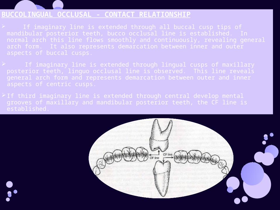
BUCCOLINGUAL OCCLUSAL - CONTACT RELATIONSHIP
If imaginary line is extended through all buccal cusp tips of mandibular posterior teeth, bucco occlusal line is established. In normal arch this line flows smoothly and continuously, revealing general arch form. It also represents demarcation between inner and outer aspects of buccal cusps.
If imaginary line is extended through lingual cusps of maxillary posterior teeth, linguo occlusal line is observed. This line reveals general arch form and represents demarcation between outer and inner aspects of centric cusps.
If third imaginary line is extended through central develop mental grooves of maxillary and mandibular posterior teeth, the CF line is established.
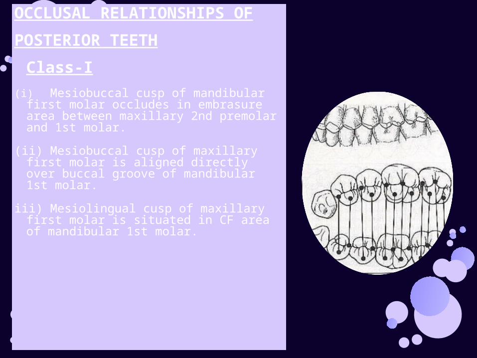
OCCLUSAL RELATIONSHIPS OF
POSTERIOR TEETH
Class-I
(i)Mesiobuccal cusp of mandibular first molar occludes in embrasure area between maxillary 2nd premolar and 1st molar.
(ii) Mesiobuccal cusp of maxillary first molar is aligned directly over buccal groove of mandibular 1st molar.
iii) Mesiolingual cusp of maxillary first molar is situated in CF area of mandibular 1st molar.
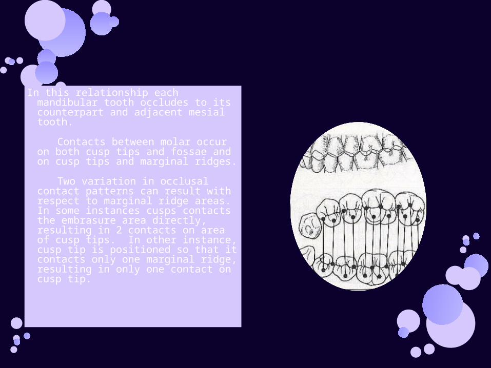
In this relationship each mandibular tooth occludes to its counterpart and adjacent mesial tooth.
Contacts between molar occur on both cusp tips and fossae and on cusp tips and marginal ridges.
Two variation in occlusal contact patterns can result with respect to marginal ridge areas. In some instances cusps contacts the embrasure area directly, resulting in 2 contacts on area of cusp tips. In other instance, cusp tip is positioned so that it contacts only one marginal ridge, resulting in only one contact on cusp tip.
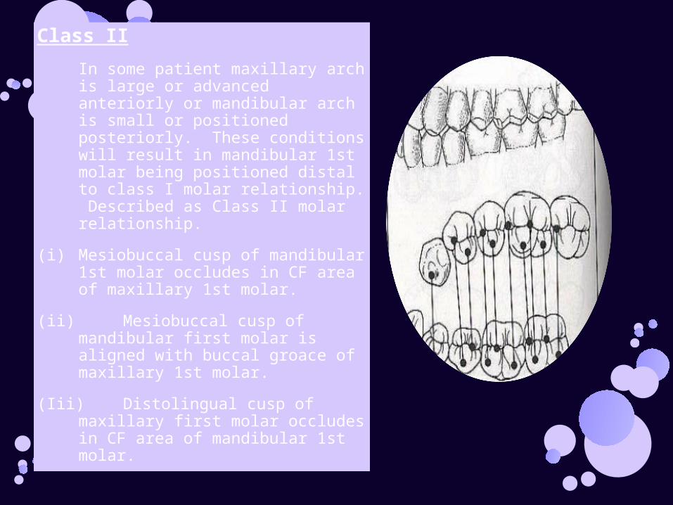
Class II
In some patient maxillary arch is large or advanced anteriorly or mandibular arch is small or positioned posteriorly. These conditions will result in mandibular 1st molar being positioned distal to class I molar relationship. Described as Class II molar relationship.
(i) Mesiobuccal cusp of mandibular 1st molar occludes in CF area of maxillary 1st molar.
(ii) Mesiobuccal cusp of mandibular first molar is aligned with buccal groace of maxillary 1st molar.
(Iii) Distolingual cusp of maxillary first molar occludes in CF area of mandibular 1st molar.
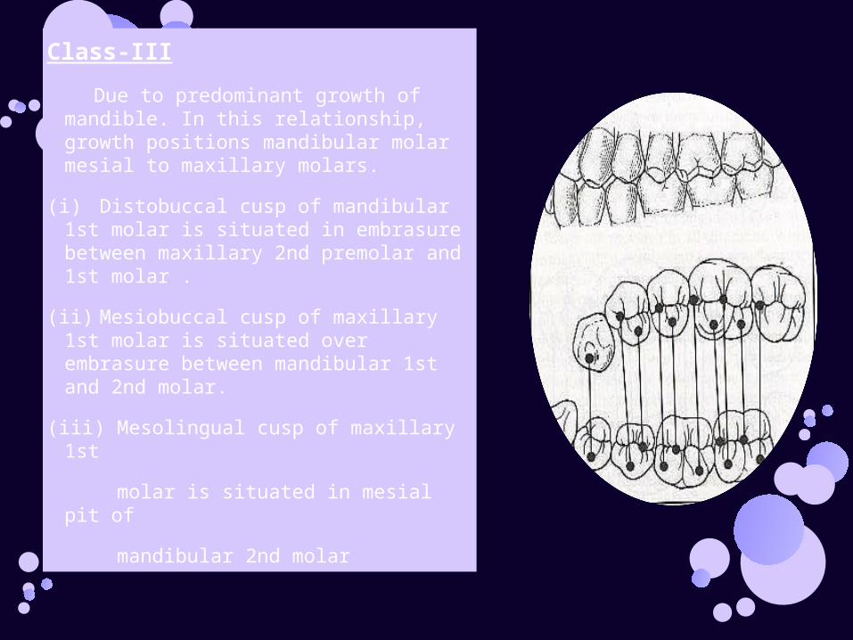
Class-III
Due to predominant growth of mandible. In this relationship, growth positions mandibular molar mesial to maxillary molars.
(i) Distobuccal cusp of mandibular 1st molar is situated in embrasure between maxillary 2nd premolar and 1st molar .
(ii)Mesiobuccal cusp of maxillary 1st molar is situated over embrasure between mandibular 1st and 2nd molar.
(iii) Mesolingual cusp of maxillary 1st
molar is situated in mesial pit of
mandibular 2nd molar
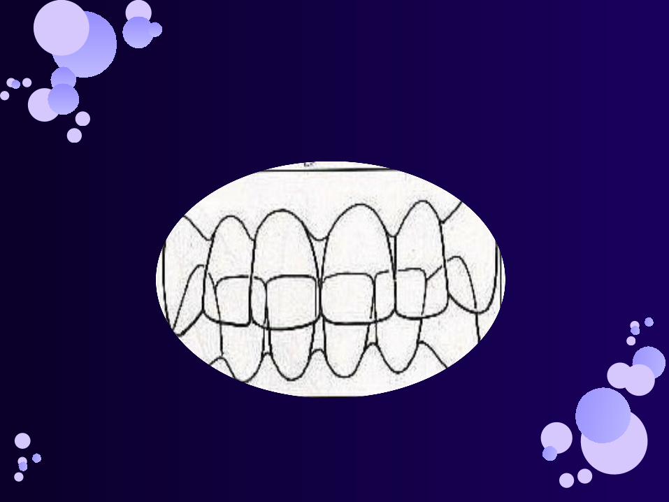
OCCLUSAL RELATIONSHIP OF ANTERIOR TEETH Maxillary anterior teeth are normally positioned labial to mandibular
anterior
teeth.
Both maxillary and mandibular anteriors are inclined to the labial, ranging
12-28º from vertical reference line.
Incisal edges of mandibular incisors contacting lingual surfaces of maxillary incisors. These contacts commonly occur in lingual fossae of maxillary incisors approximately 4mm gingival to incisal edges.
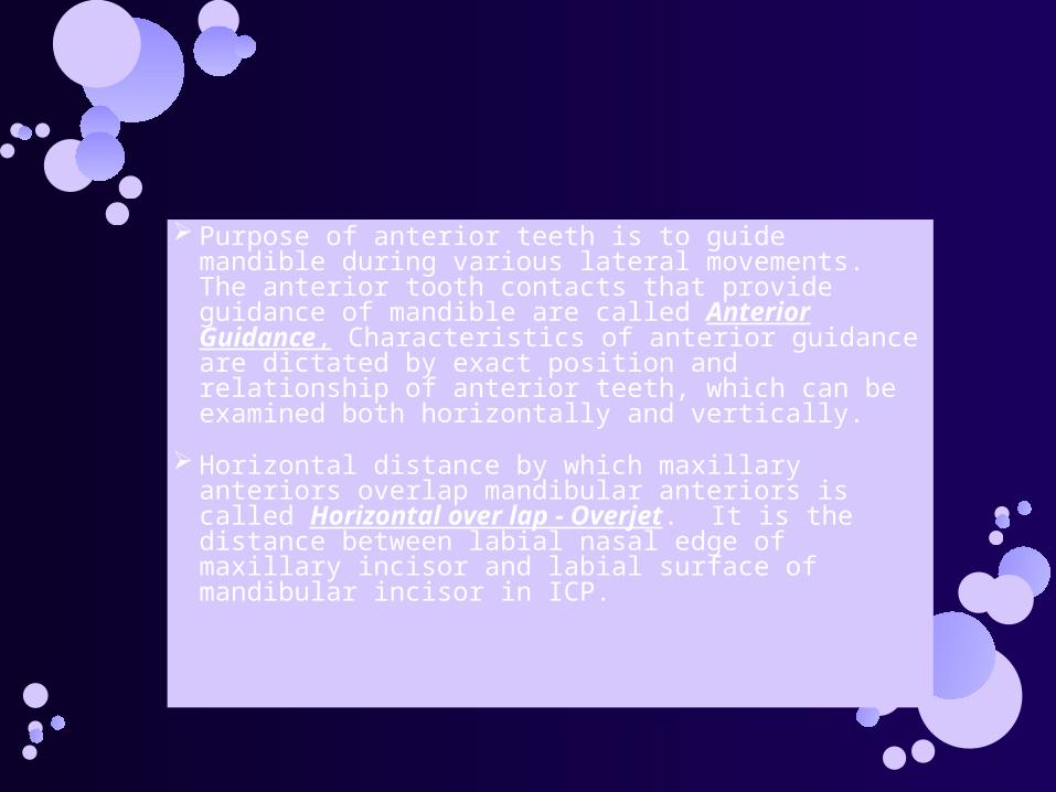
Purpose of anterior teeth is to guide mandible during various lateral movements. The anterior tooth contacts that provide guidance of mandible are called Anterior Guidance, Characteristics of anterior guidance are dictated by exact position and relationship of anterior teeth, which can be examined both horizontally and vertically.
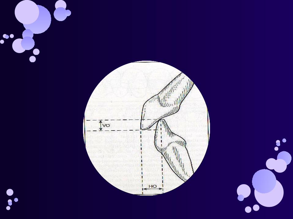
Horizontal distance by which maxillary anteriors overlap mandibular anteriors is called Horizontal over lap - Overjet. It is the distance between labial nasal edge of maxillary incisor and labial surface of mandibular incisor in ICP.

Vertical overlap is distance between incisal edges of opposing anterior teeth which is approximately 3 - 5mm.
Important function of anterior teeth is that of performing initial acts of mastication. Anterior teeth function to incise food when introduced in oral cavity.
Anterior teeth also plays significant role in speech, lip support and aesthetics.
CLASSIFICATION OF 0CCLUSION
BASED ON MANDIBULAR POSITION
CENRIC OCCLUSION-Is that position of the mandibular condyle when the teeth are in maximum intercuspation.It is also called INTERCUSPAL POSITION OR CONVENIENCE OCCLUSION.
ECCENTRIC OCCLUSION
It is defined as the occlusion other than centric occlusion.It refers to contact of teeth that occurs during movement of the mandible.
It includes:LATERAL OCCLUSION RIGHT AND LEFT LATERAL OCCLUSION
LATERAL OCCLUSION:
It is defined as the contact between opposing teeth(canines and posterior teeth)when the mandible is moved right or left of the midsaggital plane.The contacts occur on the sides towards which the mandible moves.
PROTRUDED OCCLUSION:Defined as the occlusion of the teeth when the
mandible is protruded.The position of the mandible is anterior to centric relation.Ideally the six anterior teeth contact along the lingual inclines of the max. ant. teeth while the posteriors disocclude
RETRUSIVE OCCLUSION
Occlusion of the teeth when the mandible is retruded.It is the position of the mandible posterior to centric relation.
ANDREW’S SIX KEYS TO NORMAL OCCLUSION
Andrew’s during 1970s put forward six keys to normal occlusion.
The six key are considered under the following headings:
1.MOLAR INTERARCH RELATIONSHIP.
The Mesio buccal cusp of upper first molar should occlude in the groove between mesial and medial buccal cusp of lower first molar. The mesio lingual cusp of upper first molar should occlude in the central fossa of lower first molar. The crown of upper first molar must be angulated so that distal marginal ridge occludes with the mesial marginal ridge of second molar
2.MESIO DISTAL CROWN ANGULATION
The Second Key makes use of a line that passes along the long axis of crown through the most prominent part in the center of labial or buccal surface. This line is called long axis of clinical crown.
For occlusion to be considered normal, gingival part of long axis of crown must be distal to the occlusal part of the line. Different teeth exhibit different crown angulation.
3. LABIO LINGUAL CROWN INCLINATION
The crown inclination is determined from mesial or distal view. If the gingival area of crown is more lingualy placed than the occlusal area, it is referred to as positive crown inclinication. In case the gingival area of crown is more labially or buccally placed than the occlusal area it is referred to as negative crown inclinication
The Maxillary incisors exhibit a positive crown inclination while mandibular incisors show negative crown inclination. The maxillary and mandibular posteriors have negative crown inclination.
4.ABSENCE OF ROTATION.
Normal occlusion is characterized by absence of rotation.
Rotated posteriors occupy more space in a dental arch. While rotated incisors occupy less space in dental arch.
5.TIGHT CONTACTS
For normal occlusion there should be tight contact between adjacent teeth.
6.CURVE OF SPEE
Normal occlusal plane according to Andrew’s should be flat with curve of spee not exceeding 1.5 mm.
BIO-MECHANICS OF OCCLUSION:
Dental factors of occlusion.
If dentition is functioning in health and form of dentition is esthetic, restoration of individual teeth requires duplication of existing occlusal form.
Unless there is evidence of dysfunction such as occlusal trauma, excessive tooth wear, clearly unacceptable occlusal plane orientation or joint and muscle dysfunctioning occlusion should be considered normal restorations should be patterned to follow morphology of adjacent and opposing teeth. Anatomic factors important in the development occlusal patterns for restored tooth include the following :

Cusp Height
Cusp angle
Marginal ridge height
Contact area position
Contact area height

Fossa size
Triangular and oblique ridge height.
Triangular and oblique ridge directions
Groove depth and width
Groove direction
Faciolingual dimension of occlusal table
HISTORY OF OCCLUSAL STUDIES
Historically the study of occlusion has undergone an evolution of concepts.
Bilaterally balanced, unilaterally balanced and mutually protected.
Current emphasis is on mutually protected occlusion.
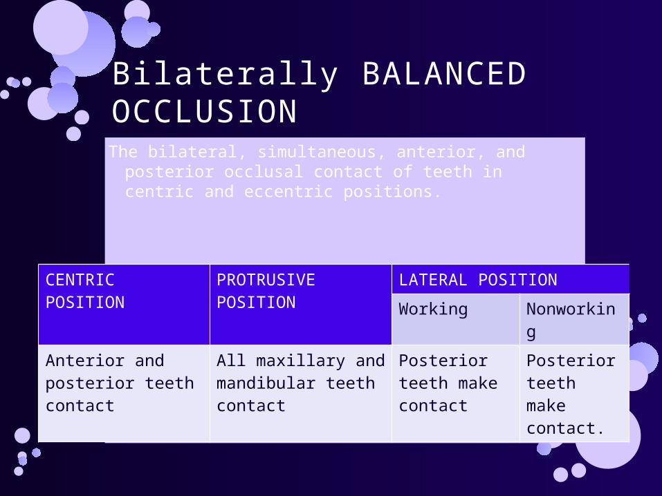
Bilaterally BALANCED OCCLUSIONThe bilateral, simultaneous, anterior, and posterior
occlusal contact of teeth in centric and eccentric positions.
CENTRIC POSITION PROTRUSIVE POSITION
LATERAL POSITION
Working Nonworking
Anterior and posterior teeth contact
All maxillary and mandibular teeth contact
Posterior teeth make contact
Posterior teeth make contact.
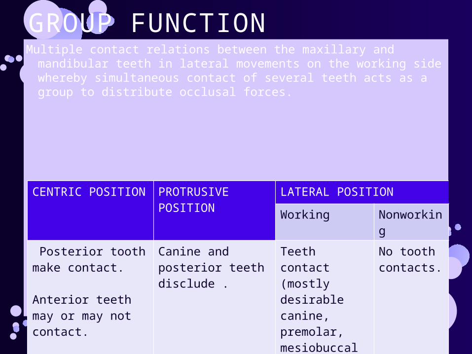
GROUP FUNCTIONMultiple contact relations between the maxillary and mandibular teeth
in lateral movements on the working side whereby simultaneous contact of several teeth acts as a group to distribute occlusal forces.
CENTRIC POSITION PROTRUSIVE POSITION
LATERAL POSITION
Working Nonworking
Posterior tooth make contact.
Anterior teeth may or may not contact.
Canine and posterior teeth disclude .
Teeth contact (mostly desirable canine, premolar, mesiobuccal cusp of 1st molar)
No tooth contacts.
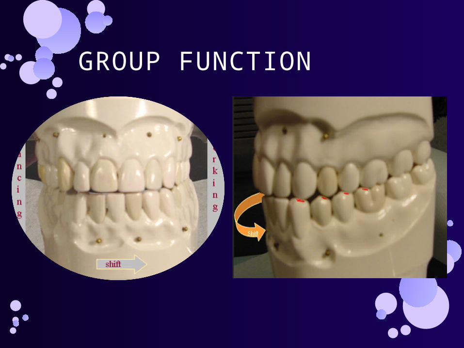
GROUP FUNCTION
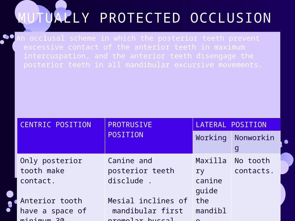
MUTUALLY PROTECTED OCCLUSIONAn occlusal scheme in which the posterior teeth prevent excessive
contact of the anterior teeth in maximum intercuspation, and the anterior teeth disengage the posterior teeth in all mandibular excursive movements.
CENTRIC POSITION PROTRUSIVE POSITION
LATERAL POSITION
Working Nonworking
Only posterior tooth make contact.
Anterior tooth have a space of minimum 30 microns.
Canine and posterior teeth disclude .
Mesial inclines of mandibular first premolar buccal cusps may contact.
Maxillary canine guide the mandible.
Posterior teeth disclude.
No tooth contacts.
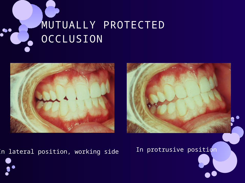
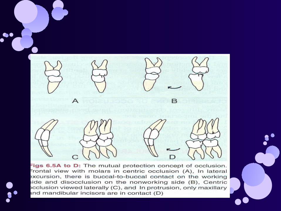
MUTUALLY PROTECTED OCCLUSION
In lateral position, working side In protrusive position
The most ideal occlusion is mutually Protected occlusion in which posterior teeth protect anterior teeth during centric closure & anterior teeth protect posterior teeth in any movement or position away from centric contact position.
CANINE GUIDED OR PROTECTED OCCLUSION
During lateral mandibular movements,the opposing upper and lower canines of the working side contact thereby causing disclusion of all posterior teeth on the working and balancing sides.This is because the mandible moves away from centric occlusion.

REFERENCES
The Glossary of Prosthodontic Terms 8th Ed. Prostho-dent
Stanley Jablonski. Jablonskis Dictionary of Dentistry. Ist Ed;
Mayor M. Ash, Stanley J. Nelson. Wheeler’s Dental Anatomy Physiology and Occlusion, 8th Ed. Saunders
Balaji S.I. Orthodontia the Art and Science 2nd Ed.
Fixed Prosthodontics: Rosential


ANY

Q. The lingual cusps of the maxillary posterior teeth are:
A. Non-supporting and Working
B. Supporting and Balancing
C. Supporting and Working
D. Non-supporting and Balancing

C. Supporting and Working
Remember: The supporting cusps are the maxillary lingual and the mandibular buc- cal. These cusps do grinding work because they occlude in a fossa or marginal ridge and are also called working cusps . They are sometimes called centric cusps because they hold the occlusion in a middle position (centric position).

Which teeth should ideally provide the predominant guidance through the full range of movement in lateral mandibular excursions?
Premolars
1ST molars
Incisors
Canines

Canines
This is called canine or cuspid protected occlusion. It is an occlusal relationship in which the vertical overlap of the maxillary and mandibular canines produces a disclusion (separation) of all of the posterior teeth when the mandible moves to either side. All other teeth, once they move from centric relation, do not contact.