observations on the development of erythrocytes in mammalian fetal
TRANSCRIPT

O B S E R V A T I O N S O N T H E D E V E L O P M E N T O F
E R Y T H R O C Y T E S I N M A M M A L I A N F E T A L L I V E R
J O S E P H A. G R A S S O , Ph.D., H E W S O N S W I F T , Ph.D., and
G. A D O L P H A C K E R M A N , M.D.
From the Whitman Laboratory, University of Chicago, Illinois, and the Department of Anatomy, The Ohio State University, Columbus
A B S T R A C T
The fine structure of the erythrocyte during development in rabbit and human fetal liver has been studied. A morphologic description of representative erythropoietic cells and their relationship to the hepatic parenchyma is presented. Erythrocyte development was accompanied by a decrease in nuclear and cell size, fragmentation and eventual loss of nucleoli, and progressive clumping of chromatin at the nuclear margin. Mitochondria, endoplasmic reticulum, and Golgi elements decreased in size or abundance and eventually disappeared. Ribosome concentration initially increased, but subsequently diminished as the cytoplasm increased in electron opacity, probably through the accumulation of hemo- globin. Similar dense material, interpreted to be hemoglobin, infiltrated the nuclear annul i and, in some cases, appeared to extend into the interchromatin regions. There was a marked decrease in the number of annuli of the nuclear envelope. Possible relationships between nucleus and cytoplasm and of RNA to hemoglobin synthesis are discussed. In rabbits, crythroid and hepatic cells were separated by a 200 to 400 A space limited by the undula- tory membranes of the respective cells. Membranes of adjacent erythropoietic cells were parallel and more closely apposed (100 to 200 A). In humans, relationship between various cells exhibited wide variation. Ferritin particles were observed within forming and formed "rhopheocytotic" vesicles.
I N T R O D U C T I O N
The differentiation of mammal ian erythrocytes provides a system favorable for the study of sequential development and progressive special- ization of a definitive cell type. During embryonic development the liver is an active site of erythro- poiesis, characterized by numerous loci of dif- ferentiating erythrocytic elements diffusely ar- ranged within the hepatic parenchyma. The histologic features of the erythropoietic process in embryonic liver have been explored exten- sively (2, 10, 21, 24, 33, 34, 36, 44, 47, 54).
In this report data are presented on erythro- cytic development in rabbit and human fetal
liver. The investigation is based upon correlated light and electron microscopic examinations and is preliminary to microphotometric and auto- radiographic studies now in progress.
M A T E R I A L S A N D M E T H O D S
Livers were obtained from 13-, 14-, 15-~ and 23- day rabbit fetuses, and from a 12-week-old human fetus. Rabbit livers were cut into small blocks and im- mersed for 1 hour in cold 1 per cent osmium tctroxide (pH 7.8) (39) or cold l0 per cent acrolein (pH 7.8) buffered in 0.2 M Tris buffer (30). After dehydration in a graded series of ethanol, tissue blocks were em- bedded in a 1:5 mixture of methyl-butyl mctha-
235

crylate, Vestopal W (26), or Epon 812 (31). Metha- crylate and Vestopal polymerization was effected by exposure to ultraviolet light and heat overnight. Epon resins were polymerized in a 60°C oven for the same length of time.
Human liver was fixed in cold 1 per cent chrome- osmium (pH 7.4) (17) for 1 to 2 hours and embedded in a 1 : 5 mixture of methyl-butyl methacrylate.
Blocks were sectioned on a Servall Porter-Blum microtome using glass knives. Silver to gray sections were mounted on Athene grids which had been coated with Formvar or carbon. The majority of sections were stained in a saturated solution of lead hydroxide for 30 minutes to 1 hour (57) or in a 2 per cent solution of uranyl acetate (52) for a similar period of time. The grids were then "sandwiched" by carbon evaporation to minimize the effects of methacrylate sublimation in the electron beam (56). All specimens were examined in an RCA EMU 3C electron microscope equipped with a 2 rail objec- tive aperture.
For light microscopy, tissues fixed in acrolein as described above and embedded in paraffin were sec- tioned and stained in 0.025 per cent solution of azure B in McIlvaine's buffer at pH 4.0 (48) for periods up to 1 hour, or with hematoxylin-eosin azure (27). Alternate thin and thick sections were prepared from the plastic-embedded specimens, where possible, in order to correlate light and electron microscopic ob- servations. Thick sections of the order of 0.5 to 1.5 micra in thickness were mounted on albumen-coated glass slides and stained in a 0.25 per cent solution of azure B for 1 to 2 hours. The sections were dehydrated in three changes of tertiary butyl alcohol and mounted in Canada balsam.
O B S E R V A T I O N S
Light Microscopy Thi r teen-day rabb i t liver revealed a moderate
degree of erythropoiet ic activity character ized by the presence of relatively undifferentiated cells distr ibuted randomly within the hepat ic paren- chyma (Fig. 1). Morphologically, these cells corresponded to the hemocytoblast , i.e., intense cytoplasmic basophilia, large spherical, slightly indented nuclei containing one or more nucleoli, and a moderate n u m b e r of mitochondria .
The ret iculoendothelial cells l ining the sinus- oids exhibited a slight degree of phagocytic activity, recognized by the presence of inclusion bodies within their cytoplasm. Presumably these phagocytized particles represented remnants of pr imary erythroblasts, a large n u m b e r of which were located within the sinusoids in various stages of degenerat ion (Fig. 1).
A thin mesenchymal capsule, partial ly sur- rounding the liver, conta ined several cells which were essentially similar in appearance to the ex- t ravascular hemocytoblasts. Groups of hepat ic cells appeared to extend into the encircling cap- sule from the limits of the parenchyma.
By 14 to 15 days' gestation, a vast increase in erythropoietic activity had occurred. Clusters of erythropoiet ic cells in varying stages of develop- men t were recognized in which the classical features of erythropoiesis were readily observed. These included (a) decrease in cell size, (b) d isappearance of nucleoli, (c) increased nuclear density with c lumping of chromat in , (d) gradual decrease in cytoplasmic basophilia, and (e) appearance of cytoplasmic acidophilia. The "checkerboard" appearance of the nuclear chro- ma t in was apparen t in the later stages of erythro- blasts. The number of pr imary red cells wi th in the sinusoids was greatly decreased or absent. Inclusions representing phagocytized particles were noted in the cytoplasm of the ret iculoendo- thelial cells.
The 12-week-old h u m a n liver displayed general similarity to the 14-15-day rabb i t with respect to the degree of erythropoiet ic activity (Fig. 2). However, the hepatic cells were more distinct and ar ranged in a t rabeculated fashion surround- ing large clusters of erythrocytic derivatives. Blood cells were noted both within the paren- chyma and in the sinusoids. The cytologic charac- teristics of erythrocyte differentiat ion were essen- tially similar to those seen in the rabbit .
Electron Microscopy Electron and light microscopic observations
were correlated by preparat ion, where possible, of al ternate thick and thin sections. Wi th this technique, it was possible to recognize the various stages of erythropoietic product ion occurring in the following sequence (35): hemocy tob l a s t -* proerythroblast --~ basophilic erythroblas t --, polychromatophi l ic erythroblast --~ normoblas t --~ reticulocyte --~ erythrocyte. A t the electron microscopic level, the hemocytoblast and pro- erythroblast were basically similar in general morphologic appearance and have been de- scribed below as a unit.
1) HEMOCYTOBIaA.ST The nucleus of the hemocytoblast was roughly
spherical to ovoid in contour wi th occasional indenta t ion and revealed a relatively homoge-
236 THE JOURNAL OF CELL BIOLOGY • VOLU2KE 14, 1962

FIOm~E 1
Light micrograph of 13-day rabbit fetal liver showing extravascular hemocytoblasts (B) and primary erythroblasts within a large sinusoid (S). Note indistinct reticuloendothelial cytoplasm (arrow) separating the two cell types. 10 per cent acrolein, azure B. X 1750.
Light micrograph of 3-month human fetal liver revealing several stages of erythroblasts. EB, basophlic erythroblast; LB, polychromatophilic erythroblast. 1 per cent chrome-osmium, methacrylate, azure B. X 2100.
J. A. GRASSO, H. Swlr% A~D G. A. ACKERMAN Development of Erythrocytes 237

neous distribution of the chromatin except along the inner aspect of the nuclear membrane. Here, peripheral clumping was observed with interruptions at the nuclear annuli (Figs. 3, 8 12). One or more large nucleoli were evident in both the rabbit and human stem cells. Frequent contact of nucleoli and nuclear membrane was encountered. An outstanding characteristic of the hemocytoblastic nucleus was the high inci- dence of connections between the nucleus and cytoplasm. Such connection was manifested in the form of nuclear annuli (Figs. 8, 12), nuclear blebbing, (Fig. 3; see also reference 20) and con- tinuity of the endoplasmic reticulum with the nuclear envelope (Figs. 8, 9).
The most striking feature of the cytoplasm was the abundance of ribosomes which were freely distributed in the cytoplasm (Figs. 9, 10) and also associated with the infrequently occurring membranes of the endoplasmic reticulum. This feature accounted for the intense basophilia observed in the light microscope after staining with basic dyes (41). The ribosomes were arranged singly or as clusters of granules forming tetrads, spirals, and rosettes (Figs. 9, 10). In some in- stances, small focal concentrations of ribosomal granules were encountered. The endoplasmic reticulum was sparsly distributed as straight or tortuous cisternae and was often observed in close proximity to the mitochondria.
The mitochondria were moderate in number and surrounded the nucleus in a perinuclear fashion (Fig. 3). In the rabbit, they were approxi- mately 0.3 ~ in width and up to 0.7 ~ long. Human mitochondria were slightly longer, being about 0.6 # in width and up to 2.0 # long
(Figs. 18, 20, 21). Numerous dense granules permeated the mitochondrial matrix, a feature most prominently seen in human proerythroblasts (Fig. 18). These were somewhat similar to ribo- somes in both arrangement and density. In some instances, invagination of both the inner and outer mitochondrial membranes and /or swelling of the mitochondria resulted in displacement of the cristae (Fig. 10). However, in most cases, mitochondrial cristae appeared normal.
The Golgi complex was not markedly developed and exhibited the usual vesicular and lamellar components (Figs. 8, 9). In its juxtanuclear loca- tion, it was frequently associated with one or a pair of centrioles (Fig. 9). Of particular interest in human proerythroblasts was the occurrence of dense bodies approximately 0.1 to 0.2 micron in length in the Golgi region (Figs. 18, 19). These bodies were limited by a double membrane and manifested a dense granulated matrix in which thin membranes were indistinctly evident. The granular matrix appeared to consist of two types of particles: infrequent densely osmiophilic particles superimposed upon a less dense com- ponent constituting the bulk of the body (Fig. 19). The nature of these bodies is undetermined.
~) BASOPHILIC ERYTHROBLAST
(EARLY EaYTKROBLAST)
The nucleus of the basophilic erythroblast exhi- bited a slight clumping of the chromatin in the cen- tral region while peripheral clumping persisted as in the hemocytoblast. Nucleoli revealed a tendency to be fragmented or inconspicuous (Fig. 4). The cytoplasm contained a large number of "free" ribosomes and only traces of endoplasmic
FIGURE
Electron micrograph of rabbit proerythroblast in which a large number of ribosomes is evident. Note sparsity of cndoplasmic reticulum and occurrence of nuclear blebbing (arrow) and annuli. 1 per cent OsO~, methacrylate. X 18,700.
FIGURE 4
Electron micrograph of rabbit basophilic erythroblast in which peripheral clumping ofchromatin is apparent. Remnant of nucleolus is visible at arrow. Compare with Fig. 3. 1 per cent OsO4, Epon. X 13,100.
FIGURE 5
Electron micrograph of rabbit normoblast revealing vast clumping of nuclear chromatin and mitochondrial polarization. The cytoplasmic matrix appears stippled by a moder- ately osmiophilic material which probably represents hemoglobin. Note ecccntricity of nucleus and decrease in ribosomal content in comparison to preceding figures. X 18,700.
238 T~E JOURNAL OF CELL BIOLOGY • VOLUME 14, 196~

J. A. GRASSO, H. SWIFT, AND G. A. ACKERMAN Development of Ergthroevtes 239
reticulum (Figs. 4, 21). Mitochondria tended to concentrate at one pole of the cell (Fig. 20). The Golgi complex was absent or poorly de- veloped. The cytoplasmic matrix appeared lightly stippled as a probable result of hemoglobin synthesis (Fig. 4). In human basophilic eryth- roblasts, large amorphous masses similar in structure to lipid droplets were occasionally noted (Fig. 20). These inclusions were limited by a moderately dense membrane and probably represent the juxtanuclear sudanophilic bodies described by Jones (23). In one case, a flask- shaped vacuole, containing several microvesicles and communicating with the cytoplasmic matrix, was closely associated with an inclusion (Fig. 20). This body is somewhat reminiscent of the multi- vesicular bodies described by other authors (37).
3) POLYCHROMATOPHILIC ERYTHROBLAST (LATE ERYTHROBLAST)
Nuclear and cytoplasmic structure of the p~ly- chromatophilic crythroblast revealed a drastic change in comparison to that of earlier cell types. The chromatin was moderately clumped and radiated centrally from the inner surface of the nuclear membrane (Fig. 6). No nuclcoli could be distlnguished at this stage. The ribosomes were moderate in number and arranged singly or in clusters of two or more granules (Fig. 7). Clear arcas separated the ribosomal clusters from a moderately electron-opaque particulate com- ponent which appeared to stipple the entire cytoplasmic matrix. This material has been inter- preted to represent hemoglobin. The mitochon- dria tended to polarize at one end of the nucleus. The Golgi complex was poorly developed or ab-
sent. No traces of the endoplasmic reticulum were recognized. Frequently, the occurrence of vesicles containing ferritin was observed (Fig. 17). In some cases, the nucleus had assumed an eccentric position in the cell or was in the process of extru- sion.
4) NORMOBLAST
Marked chromatin clumping characterized the normoblast nucleus and imparted an opaque appearance to it when seen with the light micro- scope. As in the polychromatophilic erythroblast, the chromatin radiated inwardly from the nu- clear membrane. Nuclear annuli perforated the nuclear membrane and appeared to extend into the clumps of chromatin (Fig. 5). No nucleoli were visible. Two structures chiefly composed the cytoplasm: a moderate number of ribosomal clusters scattered among a less dense particulate fraction which permeated the entire cytoplasm (Figs. 5, 11, 13). Ribosomes were surrounded by rings of low density material, the significance of which is unknown. Mitochondria were few in number and were concentrated at one pole of the nucleus. Their average width measured 0.2 while their length varied from 0.2 to 0.6 #. Both the endoplasmic reticulum and the Golgi complex were lacking at this stage of development (Fig. 5).
5) RETICULOCYTE
The reticulocyte was a non-nucleated cell in which a relatively moderate number of ribosomes could still be observed. A large concentration of the lightly stained particles was located in the cytoplasm and imparted a moderate degree of density to the cell. A few mitochondria were visible. All other organelles were absent.
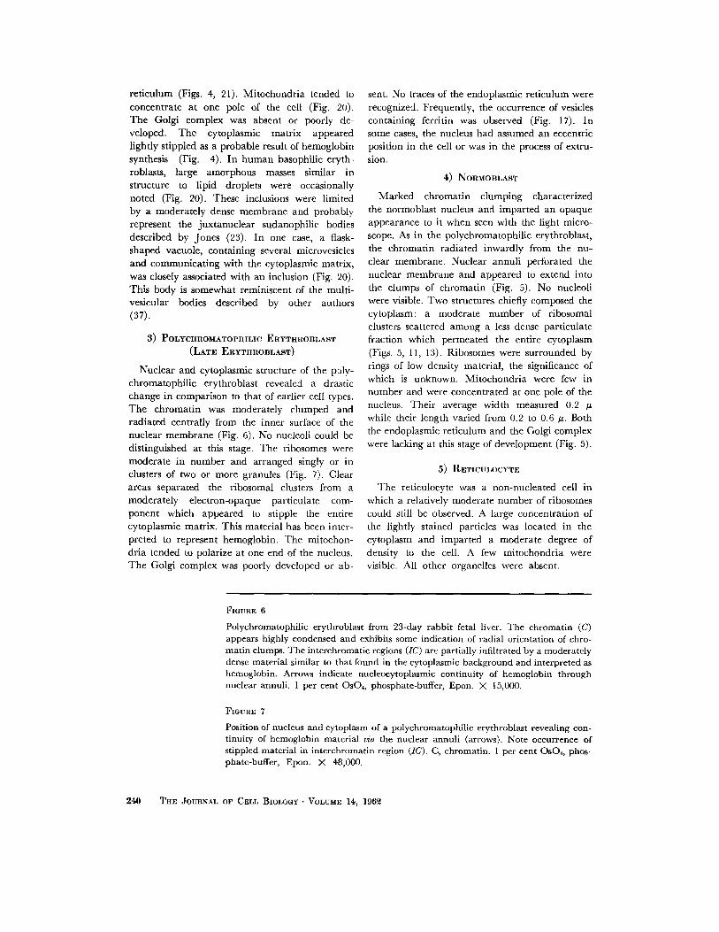
FIGURE 6
Polychromatophilic erythroblast from 23-day rabbit fetal liver. The chromatin (C) appears highly condensed and exhibits some indication of radial orientation of chro- matin clumps. The intcrchromatic regions (IC) are partially infiltrated by a moderately dense material similar to that found in the cytoplasmic background and interpreted as hemoglobin. Arrows indicate nucleocytoplasmic continuity of hemoglobin through nuclear annuli. 1 per cent OsO4, phosphate-buffer, Epon. X 15,000.
FIGURE 7
Position of nucleus and cytoplasm of a polychromatophilic erythroblast revealing con- tinuity of hcmoglobin material via the nuclear annuli (arrows). Note occurrence of stippled material in interchromatin region (IC). C, chromatin. 1 per cent OsO4, phos- phate-buffer, Epon. ;( 48,000,
240 THE JOURNAL OF CELL BIOLOGY • VOLUME 14, 196~

J. A. GaAsso, H. SWIFa', AND G. A. ACKERMAN Development of Erythrocytes 2,41

Process of Differentiation The above progressive changes, associated with
erythrocyte differentiation, may be summarized as follows, taking the hemocytoblast or stem cell as a reference point:
A marked decrease in cell size was evident with differentiation (Figs. 3 to 5). The first changes to be noted were in the structure and extent of nucleoli. In contrast to the usual single large nucleolus of hemocytoblasts and proerythroblasts, basophilic erythroblastic nucleoli appeared to undergo fragmentation and were significantly decreased in volume. Such nucleolar fragments, composed of irregular clusters of granules, were observed in one or more loci within the nucleus. Further disintegration resulted in a gradual blending of nucleolar fragments into the sur- rounding karyoplasm until the nucleoli were no longer visible. Nucleoli had disappeared in most of the basophilic erythroblasts and definitely could not be distinguished in the late or poly- chromatophilic erythroblast.
Accompanying nucleolar disintegration, a gradual decrease in nuclear volume and increase in nuclear density were encountered as clumping of chromatin occurred. The effect of clumping was most evident in polychromatophilic erythro- blasts where bars of chromatin material appeared to radiate centrally from the inner aspect of the nuclear membrane (Fig. 5). Annuli were nu- merous along the nuclear membrane of hemo- cytoblasts (Figs. 8, 9, 12). By the normoblast stage, however, annuli occurred only in isolated small groups, and stretches of several microns occurred along the nuclear membrane in which no annuli were visible (Fig. 13). These groups occurred where the interchromatin areas were in contact with the inner surface of the nuclear membrane. The distribution of chromatin clumps against the membrane, interspersed with chan-
nels of interchromatin material, is responsible for the typical "car twheel" distribution of chro- matin apparent with the light microscope. It might be added that similar changes accompany the gradual loss of synthetic activity in other cell types as well, for example in mammal ian epider- mis, and in the transformation from osteoblasts to osteoclasts in bone. Ribosomes associated with the outer nuclear membrane exhibited a gradual decrease in number and a subsequent loss (Figs. 6, 7, 13). With further development, the nuclei could occasionally be seen in the process of ex- trusion. Some evidence of karyolysis was sug- gested by nuclei which appeared to be bounded by very indistinct membranes and which blended into the cytoplasm.
Within the cytoplasm, the ribosomes, following an initial slight increase in number, exhibited a gradual diminution in concentration accom- panied by a concomitant increase in cytoplasmic density as hemoglobin concentration rose (Figs. 7, 10, 11, 14). Increased cytoplasmic density was seen to result from the deposition of a finely stippled particulate material which was diffusely spread within the matrix. It is assumed that this material was hemoglobin since these particles increased in concentration as further development occurred, eventually fusing to produce an almost uniform dense cytoplasm in the late stages. The ribosomes were separated from the hemoglobin by clear areas (Figs. 7, 11), the significance of which is uncertain. In human basophilic erythro- blasts, hemoglobin deposition appeared to be more concentrated just beneath the cell mem-
brane than in the remainder of the cell (Fig. 21). The rabbit did not reveal such a gradient in hemoglobin synthesis although occasional hetero-
geneity could be observed (Fig. 11). The endoplasmic reticulum was no longer
apparent at the late or polychromatophilic eryth-
FIGURE 8
Electron micrograph of rabbit hemocytoblast showing continuity of nuclear envelope with ER. Note the Golgi complex in association with a pair of centrioles (C). Several nuclear annuli are also apparent (arrows). Nucleolus (n) is visible in left corner of micrograph. 1 per cent OsO~, methacrylate. X 20,000.
FIGURE 9
Higher magnification of cytocentrum seen in Fig. 8. Note relative absence of ribosomes in cytocentrum. Continuity of nuclear envelope and endoplasmic reticulum is shown at arrows. 1 per cent OsO4, methacrylate. X 79,000.
242 THE JOURNAL OF CELL BIOLOGY • VOLUME ]4, 196~2

J. A. GRASSO, H. SWIFT, AND G. A. ACKERMAN Development of Erythroeytes 243

roblastic stage (Fig. 6). The Golgi complex appeared to undergo a gradual involution, although both the Golgi elements and centrioles could occasionally be seen in some polychroma- tophilic erythroblasts. In human erythroblasts, the occurrence of dense bodies in association with the Golgi complex has been previously mentioned.
Mitochondria manifested no drastic changes in basic structural qualities (40) although decrease in number, size, and volume of the chondriome was noted (Fig. 5). Furthermore, there was a tendency for mitochondrial polarization during the later stages of erythrocytic development. In no case was the presence of intramitochondrial ferritin encountered. The latter observation is in general agreement with Bessis (4) who reported the finding of intramitochondrial ferritin mainly in certain pathologic states.
Ferritin
In erythroblasts, ferritin was localized within vesicles (Fig. 17), and could not be seen free within the cytoplasmic matrix. Hepatic cells contained a large amount of freely distributed ferritin particles. The latter observation has been interpreted by some authors to indicate that the hepatic cell serves as an intermediary in ferritin transport (47). Ferritin particles were occasionally located along the plasma membrane of human erythroblasts (Fig. 16). In several cases, the membrane manifested a partial in- vagination in which ferritin particles adhered to the membrane (Fig. 16 a). This portion of the membrane exhibited a higher density than adja- cent areas of the plasma membrane. The process is similar to that described as "rhopheocytosis" by other authors (3-5, 8, 47). Forming rho-
pheocytotic vesicles were observed only in the human. In the rabbit, most of the ferritin was contained within vesicles limited by a smooth surfaced membranes and approximately 0.1 to 0.2 micron in size (Fig. 17). Presumably, these structures arose in the same manner as described in the human, In the more immature erythro- blasts, numerous vesicles were observed beneath the cell membrane which were devoid of apparent content or which contained smaller vesicles (Fig. 10). It is not unreasonable to assume that these structures represent micropinocytotic or rhopheocytotic vesicles.
Cell Relationships
Observations of rabbit and human fetal livers with the electron microscope confirmed the ex- travascular nature of the erythropoietic process. Erythrocytic derivatives were observed singly or in clusters within the hepatic parenchyma. No intervening basement membrane was imposed between erythropoietic and hepatic cells. In the rabbit, the space separating erythrocytic from hepatic cells measured approximately 200 to 400 A in width. In regions of erythrocyte-hepatocyte contact, the membranes of both cells appeared moderately undulatory with occasional indica- tions of interdigitation (Fig. 15). The erythroid cells were more closely apposed (100 to 200 A). In addition, the membranes of two adjacent erythroid cells were arranged in an essentially parallel manner with slight variation in the width of the space separating them (Fig. 13).
In the human, the width between erythropoietic and parenchymal cells varied from 200 to 1000 A. A similar degree of variation was found in the dimensions of the space between adjacent eryth- ropoietic cells (Fig. 14). The wide range of
FIGURE 10
Electron micrograph of rabbit hemocytoblast revealing relative abundance of "free '~ ribosomes. A swollen mitochondrion possessing invaginated outer and inncr mem- brances is also shown (arrow). A group of smooth surfaced vesicles (V) is visible just beneath thc cell membrane in which varied content may be seen. Note undulatory relationship of adjacent cell membranes (C). Cell in right corner is a hepatic cell. 1 per cent OsO4, methacrylate, X 79,000.
FIGURE II
Portion of cytoplasm of rabbit normoblast in which decrease in ribosomal content and appearance of hemoglobin is clearly shown. Compare with Figs. 7, 8. Note less dense areas surrounding ribosomal clusters, l per cent OsO4 methacrylate. )< 79,000.
24~ THE JOURNAL OF CELL BIOLOGY • VOLUME 14, 1962

J. A. GRAsso, H. SWIFV, AND G. A. ACKERMAN Development of Erythrocytes 245

variation can conceivably be attributed to shrink- age resulting from tissue preparation. The fre- quent occurrence of small spherical or long attenuated processes of hepatic cell cytoplasm in the intercellular space delineating adjacent cells suggested that the hepatic cell surface was thrown into numerous irregular folds by the abundant number of proliferating blood cells.
Several erythroblasts in the human were shown to be connected by a spindle remnant or the body of Flemming resulting from mitosis (Fig. 21). The membrane delimiting this connection was thicker and revealed a higher electron opacity than the adjacent plasma membrane (Fig. 21). Often, the membrane appeared highly convoluted and enclosed a dense region of cytoplasm in which occasional cisternae of endoplasmic reticulum were distinguishable. However, other cytoplasmic organelles were lacking.
D I S C U S S I O N
Several recent studies have confirmed the ex- travascular nature of erythropoiesis in the fetal liver of various mammals (2, 24, 47). The results of the present study reveal and further confirm this relationship in the rabbit and human. Pre- liminary studies by the authors on the fetal liver of the cat have shown erythropoiesis to occur in a fashion similar to that in other mammals. Some mechanism must exist whereby extravascular blood cells are released into the circulation. From observations of the present investigation, the "release" process appears to occur in several ways. First, it has been observed in several in- stances that the sinusoidal lining is discontinuous and thereby allows free communication between the extravascular and intravascular compart- ments. In this case, the escape of erythrocytic
cells may readily occur through the general con- tinuity of the two compartments. Second, the release of blood cells through diapedesis, i.e. by pushing between the processes of reticulo- endothelial cytoplasm, has been noted in a fashion similar to that noted by Bessis in some mammalian bone marrow (6). A modification of the latter mechanism can be found in the rupture of the reticuloendothelial lining as a result of pressure exerted by the underlying erythroid cells. Maximow described or postulated the re- lease process to occur in a similar manner (33, 34).
The present investigation suggests that the nucleus plays a changing role (12, 13) in the devel- opment of erythrocytic elements. The numerous structural connections evident in hemocytoblasts and early erythroblasts, i.e. nuclear blebbing, continuity of the endoplasmic reticulum with the nuclear envelope, and nuclear annnli, sug- gest the probability of material exchange be- tween the nucleus and cytoplasm. It has fre- quently been postulated that exchange could occur by means of such processes (16, 19, 42, 43, 49, 50, 51, 55).
In studies of avian and mammal ian erythro- blasts, Thorell (53) demonstrated that an inverse relationship existed between cytoplasmic R N A and hemoglobin. He noted that the percentage of cytoplasmic R N A in the stem cell was quite high and fell rapidly as maturat ion proceeded. The present morphologic results are in agree- ment with his findings since a diminution in the number of ribosomes occurred concomitant with an increase in hemoglobin concentration.
The decrease in ribosome concentration also
paralleled a fragmentation and loss of nucleoli. This is in general agreement with the concept
FIGURE 1~2
Electron micrograph of rabbit hemocytoblast exhibiting frequency of nuclear annuli (arrows). 1 per cent Os04, methacrylate. X 79,500.
FIaURE 13
Electron micrograph of rabbit normoblast showing changes in nuclear componcnts and cytoplasm. Nuclear annuli seem to have dccreased in frequency (arrows). Note that they appcar filled with a similar material to that permeating the cytoplasmic matrix. Ribosomes arc no longer present on outcr nuclcar membrane. The relationship of erythroblastic cell membranes is exhibitcd in upper right corner. N, normoblast. X 79,000.
246 THE JOURNAL OV CELL BIOLOGY • VOLUME 14,, 196~

J. A. GnASSO, ]:I. SWIFT, AND G. A. ACKERMAN Development of Erythrocyte8 247

that cytoplasmic R N A derives, at least in part, from the nucleolus (9, 11, 15, 32, 53). Ribosomes may decrease in number either through dilution by cell multiplication, or by being "used up" in their role as templates (46) during hemoglobin synthesis. It is interesting to note that throughout the period of rapid cell division the ribosomal con- centration remained high. Much of the period of decrease in ribosome concentration took place during the normoblast and reticulocyte stages, after cessation of mitosis. Ribosomes presumably decreased in number at the expense of hemoglobin synthesis, and were incapable of being replaced from the fragmenting and disappearing nucleolus.
Several recent reports have been published in which nuclear hemoglobin synthesis has been thought to occur during the early stages of eryth- rocytic development. O'Brien (38), using histochemical techniques, concluded that initial hemoglobin synthesis took place in the nucleus of chick erythroblasts. On the other hand, De- Carvalho (14), employing combined histochemical and microspectrographic methods in rat and human erythroblasts, concluded that heme was present in the nuclei of the various stages in maturation of erythroblasts, but postulated that nuclear heine subsequently passed into the cyto- plasm before it was combined with the globin.
Davies (18) reported the presence of hemo- globin within nuclei of frog and chicken eryth- throcytes. Further, a continuity of nuclear and cytoplasmic hemoglobin through the nuclear annuli was observed and found to be of equal concentration in the two compartments. Davies concluded that there was free diffusion of hemo- globin between the nucleus and cytoplasm and that this exchange took place through the nuclear "pores."
Presence of the material here interpreted as hemoglobin within the annular tubules of the nuclei (Fig. 13) tempts the speculation that ex- change of material is occurring between the nu- cleus and cytoplasm. In some cells, this dense material is located only within the annular tubules and does not extend for any great distance into the interchromatin regions which exhibit a lower density (Figs. 5, 13). However, the occurrence of finely stippled material within the interchroma- tin region similar to the cytoplasmic component has been noted (Figs. 6, 7).
Two alternative interpretations may be ap- plied to these observations. Obviously, this mate- rial, interpreted to be hemoglobin, occurs in lower concentrations or is absent from the interchroma- tin regions in some cells. Either it is emptied to the cytoplasm against a concentration gradient,
FmURE 14
Electron micrograph of human erythroblasts revealing changes in cytoplasmic ap- pearance during erythrocytic differentiation. A decrease in ribosomal concentration appears to be accompanied by increased cytoplasmic density resulting from presumable hemoglobin synthesis. Note variation in width of space separating the three cell types. E, early erythroblast; L, late erythroblast; N, normoblast. 1 per cent chrome-Os~, methacrylate. X 60,000.
FIGURE 15
Rabbit erythroblast in contact with hepatic cell (H) showing undulatory relationship of thcir adjaccnt cell membranes (arrows). X 70.500.
FIGURE 16
a and b. a, "rhopheocytotic" or "micropinocytic" vesicle (arrow) in the process of formation in two human basophilic erythroblasts. Note ferritin particles within in- vaginating portion and also along adjacent areas of cell membrane in Fig. 16 a. 1 per cent chrome-OsO4, methacrylate. X 79,000.
FIGURE 17
Ferritin-containing vesicles in a rabbit polychromatophilic erythroblast. Note empty vesicle (arrow) just beneath cell membrane. X 79,000.
248 ThE JOURNAL OF CELL BtOLOGV ' VOLUME 14, 196~

J. A. GRASSO, H. SWIFT, AND G. A. ACKEIqMAN Development of Erythrocvtes 249

changes its density on reaching the cytoplasm, or is synthesized there but diffuses part way back into the annular tubules. The latter theory would seem most plausible, particularly in view of the known ability of reticulocytes to synthesize hemo- globin in the absence of nuclei (28, 45). A second interpretation may be that free diffusion between nuclear and cytoplasmic compartments takes place via the nuclear pores in a fashion similar to the system proFosed by Davies (18). It is also interesting to note the much lower density be- tween the layers of the nuclear envelope. Dense material does not invade this area but appears limited to the confines of the annular tubules.
The dense bodies visible in the cytoplasm of some human erythroblasts are difficult to corre- late with known light microscopic findings. They may represent the azurophilic granules described in megaloblasts during various disease states (22, 23). Their relationship to the Golgi complex sug- gests that they may be the "granules" or "vacu- oles" observed in supravital preparations of rat embryonic blood (23) and in developing human erythroid cells (1). More recently Bessis (7) has described similar bodies in proerythroblasts from human bone marrow to which he has applied the term "cytosome." He interprets the granular
component to represent ferritin particles. These bodies may originate in the Golgi complex in a fashion similar to that of the granules of mega- karyocytes (25, 29, 59).
In conclusion, the production of erythrocytes appears to consist of two mutually occurring processes. First, red blood cell development in- volves the acquisition of a high degree of cell specialization in accordance with the classical definition (58) of cell differentiation. In addi- tion, erythrocyte production might be considered as a series of terminal changes characterized by the gradual decline of nuclear activity and sub- sequently by the disappearance of the nucleus and all cytoplasmic organelles. The end result is a cell type performing a highly specialized function and which is incapable of growth and reproduc- tion.
Portions of this paper were submitted in partial ful- fillment of the requirements for the doctoral degree to The Graduate School, The Ohio State University, Columbus, Ohio (1961). The work was supported by grants from the United States Public Health Service and the Abbott Memorial Fund. Dr. Grasso is pres- ently a postdoctoral research fellow of the National Heart Institute.
Received for publication, February 2, 1962.
B I B L I O G R A P H Y
SEE PAGE 252
FIGURE 18
Human proerythroblast exhibiting numerous dense bodies and several smooth surfaced eisternae of Golgi complex (G) and endoplasmic reticulum (ER). Note presence of granules within mitochondrial matrix. 1 per cent chrome-OsO4, methacrylate, X 18,700
FIGURE 19
Higher magnification of dense bodies shown in preceding figure. A double membrane surrounding a body (b) is visible. Indistinct membranes can be seen in some of the bodies (arrow). Note that two typcs of granules compose thcse structures. X 74,000.
FIovaE ~0
Lipid droplets (L) in a human basophilic erythroblast. A structure similar to a multi- vesicular body is shown (MV) and is continuous with the cytoplasmic matrix (arrow). Mitochondrial granularity is clearly shown in mitochondrion at left. Note tortuosity of endoplasmic reticulum. )< 74,000.
250 THE JOURNAL OF CELL BIOLOGY • VOLUME 14, 196~

J. A. GRAsso, H. SWIFT, AND G. A. ACKERMAN Development of Erythrocytes 251

B I B L I O G R A P H Y
1. ACI(ERMAN, G. A., and BELLIOS, N. C.. Blood, 1955, 10, 1183.
2. ACKERMAN, G. A., GRASSO, J. A., and KNOU~F, R. A., Lab. Inv., 1961, 10, 787.
3. BESSlS, M., and BRETON-GoRIUS, J., J. Biophysic. and Biochem. Cytol., 1957, 3,503.
4. BEssls, M., in Dynamics of Cellular Proliferation, (F. Stohlman, editor), New York, Grune and Stratton, 1959, 22.
5. BEssis, M., and BRETON-GORIUS, J., Rev. d'Hemat.: 1960, 15,233.
6. BESSlS, M., and BRETON-GoRIUS, J., Compt. rend Acad. sc., 1960, 251,465.
7. BESSlS, M., and BRETON-GoRIUS, J., Nouvelle Rev. Franc. d'Hemat., 1961, 1, 529.
8. BEssIs, M., and BRETON-GoRIUS, J., Nouvdle Rev. Franc. d'Hemat., 1961, 1, 569.
9. BLOCH, D. P., in Frontiers in Cytology, (S. Palay, editor), New Haven, Yale University Press, 1958, 113.
10. BLOOM, W., in Downey's Handbook of Hema- tology, New York, Hoeber, 2, 1938, 865.
11. BCNNER, J., in Protein Biosynthesis, (R. J. C. Harris, editor), New York, Academic Press, Inc., 1961, 323.
12. BRACHET, J., Biochim. et Biophysica Acta, 1955, 18, 247.
13. BRACHET, J., Biochemical Cytology, New York, Academic Press, Inc., 1957.
14. DECARVALHO, S., Estudos sobre a hemoglobino- genese no eritroblasto, Edicao da Gazeta, Medica, Lisboa, Portuguesa, 1954.
15. CASVERSSON, T., Cell Growth and Cell Function, New York, W. W. Norton & Co., 1950.
16. CLARK, W. H., J. Biophysic. and Biochem. Cytol., 1960, 7,345.
17. DALTON, A.J. , Anat. Rec., 1955, 121, 281. 18. DAVIES, H., J. BiophyHc. and Biochem. Cytol., 1961,
9, 671. 19. GAY, H., J . Biophysic. and Biochem. Cytol., 1956, 2,
No. 4 suppl., 407. 20. HADEK, R., and SWIFT, H., J. Cell Biol., in press. 21. JOLLY, .J., and SARAGEA, T., Compt. rend. Soc. biol.,
1922, 87, 434.
22..JONES, O. P., Arch. Path, 1943, 35, 752 23. JONES, O. P., J. Lab. and Clin. Med., 1947, 32, 1. 24. JONES, O. P., Anat. Rec., 1959, 133,254. 25. JONES, O. P., Anat. Rec., 1960, 138, 105. 26. KELLENBERGER, E., SCHWAS, W., and RYTER,
A., Experientia, 1956, 12, 42. 27. LILLm, R., Histopathological Technique, New
York, The Blakiston Company, 1953, 117. 28. LONDON, I. M., Harvey Lectures, 1961, 56, 151. 29. Low, F. N., and FREEMAN, J. A., Electron Micro-
scope Atlas of Normal and Leukemic Human Blood, New York, Blakiston Division, McGraw Hill, 1958.
30. LtrvT, J., Anat. Rec., 1959, 133,305. 31. LUFT, J., J. Biophysic. and Biochem. Cytol., 1961,
9,409. 32. MARSHAK, A., Lab. Inv., 1959, 8, 460. 33. MAXlMOW, A., Arch. mikr. Anat., 1909, 73,533. 34. MAXIMOW, A., Physiol. Rev., 1924, 4,533. 35. MAXlMOW, A., and BLOOM, W., Textbook of His-
tology, Philadelphia, W. B. Saunders, 7th edi- tion, 1957.
36. MICHELS, N. A., Folia haematol., 1931, 45, 75. 37. NOVIKOFF, A., in The Cell, (J. Brachet and A.
Mirsky, editors), New York, Academic Press, Inc., 1961, 2, 423.
38. O'BRmN, B. F. R., Exp. Cell Research, 1960, 26, 226.
39. PALADE, G. E., 3". Exp. Med., 1952, 95,285. 40. PALADE, G. E., Anat. Rec., 1952, 114,427. 41. PALADE, G. E., J. Biophysic. and Biochem. Cytol.,
1955, 1, 59. 42. PALAV, S., J. Biophysic. and Biochem. Cytol., 1960,
7, 391. 43. PORTER, K., Proc. IVth Internat. Conf. Electron Micr.
Berlin, Springer-Verlag, 1960, 186. 44. SCHMIDT, M., Beitr. path. Anat. u. allg. Path., 1892,
11, 212, 219. 45. SCHWEET, R. S., LAMFROM, H., and ALLEN, F.,
Proc. Nat. Acad. Sc., 1958, 44, 1029. 46. SmV.EVITZ, P., in Protein Biosynthesis, (R. J. C.
Harris, editor), New York, Academic Press, Inc., 1961. 259.
47. SORENSON, G., Am. J. Anal., 1960, 106, 27.
FIGURE ~1
Spindle remnant or body of Flemming (arrow) connecting two human basophilic erythroblasts. The increased density of the plasma membrane and its enclosed cytoplasm is readily apparent. A tubule of the endoplasmic reticulum within the body is also shown. Note concentration of electron-opaque material at Feriphery of lower cell. Cytoplasm of reticuloendothelial cells (RE) appears on each side of the connection. X 40,000.
252 THE JOURNAL OF CELl, BIOLOGY • VOLUME 14, 196~

J. A. Ga,,,sso, H. SWIFT, AND G. A. ACKERMAN Development of Erythrocytes 253

48. SWIFT, H., in The Nucleic Acids, (Chargaff and Davidson, editors), New York, Academic Press, Inc., 1955, 2, 51.
49. SWIFT, H., J. Biophysic. and Biochem. Cytol., 1956, 2, No. 4, suppl., 415.
50. SWIFT, H., in Chemical Basis of Development, (McElroy and Glass, editors), Baltimore, Johns Hopkins Press, 1958, 174.
51. SWIFT, H., Brookhaven Symp., 1959, 12, 134. 52. SWWT, H., and RASCH, E., Sc. Instr. News, 1958,
3,1.
53. TttORELL, B., Studies on the Formation of Cellular Substances during Blood Cell Produc- tion, London, Kimpton, 1947.
54. VAN DER STRICHT, 0. , Arch. biol., 1891, 11, 19. 55. WATSON, M. L., J. Biophysic. and Biochem. Cytol.,
1955, 1, 157. 56. WATSON, M. L., J. Biophysic. and Biochem. Cytol.,
1957, 3, 1017. 57. WATSON, M. L., J. Biophysic. and Biochem. Cytol.,
1958, 4, 727. 58. WEISS, P., Lab. Invest., I959, 8, 415. 59. YAMADA, E., Acta Anatomica, 1957. 29, 267.
254 T~E JOURNAL OF CELL BIOLOGY" VOLUME 14, 196~