nyuiciaphys n 3 - patient care at nyu langone health · nyuiciaphys n the ma gazine of new ... some...
TRANSCRIPT
3SPRING 2013
volume 64 • No.
NYUPhYsiciaNThe MaGazINe of New YoRk UNIveRSITY School of MedIcINe
Dr. Glenn saxe on sandy hook and childhood Trauma
Earning an MD in Three Years
how Molecular autopsies can help solve Mysterious Deaths
PlUs
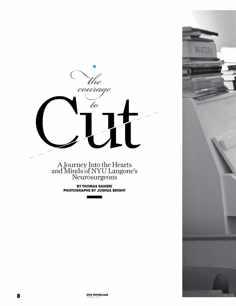
ThE cOURaGE TO cUTa Journey into the hearts and Minds of NYU langone’s Neurosurgeons

EVERY ASPIRING PHYSICIAN DREAMS OF THE DAY SOMEONE WILL CALL HIM OR HER “DOCTOR” FOR THE FIRST TIME. But getting there takes a lot more than hard work and dedication—it takes resources. By contributing to the NYU School of Medicine Alumni Campaign, you help ensure that our next generation of physicians will have access to the best teaching and research, along with a competitive financial assistance package.
When you make a gift, you help us guarantee that all of our students will have the means to complete our rigorous education. One day, you may even have the privilege of addressing them yourself as “Doctor.”
MAKE A GIFT ONLINEPlease visit www.nyu.edu/alumni.
To discuss special giving opportunities, call Anthony J. Grieco, MD, Associate Dean for Alumni Relations, at 212.263.5390.
Help Us Make Dreams Come True
Thank you for your generosity.

THE MAGAZINE OF NEW YORK UNIVERSITY SCHOOL OF MEDICINE
New York University
Martin Lipton, Esq. Chairman,
Board of Trustees
John Sexton President
Robert Berne Executive Vice President
for Health•
NYU Langone Medical Center
Kenneth G. Langone Chairman,
Board of Trustees
Robert I. Grossman, MDDean and Chief
Executive Offi cer•
NYU PHYSICIAN
Steven B. Abramson, MD Anthony J. Grieco, MD
Editors, Science and Medicine
Marjorie ShafferEditor
Nicole Dyer Contributing Editor
Sherry Zucker Print Production
Coordinator
Segal Savad DesignArt Direction
Nancy E. ShermanCopy Editor
•ON THE COVER: ILLUSTRATION BY
GREGORY MANCHESS
NYUPHYSICIAN
1
3SPRING 2013
NO. VOLUME 64
ILLU
STR
ATI
ON
: G
ETT
Y I
MA
GE
S
NYU PHYSICIAN SPRING 2013
2(S)A F C03 Fragment base #6 Base 108
The Courage to CutA journey into the hearts and minds of NYU Langone’s neurosurgeons. Missed ConnectionsAn emerging theory of autism links social isolation to faulty circuits in the brain.
A Matter of DegreesA new option to earn an MD in three years aims to help some students save time and money.
The Heart of the MatterHow molecular autopsies are solving mysterious deaths— and saving lives.
08COVER STORIES
1618 20
DEPARTMENTS
02 Message from the Dean Because It Is Brain Surgery
03 News from Medicine• When Leukemia Rebounds• A Strategy to Drive Leukemia Cells Out of Hiding• One Access Point for Two Deadly Diseases• Every Breath You Take• A Chemical Clue to Aging
14 Faculty Conversation Dr. Glenn Saxe on Sandy Hook and childhood trauma.
24 Tools of the Trade In praise of the stethoscope.
26 Faculty News• Dr. Vilcek Honored at White House Awards Ceremony• Dr. Littman Receives Inaugural Ross Prize from Molecular Medicine• NYU Langone’s Nurses Honored for Their Heroism during Hurricane Sandy
28 End Page Dr. Abramson to Chair Medicine
Molecular autopsies can help medical examiners pinpoint the cause of a mysterious death, identify lethal mutations that might run in families, and even solve crimes.

2 NYU PHYSICIAN SPRING 2013
PHOTOGRAPH BY JOHN ABBOTT
MESSAGE FROM THE DEAN & CEO
DEAN & CEO ROBERT I. GROSSMAN, MD
Because It Is Brain Surgery
I’m immensely proud of our Department of Neurosurgery, which has a long and distinguished history, and I’m delighted to see it featured in this issue. It’s a well deserved tribute to a remarkable team of surgeons. Fifty years ago, the late Joseph Ransohoff, MD, chair of our Department of Neurosurgery from 1962 to 1992, served as medical consultant to the pioneering TV series Ben Casey, whose title character was loosely modeled after the brash, brooding surgeon. For several consecutive years, U.S. News & World Report’s Best Hospital Rankings has placed our program in neurosurgery among the top 10 in America.
In the last two years, the Department has become even stronger, recruiting several outstanding neurosurgeons, some of them world-renowned for their expertise in certain subspecialties. What heartens me most about these master surgeons is their compassion and can-do attitude. As one of them puts it: “Many patients are told that their tumor is ‘inoperable,’ but then they come here and they survive.”
I think you’ll enjoy this inspirational journey into their hearts and minds. •
LONG BEFORE I BECAME DEAN & CEO OF NYU LANGONE MEDICAL CENTER, I began a residency in neurosurgery. Though I eventually changed course, deciding on the field of radiology, I never lost my fascination, my respect, my awe, for the mysteries and marvels of the human brain. Indeed, that passion for neuroscience steered me toward neuroradiology as a subspecialty. So while I try not to play favorites, I must admit that neurosurgery and neurosurgeons, whom I’ve consulted with closely for many years, hold a special place in my heart.

3NYU PHYSICIAN SPRING 2013
STE
VE
GS
CH
ME
ISS
NE
R /
SC
IEN
CE
SO
UR
CE
therapeutics at the Dana-Farber/Children’s Hospital Cancer Center in Boston, says the discovery may help clinicians create personalized treatment strategies.
“This paper identifies an abnormal enzyme that could be specifically targeted and exploited in some patients with relapsed leukemia,” Dr. Place says. Inhibiting the enzyme may also prove useful in initial ALL therapies, he says, helping doctors prevent those relapses in their pediatric leukemia patients. • —BRYN NELSON
relapse,” he says.In the new study, led by Dr.
Carroll and graduate student Julia Meyer and published in the March issue of Nature Genetics, researchers analyzed bone marrow samples from 10 pediatric ALL patients for telltale clues to the disease’s progression. After receiving samples from the Children’s Oncology Group, the medical researchers painstakingly pieced together a complete sequence of ribonucleic acid, or RNA, extracted from each patient’s bone marrow—or about 100 billion letters of RNA in all. “It took years of effort analyzing samples from the same patient at diagnosis and relapse,” Dr. Carroll says.
RNA is an essential intermediary in the cellular process that uses DNA blueprints to assemble proteins, meaning that a complete RNA sequence can give researchers a view of all active genes within a patient’s leukemia cells. By comparing the sequences at the moment of diagnosis and upon relapse, the team found that most patients had acquired multiple
mutations that changed the genetic code over the course of the disease.
Intriguingly, two patients harbored a mutation in the same gene, NT5C2, which encodes an enzyme that regulates some DNA building blocks but can also degrade an important class of drugs used in ALL therapy. When the researchers fully sequenced the NT5C2 gene in 61 other cases in which pediatric ALL patients had relapsed, they found five more mutations that had altered the gene. Experiments suggested that these seven NT5C2 mutations all made the cancer cells more resistant to chemotherapy.
By identifying a specific disease mechanism, the finding may help doctors detect the early emergence of chemotherapy-resistant leukemia cells and adjust their strategy before the disease can fully reassert itself. Andrew Place, MD, PhD, an instructor in pediatrics at Harvard Medical School and associate director of developmental
EVERY YEAR in the United States, roughly 6,000 patients are diagnosed with acute lymphoblastic leukemia (ALL), a disease in which the body’s bone marrow produces a glut of lymphocytes, or white blood cells. Improved chemotherapy and other treatments have dramatically boosted the cure rate of ALL, the most common type of childhood cancer, to about 80 percent. For the unfortunate 20 percent of children who relapse after a bout with the aggressive blood-borne disease, however, the prognosis remains dire.
“There has been no progress in curing children who relapse, in spite of giving them very high doses of chemotherapy and bone marrow transplantation,” says William L. Carroll, MD, director of NYU Langone Medical Center’s Cancer Institute and the Julie and Edward J. Minskoff Professor of Pediatrics.
Dr. Carroll’s team may now be closer to changing that grim reality with the discovery of 20 genetic mutations linked to relapse, including one that may allow ALL to reemerge months or years after the initial diagnosis. “For the first time, we have pinpointed genetic mutations that lead to chemotherapy resistance and
A micrograph of a white blood cell. In acute lymphoblastic leukemia, white blood cells turn malignant and flood the bone marrow.
Julia Meyer
William Carroll
When Leukemia ReboundsGenetic mutations linked to relapse in common childhood cancer point the way to new treatments.
NEWS FROM MEDICINE
ILLUSTRATIONS BY LEANDRO CASTELAO
A Strategy to Drive Leukemia Cells Out of Hiding
NEWS FROM MEDICINE
4
DR
. G
OPA
L M
UR
TI /
SC
IEN
CE
SO
UR
CE
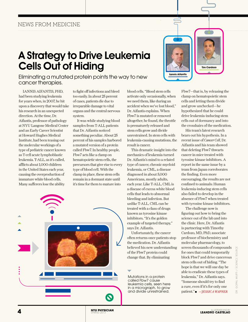
Mutations in a protein called Fbw7 cause leukemia cells, seen here in a micrograph, to grow and divide unrestrained.
Eliminating a mutated protein points the way to new cancer therapies.
to fight off infections and bleed too easily. In about 25 percent of cases, patients die due to irreparable damage to vital organs and the central nervous system.
It was while studying blood samples from T-ALL patients that Dr. Aifantis noticed something peculiar. About 25 percent of his samples harbored a mutated version of a protein called Fbw7. In healthy people, Fbw7 acts like a clamp on hematopoietic stem cells, the precursors that give rise to every type of blood cell. With the clamp in place, these stem cells remain in a dormant state until it’s time for them to mature into
blood cells. “Blood stem cells activate only occasionally, when we need them, like during an accident when we’ve lost blood,” Dr. Aifantis explains. When Fbw7 is mutated or removed altogether, he found, the throttle is prematurely released and stem cells grow and divide unrestrained. In stem cells with leukemia-causing mutations, the result is cancer.
This dramatic insight into the mechanics of leukemia turned Dr. Aifantis’s mind to a related type of cancer, chronic myeloid leukemia, or CML, a disease diagnosed in about 5,000 Americans, mostly adults, each year. Like T-ALL, CML is a disease of excess white blood cells that leads to abnormal bleeding and infection. But unlike T-ALL, CML can be treated with a class of drugs known as tyrosine kinase inhibitors. “It’s the golden example of targeted therapy,” says Dr. Aifantis.
Unfortunately, the cancer often returns once patients stop the medication. Dr. Aifantis believed his new understanding of the Fbw7 protein could change that. By eliminating
Fbw7—that is, by releasing the clamp on hematopoietic stem cells and letting them divide and grow unchecked—he hypothesized that he could drive leukemia-inducing stem cells out of dormancy and into the crosshairs of the medication.
His team’s latest research bears out his hypothesis. In a recent issue of Cancer Cell, Dr. Aifantis and his team showed that deleting Fbw7 thwarts cancer in mice treated with tyrosine kinase inhibitors. A report in the same issue by a team from Japan corroborates the finding. Even more encouraging, the results are not confined to animals: Human leukemia-inducing stem cells also failed to develop in the absence of Fbw7 when treated with tyrosine kinase inhibitors.
The challenge now is figuring out how to bring the science out of the lab and into the clinic. Here, Dr. Aifantis is partnering with Timothy Cardozo, MD, PhD, associate professor of biochemistry and molecular pharmacology, to screen thousands of compounds for ones that could temporarily block Fbw7 and drive cancerous stem cells out of hiding. “The hope is that we will one day be able to eradicate these types of leukemia,” Dr. Aifantis says. “Someone should try to find a cure, even if it’s for only one patient.”• —JESSICA WAPNER
IANNIS AIFANTIS, PHD, had been studying leukemia for years when, in 2007, he hit upon a discovery that would take his research in an unexpected direction. At the time, Dr. Aifantis, professor of pathology at NYU Langone Medical Center and an Early Career Scientist at Howard Hughes Medical Institute, had been teasing out the molecular workings of a type of pediatric cancer known as T-cell acute lymphoblastic leukemia. T-ALL, as it’s called, afflicts about 1,000 children in the United States each year, causing the overproduction of immature white blood cells. Many sufferers lose the ability
Iannis Aifantis
NYU PHYSICIAN SPRING 2013
Tim Cardozo

5NYU PHYSICIAN SPRING 2013
DA
VID
M.
PH
ILLI
PS
/ S
CIE
NC
E S
OU
RC
E
One Access Point for Two Deadly Diseases
real clinical advance.”Given the results, the CCR5
receptor could become a focal point for new anti-HIV strategies as well. Dr. Unutmaz, for exam-ple, wonders whether clinicians could protect the body’s vulner-able mucosal areas by ridding them of CCR5-bearing cells that would otherwise be targeted by the virus. His lab is also explor-ing the possibility of using the LukED toxin to target and kill CCR5-containing T cells that harbor hidden reservoirs of HIV. “These are highly speculative approaches, of course, but it’s exciting,” he says. “One break-through 17 years ago may beget another.” • —RENEE TWOMBLY
experimented with immune cells that Dr. Unutmaz had developed during his previous HIV research, including a genetically engineered T cell that has excess copies of the CCR5 receptor decorating its surface. When the scientists exposed these cells to a purified Staph toxin called LukED, all the cells died. Cells that had been completely stripped of the receptor, however, resisted the toxin’s effects and survived.
Other studies have revealed a remarkably similar phenomenon in HIV infection: Due to a genetic mutation, about 1% of Northern Europeans completely lack the CCR5 receptor on their T cells and are thereby protected against infection. The NYU Langone collaborators’ research suggested that the LukED toxin does indeed share a common target with HIV, in this case using the CCR5 receptor as a landing pad from which the toxin can lethally puncture the immune cells.
If removing CCR5 from T cells warded off Staph infections, the researchers wondered, could simply blocking access to the receptor have the same beneficial effect? To find out, they treated T cells with maraviroc—an anti-HIV medication that clings to CCR5 and makes it inaccessible to the virus—and then exposed the cells to the Staph toxin.
The result, says Dr. Unutmaz, was remarkable. “Maraviroc completely blocked the toxic effects of LukED at doses similar to those used to inhibit HIV infection.”
Dr. Torres was equally surprised. “While maraviroc does not protect other immune cells from the other leukotoxins, it might give the immune system the upper hand in controlling a Staph infection,” he says, “and that would be a
SEVENTEEN YEARS ago, Derya Unutmaz, MD, associate professor of microbiology and pathology and medicine, and Nathaniel Landau, PhD, professor of microbiology, helped identify a protein on the outer surface of the immune system’s T cells that HIV can exploit. The team discovered that the virus uses a receptor called CCR5 like a perch to penetrate T cells and launch an infection that can progress to AIDS.
A new collaboration with Victor J. Torres, PhD, assistant professor of microbiology, has implicated the same receptor as a critical access point for a toxin released by Staphylococcus aureus. The bacterial pathogen is infamous as a hospital scourge that kills an estimated 100,000 people in the U.S. every year. In a study published in the January 3 issue of Nature, the collaborators also discovered that an anti-HIV medication that blocks access to the vulnerable receptor might prevent both the virus and Staph toxin from ever gaining a toehold.
“What are the chances that a drug for HIV could possibly treat a virulent Staph infection?” asks Dr. Torres, the study’s senior author, who notes that the surprising findings could hold major implications for devising new clinical strategies.
The serendipitous discoveries began when the researchers
Nathaniel Landau
A serendipitous collaboration reveals a common link between HIV and Staph infections that could be used to treat both. Derya Unutmaz
Victor Torres
A micrograph of Staph bacteria. Staph toxins may one day help prevent HIV infection.
ILLUSTRATIONS BY LEANDRO CASTELAO
Every Breath You Take
6
WA
RR
ICK
G.
/ S
CIE
NC
E S
OU
RC
E
Scientists discover a way to trace the motor neurons central to healthy breathing.
Neuroscience that they have discovered a genetic fingerprint of PMC neurons as well as the two key genes that control their development. The study was hailed as a “major milestone” in an accompanying commentary.
“We now have a set of molecular markers that distinguish PMC cells from other populations of motor neurons so that we can study them in detail and look for ways to selectively enhance their survival,” Dr. Dasen says.
Such information could be the key to keeping these vital neurons alive in patients with amyotrophic lateral sclerosis (ALS), a debilitating and incurable disease that kills motor neurons, or in patients with injuries to nearby parts of the spine.
To find out how PMC neurons differ from their spinal neigh-bors, postdoctoral fellow Polyx-eni Philippidou, PhD, injected a fluorescent dye into one of the phrenic nerves, which wire the PMC to the diaphragm. Dr. Philippidou then looked for the spinal neurons that lit up as the tracer worked its way back up to the PMC. After noting the char-acteristic pattern of gene activity within these PMC neurons, Dr. Philippidou began to determine the genes’ specific roles.
Working with a complicated strain of transgenic mice, based partly on mice supplied by collaborator Lucie Jeannotte, PhD, at the University of Laval
in Quebec, the researchers ultimately identified two genes, Hoxa5 and Hoxc5, as the prime controllers of proper PMC development. Hox genes are well-known as regulators of animal development.
When the scientists silenced Hoxa5 and Hoxc5 in embryonic motor neurons, the PMC failed to form its usual, tightly columnar organization and failed to connect to the diaphragm, leaving newborn mice unable to breathe. “Even if you delete these genes late in fetal development, the PMC neuron population drops and the phrenic nerve doesn’t form enough branches on diaphragm muscles,” Dr. Dasen says. In other words, the genes play a crucial role in establishing and maintaining the early neuronal connections needed for every breath.
The study will help Dr. Dasen and those in his lab understand the wider circuitry of breath-ing as well—including neurons in the brain stem that regulate breathing in response to carbon dioxide levels, stress, and other environmental factors. “Now that we know something about PMC cells,” Dr. Dasen adds, “we can work our way through the broader circuit, to try to figure out how all those connections are established.” • —JIM SCHNABEL
natural rhythm of breathing.Harming the spinal cord
region where the PMC resides can instantly shut down breathing. But relatively little is known about what distinguishes these neurons from their neighbors and how PMC neurons develop and wire themselves to the diaphragm in the fetus.
Scientists are now a step closer to answering those questions. Dr. Dasen and his colleagues reported in the December issue of Nature
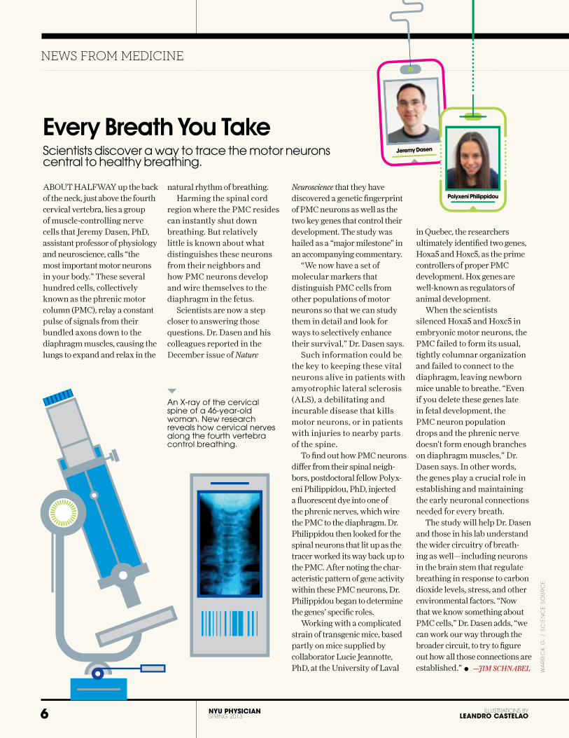
ABOUT HALFWAY up the back of the neck, just above the fourth cervical vertebra, lies a group of muscle-controlling nerve cells that Jeremy Dasen, PhD, assistant professor of physiology and neuroscience, calls “the most important motor neurons in your body.” These several hundred cells, collectively known as the phrenic motor column (PMC), relay a constant pulse of signals from their bundled axons down to the diaphragm muscles, causing the lungs to expand and relax in the
NEWS FROM MEDICINE
NYU PHYSICIAN SPRING 2013
Jeremy Dasen
Polyxeni Philippidou
An X-ray of the cervical spine of a 46-year-old woman. New research reveals how cervical nerves along the fourth vertebra control breathing.

7NYU PHYSICIAN SPRING 2013
HE
ITI
PAV
ES
/ S
CIE
NC
E S
OU
RC
E
A Chemical Clue to Aging
molecule produced by one organism can dramatically affect the physiology and even life span of another organism through direct cell signaling,” Dr. Nudler says. Given the potential for nitric oxide–generating microbes to likewise pay big dividends in the human gut, he adds, “I think it’s a very interesting direction to explore.” • —BRYN NELSON
this finding holds several important medical implications. Unlike worms, we make our own nitric oxide. However, levels gradually decrease over time, a decline he speculates could contribute to aging and could be at least partially reversed by supplemental bacteria supplying some of the missing compound.
“It’s striking that a small
“In worms, we now know that bacteria can use nitric oxide not only to their own advantage but also to provide their host with a beneficial response, and the same thing could be true in a human gut,” Dr. Nudler says. “It may well be the case that our commensal bacteria control some of our genes, at least in the gut, to protect those cells against stress and aging.”
Dr. Nudler’s lab used fluorescent labels and other techniques to document how the worm hijacks nitric oxide from a soil-dwelling bacterium called Bacillus subtilis that is both a favored food and a common colonist within its gut. Once nitric oxide penetrates the worm’s tissue, the researchers discovered, it helps activate a set of 65 genes, including some previously implicated in stress resistance, immune response, and increased life span. Crucially, the chemical’s control over these genes requires the presence of two regulatory proteins that are evolutionarily conserved from worms to humans and act like master switches to turn on or off the vast majority of the genes.
When the researchers corrected a defect that prevented mutant B. subtilis from producing nitric oxide—thereby restoring the worm’s supply of the chemical—they increased the average life span of the worm by about 15%. Dr. Nudler believes
SCIENTISTS HAVE long puzzled over the fact that some bacteria can dramatically boost the health and life spans of their hosts—the organisms in which they dwell. Evgeny Nudler, PhD, the Julie Wilson Anderson Professor of Biochemistry and newly appointed Howard Hughes Medical Institute investigator, recalls being particularly struck by research suggesting that a roundworm can survive roughly 50% longer than its average two-week life expectancy simply due to the type of bacteria it hosts.
In a study published in February in Cell, a team led by Dr. Nudler and Ivan Gusarov, PhD, research assistant professor, traces part of that difference in roundworm longevity to the ability of host bacteria to make nitric oxide, a chemical messenger implicated in everything from nerve signaling to blood flow. The new study, funded in part by Timur Artemyev, concludes that nitric oxide sends a signal to activate multiple genes in the worm that may protect it from stress and extend its life.
The worm, Caenorhabditis elegans, is a common stand-in for humans in studies of the aging process, and the discovery suggests how the microbiome, the trillions of bacterial colonists on and within our bodies, might similarly influence human health.
The roundworm, shown here beneath a fluorescence microscope, lives longer when it hosts certain bacteria.
Study shows roundworms use nitric oxide to live longer. Could humans do the same?
Evgeny Nudler
8 NYU PHYSICIAN SPRING 2013
the the tcoura�coura�coura e�e�to
A Journey Into the Hearts and Minds of NYU Langone’s
NeurosurgeonsBY THOMAS RANIERI
PHOTOGRAPHS BY JOSHUA BRIGHT
•

dr. John golfinos, chair of the department of neurosurgery, at
home with his daughter Phoebe.

pleasures, joys, laughter, and jests, as well as our sorrows, pains, grief, and fears.” Chandranath Sen, MD, professor of neurosurgery and director of the Division of Skull Base Surgery, speaks of “the sacred privilege” of being a neurosurgeon. “When I first meet a patient, that person has never seen me before, yet he or she is willing to put their life in my hands. This is the weakest moment in that person’s life—they are lost, helpless, scared to death. I have to treat this person very gently. I never take the risk. It’s the patient who takes the risk. He or she must have courage. I must have conviction. Before a big case, I meditate. It gets my mind in the zone.”
That gentleness, says Dr. Sen, must carry over into the OR. “The brain has the consistency of Jell-O. A tumor feels like a piece of meat. The nerves are like small wet noodles,” he explains. “You must peel away the meat without disrupting the Jell-O. Along the way, you must be careful not to damage any blood vessels, which could cause a stroke, or nerves, which could cause
blindness, deafness, paralysis, or other problems. To succeed, you must have the touch of a woman—and the heart of a lion. Once you are done with the operation, the brain must never know you were there.”
chair of the Department of Neurosurgery. “Either I try to remove every last bit of this tumor, or I leave some behind. Most of the time, the tumor will keep growing. But if you try to get it all, there’s often a cost to the patient. You’re constantly doing risk-benefit analysis. That’s why you have to know the patient as well as you can. You must have a sense of what he or she would be willing to sacrifice to extend their life. I’m amazed at how much more aggressive I was just 10 years ago. As you get older, all the complications and consequences start to weigh on you.”
Dr. Golfinos and his colleagues in the Department of Neurosurgery are ever mindful of their unique, profound responsibility: treating the organ that, more than any other, makes us who we are. “From the brain and the brain only,” noted Hippocrates, “arise our
T’S TAKEN HOURS of painstaking work—prepping, cutting, sawing, drilling, and clamping—to get here, but Dr. John Golfinos has finally reached his destination: the first of two benign tumors he’ll be removing from the patient’s brain. During an especially delicate part of the procedure, he shares a piece of wisdom, born of 17 years of experience as a lead surgeon, with his assisting surgeon, a seventh-year resident. “Now, when you get to this part,” he says quietly, peering through the eyepieces of a large overhead microscope, “you have to have the courage to cut.”
The courage to cut. The phrase goes to the heart of what it means to be a neurosurgeon, and what makes neurosurgeons a breed apart. “Sometimes you face a tough decision,” explains Dr. Golfinos, associate professor of neurosurgery and otolaryngology and
10 NYU PHYSICIAN SPRING 2013
•••
“ What makes a great neurosurgeon is his thinking. It’s not about technical skills. It’s about knowing what you can and can’t touch, what you can and can’t move. A great neurosurgeon can almost see through things.”— Dr. Anthony Frempong-Boadu
ABOVE Dr. Anthony Frempong-Boadu, director of the Division of Spinal Surgery, at his home in the country.

Is it any wonder that neurosurgeons speak so reverentially of this three-pound mass of pinkish-gray tissue about the size of a cantaloupe? “The brain is the most complex organ in the universe,” says Jafar Jafar, MD, professor of neurosurgery, director of the Division of Cerebrovascular Surgery, and neurosurgeon-in-chief. “We still don’t know which part of the brain we see with. It’s an enduring mystery—one of many enduring mysteries.” Dr. Golfi nos puts it another way: “We can operate on someone’s brain while they’re awake, take out part of it, and they can talk to us the entire time. Even though we’re actually damaging a small part of the brain, the rest of it couldn’t care less.”
11NYU PHYSICIAN SPRING 2013
The marvels of the human brain don’t end there, of course. An organ whose makeup is 75% water, it generates 100,000 chemical reactions every second and enough electrochemical energy to power a 10-watt lightbulb. The brain contains 100 billion neurons (nerve cells), each connected—directly or indirectly—to as many as 100,000 others. Twelve pairs of cranial nerves, some of which control numerous parts of the head, and 100,000 miles of blood vessels crisscross its terrain. Though pain is registered in the brain, the organ itself has no pain receptors and cannot feel pain. The brain demands 15% to 20% of the blood pumped from the heart. If it’s deprived of blood-borne oxygen for
as little as 8 to 10 seconds, the result is unconsciousness. After 30 seconds or so, permanent brain damage may occur. In a single day, the brain gives rise to an estimated 70,000 thoughts, processing information at a rate of up to 268 miles per hour. In a lifetime, it can retain one quadrillion separate bits of information.
Such a precious object deserves high-level security, and the brain’s protective mechanisms are truly impressive. The bony skull, or vault, is a quarter-inch thick at the top and even thicker at the base. The meninges, three layers of membranes that line the skull, shield the brain further. The brain is suspended in cerebrospinal fl uid, which functions as a shock absorber, reducing the impact of
Dr. Jafar Jafar, director of the Division of Cerebrovascular Surgery, in his home library.

12 NYU PHYSICIAN SPRING 2013
aneurysms and vascular malformations, spine ailments, epileptic seizures, deep brain stimulation for Parkinson’s disease, and a range of other conditions. “Some of our new colleagues are iconic,” says Anthony Frempong-Boadu, MD, associate professor of neurosurgery and director of the Division of Spinal Surgery. “We observe each other in the OR like it’s our first time there. The intellectual exchange has upped everyone’s game.” Dr. Jafar is so proud of his newly expanded department that he says, “I’ve visited many departments of neurosurgery around the world, and few of them rival ours. Many patients are told that their tumor is inoperable, but then they come here and they survive.”
“Our goal,” Dr. Golfinos says, “is to be the place that other neurosurgery centers refer their really difficult cases to because we can get the job done.” For neurosurgeons, that often requires what Dr. Frempong-Boadu describes as “walking the tightrope” between confidence and hubris. “Neurosurgeons are comfortable going into the unknown,” he says. “A lot of what we do is shrouded in mystery. What makes a great neurosurgeon is his thinking. It’s not about technical skills. It’s about knowing what you can and can’t touch, what you can and can’t move. A great neurosurgeon can almost see through things.”
For all the information and insights that sophisticated imaging can provide, the map is not the same as the territory itself, and danger, if not disaster, is sometimes only a millimeter away. “You’re often working in a surgical field the size of a quarter or half dollar,” Dr. Wisoff explains.
“If you don’t have fear,” adds Dr. Jafar, “you’re dangerous. You can work for hours, and in the last 10 seconds you can ruin everything. The most dangerous part of the operation is the last 5 minutes
Neurosurgery. “Before we had the benefit of detailed imaging, stereotactic navigation, and electrophysiological monitoring, you had to have incredible technical mastery,” he explains. “That said, you can’t be a klutz.”
Then what does separate good neurosurgeons from great ones? “The greats have a real balance of confidence and empathy,” Dr. Golfinos says. “The hardest thing to teach is taking ownership of the patient—realizing that this person has placed everything they have in your hands.”
Dr. Sen agrees: “If you exude a lack of confidence, your entire team senses it, and the team breaks down. If you get anxious, your hand shakes, and the situation quickly deteriorates.” For Dr. Wisoff, the key is strategy. “You plan an operation like you plan a battle,” he says. “You know where all the pieces lie, and you’re prepared for as many contingencies as possible.” Dr. Jafar sums it up in a single word: tenacity. “Hating to lose is what makes us a breed apart. I chose neurosurgery because it’s difficult. I thrive on challenges. I’m a bad loser.”
NYU Langone Medical Center’s Department of Neurosurgery has never had so many winners. In the last two years, Dr. Golfinos has recruited several outstanding neurosurgeons, some of them world-renowned for their expertise in subspecialties. The department now has 17 neurosurgeons serving patients at Tisch Hospital, Bellevue Hospital Center, and the Manhattan VA Medical Center. Its long-standing reputation for excellence encompasses surgery for brain tumors, brain
sudden blows. Isolated from the bloodstream by a blood-brain barrier, the brain has a dutiful gatekeeper, admitting some nourishing elements and forbidding entrance to others, notably toxins.
The challenge of penetrating this formidable fortress to reach the treasure that lies within has intrigued mankind for centuries. The first successful operations to remove tumors, however, took place only about a century ago. Until the 1970s and 1980s, when CT scans, overhead microscopes, high intensity illumination in the OR, and microsurgical instruments became available, a neurosurgeon’s skill relied largely on his keenness of vision and steadiness of hand. While manual dexterity, hand-eye coordination, spatial perception, and tactile memory (the ability to return your hand to exactly the same place it was before you moved it) will always be the hallmarks of a neurosurgeon, “golden hands have become less and less important,” says Jeffrey Wisoff, MD, associate professor of neurosurgery and pediatrics and director of the Division of Pediatric
NYU PHYSICIAN SPRING 2013
ABOVE Dr. Chandra Sen, director of the Division of Skull Base Surgery, after landing a Piper Archer he flies for a hobby.
•••
“ Once you are done with the operation, the brain must never know you were there.”
— Dr. Chandra Sen

13NYU PHYSICIAN SPRING 2013
because you think you’re finished. You can’t afford to let your guard down, even for a moment. I don’t listen to music while I’m operating. The night before, I listen to Mozart. In the OR, I’m listening to the patient’s heart rate, one of many things that guide me.”
The importance of total concentration is just one of the lessons Dr. Jafar tries to instill in his residents, mostly by example. Unlike many programs in neurosurgery, NYU Langone has a training period of seven years instead of six. During the last year (the second of two as chief resident), the newly minted neurosurgeon functions as the operating surgeon for more than 300 varied neurological procedures. Yet there’s one thing, says Dr. Jafar, that you can’t teach: judgment. “For a neurosurgeon,” he explains, “common sense is knowing what your limits are. Knowing how to stay out of trouble—or get yourself out of trouble. Knowing when to stop the surgery if you have to.”
Dr. Wisoff feels that the key to mastering the art of critical thinking inside the OR is to anticipate as many pitfalls as possible and carefully consider the options outside the OR. “When you’re removing a malignant tumor,” he says, “you have to be appropriately aggressive. If you go in with a timid attitude, you’ll perform a timid operation—what we call peek and shriek. If that’s not right for the patient, you’ve done him or her a tremendous disservice.” In the still fairly uncharted landscape of the brain, doing what’s right for the patient may be the greatest challenge of being a neurosurgeon, but it’s also the most critical. “If I’m not comfortable with a resident I’ve trained operating on a member of my own family,” Dr. Jafar says, “then I’ve failed.”
For even the best neurosurgeons, the possibility of failure—be it deficits or
NYU PHYSICIAN SPRING 2013
death—always looms large. “Every one of my cases is my toughest case ever,” Dr. Sen says. “That degree of respect keeps you from becoming cavalier.” The survival rates for benign brain tumors have increased dramatically over the years, so most of the time, notes Dr. Jafar, “the odds are by far in favor of the patient.” In children with malignant brain tumors, explains Dr. Wisoff, the cure rates are above 80%. “I followed in my father’s footsteps,” he explains, “and he taught me that to be a neurosurgeon, you have to be an optimist. ‘Maybe it’s a subdural hematoma that we can drain and cure the patient,’ he would say. Or ‘maybe it’s a benign tumor that we can remove and cure the patient.’ Or ‘maybe it’s a low-grade malignancy that will allow us to buy the patient a lot of time.’ Or ‘maybe we can’t save the patient, but we can bring better quality to their life.’ ”
“Some patients have a will to live that’s very, very strong,” Dr. Golfinos notes, “especially if they have children or someone else to live for.” But on those rare occasions when a neurosurgeon must accept defeat—and they can recite the names of every patient they’ve ever lost—the toll is a lingering one. “When my
kids were young,” Dr. Wisoff recalls, “I would go home and give them a big hug. Then I’d sit down with my wife and have a long talk.” For Dr. Sen, healing requires solitude. “When I have a disaster,” he says, “I go home, I sit quietly, and I have even cried.” Dr. Jafar believes that the only true measure of comfort comes from “knowing that you did your best, and that you’re at peace with yourself.”
Neurosurgeons seem to reserve their greatest optimism for their faith in nature and science. “When you think of the billions of cell divisions that take place between conception and delivery,” says Dr. Wisoff, “creating the potential for our genetic code to be misinterpreted or to go awry, it’s astonishing how few problems we actually have.” Dr. Golfinos points to improved medications, endovascular techniques, technology, and other advances that have “stacked the odds in our favor.” Fifty years ago, in Ben Casey’s day, he reminds us, “there was only one kind of tumor he could remove without killing the patient: a meningioma. Fifty years from now, the greatest advance will be the elimination of neurosurgery. Our specialty will probably be obsolete.” •
Dr. Jeffrey Wisoff, director of the Division of Pediatric Neurosurgery, cycling near his home.

NYU PHYSICIAN SPRING 2013
FACULTY CONVERSATION
14 PHOTOGRAPH BY GLORIA BAKER
Throughout his career, Dr. Saxe has dedicated
himself to understanding the impact of
traumatic events on children and adolescents
and improving their care. He pioneered a
unique community-based program, Trauma
Systems Therapy (TST), and also helped
develop the National Child Traumatic Stress
Network, a nationwide program that partners
academic research centers with community
practice sites. In February 2013, the Child
Study Center, in collaboration with the New
York City Administration for Children’s
Services and Bellevue Hospital, received grants
totaling more than $7 million to enhance
trauma services for young people. The grants
built on seed funding provided by generous
gifts from Michele and Timothy Barakett and
the Bloomgarden-Willner Family.
How do you define childhood trauma?
Trauma is when a child experiences something
that threatens life and limb. Community
violence, violence within families, abuse,
rape, disasters, war, terrorism, significant
medical illnesses—all of these fit the category
of trauma. Violence in the family and a school
shooting are very different events, but there’s
a core similarity that defines most reactions to
trauma, involving ancient, powerful survival
systems in the body. When a threat occurs, a
structure deep in the brain called the amygdala
fires. It’s a very important structure because
it prepares us to survive in the face of threat.
It makes your heart pound and your muscles
tense, preparing you to fight or flee. Following
a trauma, it’s hard for the amygdala to settle
and so it’s normal to be intensely distressed
and concerned about safety over days, and
even weeks after trauma. In most people, this
emotional state diminishes dramatically with
time, but in some people it persists: Their
amygdala keeps firing, even in situations
where there’s no real threat.
“ If Only They Could Have Been Treated When They Were Kids…”Q&A with Dr. Glenn Saxe, Chair of the Department of Child and Adolescent Psychiatry
Dr. Glenn Saxe
BY CLAUDIA KALB
HE SHOOTING at Sandy Hook Elementary School in Newtown,
Connecticut, last December left Americans desperate for answers. What
do we tell our children after such a horrific event? Will Sandy Hook’s
youngest survivors ever recover? Glenn Saxe, MD, NYU Langone’s Arnold
Simon Professor of Child and Adolescent Psychiatry and director of
the Child Study Center, quickly became an expert source for reporters
covering the tragedy. He stressed the importance of being attuned to
children’s emotional needs and answering their questions honestly, and
offered some solace when he talked about children’s capacity to heal. “Most kids, even of this age,
are resilient,” he told The New York Times.
T

NYU PHYSICIAN SPRING 2013
What happens when those fearful emotions persist?
Ordinarily, other areas of the brain such as the
hippocampus and prefrontal cortex help inhibit
the amygdala from misfiring after a trauma by
bringing context and memory to the situation.
The hippocampus is responsible for helping
people understand context. After an injury, for
example, the hippocampus will recognize that
once you’re in the hospital you’re safe. But in
some children this mechanism gets disordered
and they can no longer recognize that they’re
in a safe place. Something will remind them
of what happened and trigger those survival
systems. It might be someone using a tone of
voice similar to the one used by your stepfather
just before he raped you. You get flooded with
painful memories and may experience yourself
back in the place and time of the trauma. You’re
not under immediate threat, but you’re living
as if you are. That’s the central problem of post-
traumatic stress disorder, or PTSD.
Does trauma leave any permanent effects on the brain?
There is research that indicates kids who endure
chronic stress and PTSD early in life may
develop smaller brains and have lower IQs. If
this is true it indicates an unspeakable tragedy:
They’re starting life with a strike against them
that can influence a great many things and
have cumulative impact. It can affect learning
and school performance. It can affect how they
experience themselves, their effectiveness in the
world, and how others view them.
How big is the problem of childhood trauma and how
will your new grant help?
Obviously, what happened at Sandy Hook is a
terrible tragedy, and our hearts break for the
families who lost these young children. But there
are also many hidden cases of childhood trauma.
Do you know how many American children last
year lost their lives at the hands of their parents
or other caregivers? About 1,500. And that’s just
the tip of the iceberg: The child welfare system
gets 6 million calls about possible child abuse
annually. Any one call may end up as one of the
1500 children who die. How do they get it right?
What if they get it wrong? Our grant involves
partnering with the child welfare system and
building their capacity to get it right. We’re
helping child welfare workers better understand
trauma, training them to screen children for
trauma and working with them to implement
interventions, particularly TST, for kids at risk.
The burnout rate for child welfare workers is
very high, so we’re also working with the child
welfare system to support their workforce.
How many children develop PTSD?
Between 15% and 40% of children who
experience trauma will develop PTSD. Children
may develop other problems as well, including
depression, aggression, and self-destructive
behaviors. The bad news is that many people
have been exposed to traumatic events over
their lifetime, so a lot of people have these
disorders. The good news is that they’re in the
minority. Children are resilient.
Where does a child’s resilience come from?
We’ve found a couple of genes related to risk and
resilience to trauma, but the story is much more
complex than that. Many factors contribute
to resilience. A child’s ability to regulate their
emotions is particularly important. A child’s
ability to say, “OK, I’m getting upset now, but
this isn’t the end of the world. I need to get
through it.” If you have good skills managing
emotion and you experience a trauma, you’re
more likely to be resilient. Parents, of course,
have a critical role helping children learn how to
regulate emotion. Another key part of resilience
is community—the degree to which the people
around the child are attuned to what he or she
is going through and offer help, and the degree
to which a child feels like he or she is part of a
family and community and not alone.
What is Trauma Systems Therapy and how does it
differ from conventional therapy?
We believe traumatic stress is always about two
things: a traumatized child who isn’t able to
regulate his emotional and behavioral states, and
a social environment that isn’t sufficiently able to
help him. We began developing TST in the late
1990s. Every treatment available then involved
the child and clinician in an office. Individual
therapy is a component of caring for traumatized
children, but what was very apparent to me and
my team is that the action is way outside the
office. TST involves developing knowledge about
what triggers a child’s emotions and working
within the family, the school, and wherever else
it may be strategic to help.
What are the core components of TST?
There are four key components of TST and we
offer these components based on our assessment
of what the child needs. First, we help children
develop the skills to regulate their emotions
when they experience something that isn’t
really threatening, but that reminds them of
their trauma. These include coping skills, which
might involve activities such as deep breathing
or counting, and other skills to identify and
manage their emotional states. Second, we work
with those around the child such as families
and schools to help the child to manage emotion
and to protect them from signals that may evoke
threat responses. They may be unwittingly
emitting signals that remind a child of what
happened, so we help them identify a child’s
triggers and diminish them. Third, we provide
psychopharmacology, which is occasionally
needed to help a child take advantage of therapy.
Fourth, we use legal advocates when necessary
to help improve a child’s social environment by
helping the family gain access to needed services.
What advice would you give parents in the aftermath
of a traumatic event?
As parents, your job is to make sure your children
feel safe. After an acute trauma like Sandy Hook,
parents might say, “This is an awful thing that
happened, but we’re doing everything we can to
keep you safe.” Children need that reassurance.
Parents should also answer a child’s questions
honestly and authentically. The most important
thing is for family members to be attuned to their
loved ones and any changes in their behavior.
Is the child more isolated than before? More
irritable? If so, that’s something to track.
How did you get interested in childhood trauma?
I didn’t think I was interested in trauma when
I was going into psychiatric training in the late
1980s. Then I began seeing the devastating
consequences of childhood trauma in my adult
patients. I kept thinking, “If only they could have
been treated when they were kids.” I see child
psychiatry as a prevention field. What gives me
the most gratitude is working with people who
are dealing with really complex problems, with
families that are so vulnerable, and helping them
use a set of tools that makes a very big difference.
If you get it right, the work is life-changing. That’s
what keeps me going. •
15

CONNECTIONSAn emerging theory of autism links social
isolation to faulty circuits in the brainBY JANE BOSVELD
MISSED
17NYU PHYSICIAN SPRING 2013
ILLUSTRATION BYLUCY READING-IKKANDA
TWELVE-YEAR-OLD STEVEN RHODES (not his real name) has no shortage of interests. He can hold forth on Albert Einstein, the atomic bomb, Marvel Comics, and any number of other subjects that fascinate him. The trouble is, he doesn’t know when to stop sharing. “He can talk about Einstein for a half hour straight,” says his mother. “Sometimes people just walk away. He has a hard time reading faces.”
Steven’s monologues are especially disruptive in class, where he often blurts out information without raising his hand. And he resists group activities because he finds them frustrating and confusing. Steven is not intentionally rude, his mother and teachers know. His behavior is a classic symptom of autism-spectrum disorder (ASD), the incurable developmental condition that he was diagnosed with at age 4.
Autism affects one in 88 children in the United States, up from one in 2,000 in the 1970s. Many experts attribute at least part of this dramatic jump to a shift in the clinical definition of autism away from a single condition to a cluster of conditions that range along a spectrum of severity. Steven’s intelligence, verbal skills, and independence land him on the more functional end of the spectrum. But like all ASD patients he struggles to form social bonds.
Despite growing awareness of ASD, why the condition impairs social skills is still poorly understood. Adriana Di Martino, MD, Assistant Professor of Child and Adolescent Psychiatry at the Child Study Center at NYU Langone Medical Center, is looking to brain anatomy for answers. The brain of a child with ASD may look perfectly normal, Dr. Di Martino notes. “All the specific brain regions are right where they’re supposed to be,” she explains. “The problem is in the circuitry that connects them to each other.”
This faulty circuitry is thought to result from changes that occur while the brain is developing. By using functional MRI (fMRI) scans, which measure oxygen uptake in the brain, Dr. Di Martino and her colleagues are mapping patterns of connectivity in the brains of children with ASD and comparing them to children without the disorder. “With ASD, brain regions are poorly synchronized,” Dr. Di Martino explains. Which neural networks malfunction, however, varies from one person
to another. In people with autism, some circuits are more strongly connected than usual; in others, the connections are weaker.
One network of particular interest in autism research is the Default Mode Network, or DMN. It works a bit like an orchestra conductor, synchronizing different brain regions when the mind is at rest—as during a daydream—to help the brain marshal a coordinated response to the external world. Dr. Di Martino, among others, has found a link between the DMN and autistic traits like aloofness and social awkwardness. Using functional MRIs, she and her team have discovered that people with more ASD traits tend to have weaker connections between two distant brain regions involved in self awareness and interpreting other people’s thoughts and emotions. Thus social isolation appears to be rooted in neural isolation.
Other types of brain scans also support this idea. Using structural MRI, which reveals subtle physical abnormalities, researchers have found unusual patterns of growth in some areas of the autistic brain, including the white matter. White matter, which comprises nearly half the brain’s mass, is “the highway through which different regions of the brain communicate,” explains Dr. Di Martino. In people who are not autistic, the white matter develops at an even pace during gestation. But in autistic children there is a burst of white matter growth earlier than expected, and the growth stops before reaching expected levels. “Typically, developing children eventually have more white matter than children with autism,” Di Martino notes. These differences in how white matter develops, she adds, contribute to the wide range of problems afflicting people with autism.
Dr. Di Martino acknowledges that these recent insights raise more questions than they answer. For example, how does disconnection in the brain emerge? How does it change with age? Are specific circuits involved, or is the problem widespread in the brain? Questions abound, but fortunately the answers appear to be within reach. “It’s exciting that we now have the tools to find the relationship between brain circuits and behavior,” Dr. Di Martino says. “This relationship can guide our search for the brain mechanisms leading to autism.”•
“It’s exciting that we now have the tools to find the relationship between brain circuits and behavior. This relationship can guide our search for the brain mechanisms leading to autism.”

NYU PHYSICIAN SPRING 2013
MEDICAL EDUCATION
18 PHOTOGRAPH BY JOHN ABBOTT
without sacrificing quality. “Our new plan is based on the premise that
medical students should be offered the ability to differentiate and accelerate their careers if they were qualified to do so. We must keep in mind that you become a doctor over the entire course of your training, not just during medical school,” says Steven Abramson, MD, the Frederick H. King Professor of Internal Medicine and chairman of the Department of Medicine, senior vice president and vice dean for education, faculty, and academic affairs,
ON AN AVERAGE of once every minute, a new scholarly article is added to the medical literature—a statistic that would seem to suggest that medical schools should lengthen their curricula to better prepare physicians for 21st century clinical practice. Yet NYU School of Medicine is doing just the opposite. Starting this year, a select group of students have the option of earning an MD in three years, slicing a full year off the traditional education.
This radical rethinking of how doctors are made has prompted some to fear that NYU School of Medicine is on the road to becoming a “trade school” where training is pared down to the bare minimum. But NYU Langone Medical Center’s educators believe that accelerated study, if done properly and offered to selected highly qualified applicants, can be an effective counterbalance to ever more extensive training periods, which now average 10 years for subspecialties,
A Matter of DegreesStarting this year, a select group of students have the option of earning an MD in three years.
Dr. Steven Abramson, Dr. Melvin Rosenfeld, Dr. Vicky Harnik, and Dr. Marc Triola display images of the BioDigital Human, a 3D animated virtual dissection
tool that empowers medical students to learn how and where they wish.
BY GARY GOLDENBERG

NYU PhYSIcIaN SPRING 2013
and professor of medicine and pathology. “A four-year program for all graduates made sense when postgraduate training lasted two or three years. Now, residencies and fellowships routinely extend the postgraduate period to six years or more, which means that many physicians don’t enter practice until their early or mid-30s. Indeed, data from the AMA show that since 1975, the percentage of physicians under the age of 35 has decreased from 28% to 15%.”
With this rationale in mind, Dr. Abramson and his colleagues began looking for ways to trim the standard four-year model of medical education, which has held remarkably constant since the Flexner Report of 1910, which highlighted the era’s wide disparity in physician training and called on medical schools to enact higher admission and graduation standards. As it turns out, NyU Langone has actually laid the foundation for an abbreviated program with its Curriculum for the 21st Century, or C21. Instituted in 2009, C21 provides students with a patient-centered and disease-focused medical education, earlier-than-usual patient contact, and flexible, individualized learning experiences.
“In designing C21, we wanted a flexible curriculum, not only to accommodate students, but also to allow for new educational initiatives, even those we couldn’t anticipate,” says Melvin G. Rosenfeld, PhD, associate dean for medical education and associate professor of cell biology. “Once C21 was in place, Dr. Lynn Buckvar-keltz [associate dean of students and clinical assistant professor of medicine] led an initiative for new dual-degree pathways, including an MD-MPH program with NyU’s Washington Square campus. With some creative changes, we were able to offer this dual degree in four years instead of the usual five. At that point, it was obvious we could also offer a three-year MD pathway.”
NyU’s new pathway features 135 weeks of instruction—20 weeks less than in the traditional four-year program, but still five weeks more than required for accreditation. The time savings kick in just after year one, when three-year students
forgo their summer break, using that time to engage in an eight-week elective. Additional time is saved by waiving the usual third-year 12-week clinical/research concentration and eight weeks of electives.
In another departure from standard practice, three-year enrollees will be offered acceptance into a residency of their choice at NyU Langone at the time of admission. This, too, will save time, since students routinely spend many weeks traveling around the country for residency interviews. In addition, says Dr. Buckvar-keltz, “It’s common for students applying in the most selective fields to spend 12 to 16 weeks doing ‘audition’ electives at outside hospitals to increase their chances of matching at a preferred residency program, even though repeating these electives has limited educational value. Our three-year students will save this time and expense as well.”
“Our three-year pathway is not just about shortening training,” notes Dianna Jacob, vice president for faculty and academic affairs. “It’s really about redesigning medical education, challenging the time-based model, and focusing on competency-based assessment across the continuum of education.”
The idea for a three-year MD degree is not new. Two other American medical schools offer accelerated study, but their students are limited to primary care or family practice residencies. NyU Langone’s program is the first to allow students to choose any specialty. A new informatics technology, the Education Data Warehouse, will be used to track every aspect of the three-year students’ classroom and clinical work. “With the data warehouse, we can ensure that students get all of the experiences they need and tailor those experiences to their chosen specialty,” explains Marc Triola, MD (’98), associate dean for educational informatics and assistant professor of medicine.
The accelerated curriculum promises a number of benefits, including earlier entry into practice, lower overall tuition costs, and a seamless transition from undergraduate to graduate medical training. Beginning in year one, students will be assigned a faculty mentor from
their chosen residency program, building an unprecedented bridge between undergraduate and graduate training.
Critics argue that accelerated study will increase the likelihood of student burnout and deprive students of a “maturing” year that provides time to expand their horizons. While Dr. Abramson acknowledges that increased burnout is a possibility, he believes it can be prevented with careful monitoring and mentoring. Moreover, students will have the option of switching to the standard curriculum at any time.
“The average age of incoming students is older than in the past,” says Dr. Abramson. “Almost half come to medical school after taking one or two ‘gap’ years. So we have a more seasoned pool of applicants, some of whom know exactly what they want to do.”
“For a certain percentage of students, the fourth year is a lost year,” adds Vicky Harnik, PhD, assistant dean for curriculum and assistant professor of cell biology. “They don’t need that time to explore career possibilities or to mature. They’re anxious to get on with their training.”
Fourth-year student Tara Russell (’13) agrees. “The time you are most prepared for residency is likely toward the end of your third year, after you have completed most of your clerkships. I certainly would have considered the three-year option, especially since I took off three years between college and medical school.”
Hannah kirsch (’14) likes the idea of accelerated study, but wouldn’t have chosen it for herself. “I had an excellent summer research experience and another elective that may not have been possible had I enrolled in this program,” she says. “And I’m looking forward to exploring other specialties—or even countries—in my fourth year.”
Slots in the new program will be offered to about 10 percent of the incoming class, and perhaps more in the years ahead. “The biggest stumbling block now to expanding the program is that you have to declare your specialty so early,” says Dr. Harnik. “If we can find a way in which students could enter this pathway near the end of the first year, I think we would have a lot more takers.” •
19
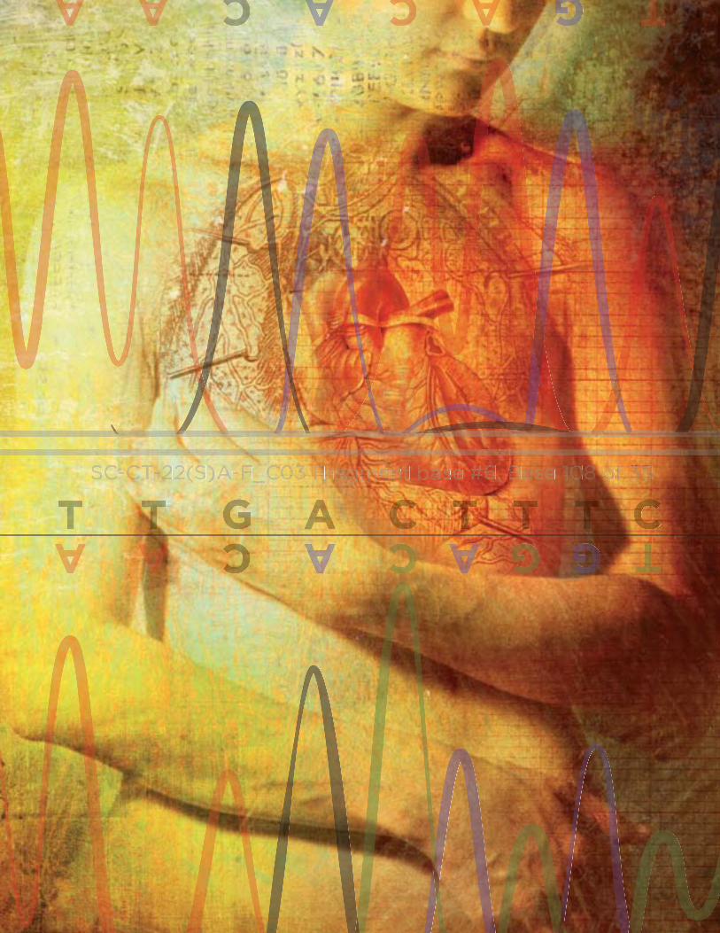
SC-CT-22(S)A-F_C03 Fragment base #6. Base 108 of 35

When seemingly healthy individuals suddenly die,
medical examiners often turn to molecular autopsies for
clues. Thanks to a multiagency collaborative effort, the results
are not only solving mysterious deaths but also saving lives.
the �eartO F T H E M AT T E R
BY BRYN NELSONPHOTOGRAPHS BY JOHN ABBOTT
proved deadly.This case and others like it highlight the power of a so-
called molecular autopsy, a method of genetic sleuthing that can determine the cause of a mysterious death, identify potentially lethal mutations that might run in families, and even help solve crimes. Now, owing to the OCME’s collaborative efforts, the technique is also spurring new insights into how stress and drugs can compound preexisting heart conditions.
Although it functions as a governmental agency, the OCME has long-standing ties to NYU Langone Medical Center’s Departments of Forensic Medicine and Pathology, and many of its medical and research staff are on the faculty of NYU School of Medicine. The agency performs about 5,000 autopsies each year, a good number of them after the sudden, unexpected death of a seemingly healthy New Yorker. In
THE SUDDEN DEATH of a 26-year-old mother of two initially seemed like an open-and-shut case: a tragic cocaine and alcohol overdose. Beyond the toxicology results, the Office of Chief Medical Examiner of the City of New York (OCME) could find no other contributing factors—until the woman’s family raised the possibility of an inherited heart condition.
An ensuing collaboration between the OCME’s molecular genetics lab and the Montefiore-Einstein Center for CardioGenetics (MECC) in the Bronx uncovered a mutation in one of her genes linked to a rare but dangerous heart arrhythmia called long QT syndrome (LQTS) that affects an estimated 1 in 7,000 people in the United States. On its own, cocaine was unlikely to cause the woman to collapse and die, the joint investigation suggested. But in combination with a genetic defect that altered her heart’s electric current, the drug
21NYU PHYSICIAN SPRING 2013
ILLU
STR
ATI
ON
: G
ETT
Y I
MA
GE
S
22 NYU PHYSICIAN SPRING 2013
Although catastrophic failure of the immune, metabolic, or central nervous systems can contribute to sudden death, most of the potentially lethal mutations have been linked to defects in the heart, particularly a subgroup known as cardiac channelopathies. A normal heartbeat derives its steady rhythm from an electrical conduction system that begins in the heart’s right atrium, where a natural pacemaker creates electrical impulses. With every beat, these bursts of energy quickly spread to the rest of the heart through a network of fibers, causing the chambers to alternately contract and relax. At the cellular level, the electrical activity results from the regulated movement of electrically charged sodium, potassium, and calcium ions back and forth through channels in the heart cells. Aberrations in these channels, or channelopathies, can prevent them from opening or closing properly, disrupting the flow and leading to dangerously irregular heartbeats.
These defects may not be apparent in a routine autopsy, but they often show up on electrocardiograms (EKGs) as abnormal patterns and in genetic analyses as DNA mutations previously linked to sudden deaths. LQTS type I, for example, disrupts a potassium channel that helps the heart muscle relax after each contraction, leading to a rapid and chaotic rhythm that can cause sudden death. Unfortunately for the young mother, the inherited channelopathy forced her heart to rely on a second major potassium channel that is exquisitely sensitive to
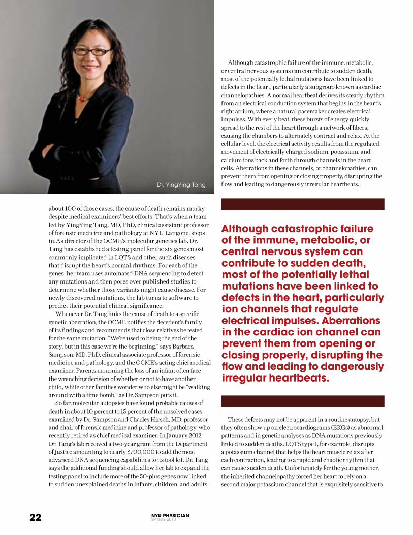
about 100 of those cases, the cause of death remains murky despite medical examiners’ best efforts. That’s when a team led by YingYing Tang, MD, PhD, clinical assistant professor of forensic medicine and pathology at NYU Langone, steps in.As director of the OCME’s molecular genetics lab, Dr. Tang has established a testing panel for the six genes most commonly implicated in LQTS and other such diseases that disrupt the heart’s normal rhythms. For each of the genes, her team uses automated DNA sequencing to detect any mutations and then pores over published studies to determine whether those variants might cause disease. For newly discovered mutations, the lab turns to software to predict their potential clinical significance.
Whenever Dr. Tang links the cause of death to a specific genetic aberration, the OCME notifies the decedent’s family of its findings and recommends that close relatives be tested for the same mutation. “We’re used to being the end of the story, but in this case we’re the beginning,” says Barbara Sampson, MD, PhD, clinical associate professor of forensic medicine and pathology, and the OCME’s acting chief medical examiner. Parents mourning the loss of an infant often face the wrenching decision of whether or not to have another child, while other families wonder who else might be “walking around with a time bomb,” as Dr. Sampson puts it.
So far, molecular autopsies have found probable causes of death in about 10 percent to 15 percent of the unsolved cases examined by Dr. Sampson and Charles Hirsch, MD, professor and chair of forensic medicine and professor of pathology, who recently retired as chief medical examiner. In January 2012 Dr. Tang’s lab received a two-year grant from the Department of Justice amounting to nearly $700,000 to add the most advanced DNA sequencing capabilities to its tool kit. Dr. Tang says the additional funding should allow her lab to expand the testing panel to include more of the 50-plus genes now linked to sudden unexplained deaths in infants, children, and adults.
Dr. YingYing Tang
Although catastrophic failure of the immune, metabolic, or central nervous system can contribute to sudden death, most of the potentially lethal mutations have been linked to defects in the heart, particularly ion channels that regulate electrical impulses. Aberrations in the cardiac ion channel can prevent them from opening or closing properly, disrupting the flow and leading to dangerously irregular heartbeats.

23NYU PHYSICIAN SPRING 2013
provide an explanation. “But slowly, through the use of this molecular testing,” she says, “we will whittle at this and gradually give more and more parents an answer.”
Dr. Tang credits Dr. Sampson and Dr. Hirsch for recognizing the importance of establishing her lab a decade ago. The publicly financed detective work, she says, is still unique among cities in the United States and would have been prohibitively expensive without the extensive infrastructure already laid within the OCME’s Department of Forensic Biology.
Despite the tragedies she encounters on a regular basis, Dr. Tang revels in her lab’s ability to help other doctors provide happier endings. In one heartbreaking case, a seven-year-old boy collapsed and died at school. After the OCME’s standard autopsy tests were inconclusive, the molecular autopsy revealed yet another mutation in a gene linked to LQTS. Two of the boy’s relatives had previously died of heart problems and his half-sister was already under the care of Montefiore-Einstein cardiologists.
Unbeknownst to her doctors, the half-sister harbored the same rare mutation, and her existing heart medication was particularly ill suited for the syndrome subtype. “We did very fast in-house molecular testing for this case,” Dr. Tang says, recalling that the lab’s post-mortem testing spurred the half-sister’s cardiologist to switch her medication immediately to a more appropriate therapy. “They were very happy with that change,” Dr. Tang says, “because that could actually be lifesaving.” •
blockage by cocaine.That knowledge can save lives. Dr. Tang and Dr. Sampson
point to their blossoming relationship with the Montefiore-Einstein Center for CardioGenetics as one example of how genetic sleuthing can open the door to lifesaving testing services and support. Thomas McDonald, MD, the center’s co-director, says the “collegial and collaborative exchange” between the groups allowed them to publish two recent case studies in the journal Pacing and Clinical Electrophysiology. Solving the cocaine-LQTS case also helped his team provide preventive care to the victim’s two children after genetic tests revealed that both had inherited the same mutation. “It’s very important because we can do things that will hopefully decrease their risk for having a heart rhythm disturbance and sudden death like their mother had,” he says.
A second collaboration between Dr. Tang and Dr. McDonald underscores the difficulty of predicting how genetic mutations might affect individual patients. When a young woman suddenly collapsed and died at home several months after undergoing a gastric banding operation to help control her obesity, investigators focused on the frequent vomiting episodes reported by her family.
Her symptoms heightened suspicions that she was dehydrated and running low on potassium. Meanwhile, EKG results that had gone unnoticed before her operation hinted at LQTS. The OCME’s molecular genetics lab discovered a mutation in a gene linked to the disorder, but the joint inquiry revealed a twist. The woman’s mother and brother harbored the same mutation but lacked any symptoms, suggesting that the fatal arrhythmia had been triggered by the additional stress of the deceased’s dehydration and potassium loss.
One of the most difficult aspects of her work, Dr. Tang says, is interpreting the clinical significance of previously unreported genetic mutations. “It’s challenging, it’s going to continue to be challenging, and it really requires teamwork,” she says. With every new mutation, however, scientists are adding to a collective database that may help forensic labs around the world solve other unexplained deaths, including cases of sudden infant death syndrome, or SIDS.
For years, Dr. Sampson explains, SIDS has been used as a catchall label for unexplained infant deaths. “It’s long been suspected that SIDS may not be one thing but a group of things that we are unable to identify from a regular autopsy,” she says. Since the 1994 debut of the national Back to Sleep campaign, which instructed parents to have babies sleep on their backs to avoid accidental suffocation, the overall SIDS rate has fallen by more than half in the U.S. But roughly one of every 2,000 infants born in America every year still dies suddenly.
After the loss of an infant, Dr. McDonald says, many parents are guilt-ridden and seeking closure. “The parents beat themselves up because they think, ‘Is it something that I did or didn’t do that caused this to happen?’” he says. Dr. Sampson shares his frustration at not being able to
Dr. Barbara Sampson

NYU PHYSICIAN SPRING 2013
TOOLS OF THE TRADE
24
Do You Hear What I Hear?The Stethoscope Symbolizes the Enduring Task of Physicians: Listening to PatientsBY THOMAS RANIERI
PH
OTO
GR
AP
H I
STO
CK
TOOLS OF THE TRADE
Do You Hear What I Hear?The Stethoscope Symbolizes the Enduring Task of Physicians: Listening to PatientsThe Stethoscope Symbolizes the Enduring Task of Physicians: Listening to PatientsThe Stethoscope Symbolizes the Enduring Task
so at the risk of being branded quacks or charlatans. In time, it evolved into two conjoined parts: on one side, a diaphragm to hear high-pitched sounds, and on the other side, a bell to hear low-pitched sounds, both connected by tubes leading to the earpieces. Those who mastered its use came to regard it as indispensible, for it enabled the physician to measure blood pressure, assess heart, lung, and bowel sounds, and detect a bruit (the harsh, rushing sound of blood through an artery that often signifi es a narrowing of that artery, as in arteriosclerosis).
“A well-trained physician with a stethoscope can do much more in diagnosis than the younger generation seems to think,” explains Martin Kahn, MD, the Joel E. and Joan L. Smilow Professor of
pneumothorax, lung abscess, hemorrhagic pleurisy, and pulmonary infarcts, and paved the way to a better understanding of heart sounds and murmurs. Yet when it came to his own ailments, he consistently mischaracterized them, quite possibly to avoid confronting the reality of tuberculosis, from which he died at the age of 45.
Like most great medical advances, the stethoscope was not immediately accepted, and those who embraced it did
ONE SEPTEMBER DAY in 1816, Dr. Rene Laennec, a 35-year-old physician who had trained under several distinguished mentors, including Napoleon Bonaparte’s personal physician, was scheduled to visit a patient at the Necker Hospital in Paris. Running late, he took a shortcut through the courtyard of the Louvre, where he saw children playing with a wooden beam. As one child pressed his ear up to one end of the beam, another tapped nails into the opposite end, transmitting sound through the wood.
When Dr. Laennec examined the patient, an obese young woman with heart disease, he tried to use percussion (tapping with his fi ngers) to gain information, but her many layers of insulation muffl ed the beats of her heart. He briefl y considered the ancient technique of auscultation (placing an ear to the chest), but dismissed the idea as improper because of the patient’s gender. Then, recalling his observation in the playground, he rolled some papers into a cylinder and held one end against the patient’s chest. Marveling at the “clear and distinct” manner in which he could perceive the sounds of the heart, he fashioned a similar device from cedar, creating an instrument similar to an “ear trumpet,” or hearing aid. Thus was born the stethoscope (from the Greek words “stethos,” meaning chest, and “skopos,” meaning view).
Dr. Laennec, perhaps the most brilliant diagnostician of his time, became the fi rst physician to distinguish reliably among bronchiectasis, emphysema,

NYU PHYSICIAN SPRING 2013 25
Cardiology, “and there is the potential to eliminate a lot of costly and unnecessary procedures that way.” Beno Oppenheimer, MD, assistant professor of medicine and director of the Surgical Intensive Care Unit at the Manhattan VA Medical Center, notes an additional benefi t. “With a stethoscope,” he explains, “you can monitor your patients continuously, which you can’t always do with machine-based diagnostic technology. You can’t perform numerous CT scans, X-rays, or echocardiograms in one day to see if your therapeutic interventions are successful, but you can use a stethoscope 24/7 if you have to.”
Master clinicians emphasize that identifying and discerning heart sounds, so faint and fl eeting, is as much art as science—perhaps the most challenging diagnostic skill a doctor must learn. Distinguishing signifi cant sounds from all the others requires a keen ear for subtle differences in pitch and rhythm. Moreover,
Master clinicians emphasize that identifying and discerning heart sounds, so faint and fl eeting, is as much art as science—perhaps the most challenging diagnostic skill a doctor must learn.
certain sounds occur only at certain points during the heartbeat, and are detectable only if the stethoscope is placed at just the right spot on the chest. “In 0.8 seconds,” one expert explains, “you have four or fi ve acoustic events at the threshold of audibility. You need to be able to separate them, and pick them up as a pattern.” Dr. Oppenheimer notes that “even something as simple as the clean contact of the head of the stethoscope against the skin, without interference from clothing, can make a big difference.”
In a study published in the Journal of the American Medical Association in 1997, researchers recruited 453 residents in internal medicine and family practice programs, played for them sound sequences (recorded via stethoscope) during 12 common cardiac events, and asked them to diagnose the events. The average identifi cation rate was about 20%, which the authors termed “disturbingly low.” Subsequent studies have confi rmed these fi ndings.
Can medical schools train physicians to boost their auscultatory abilities? “We can and we are,” says Dr. Kahn. Under the Merrin Bedside Teaching Faculty Development Program, Dr. Kahn and other senior faculty members are training younger colleagues to improve their bedside diagnostic skills, including auscultation. “Their participation has been very enthusiastic,” he reports, “and they are making tremendous strides in terms of their skills, which they are then passing to residents and medical students.”
“Noth ing prom ises Doc tor like a steth-o scope,” notes Steven Church in his essay Auscultation. “Draped around an ex posed neck or curled over a pressed, collared
shirt, per haps tucked neatly in to the pock-et of a white lab coat, or clutched fi rmly in hand … the only tool for a spe cifi c job. Think of the things you’ve al lowed an oth er per son to do and say to you, mainly be-cause he or she car ried a steth o scope. We don’t check re sumes or cre den tials, don’t ask for ser vice re views or cer ti fi c ates. We ex pect and ac cept the ob ject, even if it’s nev-er used. But it’s al ways used. Re gard less of phys ic al con text or at tire, the steth o scope speaks. It says, ‘I am a doc tor,’ and in so do ing it grants rights and re spons ib il it ies, ob lig a tions and ex pect a tions. It tells us you will do no harm. It tells us you know what you are talk ing about.”
Though the stethoscope has remained essentially unchanged over the last half century, it has gotten noticeably elongated over the last decade or two. Compared to an original length of 8 to 12 inches, some models now measure nearly 30 inches—an arm’s length. The change, alas, is merely a sign of the times, a concession to antibiotic-resistant bacteria.
What remains immutable, however, insists Howard Markel, MD, PhD, in his essay “The Stethoscope and the Art of Listening,” published in The New England Journal of Medicine in 2006, is that the stethoscope symbolizes “the enduring task of physicians—listening to our patients. Whether absentmindedly worn around the neck like an amulet or coiled gunslinger-style in the pocket, ever ready for the quick draw, the stethoscope is much more than a tool that allows us to eavesdrop on the workings of the body. Indeed, it embodies the essence of doctoring: using science and technology in concert with the human skill of listening to determine what ails a patient.” •
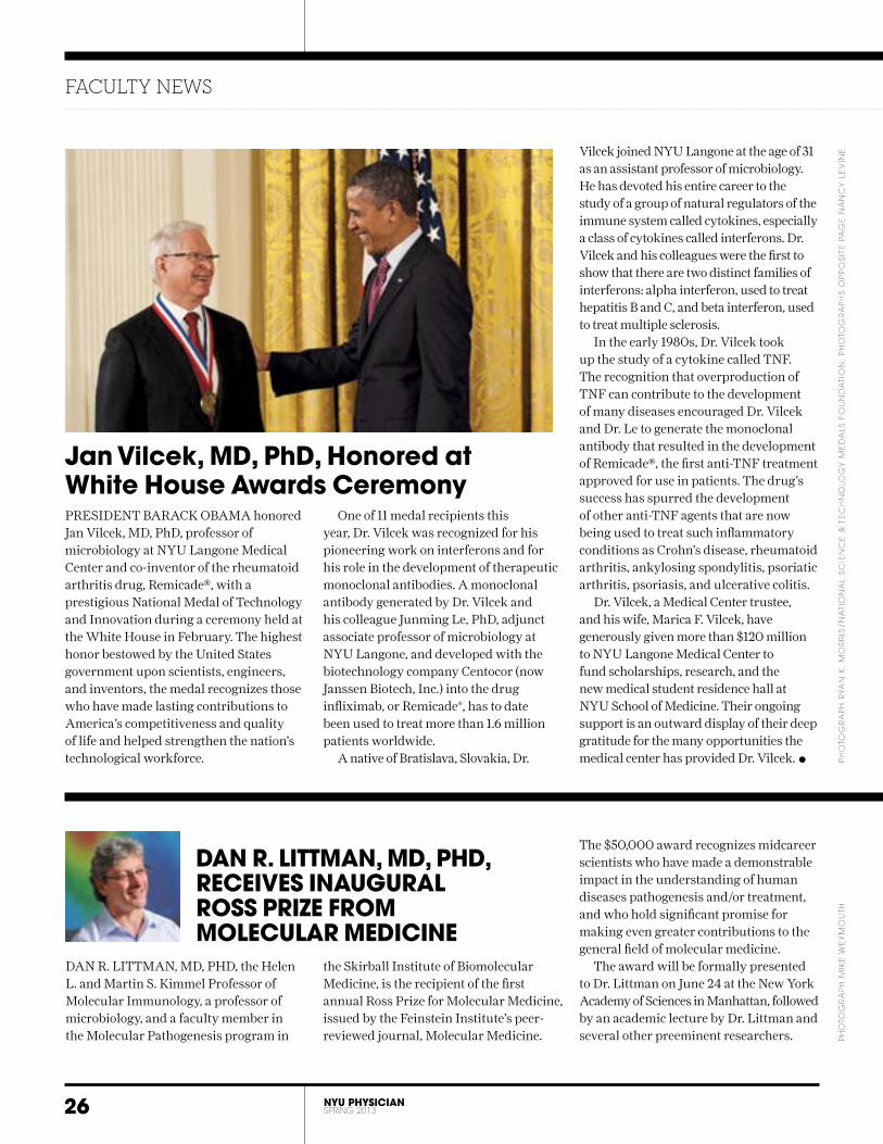
26
FACULTY NEWS
NYU PHYSICIAN SPRING 2013
PH
OTO
GR
AP
H R
YAN
K.
MO
RR
IS/
NA
TIO
NA
L S
CIE
NC
E &
TE
CH
NO
LOG
Y M
ED
ALS
FO
UN
DA
TIO
N;
PH
OTO
GR
AP
HS
OP
PO
SIT
E P
AG
E N
AN
CY
LE
VIN
E
PRESIDENT BARACK OBAMA honored Jan Vilcek, MD, PhD, professor of microbiology at NYU Langone Medical Center and co-inventor of the rheumatoid arthritis drug, Remicade®, with a prestigious National Medal of Technology and Innovation during a ceremony held at the White House in February. The highest honor bestowed by the United States government upon scientists, engineers, and inventors, the medal recognizes those who have made lasting contributions to America’s competitiveness and quality of life and helped strengthen the nation’s technological workforce.
One of 11 medal recipients this year, Dr. Vilcek was recognized for his pioneering work on interferons and for his role in the development of therapeutic monoclonal antibodies. A monoclonal antibody generated by Dr. Vilcek and his colleague Junming Le, PhD, adjunct associate professor of microbiology at NYU Langone, and developed with the biotechnology company Centocor (now Janssen Biotech, Inc.) into the drug infliximab, or Remicade®, has to date been used to treat more than 1.6 million patients worldwide.
A native of Bratislava, Slovakia, Dr.
Vilcek joined NYU Langone at the age of 31 as an assistant professor of microbiology. He has devoted his entire career to the study of a group of natural regulators of the immune system called cytokines, especially a class of cytokines called interferons. Dr. Vilcek and his colleagues were the first to show that there are two distinct families of interferons: alpha interferon, used to treat hepatitis B and C, and beta interferon, used to treat multiple sclerosis.
In the early 1980s, Dr. Vilcek took up the study of a cytokine called TNF. The recognition that overproduction of TNF can contribute to the development of many diseases encouraged Dr. Vilcek and Dr. Le to generate the monoclonal antibody that resulted in the development of Remicade®, the first anti-TNF treatment approved for use in patients. The drug’s success has spurred the development of other anti-TNF agents that are now being used to treat such inflammatory conditions as Crohn’s disease, rheumatoid arthritis, ankylosing spondylitis, psoriatic arthritis, psoriasis, and ulcerative colitis.
Dr. Vilcek, a Medical Center trustee, and his wife, Marica F. Vilcek, have generously given more than $120 million to NYU Langone Medical Center to fund scholarships, research, and the new medical student residence hall at NYU School of Medicine. Their ongoing support is an outward display of their deep gratitude for the many opportunities the medical center has provided Dr. Vilcek. •
DAN R. LITTMAN, MD, PHD, RECEIVES INAUGURAL ROSS PRIZE FROM MOLECULAR MEDICINE
Jan Vilcek, MD, PhD, Honored at White House Awards Ceremony
the Skirball Institute of Biomolecular Medicine, is the recipient of the first annual Ross Prize for Molecular Medicine, issued by the Feinstein Institute’s peer-reviewed journal, Molecular Medicine.
The $50,000 award recognizes midcareer scientists who have made a demonstrable impact in the understanding of human diseases pathogenesis and/or treatment, and who hold significant promise for making even greater contributions to the general field of molecular medicine.
The award will be formally presented to Dr. Littman on June 24 at the New York Academy of Sciences in Manhattan, followed by an academic lecture by Dr. Littman and several other preeminent researchers. P
HO
TOG
RA
PH
MIK
E W
EY
MO
UTH
DAN R. LITTMAN, MD, PHD, the Helen L. and Martin S. Kimmel Professor of Molecular Immunology, a professor of microbiology, and a faculty member in the Molecular Pathogenesis program in

27NYU PHYSICIAN SPRING 2013
NEAR THE END of President Barack Obama’s State of the Union address last February 12, he turned everyone’s attention to the balcony, where First Lady Michelle Obama was joined by 23 special guests. Seated between the First Lady and the vice president’s wife, Jill Biden, was Menchu de Luna Sanchez, RN, senior nurse clinician at NYU Langone Medical Center.
“We were sent here to look out for our fellow Americans,” President Obama told those assembled in the House chamber, “the same way they look out for one another, every single day, usually without fanfare, all across this country. We should follow the example of a New York City nurse named Menchu Sanchez. When Hurricane Sandy plunged her hospital into darkness, her thoughts were not with how her own home was faring. They were with the 20 precious newborns in her care and the rescue plan she devised that kept them all safe.”
The president was referring, of course, to Monday, October 29, 2012, the night some 1,000 medical and professional person-nel at NYU Langone safely evacuated 322 patients, including the most vulnerable:
NYU Langone’s Nurses Honored in Washington for Their Heroism During Hurricane Sandy
Her leadership caught the attention of President Obama, who called Robert I. Grossman, MD, NYU Langone’s dean and CEO, and Kimberly Glassman, PhD, RN, vice president for patient care services and chief nursing officer, two days after the storm. Sanchez, who emigrated from the Philippines in the 1980s and has worked at NYU Langone since 2010, was one of 23 Americans invited to attend the State of the Union address for their humanitarian actions. •
A renowned immunologist and molecular biologist, Dr. Littman has made seminal contributions to numerous fields, including understanding the molecular basis of immune recognition, HIV pathogenesis, T-cell differentiation and selection, and the role of commensal bacteria in immune system development and regulation.
Dr. Littman is a leader in applying molecular biology and mouse genetics to study specification of T lymphocyte
lineages and the differentiation of inflammatory T helper cells. In recent work, Dr. Littman discovered that the nuclear receptor ROR-gamma-t regulates differentiation of Th17 cells and lymphoid tissue inducer cells, and identified compounds that inhibit its activity and may be effective for autoimmune disease therapy. He identified a commensal gut bacterium that selectively induces Th17 cells and promotes autoimmunity in mice, which may be relevant to human diseases
like rheumatoid arthritis, thought to be influenced by imbalanced microbiota.
Dr. Littman is a Howard Hughes Medical Institute investigator, a member of the National Academy of Sciences and the prestigious Institute of Medicine, and a fellow in both the American Academy of Arts and Sciences and the American Academy of Microbiology.
The Ross Prize was made possible by the generosity of Feinstein Institute board members Robin and Jack Ross. •
Left to right: Maureen Halligan, RN, nurse clinician; Geraldine Cillo-Gillen, RN, senior nurse clinician; Claudia Roman, RN, senior nurse clinician; Sandra Kyong, RN, nurse clinician; Margot Condon, RN, senior nurse clinician; Beth Largey, RN, nurse clinician; Alison Coluccio, RN.
Menchu Sanchez, RN
those in the KiDS of NYU Langone Neonatal Intensive Care Unit. It was Sanchez who pro-posed that the saf-est way to evacuate the infants would
be to carry them down the stairs without incubators. As the nurse caring for the unit’s sickest baby, she went first. Nurses and phy-sicians carried their tiny charges, held snug-ly against the skin of their chests, down nine flights of stairs, while manually squeezing bags to deliver oxygen to the babies’ lungs.
As Sanchez and her colleagues escorted the newborns to other hospitals, her Secaucus, New Jersey, home was flooding.
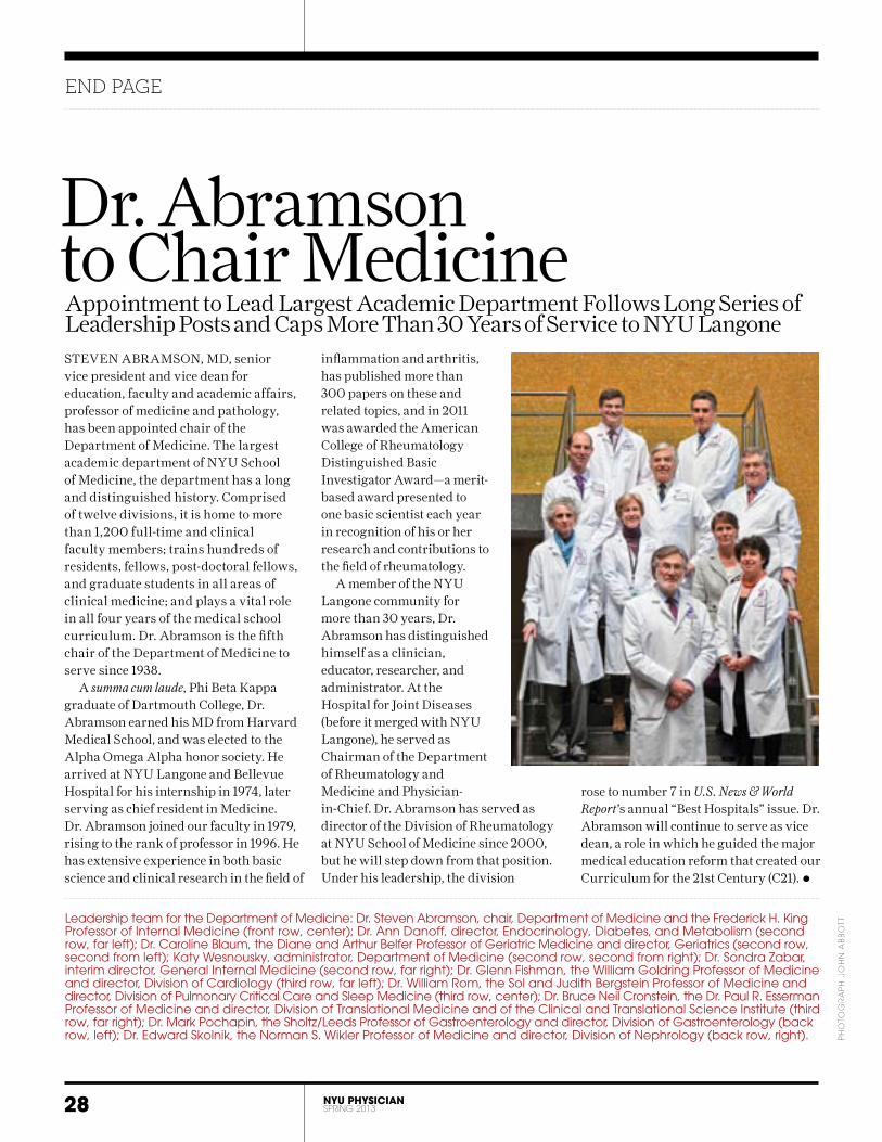
END PAGE
28 NYU PHYSICIAN SPRING 2013
PH
OTO
GR
AP
H J
OH
N A
BB
OTT
rose to number 7 in U.S. News & World Report’s annual “Best Hospitals” issue. Dr. Abramson will continue to serve as vice dean, a role in which he guided the major medical education reform that created our Curriculum for the 21st Century (C21). •
STEVEN ABRAMSON, MD, senior vice president and vice dean for education, faculty and academic affairs, professor of medicine and pathology, has been appointed chair of the Department of Medicine. The largest academic department of NYU School of Medicine, the department has a long and distinguished history. Comprised of twelve divisions, it is home to more than 1,200 full-time and clinical faculty members; trains hundreds of residents, fellows, post-doctoral fellows, and graduate students in all areas of clinical medicine; and plays a vital role in all four years of the medical school curriculum. Dr. Abramson is the fifth chair of the Department of Medicine to serve since 1938.
A summa cum laude, Phi Beta Kappa graduate of Dartmouth College, Dr. Abramson earned his MD from Harvard Medical School, and was elected to the Alpha Omega Alpha honor society. He arrived at NYU Langone and Bellevue Hospital for his internship in 1974, later serving as chief resident in Medicine. Dr. Abramson joined our faculty in 1979, rising to the rank of professor in 1996. He has extensive experience in both basic science and clinical research in the field of
inflammation and arthritis, has published more than 300 papers on these and related topics, and in 2011 was awarded the American College of Rheumatology Distinguished Basic Investigator Award—a merit-based award presented to one basic scientist each year in recognition of his or her research and contributions to the field of rheumatology.
A member of the NYU Langone community for more than 30 years, Dr. Abramson has distinguished himself as a clinician, educator, researcher, and administrator. At the Hospital for Joint Diseases (before it merged with NYU Langone), he served as Chairman of the Department of Rheumatology and Medicine and Physician-in-Chief. Dr. Abramson has served as director of the Division of Rheumatology at NYU School of Medicine since 2000, but he will step down from that position. Under his leadership, the division
Dr. Abramson to Chair MedicineAppointment to Lead Largest Academic Department Follows Long Series of Leadership Posts and Caps More Than 30 Years of Service to NYU Langone
Leadership team for the Department of Medicine: Dr. Steven Abramson, chair, Department of Medicine and the Frederick H. King Professor of Internal Medicine (front row, center); Dr. Ann Danoff, director, Endocrinology, Diabetes, and Metabolism (second row, far left); Dr. Caroline Blaum, the Diane and Arthur Belfer Professor of Geriatric Medicine and director, Geriatrics (second row, second from left); Katy Wesnousky, administrator, Department of Medicine (second row, second from right); Dr. Sondra Zabar, interim director, General Internal Medicine (second row, far right); Dr. Glenn Fishman, the William Goldring Professor of Medicine and director, Division of Cardiology (third row, far left); Dr. William Rom, the Sol and Judith Bergstein Professor of Medicine and director, Division of Pulmonary Critical Care and Sleep Medicine (third row, center); Dr. Bruce Neil Cronstein, the Dr. Paul R. Esserman Professor of Medicine and director, Division of Translational Medicine and of the Clinical and Translational Science Institute (third row, far right); Dr. Mark Pochapin, the Sholtz/Leeds Professor of Gastroenterology and director, Division of Gastroenterology (back row, left); Dr. Edward Skolnik, the Norman S. Wikler Professor of Medicine and director, Division of Nephrology (back row, right).

One patient at a time. When you include a bequest in your will to NYU Langone Medical Center you help us deliver outstanding health care to the many patients and families who rely upon us to improve their lives. Superb physicians, an award-winning nursing staff and internationally ranked scientists make the difference. Join our community, and create your legacy today.
To learn more about making your planned gift to NYU Langone, please contact Marilyn Van Houten at 212.404.3653 or [email protected].
www.nyUlmc.org
John
Abb
ott
the world…Change Change Change
Change Change ChangeChange Change Change

Office of Communications and Public Affairs550 First Avenue, New York, NY 10016