nutritional treatment for leagnous conjuntivitis: case ... · nutritional treatment for leagnous...
TRANSCRIPT

476 CASE REPORT
Received for publication 18/07/2015 - Accepted for publication 11/12/2015The authors declare no conflicts of interests.
Terapia nutricional proposta para conjuntivite lenhosa:estudo de caso e revisão de literatura
Nutritional treatment for leagnous conjuntivitis:case report and literature review and case report
RESUMO
A conjuntivite lenhosa é resultante de um raro distúrbio autossômico recessivo hereditário, a deficiência de plasminogênio. Estaapresenta sintomas crônicos, como lesões conjuntivais membranosas características, inicialmente finas e com a persistência dainflamação evoluem se tornando esbranquiçadas, espessas e enrijecidas, lacrimejamento, secreção mucosa e hiperemia ocular acom-panhados de espessas pseudomembranas lenhosa (PL) que recobrem a parte interior da conjuntiva tarsal. A literatura apresentaalguns tratamentos, entretanto nenhum deles alcançou a cura da doença. A terapia nutricional abordada neste estudo trata-se dacombinação de nutrientes dentro dos limites estabelecidos para ingestão diária, baseada na nutrição ortomolecular, visando aoaumento da taxa de plasminogênio funcional, e a consequente redução dos sintomas associados à sua deficiência. Notou-se odesaparecimento de sintomas associados e redução do crescimento da PL, e também um aumento de 25% do plasminogêniofuncional. Um aumento de 25% na dosagem de plasminogênio pode não ser altamente significativo, mas abre um respaldo paramaiores estudos, pois já apresentou minimização dos sintomas da paciente.
Descritores:: Conjuntivite/etiologia; Plasminogênio/deficiência; Oftalmopatias; Terapia nutricional; Relato de casos
ABSTRACT
Ligneous conjunctivitis is the result of a rare inherited autosomal recessive disorder, the plasminogen deficiency. This presents chronicsymptoms such as growth spongiosa meat, tearing, mucous discharge and ocular reddening accompanied by ligenous pseudomembranes(PL) coat the inside of the tarsal conjunctiva. The literature presents some treatments, but none of them reached a cure. The nutritionaltherapy addressed in this study is a combination of nutrients within the limits of daily intake, based on orthomolecular nutrition, aimedat increasing functional plasminogen rate, and the consequent reduction of symptoms associated with their disability. It was noteddisappearance of the symptoms associated and a 25% increase in the functional plasminogen. A 25% increase in plasminogen dosagemay not be highly significant, but it opens up a support for further study, as already presented reduction of symptoms of the patient.
Keywords: Conjunctivitis/etiology; Plasminogen/deficiency; Eye disease; Nutrition therapy; Case reports
Mariana Granito¹, João Maria Ferreira²
Rev Bras Oftalmol. 2016; 75 (6): 476-80
Study carried out at Centro Universitário Serra dos Órgãos (UniFeso), Teresópolis, RJ, Brazil.
¹ Nutritionist; Master’s Degree in Medical Sciences from UFF² Medicine School, Centro Universitário Serra dos Órgãos (UniFeso), Teresópolis, RJ, Brazil.
Rev RBO _Nov_Dez_2016 _Inglês.pmd 18/11/2016, 04:56476
DOI 10.5935/0034-7280.20160096

477
INTRODUCTION
Leagnous conjunctivitis is a rare autosomal recessivedisease. Its diagnosis is based on clinical and histologicaldata, and eventually on positive family history. The clinical
findings usually are: chronic tearing, conjunctival hyperemia,growth of leagnous pseumembranes (PL) in the conjunctiva and/or other mucosal sites in the body, possibly in combination withmilium colloid or obstructive congenital hydrocephalus.(1-3)
It is caused because there is a deficiency of plasminogen,it means that there is accumulation of mucus threads and fibrinmembranes, stimulating fibroblasts and inflammatory cells ofthe ocular surface (but can also accumulate in other surfacessuch as the gums, causing leagnous gingivitis.(2,3)
CASE REPORT
Patient history
Patient C.G.C.P., 13 years old, female, diagnosed at 3years old with leagnous conjunctivitis due to a symptomatichomozygous deficiency of plasminogen (FunctionalPlasminogen 20%; Reference threshold according to themedical diagnostic laboratory: 70% to 140%). Concomitantto the disease, the same featured low immunity with frequentappearance of skin infections due to atopic dermatitis, and inthe respiratory tract, constant rashes, excessive production ofsecretions in the mucosa of the eyelid, with growth of leagnouspseudomembrane (PL).
Since the beginning of the lesions, around 2 years of age,the same underwent surgical procedures several times for theremoval of the PL, which were recurrent weeks after removal.This process was carried out as reported in the present study,without the use of the specific medication described. She onlyused anesthetic eyedrops in the post-operative period, andtopical use of loteprednol etabonate (2 mg applied 3x/day).The treatment with about 1g/day of calf thymus acid lysatewas carried out, equivalent to 20 mg of the active ingredientthymomodulin in an attempt to improve the immune systemwithout great results.
Due to ignorance about the disease by all healthprofessionals consulted, for nearly four years no specifictreatment was used for the disease, even after its diagnosis.
At age 6, she began a treatment using autologous serum,where the paternal blood was extracted and centrifuged, andthe plasma decanted was stored in sterile packaging of theeyedropper type and frozen for topical use on PL. It seemedto alleviate the symptoms of mucus production and hyperemia.It was used for 4 years, but with no significance in thetreatment. Concomitant to the use of the serum, the removalsurgery was performed at intervals of about 3 months.
At age 10, in the year 2011, the patient began thetreatment reported in the present study. A nutritional therapywas developed by a professional Nutritionist according tostudies carried out in respect of each nutrient and theirfunction. The surgical removal was made by anophthalmologist in the operating room at Hospital São José,in Teresópolis. The histopathological exam was made by thelaboratory of the same hospital where the surgery wasperformed.
Nutritional therapy
The nutritional therapy applied was divided into 3 phases.Phase 1 addresses the nutritional complementation with 1g ofcollagen, collaborating on the protein structure, and 1g ofcolostrum, for a better immune balance. This phase lasted a year,and was followed by phase 2. In this second phase 100mg ofCoQ10, magnesium and calcium were administered as proposedby the orthomolecular practice [3].
And in both phases, the reduction of lactose was proposedfor the improvement of rash, while the other nutrients wereused directly for the treatment of plasminogen deficiency, thecause of leagnous conjunctivitis.[3]. The treatment was drawn upaccording to the table below:
Table 1 Administration of orthomolecular nutrition
treatment for deficiency of plasminogen
Nutrient administered Daily dose Phase
Collagen 1g 1
Colostrum 1g
Coenzyme Q10 200mg
Magnesium 200mg 2
Calcium 200mg
Reduction of lactose Milk exemption 1and 2
Nutritional therapy for food
Three years after orthomolecular therapy, the functionalnutritional therapy began concomitantly to promote anti-inflammatory action, focusing on bioactive compounds.
The elements shown in table 2 were added to the diet.
Table 2Administration of functional nutrition treatment for
deficiency of plasminogen
Element administered Dose Desired compound
Olive oil 7g 2x/day Omega 9 Full red grape juice 150ml 1x/day Resveratrol
Oats 2x/day PolyphenolsFlaxseed 1x/day Polyphenols
In addition to the administration of nutrients describedabove, the consumption of foods rich in saturated fats like friedfoods, chocolates, yellow cheese, etc., was reduced. Propolis wasalso administered in the form of topical application on the gums1x day.
Nutritional therapy for complementation
A supplement of vitamins and minerals wasadministered, besides some bioactive compounds bymanipulated capsules administration (by compoundingpharmacy properly regularized). The compoundsadministered are specified in table 3
Rev Bras Oftalmol. 2016; 75 (6): 476-80
Nutritional treatment for leagnous conjuntivitis:case report and literature review and case report
Rev RBO _Nov_Dez_2016 _Inglês.pmd 18/11/2016, 04:56477
478 Granito M , Ferreira JM
Table 3Administration of nutrition complement
for deficiency of plasminogen
Element administered Daily dose
Omega 3 3000mg
Iron 14mg
Turmeric 500mg
Vitamin B12 2,4mcg
Zinc 30mg
Retinol 500ui
Acid 50mg
Magnesium 100 mg
Calcium 200 mg
Copper 800mcg
Selenium 60mcg
Alpha Tocopherol 400mg
Thiamin 5mg
Nicotiamide 20mg
Pyridoxine 2mg
Riboflavin 5mg
Calcium pantothenate 3mg
Folic acid 300mcg
Turmeric 400mg
The surgical excision of the PL is necessary due to therapid growth, leading consequently to the trauma of the corneas,although it is known that relapses are inevitable. However, wenoticed significant reduction of growth of PL, having extendedthe period between surgeries, from 3 months to 1 year, onaverage. After physical examination, we observed thedisappearance of recurring rashes and skin infections. It isbelieved to be due to relief of lactose and the use of anti-inflammatory and antioxidant nutrients.
Regarding biochemistry, there was an increase of 25% inthe dosage of the patient’s functional plasminogen, that is, atthe onset of treatment the patient presented 20% of serumplasminogen, and three years after the onset she presented25% in the total dose, representing an increase of 25%.
It should be noted that there was no treatmentconcomitant to the one described.
Surgical treatment
When PLs are bothersome to the patient, they are surgicallyremoved. It is usually necessary when PLs grow, assaulting thecornea (figures 1 and 2), but not generating direct injuries tothis. A surgical debridement (Figure 3) of the PLs located on thetarsal conjunctiva of the upper and lower eyelids of both eyeswas carried out. For this removal, the patient is subjected to theprocess of local anesthesia associated to assisted sedation.
After removing the tissue, the conjunctiva cauterization ismade with the aid of a bipolar cautery. It is also carried out theapplication of mitomycin 0.02% soaked in flexible cotton swab,being in contact with the conjunctival surface injured for a periodof 45 seconds.
After surgery (figure 4), eyedrops cyclosporine 0.05% wasused for 15 days. Eyedrops loteprednol etabonate (2.0mg) isused topically as an adjunct to treatment (2 drops, 2x/day).
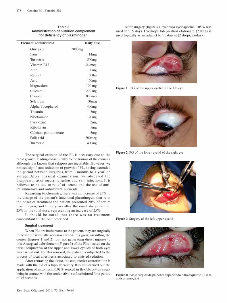
Figure 1: PG of the upper eyelid of the left eye
Figure 2: PG of the lower eyelid of the right eye
Figure 4: Pós-cirurgico da pálpebra superior do olho esquerdo (2 diasapós a remoção)
Figure 3: Surgery of the left upper eyelid
Rev Bras Oftalmol. 2016; 75 (6): 476-80
Rev RBO _Nov_Dez_2016 _Inglês.pmd 18/11/2016, 04:56478

479
The histopathological analysis of the tissue removed carriedout by the laboratory of the hospital where the surgery wasperformed showed acute inflammatory process in theMeibonium glands, with absence of malignancy.
LITERATURE REVIEW
Leagnous conjunctivitis (CL) is a rare pathology, withoutspecific ethnic predilection. However, it is considered that it mayhave higher prevalence in areas where consanguinity is morecommon[1,2]. PLs are deeply attached to the conjunctiva of theeyelid, and with its ends free. Removal tends to cause bleeding,because the area is vascularized.[4,5]
It is possible to observe the presence of mast cells andeosinophils in the lesions, which makes us believe that thesecretions deposition would be by degranulation of these mastcells. It is understood that the massive extravasation of plasmaconstituents, fibrin in particular, ends up forming a kind ofgranuloma.[6]. In addition to the ocular region, other mucosasmay be involved, such as oral, nasopharyngeal, gums, throat andvagina[7,8]. Several cases of leagnous conjunctivitis and/or leagnousperiodontitis were found in association with plasminogendeficiency type I[9,10].
CL is caused by deficiency of plasminogen. It is linked tothe dissolution of fibrin, and consequently, thrombus. When fibrinis not properly removed, it accumulates in the mucous tissues,creating a sort of leagnous pseudomembrane[2,3]. It plays animportant role in the recruitment of macrophages during theinflammatory response, in invasion and metastasis of tumor cells,wound healing, tissue remodeling, growth of neurites and skeletalmuscle tissue.[10, 11]. The reference values vary according to thelaboratory. In the present study, the reference value establishedwas 70% to 140%, as provided by the laboratory where the testwas carried out.
Plasminogen deficiency is a rare autosomal recessive diseasewhich can be asymptomatic heterozygous or symptomatichomozygous. It can be linked to the onset of low levels ofplasminogen in children whose parents present reducedvalues[12,13]. It can be classified in two ways: Type I(hipoplasminogenemia) or type II (displasminogenemia). Thetype I addressed in this study is characterized by great difficultyin healing, and has leagnous conjunctivitis as its mainmanifestation[14], but type II has thrombosis as the main risk[15].
Leagnous conjunctivitis is the most common presentation ofclinical syndromes associated to plasminogen deficiency type I[1,2].
The prevalence of this disease has not yet been established.The theoretical prevalence of homozygous or heterozygous(associated) patients was calculated as being of 1.6 by 1.000.000.[6].
However, other epidemiological studies are needed to betterdetermine the prevalence of this deficiency.[2].
In the literature, there are reports of surgery performedto remove PL, topical use of plasmin or plasminogen in thearea[14,16], immunosuppressants such as cyclosporine and heparinand positive autologous serum [5] showing poor positive results.However, the granulomas returned at the end of the treatment.It should be noted that no treatment achieved the cure of thedisease. The use of ophthalmologic concentrate of plasminogenand fresh frozen plasma systemically proved to be efficient, butare not accessible products to patients, not available on themarket.[2].
DISCUSSION
The current literature is very scarce as to possibletreatments to be carried out, none of them showing a possiblecure for the disease, not even to alleviate the symptoms[2,5].
The nutritional treatment after three years of treatment ofphases 1 and 2 showed an increase of 25% in the dosage ofplasminogen. This may not be highly significant at first, but givessupport for further studies, because it shows improvement in thegeneral condition of the patient, and can be related to this increase.This result was not found in any study in this literature[3]. Also,there was reduction in the production of mucus secretion after 15days of administration of nutritional therapy for completion.
Regarding surgical treatment, it is palliative, because a fewmonths after surgery there is recurrence of conjunctivalmembranes[2]. It was noted an increase in the interval betweensurgical procedures (from 3 months to1 year) that may havebeen related to the increase of the percentage rate of serumplasminogen.
Regarding the acute inflammatory process in the glands ofMeibonium, studies show that this can have an effect on hormonessuch as estrogen[17]. Thus, it was speculated that this conjunctivallesion may have increased due to the influence of increasedestrogen rate during puberty. Given the circumstance, a hormonalsupplementation therapy started to further analyze the resultsgenerated.
CONCLUSION
The treatment reported is new in the literature becauseuntil then, no article presents increased organic production ofplasminogen. This supports further studies in the area. It isnecessary to continue to assess over time whether there will bea better patient response, and I suggest to seek alternatives tocomplement the treatment, aiming at better results.
REFERENCES
1. Kraft J, Lieb W, Zeitler P, Schuster V. Ligneous conjunctivitis in agirl with severe type I plasminogen deficiency. Graefes Arch ClinExp Ophthalmol. 2000;238(9):797-800.
2. Mehta R, Shapiro AD. Plasminogen deficiency. Haemophilia.2008;14(6):1261-8. Review.
3. Granito M, Schmidt B. Orthomolecular nutritional therapy forplasminogen deficiency: report of a case that showed positiveresults. Haemophilia. 2015;21(2):e139-41.
4. Holland EJ, Schwartz GS. Ligneous conjunctivis. In: Krachmer JH,Mannis MJ. Cornea and extrenal disease: clinical diagnosis andmanagement. St. Louis: Mosby; 1997. 863-89.
5. Ang AY, Neff KD, Schwartz GS, Holland EJ. Conjuntivite lenhosain: Holland EJ, Mannis MJ, Lee WD. Doenças da superfície ocu-lar: Córnea, Conjuntiva e Filme Lacrimal. Elsevier, 1°ed, Rio deJaneiro, 2013. p. 183-188.
6. Schuster V, Seregard S. Ligneous conjunctivitis. Surv Ophthalmol.2003;48(4):369-88. Review.
7. Heinz C, Kremmer S, Externbrink P, Steuhl KP. [Ligneous con-junctivitis in a patient with plasminogen type I deficiency—casereport with review of literature]. Klin Monbl Augenheilkd.2002;219(3):156-8. German.
Rev Bras Oftalmol. 2016; 75 (6): 476-80
Nutritional treatment for leagnous conjuntivitis:case report and literature review and case report
Rev RBO _Nov_Dez_2016 _Inglês.pmd 18/11/2016, 04:56479

480
8. Tefs K, Gueorguieva M, Klammt J, Allen CM, Aktas D, AnlarFY,et al. Molecular and clinical spectrum of type I plasminogendeficiency: A series of 50 patients. Blood. 2006;108(9):3021-6..
9. El-Darouti M, Zayed AA, El-Kamah GY, Mostafa MI. Ligneousconjunctivitis with oral mucous membrane involvement and de-creased plasminogen level. Pediatr Dermatol. 2009;26(4):448-51.
10. Yohe SL, Reyes M, Johnson DA, Fry CL, Scribbick FW, KinneyMC. Plasminogen deficiency as a rare cause of conjunctivitis andlymphadenopathy. Am J Surg Pathol. 2009 ;33(2):313-9.
11. Braunwald E, Zipes D, Libby P. Tratado de medicina cardiovas-cular. 6a ed. Rio de Janeiro: Roca; 2003
12. Miles LA, Plow EF, Waisman DM, Parmer RJ. Plasminogen re-ceptors. J Biomed Biotechnol. 2012;2012:130735.
13. Lourenço DM. Trombofilia. In: Pitta GB, Castro AA, Burihan E.Angiologia e cirurgia vascular: guia ilustrado. Maceió: Uncisal/Ecmal & Lava; 2003.
14. Cohen J, Cohen S, Cymberknoh MC, Gross M, Hirshoren N,Shoseyov D. Laryngeal obstruction in congenital plasminogendeficiency. Pediatr Pulmonol. 2012;47(9):923-5.
Corresponding author:João Maria FerreiraAv. Alberto Torres, 111 - AltoTeresópolis, RJ, Brasil.Phone.: (21) 2641-7128.E-mail: [email protected]
15. Pergantou H, Likaki D, Fotopoulou M, Katsarou O, Xafaki P,Platokouki H. Management of ligneous conjunctivitis in a childwith plasminogen deficiency. Eur J Pediatr. 2011;170(10):1333-6.
16. Schuster V, Hügle B, Tefs K. Plasminogen deficiency. J ThrombHaemost. 2007;5(12):2315-22.Review.
17. Heidemann DG, Williams GA, Hartzer M, Ohanian A, Citron ME.Treatment of ligneous conjunctivitis with topical plasmin andtopical plasminogen. Cornea. 2003;22(8):760-2.
18. Del Castillo JM. Ojo seco. Granada: Tecnimedia; 1997.
Rev Bras Oftalmol. 2016; 75 (6): 476-80
Granito M , Ferreira JM
Rev RBO _Nov_Dez_2016 _Inglês.pmd 18/11/2016, 04:56480