nucleotide sequence, transcriptional analysis, and expression of
TRANSCRIPT

THE JOURNAL OF BIOLOGICAL CHEMISTRY (0 1991 by The American Society for Biochemistry and Molecular ’ Biology, Inc
Vol. 266, No. 22, Issue of August 5, PP . 14646-14653,1991 Printed in U. S. A.
Nucleotide Sequence, Transcriptional Analysis, and Expression of Genes Encoded within the Form I COa Fixation Operon of Rhodobacter sphaeroides”
(Received for publication, February 1, 1990)
Janet L. Gibson$, Deane L. Falcone$, and F. Robert Tabita$§ From the $Department of Microbiology and The Biotechnology Center, The Ohio State Uniuersity, Columbus, Ohio 43210
In Rhodobacter sphaeroides, many of the structural genes encoding enzymes of the Calvin cycle are dupli- cated and grouped within two separate clusters. In this study, the nucleotide sequence of a 5627-base pair region of DNA that contains the form I Calvin cycle gene cluster has been determined. The five open read- ing frames are arranged in the order, fbpA prkA cfxA rbcL rbcS and are tightly linked and oriented in the same direction. The results of insertional mutagenesis studies suggest the genes are organized within an op- eron. Consistent with this proposal, the cfxA gene has been tentatively identified as a gene encoding the Cal- vin cycle enzyme, aldolase. Measurement of the activi- ties of various Calvin cycle enzymes in the insertion mutants showed that inactivation of genes within one COa fixation cluster affected expression of genes within the second cluster, revealing a complex regu- latory network.
In purple non-sulfur photosynthetic bacteria, CO, fixation via the Calvin cycle has multiple functions. In addition to its role in providing carbon for cells during photo- or chemoli- thoautotrophic growth, C02 can also serve as an electron acceptor to maintain redox balance in cells growing on re- duced carbon sources such as malic or butyric acid. In some photosynthetic bacteria, a complex system has evolved to accommodate the different modes of CO, fixation. For one, Rhodobacter sphaeroides synthesizes discrete forms of ribulose 1,5-bisphosphate carboxylase/oxygenase (Rbu-P2 carboxyl- ase),’ the enzyme that catalyzes the primary COz fixation reaction (Gibson and Tabita, 1977). The structural and cata- lytic properties of the form I and form I1 Rbu-Pz carboxylases differ substantially and they exhibit distinct inductive re- sponses under varying growth conditions (Gibson and Tabita, 1977; Jouanneau and Tabita, 1986). I t is known that the form I Rbu-Pz carboxylase predominates under conditions where
* This work was supported by National Institutes of Health Grant GM 24497 and United States Department of Agriculture Grants 89- 37262-4566 and 87-CRCR-1-2591. The costs of publication of this article were defrayed in part by the payment of page charges. This article must therefore be hereby marked “aduertisement” in accord- ance with 18 U.S.C. Section 1734 solely to indicate this fact.
The nucleotide sequence(s) reported in thispaper has been submitted to the GenBankTM/EMBL Data Bank with accession number(s) M64624.
To whom correspondence should be addressed: Dept. of Micro- biology, The Ohio State University, 484 West 12th Ave., Columbus, OH 43210.
’ The abbreviations used are: Rbu-P2 carboxylase, ribulose bis- phosphate carboxylase/oxygenase; FBPase, fructose 1,6-bisphospha- tase; PRK, phosphoribulokinase; SDS, sodium dodecyl sulfate; kb, kilobase pair; ORF, open reading frame.
COz is limiting, whereas the form I1 Rbu-P2 carboxylase predominates under conditions where carbon is saturating and electrons are in excess (Jouanneau and Tabita, 1986; Falcone et al., 1988; Hallenbeck et al., 1990a, 1990b). The functional and regulatory differences suggest diverse roles for the two enzymes in uiuo. However, the inactivation of either Rbu-P2 carboxylase in R. sphaeroides does not preclude growth under any condition tested (Falcone et al., 1988). Therefore, the requirement of one form or the other does not appear to be absolute.
The form I and form I1 Rbu-P2 carboxylase coding se- quences in R. sphaeroides have been localized to physically distinct loci and are clustered with structural genes encoding other Calvin cycle enzymes (Gibson and Tabita, 1987, 1988; Gibson et al., 1990; Hallenbeck and Kaplan, 1987). Some of these genes are present in both clusters, such as the genes coding for fructose 1,6-bisphosphatase ( fbpA, fbpB), phospho- ribulokinase ( p r M , p r k B ) , and genes encoding a protein of unknown function, termed cfxA and cfxB. However, coding sequences for transketolase ( tklB) and glyceraldehyde-3- phosphate dehydrogenase (gapB) are solely present within the form I1 or B cluster (Gibson and Tabita, 1988; Gibson, et al., 1990). The duplication and clustering of genes encoding Calvin cycle enzymes in R. sphaeroides raise intriguing ques- tions concerning their regulation and expression. In attempts to further elucidate the molecular mechanisms underlying the control of the two gene clusters we have analyzed the primary sequence of these two regions. In the present investigation we have determined the nucleotide sequence and the deduced primary structure of all the structural genes of the form I Calvin cycle gene cluster; insertional mutagenesis studies indicate that the genes belong to a single transcriptional unit. In addition, on the basis of DNA sequence comparisons, we conclude that cfxA encodes aldolase, an additional structural gene of the Calvin cycle.
EXPERIMENTAL PROCEDURES
Bacterial Strains, Plasmids, and Growth Conditions-Escherichia coli strain JM107 was used as the host strain for all pUC-derived plasmid constructs (Yanisch-Perron et al., 1985), and E. coli SMlO (Simon et al.., 1983) was used for delivery of pSUP202 constructs into R. sphaeroides. E . coli strains were routinely grown in LB broth a t 37 “C. R. sphaeroides HR was grown aerobically in a peptone-yeast extract medium (PYE) (Weaver and Tabita, 1983), photoheterotroph- ically on 0.4% malate or photolithoautotrophically in an atmosphere of 1.5% CO,, 98.5% Hz, as described previously (Jouanneau and Tabita, 1986). Antibiotics were added to growth media at the following concentrations (pg/ml): for E. coli, ampicillin, 50; tetracycline, 25; trimethoprim, 200; kanamycin 25; for R. sphueroides, streptomycin, 25; tetracycline, 12.5; trimethoprim, 200; kanamycin, 25.
DNA Sequencing-DNA sequencing was performed on double stranded DNA templates by the dideoxy chain termination method of Sanger et al. (1977) using the Sequenase kit obtained from U. S.
14646

Sequence of Calvin Cycle Genes of R. sphaeroides 14647
Biochemicals and [LY-~'S]~ATP from Du Pont-New England Nuclear. In some cases G + C compressions were resolved by replacing dGTP with dITP or deaza-GTP in the sequencing reactions.
Defined restriction fragments cloned into pUC vectors were se- quenced using commercial primers from United States Biochemical Corp. Additional oligonucleotide primers were obtained from Operon Technologies, Inc., Alameda, CA. DNA sequences were determined completely on both strands of DNA within the coding regions. The 5' end of the sequence up to nucleotide 190 and the 3' end of the sequence beyond nucleotide 5355 were determined on only one strand.
Enzyme Assays-Cell extracts for enzyme assays were prepared as described previously (Jouanneau and Tabita, 1986). The Rbu-Pz carboxylase assay was based on the incorporation of 14C02 into acid- stable radioactivity (Gibson and Tabita, 1977). Phosphoribulokinase (PRK) activity was measured using an assay coupled to the activity of added Rbu-P2 carboxylase as described by Tabita (1980). Fructose 1,6-bisphosphatase (FBPase) activity was determined as described previously (Gibson and Tabita, 1988). Total protein measurements were made according to a modified Lowry protocol described by Markwell et al.. (1978).
Amino-terminal Amino Acid Analysis-The amino-terminal se- quence of the form I Rbu-P2 carboxylase large and small subunits was determined as described previously (Tabita et al., 1986) on purified subunits (Gibson and Tabita, 1985).
Insertional Mutagenesis-To inactivate fbpA, a 2.7-kb SalI tri- methoprim resistance cartridge, derived from pUC1889 (Falcone and Tabita, 1991) from which an internal EcoRI site was deleted, was cloned into the SalI site of pJG1244 (Gibson and Tabita, 1988). The disruptedfbpA gene was moved as a PstI fragment into the mobilizable vector pSUP202, and conjugated into R. sphaeroides using the dipar- ental mating procedure (Simon et al.. 1983).
For mutagenesis of c f d , the 3.4-kb EcoRI fragment of pJG6 (Gibson and Tabita, 1987) which carries p r M , c f d , and part of rbcL was recloned into pUC1318 (Kay and McPherson, 1987) to eliminate the vector PstI site. The resulting construct, pJG136, was digested with PstI and religated to remove a 581-base pair PstI fragment within the c f d open reading frame (ORF), generating the deletion derivative, pJG1361. A trimethoprim resistance cartridge was inserted into the unique PstI site of pJG1361. The 5.5-kb EcoRI fragment containing the deletion-insertion mutation was cloned into pSUP202 (Simon et al., 1983) and the resulting construct, pSUP1361, was mated into R. sphaeroides as described above. Streptomycin was used to select against the E. coli donor strains. Trimethoprim-resistant exconjugants were treated for tetracycline sensitivity, indicative of loss of vector sequences.
Immunological Methods-Rocket immunoelectrophoresis was per- formed according to the protocol described previously (Jouanneau and Tabita, 1986). The use of antibodies directed against the form I and form I1 Rbu-Pn carboxylase of R. sphaeroides allowed quantitation of each protein in crude extracts by comparison to standards of purified Rbu-P2 carboxylase. Western immunoblot analsyis was per- formed as described previously using antibodies directed against recombinant form I PRK (Gibson and Tabita, 1987).
RNA Isolation and Northern Blot Analyses-RNA was isolated from A. sphueroides according to the procedure described by Zhu and Kaplan (1985). For Northern analysis, RNA was electrophoresed in formaldehyde-agarose gels, electroblotted to Genescreen Plus (Du Pont-New England Nuclear Corp.), and hybridized in 50% formamide at 42 "C as described in protocols supplied with the Genescreen Plus membranes. The restriction fragments used as gene probes in North- ern blot analysis of the form I clusters are indicated in Fig. 1. Additional probes included a 0.9-kb PstI fragment of pRQ53 (Quivey and Tabita, 1984) for rbpL and a 0.3-kb SmaI-Sal1 fragment of pJG5410 (Gibson and Tabita, 1988) for gapB.
RESULTS
Organization and Sequence Analysis of the Form I Gene Cluster-Five genes within the form I Calvin cycle gene cluster were previously identified by Southern hybridization analyses and shown to be arranged in the order: fbpA prkA c f d rbcL rbcS (Gibson and Tabita, 1988). Expression studies in E. coli demonstrated that the genes were transcribed in the same direction (Gibson and Tabita, 1986, 1987, 1988). In this study, the complete nucleotide sequence of the 5.5-kb gene cluster has been determined. A physical map of the form I
cluster is shown in Fig. 1 along with relevant restriction sites and the sequencing strategy employed in this study. The complete nucleotide sequence and deduced amino acid se- quences are shown in Fig. 2. The assignment of the four open reading frames corresponding to fbpA, prkA, rbcL, and rbcS was based on computer alignment with published sequences from other organisms and on appropriately positioned ribo- some-binding sites and initiation codons. In the case of prkA, rbcL, and rbcS, amino-terminal sequence data of purified proteins were available, thus making assignment of coding regions unequivocal (Fig. 3).
The first gene in the cluster, fbpA, codes for the form I FBPase. Although the overall fbpA coding region was easily aligned with other FBPase sequences, the assignment of the putative start codon was difficult as the amino terminus of FBPase is not highly conserved with regard to either length or sequence. Examination of the amino-terminal region of the fbpA sequence revealed two possible start sites including one in-frame GTG codon at position 319 and an ATG codon at position 373. The GTG codon is separated from a consensus ribosome binding sequence by the usual distance observed in E. coli, whereas the ATG codon is separated from a potential ribosome-binding site by only four nucleotides. Initiation at position 319 would produce a translation product of approxi- mately the same length as that proposed for the R. sphaeroides fbpB gene product (Gibson et al., 1990).
If the fbpA translational start site is assigned to the codon GTG at position 319, then the form I FBPase protein is 1002 nucleotides long, ending in a TGA codon at position 1320 (Fig. 2). The deduced amino acid sequence reveals a polypep- tide of 333 amino acids with a calculated molecular weight of 35,962. The deduced amino acid sequence derived from the fbpA gene has recently been compared to the R. sphaeroides fbpB gene product as well as to other FBPase sequences and therefore will not be discussed here (Gibson et al., 1990).
The second gene in the cluster, prkA, starts at an ATG codon six nucleotides downstream from the stop codon of fbpA. As noted previously, protein sequence data of the amino terminus were available for the form I PRK (Hallenbeck and Kaplan, 1987) and the known amino terminus aligns exactly with that deduced from the nucleotide sequence (Fig. 3). The putative ribosome-binding site GGAG, actually lies within the fbpA coding sequence. This feature was also observed in the fbpB prkB sequence where the intergenic region was only 3 nucleotides long (Gibson et al., 1990). The prkA gene is 873 nucleotides long and ends in a TGA codon. The translation product is a polypeptide of 290 amino acids with a predicted
u ikb
FIG. 1. Physical and genetic map of the form I CO, fixation gene cluster and probes used in Northern blot analysis. The restriction map of the region of DNA containing the form I operon is shown. Various restriction fragments were cloned into pUC vectors and sequenced using the commercially available universal and reverse primers (+) or custom primers (e). The dark bars under the map indicate the restriction fragments used as probes in Northern blot hybridizations. Gene designations are shown above corresponding open reading frames. Restriction enzymes are: X, XhoI; S, SalI; E, EcoRI; Bg, BglII; B, BamHI; Sm, SnaI; P, PstI.

Sequence of Calvin Cycle Genes of R. sphaeroides 14649
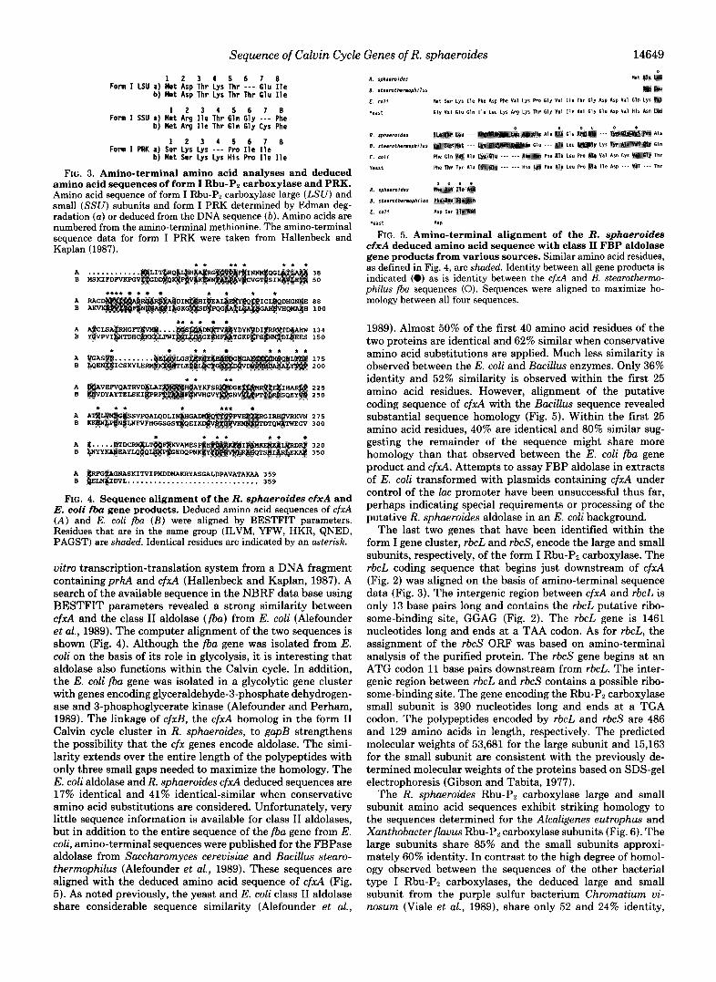
Forn I LSU a) Met Asp Thr Lys Thr --- Glu Ile 1 2 3 1 5 6 7 8
b ) Met Asp Thr Lys Thr Thr Glu Ile
Forn I SSU a) Met Arg Ile Thr Gln Gly - - - Phe b ) Met Arg Ile Thr Gln Gly Cys Phe
1 2 3 1 5 6 7 8
Forn I PRK a) Ser Lys Lys - - - Pro Ile Ile 1 2 9 4 5 6 7 8
b ) Met Ser Lys Lys H I S Pro Ile Ile
FIG. 3. Amino-terminal amino acid analyses and deduced amino acid sequences of form I Rbu-Pz carboxylase and PRK. Amino acid sequence of form I Rbu-Pz carboxylase large (LSU) and small ( S S U ) subunits and form I PRK determined by Edman deg- radation ( a ) or deduced from the DNA sequence ( b ) . Amino acids are numbered from the amino-terminal methionine. The amino-terminal sequence data for form I PRK were taken from Hallenbeck and Kaplan (1987).
A B
A B
A B
A B
A B
A B
A B
A B
.* ..* t. * ............ MSKIFDWXPGV
**** t * . t *ig * * * * ....
LPWI YDY TGK
I * * * * *
.. .*
t *.*
* t t t * *
GNASKITVIPHDDMAKRYASGALDPAVATAKAA 359 DVL .............................. 359
38 5 0
88 100
1 3 4 150
175 200
225 250
275 300
320 350
FIG. 4. Sequence alignment of the R. sphaeroides cfxA and E. coli fba gene products. Deduced amino acid sequences of c f d ( A ) and E. coli fba ( B ) were aligned by BESTFIT parameters. Residues that are in the same group (ILVM, YFW, HKR, QNED, PAGST) are shaded. Identical residues are indicated by an asterisk.
uitro transcription-translation system from a DNA fragment containingpru and cfxA (Hallenbeck and Kaplan, 1987). A search of the available sequence in the NBRF data base using BESTFIT parameters revealed a strong similarity between cfxA and the class I1 aldolase (fba) from E. coli (Alefounder e t al., 1989). The computer alignment of the two sequences is shown (Fig. 4). Although the fba gene was isolated from E. coli on the basis of its role in glycolysis, it is interesting that aldolase also functions within the Calvin cycle. In addition, the E. coli fba gene was isolated in a glycolytic gene cluster with genes encoding glyceraldehyde-3-phosphate dehydrogen- ase and 3-phosphoglycerate kinase (Alefounder and Perham, 1989). The linkage of cfxB, the cfxA homolog in the form I1 Calvin cycle cluster in R. sphaeroides, to gapB strengthens the possibility that the cfx genes encode aldolase. The simi- larity extends over the entire length of the polypeptides with only three small gaps needed to maximize the homology. The E. coli aldolase and R. sphaeroides cfxA deduced sequences are 17% identical and 41% identical-similar when conservative amino acid substitutions are considered. Unfortunately, very little sequence information is available for class I1 aldolases, but in addition to the entire sequence of the fba gene from E. coli, amino-terminal sequences were published for the FBPase aldolase from Saccharomyces cereuisiae and Bacillus stearo- therrnophilus (Alefounder et al., 1989). These sequences are aligned with the deduced amino acid sequence of cfxA (Fig. 5). As noted previously, the yeast and E. coli class I1 aldolase share considerable sequence similarity (Alefounder et al.,
R . sphaemides
8. s t e l m t h e m p h i l u s
E . coli
veart GIY v11 61" Gln Ile Leu Lyr Arp Lyr Thr Gly Val I l r Val GlY Glu ASP V I 1 H I S A m
8 . I t earo themphi lUS
E . coli
Yeast
R . sph,cmidcr
8 . r t e a m t h c m p h i l u r
E . coli ASP Ser
veart ASP
FIG. 5. Amino-terminal alignment of the R. sphaeroides cfxA deduced amino acid sequence with class I1 FBP aldolase gene products from various sources. Similar amino acid residues, as defined in Fig. 4, are shaded. Identity between all gene products is indicated (0) as is identity between the cfrA and B. stearotherrno- philus fba sequences (0). Sequences were aligned to maximize ho- mology between all four sequences.
1989). Almost 50% of the first 40 amino acid residues of the two proteins are identical and 62% similar when conservative amino acid substitutions are applied. Much less similarity is observed between the E. coli and Bacillus enzymes. Only 36% identity and 52% similarity is observed within the first 25 amino acid residues. However, alignment of the putative coding sequence of cfxA with the Bacillus sequence revealed substantial sequence homology (Fig. 5). Within the first 25 amino acid residues, 40% are identical and 80% similar sug- gesting the remainder of the sequence might share more homology than that observed between the E. coli fba gene product and cfwl . Attempts to assay FBP aldolase in extracts of E. coli transformed with plasmids containing cfwl under control of the lac promoter have been unsuccessful thus far, perhaps indicating special requirements or processing of the putative R. sphaeroides aldolase in an E. coli background.
The last two genes that have been identified within the form I gene cluster, rbcL and rbcS, encode the large and small subunits, respectively, of the form I Rbu-P2 carboxylase. The rbcL coding sequence that begins just downstream of cfxA (Fig. 2) was aligned on the basis of amino-terminal sequence data (Fig. 3). The intergenic region between cfxA and rbcL is only 13 base pairs long and contains the rbcL putative ribo- some-binding site, GGAG (Fig. 2). The rbcL gene is 1461 nucleotides long and ends at a TAA codon. As for rbcL, the assignment of the rbcS ORF was based on amino-terminal analysis of the purified protein. The rbcS gene begins at an ATG codon 11 base pairs downstream from rbcL. The inter- genic region between rbcL and rbcS contains a possible ribo- some-binding site. The gene encoding the Rbu-P2 carboxylase small subunit is 390 nucleotides long and ends at a TGA codon. The polypeptides encoded by rbcL and rbcS are 486 and 129 amino acids in length, respectively. The predicted molecular weights of 53,681 for the large subunit and 15,163 for the small subunit are consistent with the previously de- termined molecular weights of the proteins based on SDS-gel electrophoresis (Gibson and Tabita, 1977).
The R. sphaeroides Rbu-P2 carboxylase large and small subunit amino acid sequences exhibit striking homology to the sequences determined for the Alcaligenes eutrophus and Xanthobacterflauus Rbu-P2 carboxylase subunits (Fig. 6). The large subunits share 85% and the small subunits approxi- mately 60% identity. In contrast to the high degree of homol- ogy observed between the sequences of the other bacterial type I Rbu-P2 carboxylases, the deduced large and small subunit from the purple sulfur bacterium Chromatium vi- nosum (Viale et al., 1989), share only 52 and 24% identity,

14650 Sequence of Calvin Cycle Genes of R. sphaeroides
respectively. A high degree of similarity has been noted be- tween the sequence of the R. sphaeroides Rbu-Pp carboxylase genes and sequences determined for Rbu-Pp carboxylase from Rhodophyta and Chromophytu (Hwang and Tabita, 1991). In this case, the large subunits from a red alga and a marine diatom share approximately 70% and the small subunits 50% identity, respectively, with the large subunit of R. sphaeroides Rbu-Pz carboxylase.
Several lines of evidence, including peptide mapping and immunological techniques (Gibson and Tabita, 1977, 1985) had previously indicated the lack of similarity between the form I and form I1 Rbu-Pp carboxylase large subunits. There- fore, the low homology observed between the deduced se- quences of the R. sphaeroides form I and form I1 Rbu-Pz carboxylase large subunits was not surprising. The two large subunits share only 25% identity at the amino acid level, the same as that shared between the form I rbcL and the R. rubum Rbu-Pz carboxylase large subunit (Nargang et al., 1984). Many of the identical residues are clustered around sites known to be involved in the activation or catalysis of Rbu-Pz carbox- ylase (Fig. 6).
Codon Usage-The codon usage for genes within the R. sphaeroides Calvin cycle A and B clusters is highly skewed in favor of G or C in the third position as expected for an organism containing DNA with an overall 67% G + C content (Triiper and Pfennig, 1979). The pattern was virtually iden- tical for all of the genes within the A cluster as well as for the previously published sequences of fbpB, prkB, and rbpL, from the B cluster (Wagner et al., 1988; Gibson et al., 1990). Only three codons, CCA, CTA, and TTA were never used, and six codons, all of which end in A or T, appeared only once. Of those six, ATA appeared within the amino terminus of the fbpA gene, hence its usage must be considered tentative until definitive assignment of the fbpA start codon.
Insertional Mutagenesis of Genes within the Form I Clus- ter-The genes within the form I cluster are tightly linked. As noted previously the putative ribosome-binding site of prkA lies within the coding sequence of fbpA. Similarly, ribo- some-binding sites of rbcL and rbcS are located within the small intergenic space separating those genes from cfxA and rbcL, respectively. Because of the tight spacing between cod- ing sequences and the fact that all of the genes are oriented in the same direction, we utilized interposon mutagenesis to investigate the possibility that the five genes form part of a large operon. A trimethoprim resistance cartridge was used to disrupt fbpA and cfxA as described under “Experimental Pro- cedures.”
In preliminary experiments, the fbpA- and c f d - strains were tested for the ability to grow photosynthetically. For comparison, the wild-type strain HR, two Rbu-Pp carboxylase deletion derivatives (rbcLrbcS- and rbpL-) (Falcone and Ta- bita, 1991), and an fbpB- strain (Gibson et d., 1990) were grown in parallel. All of the strains tested had similar growth rates in malate-supplemented standing cultures, and in cul- tures sparged with 1.5% Cop, 98.5% Hz (data not shown). Interestingly, the cfxA- strain was incapable of photohetero- trophic growth on malate when the culture was bubbled with argon until after a lag of several days, whereas the other strains exhibited growth rates comparable to the wild-type strain. The reason for this is not known, but is probably related to the decreased concentration of COz in the medium when the culture was bubbled with argon. It should be noted that in a recent study that examined growth characteristics of cfx mutants, it was reported that even the wild-type R. sphaeroides was incapable of photoheterotrophic growth on succinate and malate without Cop (Hallenbeck et aL, 1990a).
OPV 53 DW 56 OW 5k
GYL 39 PRE 43
I X A C
I1
ff
..
I X A C
I 1
434 437 435 425 424
PEILVEAAKUCQ-PLRRIILDWGEVTFNYASTDTSDFVPTASVA 187 PEILRAAAKUCK-PLEIULDTVW(ITFNVTSTDTSDfVPTASV~ 486
PEILRDPRRA~PLRARARYUGDITFNYTPTDTSDFVPTASVA 489 KDVLTWIAISSP-ELKlWENKEIKFEFDNDLDlAM 469 REURAFESFPMIDKFYPGYRDRLHRM 461
I PSLRnERTEVDGRSlRYTHSIVR I29 X DGFRLDRTEGPGRTQRYALQHRSYRAG 133 A PGFRLVRQEEPGRTLRYSIESYAVQAGPK 135
FIG. 6. Sequence comparisons of the Rbu-Pz carboxylase large and small subunits of various bacteria. Deduced amino acid sequences of rbcL and rbcS of R. sphaeroides form I Rbu-Pz carboxylase compared with sequences from X . fluuus ( X ) (Meijer et al., 1991), A. eutrophus ( A ) (Andersen and Caton, 1987), C. uinosum (C) (Viale et al., 1989), and the R. sphaeroides form I1 Rbu-Pz carboxylase (large subunit only) (11) (Wagner et al., 1988). Identical residues are shaded. Residues implicated in activation and catalysis are shown (*) (Knight et al., 1990).
We found that R. sphaeroides grows on malate or succinate in cultures bubbled with argon, albeit at a reduced rate of 7 versus 4 h in a standing culture, i.e. not bubbled with argon. The reason for this discrepancy is not clear but may reflect differences in strain or preculture techniques.
Further characterization of the mutant strains involved analysis of the various enzymes in cell extracts prepared from photoheterotrophically and photolithoautotrophically grown cells. Because R. sphueroides synthesizes two forms of these enzymes, it is impossible to assess the contribution of each isozyme to the overall activity based on enzyme assays alone. However, the form I and form I1 PRK and Rbu-Pp carboxylase enzymes can be distinguished by immunological methods. Therefore, initial characterization of the fbpA- and c f d - strains utilized rocket immunoelectrophoresis to detect and quantitate the antigenically distinct form I and form I1 Rbu- Pp carboxylase, and Western immunoblot analysis to distin-

Sequence of Calvin Cycle
guish between form I and form I1 PRK polypeptides which are separable by SDS-gel electrophoresis (Gibson and Tabita, 1987).
Rocket immunoelectrophoresis revealed a complete absence of form I Rbu-P2 carboxylase in extracts of the fbpA-, cfd- , and rbcLrbcS- strains grown photolithoautotrophically. A cor- responding increase in form I1 Rbu-P2 carboxylase was ob- served in these strains compared to the level present in the wild-type strain HR. The actual levels of Rbu-P2 carboxylase protein as measured by rocket immunoelectrophoresis are shown in Table I. When these extracts were examined by Western blotting and immunodetection using antiserum di- rected against PRK, no form I PRK could be detected in the fbpA- strain (Fig. 7). No apparent difference in the relative amounts of form I and form I1 PRK was observed in the c f d - or rbcL rbcS- strain (data not shown). Similar results were reported previously for mutations within the B cluster (Gib- son et al., 1990). In these experiments, Western blot analyses showed an absence of form I1 Rbu-P2 carboxylase and PRK in the fbpB- strain, and an absence of form I1 Rbu-Pp carbox- ylase, but an unaltered PRK pattern in the rbpL- strain. In this investigation, those findings were extended by quantitat-
TABLE I Specific activities of Calvin cycle enzymes in mutant strains
R ~ u - P ~
Strain condition protein" FBPase PRK carboxylase G~~~~ carboxylase Rbu-P2
I I1
HR HE'P A U T
rbcLrbcS- HET AUT
rbpL- HET AUT
&A- HET AUT
fW- HET AUT
c f d - HET AUT
1.8 0.54 5.6 1.9
0.73 3.1
3.8 5.8
2.1 21.0
4.8 14.2
0.17 2.8
0.03 0.10 0.05 0.18 0.08 0.08 0.1 1 0.21 0.05 0.12 0.03 0.11
units f mg protein 0.02 0.05 0.13 0.13 0.05 0.04 0.22 0.17 0.01 0.04 0.09 0.09 0.04 0.08 0.17 0.36 0.03 0.06 0.13 0.17 0.02 0.05 0.17 0.09
a Rbu-P2 carboxylase antigen as determined by rocket immunoe- lectrophoresis expressed as percent total soluble protein. I, form I Rbu-Pz carboxylase; 11, form I1 Rbu-P2 carboxylase.
* Photoheterotrophic growth on 0.4% malate bubbled with argon. ' Photolithoautotrophic growth on 1.5% CO2, 98.5% H2.
1 2 3 4 5 6 7 8 9
FIG. 7. Western immunoblot analysis of wild-type strain HR, and the fbpA- and fbpB- strains of R. sphaeroides. Cell extracts were prepared from photolithoautotrophically grown wild- type strain HR (lanes I , 4, and 7), the fbpA- strain (lunes 2, 5, and 81, and the fbpB- strain (lanes 3,6, and 9). Extracts were electropho- resed in SDS gels, electroblotted to nitrocellulose, and probed with antibodies raised against form I Rbu-Pz carboxylase (lanes I-3), form I1 Rbu-P2 carboxylase (lanes 4-6), or form I PRK (lanes 7-9).
Genes of R. sphaeroides 14651
ing the amount of Rbu-Pp carboxylase produced based on rocket immunoelectrophoresis. The levels of form I Rbu-Pp carboxylase increased over that observed in HR in both the fbpB- and rbpL- strains (Table I). The increase of form I Rbu-Pp carboxylase in these strains is not as dramatic, how- ever, as that observed for the form I1 Rbu-P2 carboxylase in the strains containing mutations within the A cluster. In every case, mutations within the A and B clusters exert polar effects on downstream genes, whereas genes positioned tran- scriptionally upstream are still expressed. As noted previously, the simplest interpretation of these results is that the genes within each cluster form part of an operon.
The increased production of the form I and form I1 Rbu-P2 carboxylase in the insertion strains observed here and in other studies (Hallenbeck et al., 1990a, 1990b) indicated that mu- tations within one operon could affect expression of genes within the second operon. In order to examine the effect of the mutations on expression of the various Calvin cycle en- zymes more closely, the extracts were assayed for FBPase, PRK, and Rbu-P2 carboxylase activities (Table I). The de- repression of Rbu-Pz carboxylase in wild-type cells grown under C02-limiting conditions is well documented (Jouanneau and Tabita, 1986; Hallenbeck et aZ., 1990a, 1990b). All of the mutants retain the C02-regulated expression of Rbu-P2 car- boxylase as evidenced by the increase in Rbu-Pp carboxylase activity in Cop-grown cells compared to malate-grown cells. The activities of FBPase and PRK are also much higher in Cop-grown cells, suggesting the genes are subject to coordi- nate regulation. Unlike the wild-type, the mutants exhibit altered levels of these enzymes. For example, in the fbpA- strain, the specific activities of all three enzymes are higher than those measured in wild-type strain HR in both malate- and Con-grown cells. These increases are even more dramatic considering only the form I1 enzymes are present in the fbpA- strain. In the C02-grown cells, the form I1 Rbu-P2 carboxylase normally accounts for approximately 33% of total Rbu-P2 carboxylase (Joaunneau and Tabita, 1986). Therefore, the 2.8-fold increase in Rbu-PZ carboxylase activity in the fbpA- strain actually represents a 8.5-fold increase in the expression of form I1 Rbu-P2 carboxylase and is higher than the combined form I and form I1 Rbu-P:! carboxylase activity in wild-type strain HR. Similar results were obtained with the fbpB- strain in which only the form I enzymes are expressed. Although the increases in activity of FBPase, PRK, and Rbu-Pz carboxylase are not as pronounced as those observed in the fbpA- strain, the levels of the form I enzymes are still substantially higher than in strain HR.
The results are more difficult to interpret in the Rbu-P2 carboxylase deletion derivatives and the c f d - strain. These mutants retain both forms of FBPase and PRK, but express only one form of Rbu-Pp carboxylase. The rbcLrbcS- strain basically follows the same pattern as the fbpA- strain, except that in the fbpA- strain the Rbu-P2 carboxylase activity in C02-grown cells represents a greater increase in the amount of form I1 Rbu-P2 carboxylase compared to wild-type cells. The rbpL- strain exhibits a pattern of activities distinct from the other mutant strains in that PRK and Rbu-Pp carboxylase levels are quite low in CO2-grown cells. Based on specific activity, the form I Rbu-P2 carboxylase is actually lower in this strain than in the wild-type strain. In the c f d - strain, the level of the form I1 Rbu-P2 carboxylase was higher than that observed in the wild-type strain, whereas the level of PRK was relatively unaffected.
Northern Blot Analysis-Because the insertional mutagen- esis studies indicated the genes within each cluster were co- transcribed, Northern hybridizations were performed to de-
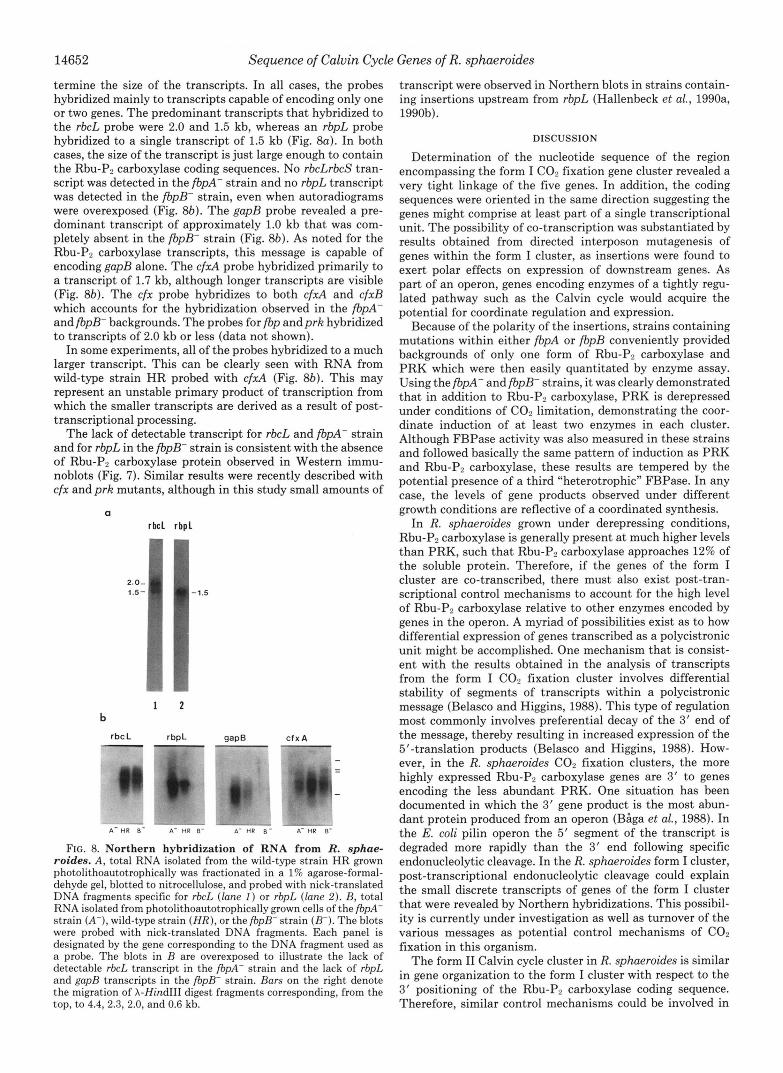
14652 Sequence of Calvin Cycle Genes of R. sphaeroides
termine the size of the transcripts. In all cases, the probes hybridized mainly to transcripts capable of encoding only one or two genes. The predominant transcripts that hybridized to the rbcL probe were 2.0 and 1.5 kb, whereas an rbpL probe hybridized to a single transcript of 1.5 kb (Fig. 8a). In both cases, the size of the transcript is just large enough to contain the Rbu-P, carboxylase coding sequences. No rbcLrbcS tran- script was detected in the fbpA- strain and no rbpL transcript was detected in the fbpB- strain, even when autoradiograms were overexposed (Fig. 8b). The gapB probe revealed a pre- dominant transcript of approximately 1.0 kb that was com- pletely absent in the fbpB- strain (Fig. 8b). As noted for the Rbu-P, carboxylase transcripts, this message is capable of encoding gapB alone. The c f d probe hybridized primarily to a transcript of 1.7 kb, although longer transcripts are visible (Fig. 8b). The cfx probe hybridizes to both c f d and cfxB which accounts for the hybridization observed in the fbpA- andfbpB- backgrounds. The probes for fbp andprk hybridized to transcripts of 2.0 kb or less (data not shown).
In some experiments, all of the probes hybridized to a much larger transcript. This can be clearly seen with RNA from wild-type strain HR probed with cfxA (Fig. 8b). This may represent an unstable primary product of transcription from which the smaller transcripts are derived as a result of post- transcriptional processing.
The lack of detectable transcript for rbcL and fbpA- strain and for rbpL in the fbpB- strain is consistent with the absence of Rbu-Pp carboxylase protein observed in Western immu- noblots (Fig. 7). Similar results were recently described with cfx and prk mutants, although in this study small amounts of
a rbc l rbpl
2.0- 1.5- -1.5
i r
1 2 b
rbcL rbpL -
FIG. 8. Northern hybridization of RNA from R. sphae- roides. A , total RNA isolated from the wild-type strain HR grown photolithoautotrophically was fractionated in a 1% agarose-formal- dehyde gel, blotted to nitrocellulose, and probed with nick-translated DNA fragments specific for rbcL (lane I ) or rbpL (lune 2) . B, total RNA isolated from photolithoautotrophically grown cells of the fbpA- strain ( A - ) , wild-type strain ( H R ) , or the fbpB- strain (B-). The blots were probed with nick-translated DNA fragments. Each panel is designated by the gene corresponding to the DNA fragment used as a probe. The blots in B are overexposed to illustrate the lack of detectable rbcL transcript in the fbpA- strain and the lack of rbpL and gupB transcripts in the fbpB- strain. Burs on the right denote the migration of X-Hind111 digest fragments corresponding, from the top, to 4.4, 2.3, 2.0, and 0.6 kb.
transcript were observed in Northern blots in strains contain- ing insertions upstream from rbpL (Hallenbeck et al., 1990a, 1990b).
DISCUSSION
Determination of the nucleotide sequence of the region encompassing the form I Cop fixation gene cluster revealed a very tight linkage of the five genes. In addition, the coding sequences were oriented in the same direction suggesting the genes might comprise at least part of a single transcriptional unit. The possibility of co-transcription was substantiated by results obtained from directed interposon mutagenesis of genes within the form I cluster, as insertions were found to exert polar effects on expression of downstream genes. As part of an operon, genes encoding enzymes of a tightly regu- lated pathway such as the Calvin cycle would acquire the potential for coordinate regulation and expression.
Because of the polarity of the insertions, strains containing mutations within either fbpA or fbpB conveniently provided backgrounds of only one form of Rbu-P2 carboxylase and PRK which were then easily quantitated by enzyme assay. Using the fbpA- and fbpB- strains, it was clearly demonstrated that in addition to Rbu-Pp carboxylase, PRK is derepressed under conditions of CO, limitation, demonstrating the coor- dinate induction of at least two enzymes in each cluster. Although FBPase activity was also measured in these strains and followed basically the same pattern of induction as PRK and Rbu-P, carboxylase, these results are tempered by the potential presence of a third “heterotrophic” FBPase. In any case, the levels of gene products observed under different growth conditions are reflective of a coordinated synthesis.
In R. sphueroides grown under derepressing conditions, Rbu-Pa carboxylase is generally present at much higher levels than PRK, such that Rbu-Pp carboxylase approaches 12% of the soluble protein. Therefore, if the genes of the form I cluster are co-transcribed, there must also exist post-tran- scriptional control mechanisms to account for the high level of Rbu-Pp carboxylase relative to other enzymes encoded by genes in the operon. A myriad of possibilities exist as to how differential expression of genes transcribed as a polycistronic unit might be accomplished. One mechanism that is consist- ent with the results obtained in the analysis of transcripts from the form I Con fixation cluster involves differential stability of segments of transcripts within a polycistronic message (Belasco and Higgins, 1988). This type of regulation most commonly involves preferential decay of the 3‘ end of the message, thereby resulting in increased expression of the 5’-translation products (Belasco and Higgins, 1988). How- ever, in the R. sphueroides CO, fixation clusters, the more highly expressed Rbu-P, carboxylase genes are 3’ to genes encoding the less abundant PRK. One situation has been documented in which the 3‘ gene product is the most abun- dant protein produced from an operon (Blga et al., 1988). In the E. coli pilin operon the 5’ segment of the transcript is degraded more rapidly than the 3’ end following specific endonucleolytic cleavage. In the R. sphueroides form I cluster, post-transcriptional endonucleolytic cleavage could explain the small discrete transcripts of genes of the form I cluster that were revealed by Northern hybridizations. This possibil- ity is currently under investigation as well as turnover of the various messages as potential control mechanisms of COn fixation in this organism.
The form I1 Calvin cycle cluster in R. sphueroides is similar in gene organization to the form I cluster with respect to the 3‘ positioning of the Rbu-Pp carboxylase coding sequence. Therefore, similar control mechanisms could be involved in

Sequence of Calvin Cycle Genes of R. sphaeroides 14653
post-transcriptional regulation of the form I and form I1 operons. In this context, it is interesting to note that the spatial arrangement of genes within other bacterial COP fix- ation clusters differs from that found in R. sphueroides. In A. eutrophus, a facultative chemolithoautotrophic bacterium, the genes encoding several Calvin cycle enzymes are duplicated and clustered within two very similar operons (Windhovel and Bowien, 1990). As in R. sphaeroides, the FBPase and PRK coding sequences are tandemly arranged in a tight linkage. However, these genes are situated downstream from the Rbu-Pz carboxylase coding sequences. The COP fixation genes in X. f l a w u s have also been mapped within a single cluster (Meijer et al., 1990). In this organism, the gene ar- rangement is similar, although not identical to that of A. eutrophus. Although the functional significance of the differ- ent arrangements of genes within the COz fixation clusters is not known, gene order may turn out to play a crucial role in regulation of gene expression in these systems.
Several lines of evidence suggest the genes within the form I and form I1 Calvin cycle clusters constitute two operons. In this regard, the tentative identification of the cfxA gene prod- uct as aldolase, on the basis of sequence comparisons, was not surprising in view of its position amidst other structural genes of the same pathway. Although the degree of similarity of cfxA to the E. coli fba is not high, the relatedness does extend over the full length of both reading frames. Moreover, the high degree of identity observed between the amino terminus of the B. stearothermophilus aldolase and the R. sphaeroides cfxA gene product suggests these sequences may be more closely related than are the E. coli fba and R. sphaeroides cfxA products. Alternatively, the low homology may represent a photosynthetic aldolase specifically adapted in functional and regulatory characteristics to the needs of the Calvin cycle in R. sphaeroides. Finally, the linkage of fba to gap in E. coli further strengthens the possibility that cfx is aldolase. Al- though cfxA is not linked to gap in the form I cluster, its homolog, cfxB, is situated immediately downstream of gapB in the form I1 cluster.
In previous studies, cfxA andprkA mutants exhibited drast- ically decreased growth rates during COP-limited growth com- pared to the wild-type strain (Hallenbeck et al., 1990a, 1990b). The differences in growth were attributed to the absence of form I Rbu-Pz carboxylase, the rationale being that the high affinity of form I Rbu-PP carboxylase for COP makes it essen- tial for COP limited growth. However, in this study, the rbcLrbcS- strain exhibited wild-type growth rates under ma- late-argon growth conditions. In addition, a requirement of form I Rbu-PP carboxylase for COP-limited growth cannot explain the wild-type growth characteristics of the fbpA- strain. One possible explanation of these seemingly discrepant observations may lie in the identity of cfx as aldolase. FBPase and aldolase catalyze diametrically opposed reactions in the Calvin cycle, the former being responsible for the breakdown of FBP and sedoheptulose 1,7-bisphosphate, the latter for their synthesis. In the fbpA- and rbcLrbcS- mutants, the balance of FBPase and aldolase is maintained, whereas in the prk and cfx mutants there are two copies of “photosynthetic”
FBPase to one copy of the putative aldolase. Disruption of the relative amounts of these two antagonistic enzymes may lead to futile cycling of metabolites.
Acknowledgment-We are grateful to Sandy Smith of the Amino Acid Sequencing Facility of the University of Texas at Austin.
REFERENCES Alefounder, P. R., and Perham, R. N. (1989) Mol. Microbiol. 3 , 723-
Alefounder, P. R., Baldwin, S. A., Perham, R. N., and Short, N. J.
Andersen, K., and Caton, J. (1987) J. Bacteriol. 169,4547-4558 BHga, M., Goransson, M., Normark, S., and Uhlin, B. E. (1988) Cell
Belasco, J. G., and Higgins, C. F. (1988) Gene (Amst.) 7 2 , 15-23 Falcone, D. L., and Tabita, F. R. (1991) J. Bacteriol. 173,2099-2108 Falcone, D. L., Quivey, R. G., Jr., and Tabita, F. R. (1988) J. Bacteriol.
Gibson, J. L., and Tabita, F. R. (1977) J. Biol. Chern. 252,943-949 Gibson, J . L., and Tabita, F. R. (1985) J. Bacteriol. 164 , 11W-1193 Gibson, J. L., and Tabita, F. R. (1986) Gene (Amst.) 4 4 , 271-278 Gibson, J . L., and Tabita, F. R. (1987) J. Bacteriol. 169,3685-3690 Gibson, J. L., and Tabita, F. R. (1988) J. Bacteriol. 170, 2153-2158 Gibson, J . L., Chen, J.-H., Tower, P. A., and Tabita, F. R. (1990)
Hallenbeck, P. L., and Kaplan, S. (1987) J. Bacteriol. 169 , 3669-
Hallenbeck, P. L., Lerchen, R., Hessler, P., and Kaplan, S. (1990a)
Hallenbeck, P. L., Lerchen, R., Hessler, P., and Kaplan, S. (1990b)
Hwang, S.-R., and Tabita, F. R. (1991) J. Biol. Chern. 266, 6271-
Jouanneau, Y., and Tabita, F. R. (1986) J. Bacteriol. 165,620-624 Kay, R., and McPherson, J. (1987) Nucleic Acids Res. 15 , 2778 Knight, S., Anderssen, I., and Branden, C.-I. (1990) J. Mol. Biol.
Markwell, M. A., Haas, S. M., Bieber, L. L., and Tolbert, N. E. (1978) Anal. Biochem. 87,206-210
Meijer, W. G., Enequist, H. G., Terpstra, P., and Dijkhuizen, L. (1990) J. Gen. Microbiol. 136 , 2225-2230
Meijer, W. G., Arnberg, A. C., Enequist, H. G., Terpstra, P., Lidstrom, M. E., and Dijkhuizen, L. (1991) Mol. Gen. Genet. 225,320-330
Nargang, F., McIntosh, L., and Sommerville, C. (1984) Mol. Gen. Genet. 193,220-224
Quivey, R. G., Jr., and Tabita, F. R. (1984) Gene (Amst.) 31 , 91-101 Sanger, F., Nicklen, S., and Coulson, A. R. (1977) Proc. Natl. Acad.
Simon, R., Priefer, V., and Puhler, A. (1983) Bioltechnology 1 , 784-
Tabita, F. R. (1980) J. Bacteriol. 143 , 1275-1280 Tabita, F. R., Gibson, J. L., Mandy, W. J., and Quivey, R. G., Jr.
(1986) BiolTechnology 4, 138-141 Triiper, H. G., and Pfennig, N. (1979) in The Photosynthetic Bacteria
(Clayton, R. K., and Sistrom, W. R., eds) pp. 19-27, Plenum Publishing Corp., New York
Viale, A. M., Kobayashi, H., and Akazawa, T. (1989) J. Bacteriol. 171,2391-2400
Wagner, S. J., Stevens, S. E., Jr., Nixon, B. T., Lambert, D. H., Quivey, R. G., Jr., and Tabita, F. R. (1988) FEMS Microbiol. Lett.
732
(1989) Biochem. J. 257,529-534
52,197-206
170,5-11
Biochemistry 29,8085-8093
3678
J. Bacteriol. 172 , 1736-1748
J. Bacteriol. 172, 1749-1761
6279
2 1 5 , 113-160
Sci. U. S. A. 74,5463-5467
791
55,217-222 Weaver, K. E., and Tabita, F. R. (1983) J. Bacteriol. 165 , 507-515 Windhovel, U., and Bowien, B. (1990) Arch. Microbiol. 154,85-91 Yanisch-Perron, C., Vieira, J., and Messing, J . (1985) Gene (Amst.)
Zhu, Y. S., and Kaplan, S. (1985) J. Bacteriol. 162,925-932 33,103-119