nrf2 activation by antioxidant anti-diabetic agents accelerates...
TRANSCRIPT

NRF2 activation by antioxidant antidiabeticagents accelerates tumor metastasis.
Item Type Article
Authors Wang, Hui; Liu, Xiufei; Long, Min; Huang, Yi; Zhang, Linlin; Zhang,Rui; Zheng, Yi; Liao, Xiaoyu; Wang, Yuren; Liao, Qian; Li, Wenjie;Tang, Zili; Tong, Qiang; Wang, Xiaocui; Fang, Fang; Rojo de laVega, Montserrat; Ouyang, Qin; Zhang, Donna D; Yu, Shicang;Zheng, Hongting
Citation NRF2 activation by antioxidant antidiabetic agents acceleratestumor metastasis. 2016, 8 (334):334ra51 Sci Transl Med
DOI 10.1126/scitranslmed.aad6095
Publisher AMER ASSOC ADVANCEMENT SCIENCE
Journal Science translational medicine
Rights Copyright © 2016, American Association for the Advancement ofScience
Download date 12/06/2018 01:39:58
Link to Item http://hdl.handle.net/10150/615617

Submitted Manuscript: Confidential template updated: February 28 2012
NRF2 activation by antioxidant anti-diabetic agents accelerates tumor
metastasis
Hui Wang1,†, Xiufei Liu1,†, Min Long1,†, Yi Huang4,†, Linlin Zhang1, Rui Zhang1, Yi Zheng1,
Xiaoyu Liao1, Yuren Wang1, Qian Liao1, Wenjie Li1, Zili Tang5, Qiang Tong1, Xiaocui Wang1,
Fang Fang1, Montserrat Rojo de la Vega2, Qin Ouyang6, Donna D. Zhang2,*, Shicang Yu3,*, and
Hongting Zheng1,*
1Department of Endocrinology, Xinqiao Hospital, Third Military Medical University, Chongqing
400037, China.
2Department of Pharmacology and Toxicology, College of Pharmacy, University of Arizona,
Tucson, AZ 85721, USA.
3Institute of Pathology and Southwest Cancer Center, Southwest Hospital, Third Military
Medical University, Chongqing 400038, China.
4Ministry of Education Key Laboratory of Child Development and Disorders, Chongqing
key Laboratory of pediatrics, Chongqing key Laboratory of Immunity and Infectious Diseases,
Children's Hospital of Chongqing Medical University, Chongqing 400014, China.
5Molecular & Translational Radiation Oncology, Heidelberg Ion Therapy Center (HIT),
Heidelberg Institute of Radiation Oncology (HIRO), University of Heidelberg Medical School
and National Center for Cancer Diseases (NCT), German Cancer Research Center (DKFZ),
Heidelberg 69120, Germany.
6College of Pharmacy, Third Military Medical University, Chongqing 400038, China.
†These authors contributed equally to this work.

* Correspondence addressed to:
Hongting Zheng, M.D.
Department of Endocrinology, Xinqiao Hospital
Third Military Medical University, Chongqing 400037, China
E-mail: [email protected], Phone: +86 02368755709, Fax: +86 02368755707
Shicang Yu, Ph.D.
Institute of Pathology and Southwest Cancer Center, Southwest Hospital
Third Military Medical University, Chongqing 400037, China
E-mail: [email protected]
Donna D. Zhang, Ph.D.
Department of Pharmacology and Toxicology, College of Pharmacy
University of Arizona, Tucson, AZ 85721, USA
E-mail: [email protected]
One Sentence Summary: NRF2 activation promotes tumor metastasis.
Disclosure: The authors have nothing to disclose.

Abstract
Cancer is a common comorbidity of diabetic patients; however, little is known about the effects
that anti-diabetic drugs have on tumors. We discovered that common classes of drugs used in
type 2 diabetes mellitus, the hypoglycemic dipeptidyl peptidase-4 inhibitors (DPP-4i) saxagliptin
and sitagliptin, as well as the antineuropathic alpha-lipoic acid (ALA), do not increase tumor
incidence but increase the risk of metastasis of existing tumors. Specifically, these drugs induce
prolonged activation of the nuclear factor-E2-related factor 2 (NRF2)-mediated antioxidant
response via inhibition of KEAP1-C151-dependent ubiquitination and subsequent degradation of
NRF2, resulting in up-regulated expression of metastasis-associated proteins, increased cancer
cell migration, and promotion of metastasis in xenograft mouse models. Accordingly,
knockdown of NRF2 attenuated naturally-occurring and DPP-4i-induced tumor metastasis,
whereas NRF2 activation accelerated metastasis. Furthermore, in human liver cancer tissue
samples, increased NRF2 expression correlated with metastasis. Our findings suggest that
antioxidants that activate NRF2 signaling may need to be administered with caution in cancer
patients, such as diabetic patients with cancer. Moreover, NRF2 may be a potential biomarker
and therapeutic target for tumor metastasis.

Introduction
Accumulating epidemiological evidence suggests that diabetes increases the risk of multiple
cancers, including colon, liver, and breast cancers (1). Therefore, the increased prevalence of
diabetes suggests that the incidence of individuals with both diabetes and cancer is also rising.
Anti-diabetic drugs modulate glucose metabolism, the insulin-like growth factor-1 axis, or other
factors associated with tumor initiation and progression, and therefore they may affect tumor
behavior (1, 2). Moreover, because diabetic patients are chronically exposed to anti-diabetic
drugs, this long-term exposure as well as drug accumulation resulting from compromised renal
and hepatic functions commonly seen in these patients may amplify toxic effects of anti-diabetic
drugs (3). Therefore, understanding the effects of anti-diabetic agents on tumor biology is
indispensable for the development of specialized drug therapy that is safe to treat diabetic
patients with cancer.
Few studies have investigated the effects that anti-diabetic drugs have on tumors; some
have suggested that anti-diabetic agents affect the incidence of cancer. Lewis and Piccinni
reported a positive correlation between the dose and time of pioglitazone administration and
bladder cancer risk (4, 5). Whether insulin analogs (especially glargine) increase cancer risk is
currently controversial (6). On the other hand, metformin was found to reduce the risk of
multiple cancers (such as breast, colon, and pancreatic cancers) and to inhibit the proliferation of
cancer cells by activating the LKB1/AMPK pathway, indicating its potential as a cancer
chemoprevention agent (7, 8). However, little is known about the effect of anti-diabetic agents
on comorbid tumors in diabetic patients. To study the effects of anti-diabetic drugs on the
biological behavior of existing tumors, we screened common clinical anti-diabetic agents such as
metformin, various insulin analogs, and dipeptidyl peptidase-4 inhibitors (DPP-4i) in vitro.

Specifically, we assessed the proliferation and migration of different cancer cells after treatment
with these drugs. We discovered that the DPP-4i saxagliptin (Sax), which is currently
recommended by the American Association of Clinical Endocrinologists (AACE) as first-line
hypoglycemic treatment in type 2 diabetes mellitus (T2DM) (9), potentially promoted migration
of cancer cells. To clarify the correlation between DPP-4i and tumor metastasis and to reveal the
mechanism underlying our preliminary observations, we further performed a series of in vitro
and in vivo experiments. Here we report that DPP-4i Sax and sitagliptin (Sit), as well as alpha-
lipoic acid (ALA, an antioxidant used to treat diabetic neuropathy), promote tumor metastasis
through activation of the nuclear factor-E2-related factor 2 (NRF2)-mediated antioxidant
response, though they don’t increase tumor incidence. Additionally, NRF2 pathway activation
correlated with tumor metastasis and may thus serve as a potential biomarker and therapeutic
target.

Results
DPP-4i anti-diabetic treatment does not increase cancer risk
Previous studies have suggested that anti-diabetic treatments may modify neoplastic risk factors
or modulate existing tumors’ biological behavior. To examine the effect of DPP-4i on cancer
risk, we performed a meta-analysis of randomized clinical trials on DPP-4i up to July 2015. We
did not find any correlation between DPP-4i monotherapy (saxagliptin, sitagliptin, vildagliptin,
linagliptin, or alogliptin; n=29, OR=0.80, 95% CI=0.52-1.24) or combinations with other anti-
diabetic drugs and total cancer incidence (n=88, OR=1.07, 95% CI=0.91-1.26). These results
were consistent irrespective of the anti-diabetic drug used alone (table S1) or in combination
(table S2). Specifically, DPP-4i treatment did not increase the risk of digestive (n=18, OR=0.89,
95% CI=0.42-1.85), dermal (n=9, OR=1.05, 95% CI=0.40-2.71), or urological cancers (n=11,
OR=0.94, 95% CI=0.38-2.37) (table S3), although previous studies reported that DPP-4i may be
associated with increased incidence in these tumor types (10, 11). This meta-analysis suggests
that DPP-4i alone or in combination with other anti-diabetic drugs do not increase cancer risks.
DPP-4i (Sax and Sit) enhance tumor metastasis
Currently, it remains unclear whether DPP-4i treatment is detrimental to diabetic patients with
existing tumors. To address this, we tested two DPP-4i compounds, Sax and Sit, with SW480,
HCT116, and HuH-7 cancer cells and determined that within the therapeutic range (0.1 μM or
0.6 μM, respectively), DPP-4 activity was inhibited (fig. S1). Sax and Sit did not affect cell
proliferation (fig. S2A) or alter cancer cell sensitivity to cisplatin (fig. S2B). However, Sax and
Sit markedly increased cell migration and invasion of multiple cancer cell lines (colon SW480
and HCT116, hepatic HuH-7, breast MDA-MB-231, lung A549, ovary SKOV-3, and melanoma
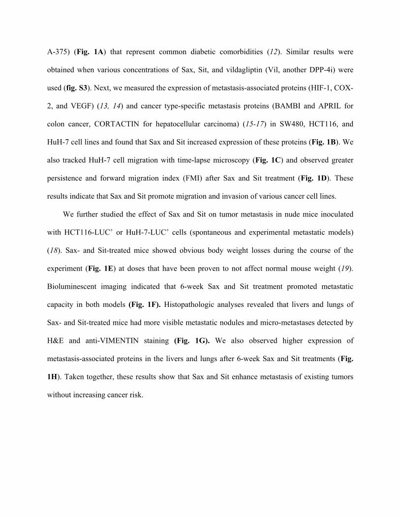
A-375) (Fig. 1A) that represent common diabetic comorbidities (12). Similar results were
obtained when various concentrations of Sax, Sit, and vildagliptin (Vil, another DPP-4i) were
used (fig. S3). Next, we measured the expression of metastasis-associated proteins (HIF-1, COX-
2, and VEGF) (13, 14) and cancer type-specific metastasis proteins (BAMBI and APRIL for
colon cancer, CORTACTIN for hepatocellular carcinoma) (15-17) in SW480, HCT116, and
HuH-7 cell lines and found that Sax and Sit increased expression of these proteins (Fig. 1B). We
also tracked HuH-7 cell migration with time-lapse microscopy (Fig. 1C) and observed greater
persistence and forward migration index (FMI) after Sax and Sit treatment (Fig. 1D). These
results indicate that Sax and Sit promote migration and invasion of various cancer cell lines.
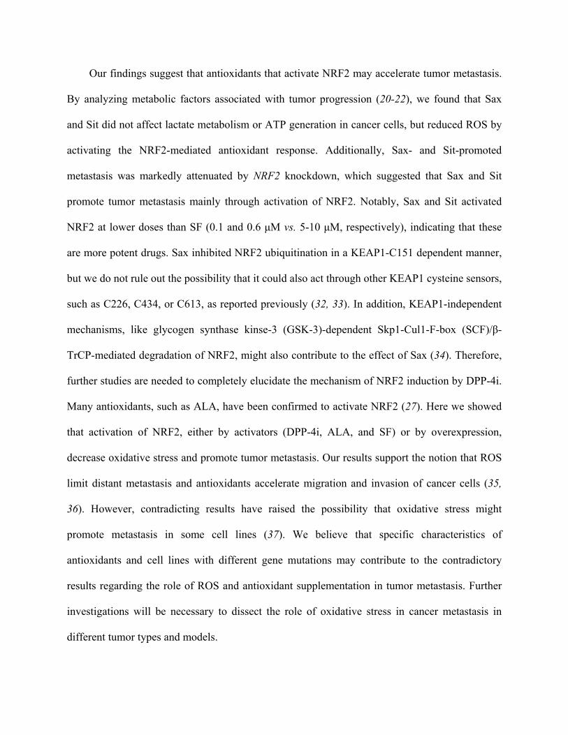
We further studied the effect of Sax and Sit on tumor metastasis in nude mice inoculated
with HCT116-LUC+ or HuH-7-LUC+ cells (spontaneous and experimental metastatic models)
(18). Sax- and Sit-treated mice showed obvious body weight losses during the course of the
experiment (Fig. 1E) at doses that have been proven to not affect normal mouse weight (19).
Bioluminescent imaging indicated that 6-week Sax and Sit treatment promoted metastatic
capacity in both models (Fig. 1F). Histopathologic analyses revealed that livers and lungs of
Sax- and Sit-treated mice had more visible metastatic nodules and micro-metastases detected by
H&E and anti-VIMENTIN staining (Fig. 1G). We also observed higher expression of
metastasis-associated proteins in the livers and lungs after 6-week Sax and Sit treatments (Fig.
1H). Taken together, these results show that Sax and Sit enhance metastasis of existing tumors
without increasing cancer risk.

DPP-4i activate NRF2 via KEAP1-C151-dependent inhibition of ubiquitination
To understand how Sax and Sit promote tumor metastasis, we measured tumor progression-
associated factors such as lactate production, ATP metabolism, and oxidative stress (20-22).
Neither intracellular nor secreted lactate (fig. S4A), nor ATP production (fig. S4B) were affected
by treatment, but Sax and Sit greatly reduced reactive oxygen species (ROS) production (Fig.
2A), increased GSH/GSSG ratios (Fig. 2B and fig. S5), and decreased 8-oxo-dG in cancer cells
(Fig. 2C), suggesting that Sax and Sit may reduce oxidative stress in cancer cells. Furthermore,
Sax- and Sit-induced cell migration and invasion were prevented by administration of buthionine
sulfoximine (BSO), an inhibitor of new glutathione synthesis, indicating that glutathione
synthesis is required for the effects of Sax and Sit on tumor metastasis (fig. S6). Commonly,
ROS concentrations are suppressed by activation of the transcription factor NRF2 and
subsequent expression of antioxidant systems (22); therefore, we measured the protein
expression of NRF2 and its downstream targets NQO1 and GCLM. We observed that the NRF2
pathway was markedly activated by Sax and Sit treatment in vitro (Fig. 2D) and in vivo (Fig. 2,
E and F). Similar results were obtained by treating cancer cells with commercial human drugs
Sax or Sit (fig. S7). Thus, these results suggest that Sax and Sit activate NRF2 to increase
intracellular antioxidants and neutralize ROS in cancer cells.
DPP-4i are structurally diverse drugs, so we next investigated if other members of this class
activate NRF2. Our results showed that Sax, Sit, Vil, alogliptin (Alo), and linagliptin (Lin)
induced NRF2 and its downstream targets (fig. S8A), as well as antioxidant response element
(ARE)-driven luciferase reporter gene expression (fig. S8B). Furthermore, we performed a 2D
similarity search in SYBYL-X 2.0 and found that although the similarity of Sax and Vil is
72.8%, the other 5 DPP-4i have less than 40% similarity, which means that these DPP-4i do not

share a common scaffold (fig. S8C). The fact that structurally diverse DPP-4i inhibited DPP-4
and induced NRF2 indicates that their ability to activate NRF2 may be a consequence of
inhibiting DPP-4.
Next, we further explored the molecular mechanism of DPP-4i-induced NRF2 activation
focusing on Sax. We measured mRNA and protein expression of NRF2 and its targets in HuH-7
cells treated with Sax. Both mRNA and protein expression of these targets increased after
treatment; however, NRF2 transcription did not apparently change, suggesting that Sax
modulates NRF2 post-transcriptionally (Fig. 2G). Because expression of NRF2 is primarily
controlled at the protein level by the KELCH-like ECH-associated protein-1 (KEAP1)-
containing E3 ubiquitin ligase complex through ubiquitination and subsequent proteasomal
degradation of NRF2 (23), we next assessed the ubiquitination and half-life of NRF2. We
observed decreased NRF2 ubiquitination and a three-fold half-life increase (from 13.7 to 50.4
min) after Sax treatment (Fig. 2, H and I). Furthermore, because cysteine151 in KEAP1 is
specifically required for NRF2 activation by canonical NRF2 inducers sulforaphane (SF) and
tert-butylhydroquinone (tBHQ) (24), we examined the effect of this cysteine in NRF2 induction
by Sax. We observed that NRF2 induction by Sax was inhibited in KEAP1-C151S mutant cells
(Fig. 2J), indicating that Sax activates NRF2 in a KEAP1-C151 dependent manner.
NRF2 inhibition attenuates tumor metastases
Recent data indicate that persistent activation of NRF2, the “dark side of NRF2”, promotes
tumorigenesis (22); however, whether persistent NRF2 activation promotes tumor metastasis is
unclear. Because Sax and Sit promote tumor metastasis and also activate NRF2 signaling, we
tested whether Sax and Sit promote tumor metastasis via activation of NRF2. We knocked down

NRF2 in SW480, HCT116, and Huh-7 cells and observed a marked decrease of Sax and Sit-
induced metastasis-associated proteins (Fig. 3A). Moreover, NRF2 silencing reduced Sax- and
Sit-driven cancer cell migration (Fig. 3B) and inhibited persistence and FMI (Fig. 3C), as well as
intrinsic migratory capacity (Fig. 3D). Similarly, knockdown of NRF2 with lentiviral shRNA
targeting NRF2 in HuH-7-LUC+ greatly reduced the expression of NRF2 and its downstream
targets at the protein level in cells (Fig. 3E) and mRNA in metastatic nodules in vivo (Fig. 3F). It
also decreased intrinsic (Fig. 3G) and Sax- and Sit-induced (Fig. 3H) metastases in mice
inoculated with these cells. Together, these results suggest that NRF2 inhibition attenuates tumor
metastases.
Activation of NRF2 promotes tumor metastasis
To further confirm the role of NRF2 in tumor metastasis, we investigated the effects of NRF2
activation on migration and metastasis. We noted that the expression of metastasis-associated
proteins increased upon genetic (by NRF2 overexpression) or pharmacologic [treatment with SF,
a well-characterized NRF2 activator (25)] NRF2 activation (Fig. 4, A and B). Similarly, cell
migration was also enhanced (Fig. 4C). Moreover, time-lapse microscopy confirmed increased
directed and persistent migration of cells treated with SF (Fig. 4D). In vivo, SF treatment also
activated the NRF2 pathway (Fig. 4, E and F). We observed that the normally nontoxic low-dose
SF treatment (25) (10 mg/kg, three times per week) caused animal weight loss (Fig. 4G), which
might be related to an increased metastatic tumor burden (Fig. 4H), similar to what was observed
with Sax and Sit treatments. Consistently, an increase in micro-metastasis was confirmed by
H&E staining and IHC analysis of VIMENTIN in liver and lung tissues (Fig. 4I). These results

indicate that NRF2 activation increases the ability of cancer cells to migrate, which results in
increased metastasis.
Pharmacological activation of NRF2 by ALA promotes tumor metastasis
Oxidative stress contributes to the onset of diabetes and its associated complications, making the
prescription of antioxidants a regular clinical practice (26). Some antioxidants used for diabetic
treatment, such as ALA, are known NRF2 activators (27). Because our results showed that NRF2
activation accelerates tumor metastasis, we investigated whether ALA also promotes tumor
metastasis. Indeed, ALA treatment activated the NRF2 pathway, increased metastasis-associated
protein expression (Fig. 5A), and promoted migration in SW480 and HCT116 cells (Fig. 5B). In
vivo, low-dose ALA, which also does not affect normal mouse weight (28), increased NRF2 and
downstream protein expression (Fig. 5, C and D) and caused weight loss in tumor bearing-mice
(Fig. 5E), similar to what we previously observed for Sax, Sit, and SF treatments. Moreover,
ALA increased total metastatic tumor burden (Fig. 5F) and increased micro-metastasis to the
liver and lungs (Fig. 5G). Thus, these results indicate that pharmacological activation of NRF2
by ALA also promotes cancer cell migration and tumor metastasis.
NRF2 is a potential metastatic marker in human liver cancer
To further characterize the value of NRF2 expression in cancer metastasis, we analyzed human
liver cancer tissue microarrays, including 176 primary tumor samples (105 non-metastatic, 69
lymph node metastases, 2 distant metastases) and 19 metastatic tumors, to correlate the
expression of NRF2 and its downstream target GCLM with patient clinicopathological features
(images of H&E staining and clinical information can be obtained from

http://www.biomax.us/tissue-arrays/Liver/LV1504, http://www.biomax.us/tissue-
arrays/Liver/LV1505, and http://www.biomax.us/tissue-arrays/Liver/LV1221). Overall,
compared to non-metastatic cancer, metastatic cancer (either primary tumor or metastatic lesion)
had greater NRF2 and GCLM expression, P < 0.0001 (Fig. 6, A and B). NRF2 and GCLM
expression were associated with lymph node metastasis and tumor stage (P < 0.0001), but were
not correlated to sex, age, pathology type, tumor size, or grade (Fig. 6, C and D; table S4 and
S5). Therefore, our data suggest that NRF2 activation contributes to enhanced metastasis in
human liver cancer, and as such, may be a promising biomarker as well as a therapeutic target.

Discussion
Recent evidence has shown that diabetes may increase the risk of many cancers, and the
incidence of diabetic patients with cancer has increased (1). In this subpopulation, long-term
anti-diabetic therapy may potentially have an impact on existing tumors (1-3). Therefore, there is
a public health concern to choose anti-diabetic drugs that maximize anti-diabetic benefits but
have no tumor-promoting effects.
Here, we determined that the hypoglycemic drugs used in T2DM, DPP-4i Sax and Sit (9)
and the anti-neuropathic antioxidant ALA (28), increased the risk of metastasis of multiple
tumors which represent common comorbidities of diabetes. Our meta-analysis of the existing
clinical evidence showed that DPP-4i used alone or in combination with other drugs, did not
increase the risk of cancers, indicating that these drugs have no carcinogenic effects. However,
the results we present here indicate that Sax and Sit enhance cancer cells’ mobility and invasive
capacity and may promote tumor metastasis. Consistent with our results, a recent case report
showed that Sax aggravated the condition of a patient with metastatic carcinoid tumor (29).
However, DPP-4 inhibition was also recently reported to improve anti-tumor immunity by
regulating CXCL10-mediated lymphocyte trafficking, showing potential for tumor
immunotherapy applications (30). Future studies that fully characterize the effects of DPP-4i on
tumors will be required for a comprehensive evaluation of the administration of DPP-4i in cancer
patients. Additionally, we showed that ALA also promoted tumor metastasis, whereas some
reports showed that ALA can inhibit tumor cell proliferation and induce apoptosis (31),
highlighting the need for a deep and comprehensive understanding of anti-diabetic drugs’ effects
on cancer biology.
Our findings suggest that antioxidants that activate NRF2 may accelerate tumor metastasis.
By analyzing metabolic factors associated with tumor progression (20-22), we found that Sax
and Sit did not affect lactate metabolism or ATP generation in cancer cells, but reduced ROS by
activating the NRF2-mediated antioxidant response. Additionally, Sax- and Sit-promoted
metastasis was markedly attenuated by NRF2 knockdown, which suggested that Sax and Sit
promote tumor metastasis mainly through activation of NRF2. Notably, Sax and Sit activated
NRF2 at lower doses than SF (0.1 and 0.6 μM vs. 5-10 μM, respectively), indicating that these
are more potent drugs. Sax inhibited NRF2 ubiquitination in a KEAP1-C151 dependent manner,
but we do not rule out the possibility that it could also act through other KEAP1 cysteine sensors,
such as C226, C434, or C613, as reported previously (32, 33). In addition, KEAP1-independent
mechanisms, like glycogen synthase kinse-3 (GSK-3)-dependent Skp1-Cul1-F-box (SCF)/β-
TrCP-mediated degradation of NRF2, might also contribute to the effect of Sax (34). Therefore,
further studies are needed to completely elucidate the mechanism of NRF2 induction by DPP-4i.
Many antioxidants, such as ALA, have been confirmed to activate NRF2 (27). Here we showed
that activation of NRF2, either by activators (DPP-4i, ALA, and SF) or by overexpression,
decrease oxidative stress and promote tumor metastasis. Our results support the notion that ROS
limit distant metastasis and antioxidants accelerate migration and invasion of cancer cells (35,
36). However, contradicting results have raised the possibility that oxidative stress might
promote metastasis in some cell lines (37). We believe that specific characteristics of
antioxidants and cell lines with different gene mutations may contribute to the contradictory
results regarding the role of ROS and antioxidant supplementation in tumor metastasis. Further
investigations will be necessary to dissect the role of oxidative stress in cancer metastasis in
different tumor types and models.

Our findings also offer a potential therapeutic target for tumor metastasis. The dark side of
NRF2 in tumor biology was recently discovered. Many studies, including ours, showed that
NRF2 expression was increased in various human cancers (38), and this aberrant accumulation
of NRF2 correlated with oncogene expression, cell proliferation, and cancer chemoresistance
(22, 39). Here, we identified that persistent NRF2 activation also increased tumor metastasis.
Consistent with our results, osteopontin induced glioma cell migration and invasion via NRF2
activation (40). Most importantly, NRF2 knockdown decreased cancer cell migration and tumor
metastasis, suggesting that NRF2 may serve as a promising therapeutic target for tumor
metastasis. The detailed mechanism by which NRF2 regulates metastasis-associated proteins is
currently unclear. Studies have shown that NRF2 knockdown correlates with reduced HIF-1α
and low VEGF expression; however, NRF2 does not regulate HIF-1α transcriptionally but
through indirect mechanisms (41, 42). Moreover, a recent study identified ARE and ARE-like
sequences in the promoter region of COX-2, suggesting that NRF2 may directly control COX-2
transcription (43). Further investigation of how these metastasis-associated proteins are regulated
by NRF2 is still needed.
Our human tissue microarray analysis provides clinicopathological insight into the
association of NRF2 expression and metastasis. In liver cancer tissue samples, the expression of
NRF2 and GCLM in metastasis-positive lesions (lymph nodes and distant metastases) was higher
than in metastasis-negative lesions. The expression of NRF2 and GCLM also correlated with
tumor stage. These results showed that NRF2 may not only be used to evaluate the sensitivity
and response to chemo and radiotherapy as previously reported (44), but may also serve as a
prognostic biomarker for tumor metastasis.

One limitation of our study is the weakness of using cell lines to study the metastatic
process. Cancer cell lines are often derived from the primary site or distant metastases of cancer
patients. Tumorigenicity of these cells mainly reflects their ability to form tumors at different
sites if they are injected intravenously. Even if these cells are injected in the paw, they could still
rapidly enter the circulation by mechanisms that are distinct from those that trigger normal
metastasis. This is the inherent weakness of using cancer cell lines in xenograft models to study
the metastasis process. Moreover, the variation among different cancer cell lines with regard to
their origins and mutations (table S6) might influence the interpretation of the results. Future
studies of tumor metastasis in diabetic mice will more adequately reflect the current clinical
application of DPP-4i.
In summary, our study shows that the anti-diabetic DPP-4i (Sax and Sit) and the anti-
neuropathic ALA promote tumor metastasis via NRF2 activation by inhibition of NRF2
ubiquitination in a KEAP1-C151-dependent manner, although they do not increase cancer
incidence. In human cancer tissues, NRF2 expression positively correlated with tumor
metastasis, suggesting that NRF2 may be a potential biomarker and therapeutic target. Our
findings indicate that antioxidants that activate NRF2 signaling should be administered with
caution in cancer patients. Our study also argues for a need to perform more comprehensive
preclinical and clinical studies in cancer patients, such as the subpopulation of diabetic patients
with cancer, to ensure the safety of antioxidants.

Supplementary materials
Materials and Methods
Fig. S1. Sax and Sit inhibit the activity of DPP-4.
Fig. S2. Sax and Sit do not affect tumor cell proliferation or sensitivity to chemotherapeutics.
Fig. S3. Sax, Sit and Vil at different dosages accelerate tumor cell migration.
Fig. S4. Sax and Sit do not change the levels of lactate and ATP production in tumor cells.
Fig. S5. The effect of Sax and Sit on GSH and GSSG levels.
Fig. S6. BSO prevents Sax- and Sit-induced cancer cell migration and invasion.
Fig. S7. Commercial forms of Sax and Sit activate NRF2 and promote tumor cell migration.
Fig. S8. Sax, Sit, Vil, Alo and Lin do not share a common scaffold, but they all activate the
NRF2 pathway.
Table S1. DPP-4i monotherapy does not increase the overall risk of tumors.
Table S2. DPP-4i do not increase the overall risk of tumors.
Table S3. DPP-4i monotherapy does not increase the risk of digestive system tumors, skin
tumors, or urinary system tumors.
Table S4. Clinical characteristics of NRF2 expression in liver cancer.
Table S5. Clinical characteristics of GCLM expression in liver cancer.
Table S6. The tissue origin and mutational profile of cancer cell lines used.
Table S7. Exact p values (provided as an Excel file).
Table S8. List of primers and siRNA/shRNA sequences.
Table S9. Original data (provided as an Excel file).
References (45-113)

References and Notes
1. E. Giovannucci, D. M. Harlan, M. C. Archer, R. M. Bergenstal, S. M. Gapstur, L. A. Habel, M. Pollak, J. G.
Regensteiner, D. Yee, Diabetes and cancer: a consensus report. CA Cancer J Clin 60, 207-221 (2010).
2. U. Smith, E. A. Gale, Does diabetes therapy influence the risk of cancer? Diabetologia 52, 1699-1708 (2009).
3. J. J. Walker, J. A. Johnson, S. H. Wild, Diabetes treatments and cancer risk: the importance of considering aspects
of drug exposure. Lancet Diabetes Endocrinol 1, 132-139 (2013).
4. J. D. Lewis, A. Ferrara, T. Peng, M. Hedderson, W. B. Bilker, C. P. Quesenberry, Jr., D. J. Vaughn, L. Nessel, J.
Selby, B. L. Strom, Risk of bladder cancer among diabetic patients treated with pioglitazone: interim report of a
longitudinal cohort study. Diabetes Care 34, 916-922 (2011).
5. C. Piccinni, D. Motola, G. Marchesini, E. Poluzzi, Assessing the association of pioglitazone use and bladder
cancer through drug adverse event reporting. Diabetes Care 34, 1369-1371 (2011).
6. E. Mannucci, M. Monami, D. Balzi, B. Cresci, L. Pala, C. Melani, C. Lamanna, I. Bracali, M. Bigiarini, A.
Barchielli, N. Marchionni, C. M. Rotella, Doses of insulin and its analogues and cancer occurrence in insulin-
treated type 2 diabetic patients. Diabetes Care 33, 1997-2003 (2010).
7. N. Sadeghi, J. L. Abbruzzese, S. C. Yeung, M. Hassan, D. Li, Metformin use is associated with better survival of
diabetic patients with pancreatic cancer. Clin Cancer Res 18, 2905-2912 (2012).
8. M. Zakikhani, R. Dowling, I. G. Fantus, N. Sonenberg, M. Pollak, Metformin is an AMP kinase-dependent
growth inhibitor for breast cancer cells. Cancer Res 66, 10269-10273 (2006).
9. Y. Handelsman, Z. T. Bloomgarden, G. Grunberger, G. Umpierrez, R. S. Zimmerman, T. S. Bailey, L. Blonde, G.
A. Bray, A. J. Cohen, S. Dagogo-Jack, J. A. Davidson, D. Einhorn, O. P. Ganda, A. J. Garber, W. T. Garvey, R.
R. Henry, I. B. Hirsch, E. S. Horton, D. L. Hurley, P. S. Jellinger, L. Jovanovic, H. E. Lebovitz, D. LeRoith, P.
Levy, J. B. McGill, J. I. Mechanick, J. H. Mestman, E. S. Moghissi, E. A. Orzeck, R. Pessah-Pollack, P. D.
Rosenblit, A. I. Vinik, K. Wyne, F. Zangeneh, American association of clinical endocrinologists and american
college of endocrinology - clinical practice guidelines for developing a diabetes mellitus comprehensive care
plan - 2015. Endocr Pract 21 Suppl 1, 1-87 (2015).
10. M. Monami, I. Dicembrini, E. Mannucci, Dipeptidyl peptidase-4 inhibitors and pancreatitis risk: a meta-analysis
of randomized clinical trials. Diabetes Obes Metab 16, 48-56 (2014).
11. K. Nakatani, T. Kurose, T. Hyo, K. Watanabe, D. Yabe, T. Kawamoto, Y. Seino, Drug-induced generalized skin
eruption in a diabetes mellitus patient receiving a dipeptidyl peptidase-4 inhibitor plus metformin. Diabetes Ther
3, 14 (2012).
12. X. Yang, H. Zhao, Y. Sui, R. C. Ma, W. Y. So, G. T. Ko, A. P. Kong, R. Ozaki, C. Y. Yeung, G. Xu, P. C.
Tong, J. C. Chan, Additive interaction between the renin-angiotensin system and lipid metabolism for cancer in
type 2 diabetes. Diabetes 58, 1518-1525 (2009).
13. C. Branco-Price, N. Zhang, M. Schnelle, C. Evans, D. M. Katschinski, D. Liao, L. Ellies, R. S. Johnson,
Endothelial cell HIF-1alpha and HIF-2alpha differentially regulate metastatic success. Cancer cell 21, 52-65
(2012).

14. G. Zhang, D. Panigrahy, S. H. Hwang, J. Yang, L. M. Mahakian, H. I. Wettersten, J. Y. Liu, Y. Wang, E. S.
Ingham, S. Tam, M. W. Kieran, R. H. Weiss, K. W. Ferrara, B. D. Hammock, Dual inhibition of
cyclooxygenase-2 and soluble epoxide hydrolase synergistically suppresses primary tumor growth and
metastasis. Proc. Natl. Acad. Sci. U. S. A. 111, 11127-11132 (2014).
15. J. Fritzmann, M. Morkel, D. Besser, J. Budczies, F. Kosel, F. H. Brembeck, U. Stein, I. Fichtner, P. M. Schlag,
W. Birchmeier, A colorectal cancer expression profile that includes transforming growth factor beta inhibitor
BAMBI predicts metastatic potential. Gastroenterology 137, 165-175 (2009).
16. G. Wang, F. Wang, W. Ding, J. Wang, R. Jing, H. Li, X. Wang, Y. Wang, S. Ju, H. Wang, APRIL induces
tumorigenesis and metastasis of colorectal cancer cells via activation of the PI3K/Akt pathway. PLoS One 8,
e55298 (2013).
17. M. Chuma, M. Sakamoto, J. Yasuda, G. Fujii, K. Nakanishi, A. Tsuchiya, T. Ohta, M. Asaka, S. Hirohashi,
Overexpression of cortactin is involved in motility and metastasis of hepatocellular carcinoma. J Hepatol 41,
629-636 (2004).
18. C. Khanna, K. Hunter, Modeling metastasis in vivo. Carcinogenesis 26, 513-523 (2005).
19. M. Sauve, K. Ban, M. A. Momen, Y. Q. Zhou, R. M. Henkelman, M. Husain, D. J. Drucker, Genetic deletion or
pharmacological inhibition of dipeptidyl peptidase-4 improves cardiovascular outcomes after myocardial
infarction in mice. Diabetes 59, 1063-1073 (2010).
20. F. Hirschhaeuser, U. G. Sattler, W. Mueller-Klieser, Lactate: a metabolic key player in cancer. Cancer Res 71,
6921-6925 (2011).
21. R. Kang, D. Tang, N. E. Schapiro, T. Loux, K. M. Livesey, T. R. Billiar, H. Wang, B. Van Houten, M. T. Lotze,
H. J. Zeh, The HMGB1/RAGE inflammatory pathway promotes pancreatic tumor growth by regulating
mitochondrial bioenergetics. Oncogene 33, 567-577 (2014).
22. G. M. DeNicola, F. A. Karreth, T. J. Humpton, A. Gopinathan, C. Wei, K. Frese, D. Mangal, K. H. Yu, C. J.
Yeo, E. S. Calhoun, F. Scrimieri, J. M. Winter, R. H. Hruban, C. Iacobuzio-Donahue, S. E. Kern, I. A. Blair, D.
A. Tuveson, Oncogene-induced Nrf2 transcription promotes ROS detoxification and tumorigenesis. Nature 475,
106-109 (2011).
23. D. D. Zhang, M. Hannink, Distinct cysteine residues in Keap1 are required for Keap1-dependent ubiquitination
of Nrf2 and for stabilization of Nrf2 by chemopreventive agents and oxidative stress. Mol Cell Biol 23, 8137-
8151 (2003).
24. S. Tao, Y. Zheng, A. Lau, M. C. Jaramillo, B. T. Chau, R. C. Lantz, P. K. Wong, G. T. Wondrak, D. D. Zhang,
Tanshinone I activates the Nrf2-dependent antioxidant response and protects against As(III)-induced lung
inflammation in vitro and in vivo. Antioxid Redox Signal 19, 1647-1661 (2013).
25. H. Zheng, S. A. Whitman, W. Wu, G. T. Wondrak, P. K. Wong, D. Fang, D. D. Zhang, Therapeutic potential of
Nrf2 activators in streptozotocin-induced diabetic nephropathy. Diabetes 60, 3055-3066 (2011).
26. N. Kashihara, Y. Haruna, V. K. Kondeti, Y. S. Kanwar, Oxidative stress in diabetic nephropathy. Curr Med
Chem 17, 4256-4269 (2010).
27. J. H. Suh, S. V. Shenvi, B. M. Dixon, H. Liu, A. K. Jaiswal, R. M. Liu, T. M. Hagen, Decline in transcriptional
activity of Nrf2 causes age-related loss of glutathione synthesis, which is reversible with lipoic acid. Proc Natl
Acad Sci U S A 101, 3381-3386 (2004).
28. X. Cui, P. Zuo, Q. Zhang, X. Li, Y. Hu, J. Long, L. Packer, J. Liu, Chronic systemic D-galactose exposure
induces memory loss, neurodegeneration, and oxidative damage in mice: protective effects of R-alpha-lipoic
acid. J Neurosci Res 84, 647-654 (2006).
29. V. Pech, K. Abusaada, C. Alemany, Dipeptidyl Peptidase-4 Inhibition May Stimulate Progression of Carcinoid
Tumor. Case Rep Endocrinol 2015, 952019 (2015).
30. R. Barreira da Silva, M. E. Laird, N. Yatim, L. Fiette, M. A. Ingersoll, M. L. Albert, Dipeptidylpeptidase 4
inhibition enhances lymphocyte trafficking, improving both naturally occurring tumor immunity and
immunotherapy. Nat Immunol 16, 850-858 (2015).
31. H. Michikoshi, T. Nakamura, K. Sakai, Y. Suzuki, E. Adachi, S. Matsugo, K. Matsumoto, alpha-Lipoic acid-
induced inhibition of proliferation and met phosphorylation in human non-small cell lung cancer cells. Cancer
Lett 335, 472-478 (2013).
32. M. McMahon, D. J. Lamont, K. A. Beattie, J. D. Hayes, Keap1 perceives stress via three sensors for the
endogenous signaling molecules nitric oxide, zinc, and alkenals. Proc Natl Acad Sci U S A 107, 18838-18843
(2010).
33. S. Fujii, T. Sawa, H. Ihara, K. I. Tong, T. Ida, T. Okamoto, A. K. Ahtesham, Y. Ishima, H. Motohashi, M.
Yamamoto, T. Akaike, The critical role of nitric oxide signaling, via protein S-guanylation and nitrated cyclic
GMP, in the antioxidant adaptive response. J Biol Chem 285, 23970-23984 (2010).
34. S. Chowdhry, Y. Zhang, M. McMahon, C. Sutherland, A. Cuadrado, J. D. Hayes, Nrf2 is controlled by two
distinct beta-TrCP recognition motifs in its Neh6 domain, one of which can be modulated by GSK-3 activity.
Oncogene 32, 3765-3781 (2013).
35. K. Le Gal, M. X. Ibrahim, C. Wiel, V. I. Sayin, M. K. Akula, C. Karlsson, M. G. Dalin, L. M. Akyurek, P.
Lindahl, J. Nilsson, M. O. Bergo, Antioxidants can increase melanoma metastasis in mice. Sci Transl Med 7,
308re308 (2015).
36. E. Piskounova, M. Agathocleous, M. M. Murphy, Z. Hu, S. E. Huddlestun, Z. Zhao, A. M. Leitch, T. M.
Johnson, R. J. DeBerardinis, S. J. Morrison, Oxidative stress inhibits distant metastasis by human melanoma
cells. Nature 527, 186-191 (2015).
37. F. L. Meyskens, Jr., S. E. McNulty, J. A. Buckmeier, N. B. Tohidian, T. J. Spillane, R. S. Kahlon, R. I.
Gonzalez, Aberrant redox regulation in human metastatic melanoma cells compared to normal melanocytes.
Free Radic Biol Med 31, 799-808 (2001).
38. T. Jiang, N. Chen, F. Zhao, X. J. Wang, B. Kong, W. Zheng, D. D. Zhang, High levels of Nrf2 determine
chemoresistance in type II endometrial cancer. Cancer Res 70, 5486-5496 (2010).
39. M. C. Jaramillo, D. D. Zhang, The emerging role of the Nrf2-Keap1 signaling pathway in cancer. Genes Dev 27,
2179-2191 (2013).

40. D. Y. Lu, W. L. Yeh, S. M. Huang, C. H. Tang, H. Y. Lin, S. J. Chou, Osteopontin increases heme oxygenase-1
expression and subsequently induces cell migration and invasion in glioma cells. Neuro Oncol 14, 1367-1378
(2012).
41. X. Ji, H. Wang, J. Zhu, L. Zhu, H. Pan, W. Li, Y. Zhou, Z. Cong, F. Yan, S. Chen, Knockdown of Nrf2
suppresses glioblastoma angiogenesis by inhibiting hypoxia-induced activation of HIF-1alpha. Int J Cancer 135,
574-584 (2014).
42. T. H. Kim, E. G. Hur, S. J. Kang, J. A. Kim, D. Thapa, Y. M. Lee, S. K. Ku, Y. Jung, M. K. Kwak, NRF2
blockade suppresses colon tumor angiogenesis by inhibiting hypoxia-induced activation of HIF-1alpha. Cancer
Res 71, 2260-2275 (2011).
43. O. Gjyshi, V. Bottero, M. V. Veettil, S. Dutta, V. V. Singh, L. Chikoti, B. Chandran, Kaposi's sarcoma-
associated herpesvirus induces Nrf2 during de novo infection of endothelial cells to create a microenvironment
conducive to infection. PLoS Pathog 10, e1004460 (2014).
44. T. Ishikawa, Genetic polymorphism in the NRF2 gene as a prognosis marker for cancer chemotherapy. Front
Genet 5, 383 (2014).
45. Y. Fang, Y. Chen, L. Yu, C. Zheng, Y. Qi, Z. Li, Z. Yang, Y. Zhang, T. Shi, J. Luo, M. Liu, Inhibition of breast
cancer metastases by a novel inhibitor of TGFbeta receptor 1. J Natl Cancer Inst 105, 47-58 (2013).
46. C. Christoforides, E. Rainero, K. K. Brown, J. C. Norman, A. Toker, PKD controls alphavbeta3 integrin
recycling and tumor cell invasive migration through its substrate Rabaptin-5. Dev Cell 23, 560-572 (2012).
47. M. Long, S. Tao, M. Rojo de la Vega, T. Jiang, Q. Wen, S. L. Park, D. D. Zhang, G. T. Wondrak, Nrf2-
dependent suppression of azoxymethane/dextran sulfate sodium-induced colon carcinogenesis by the cinnamon-
derived dietary factor cinnamaldehyde. Cancer Prev Res (Phila) 8, 444-454 (2015).
48. T. Shen, T. Jiang, M. Long, J. Chen, D. M. Ren, P. K. Wong, E. Chapman, B. Zhou, D. D. Zhang, A Curcumin
Derivative That Inhibits Vinyl Carbamate-Induced Lung Carcinogenesis via Activation of the Nrf2 Protective
Response. Antioxid Redox Signal 23, 651-664 (2015).
49. R. Frederich, R. McNeill, N. Berglind, D. Fleming, R. Chen, The efficacy and safety of the dipeptidyl peptidase-
4 inhibitor saxagliptin in treatment-naive patients with type 2 diabetes mellitus: a randomized controlled trial.
Diabetol Metab Syndr 4, 36 (2012).
50. M. Jadzinsky, A. Pfutzner, E. Paz-Pacheco, Z. Xu, E. Allen, R. Chen, C. V. Investigators, Saxagliptin given in
combination with metformin as initial therapy improves glycaemic control in patients with type 2 diabetes
compared with either monotherapy: a randomized controlled trial. Diabetes Obes Metab 11, 611-622 (2009).
51. J. Rosenstock, C. Aguilar-Salinas, E. Klein, S. Nepal, J. List, R. Chen, C. V. S. Investigators, Effect of
saxagliptin monotherapy in treatment-naive patients with type 2 diabetes. Curr Med Res Opin 25, 2401-2411
(2009).
52. J. C. Arjona Ferreira, M. Marre, N. Barzilai, H. Guo, G. T. Golm, C. M. Sisk, K. D. Kaufman, B. J. Goldstein,
Efficacy and safety of sitagliptin versus glipizide in patients with type 2 diabetes and moderate-to-severe chronic
renal insufficiency. Diabetes Care 36, 1067-1073 (2013).

53. J. C. Arjona Ferreira, D. Corry, C. E. Mogensen, L. Sloan, L. Xu, G. T. Golm, E. J. Gonzalez, M. J. Davies, K.
D. Kaufman, B. J. Goldstein, Efficacy and safety of sitagliptin in patients with type 2 diabetes and ESRD
receiving dialysis: a 54-week randomized trial. Am J Kidney Dis 61, 579-587 (2013).
54. P. Aschner, H. L. Katzeff, H. Guo, S. Sunga, D. Williams-Herman, K. D. Kaufman, B. J. Goldstein, G.
Sitagliptin Study, Efficacy and safety of monotherapy of sitagliptin compared with metformin in patients with
type 2 diabetes. Diabetes Obes Metab 12, 252-261 (2010).
55. N. Barzilai, H. Guo, E. M. Mahoney, S. Caporossi, G. T. Golm, R. B. Langdon, D. Williams-Herman, K. D.
Kaufman, J. M. Amatruda, B. J. Goldstein, H. Steinberg, Efficacy and tolerability of sitagliptin monotherapy in
elderly patients with type 2 diabetes: a randomized, double-blind, placebo-controlled trial. Curr Med Res Opin
27, 1049-1058 (2011).
56. J. C. Chan, R. Scott, J. C. Arjona Ferreira, D. Sheng, E. Gonzalez, M. J. Davies, P. P. Stein, K. D. Kaufman, J.
M. Amatruda, D. Williams-Herman, Safety and efficacy of sitagliptin in patients with type 2 diabetes and
chronic renal insufficiency. Diabetes Obes Metab 10, 545-555 (2008).
57. P. Hartley, Y. Shentu, P. Betz-Schiff, G. T. Golm, C. M. Sisk, S. S. Engel, R. R. Shankar, Efficacy and
Tolerability of Sitagliptin Compared with Glimepiride in Elderly Patients with Type 2 Diabetes Mellitus and
Inadequate Glycemic Control: A Randomized, Double-Blind, Non-Inferiority Trial. Drugs Aging 32, 469-476
(2015).
58. M. Roden, J. Weng, J. Eilbracht, B. Delafont, G. Kim, H. J. Woerle, U. C. Broedl, E.-R. M. t. investigators,
Empagliflozin monotherapy with sitagliptin as an active comparator in patients with type 2 diabetes: a
randomised, double-blind, placebo-controlled, phase 3 trial. Lancet Diabetes Endocrinol 1, 208-219 (2013).
59. D. Russell-Jones, R. M. Cuddihy, M. Hanefeld, A. Kumar, J. G. Gonzalez, M. Chan, A. M. Wolka, M. K.
Boardman, D.-S. Group, Efficacy and safety of exenatide once weekly versus metformin, pioglitazone, and
sitagliptin used as monotherapy in drug-naive patients with type 2 diabetes (DURATION-4): a 26-week double-
blind study. Diabetes Care 35, 252-258 (2012).
60. K. Stenlof, W. T. Cefalu, K. A. Kim, E. Jodar, M. Alba, R. Edwards, C. Tong, W. Canovatchel, G. Meininger,
Long-term efficacy and safety of canagliflozin monotherapy in patients with type 2 diabetes inadequately
controlled with diet and exercise: findings from the 52-week CANTATA-M study. Curr Med Res Opin 30, 163-
175 (2014).
61. M. C. Bunck, M. Poelma, E. M. Eekhoff, A. Schweizer, R. J. Heine, G. Nijpels, J. E. Foley, M. Diamant, Effects
of vildagliptin on postprandial markers of bone resorption and calcium homeostasis in recently diagnosed, well-
controlled type 2 diabetes patients. J Diabetes 4, 181-185 (2012).
62. B. Goke, K. Hershon, D. Kerr, A. Calle Pascual, A. Schweizer, J. Foley, Q. Shao, S. Dejager, Efficacy and
safety of vildagliptin monotherapy during 2-year treatment of drug-naive patients with type 2 diabetes:
comparison with metformin. Horm Metab Res 40, 892-895 (2008).
63. J. Rosenstock, M. Niggli, M. Maldonado-Lutomirsky, Long-term 2-year safety and efficacy of vildagliptin
compared with rosiglitazone in drug-naive patients with type 2 diabetes mellitus. Diabetes Obes Metab 11, 571-
578 (2009).

64. A. H. Barnett, S. Patel, R. Harper, R. Toorawa, S. Thiemann, M. von Eynatten, H. J. Woerle, Linagliptin
monotherapy in type 2 diabetes patients for whom metformin is inappropriate: an 18-week randomized, double-
blind, placebo-controlled phase III trial with a 34-week active-controlled extension. Diabetes Obes Metab 14,
1145-1154 (2012).
65. S. Del Prato, A. H. Barnett, H. Huisman, D. Neubacher, H. J. Woerle, K. A. Dugi, Effect of linagliptin
monotherapy on glycaemic control and markers of beta-cell function in patients with inadequately controlled
type 2 diabetes: a randomized controlled trial. Diabetes Obes Metab 13, 258-267 (2011).
66. T. Haak, T. Meinicke, R. Jones, S. Weber, M. von Eynatten, H. J. Woerle, Initial combination of linagliptin and
metformin improves glycaemic control in type 2 diabetes: a randomized, double-blind, placebo-controlled study.
Diabetes Obes Metab 14, 565-574 (2012).
67. M. Laakso, J. Rosenstock, P. H. Groop, A. H. Barnett, B. Gallwitz, U. Hehnke, I. Tamminen, S. Patel, M. von
Eynatten, H. J. Woerle, Treatment with the dipeptidyl peptidase-4 inhibitor linagliptin or placebo followed by
glimepiride in patients with type 2 diabetes with moderate to severe renal impairment: a 52-week, randomized,
double-blind clinical trial. Diabetes Care 38, e15-17 (2015).
68. R. A. DeFronzo, P. R. Fleck, C. A. Wilson, Q. Mekki, G. Alogliptin Study, Efficacy and safety of the dipeptidyl
peptidase-4 inhibitor alogliptin in patients with type 2 diabetes and inadequate glycemic control: a randomized,
double-blind, placebo-controlled study. Diabetes Care 31, 2315-2317 (2008).
69. J. Rosenstock, S. E. Inzucchi, J. Seufert, P. R. Fleck, C. A. Wilson, Q. Mekki, Initial combination therapy with
alogliptin and pioglitazone in drug-naive patients with type 2 diabetes. Diabetes Care 33, 2406-2408 (2010).
70. J. Rosenstock, C. Wilson, P. Fleck, Alogliptin versus glipizide monotherapy in elderly type 2 diabetes mellitus
patients with mild hyperglycaemia: a prospective, double-blind, randomized, 1-year study. Diabetes Obes Metab
15, 906-914 (2013).
71. Y. Seino, T. Fujita, S. Hiroi, M. Hirayama, K. Kaku, Efficacy and safety of alogliptin in Japanese patients with
type 2 diabetes mellitus: a randomized, double-blind, dose-ranging comparison with placebo, followed by a
long-term extension study. Curr Med Res Opin 27, 1781-1792 (2011).
72. A. H. Barnett, B. Charbonnel, J. Li, M. Donovan, D. Fleming, N. Iqbal, Saxagliptin add-on therapy to insulin
with or without metformin for type 2 diabetes mellitus: 52-week safety and efficacy. Clin Drug Investig 33, 707-
717 (2013).
73. A. R. Chacra, G. H. Tan, S. Ravichandran, J. List, R. Chen, C. V. Investigators, Safety and efficacy of
saxagliptin in combination with submaximal sulphonylurea versus up-titrated sulphonylurea over 76 weeks.
Diab Vasc Dis Res 8, 150-159 (2011).
74. R. A. DeFronzo, M. N. Hissa, A. J. Garber, J. Luiz Gross, R. Yuyan Duan, S. Ravichandran, R. S. Chen, G.
Saxagliptin 014 Study, The efficacy and safety of saxagliptin when added to metformin therapy in patients with
inadequately controlled type 2 diabetes with metformin alone. Diabetes Care 32, 1649-1655 (2009).
75. B. Goke, B. Gallwitz, J. G. Eriksson, A. Hellqvist, I. Gause-Nilsson, Saxagliptin vs. glipizide as add-on therapy
in patients with type 2 diabetes mellitus inadequately controlled on metformin alone: long-term (52-week)
extension of a 52-week randomised controlled trial. Int J Clin Pract 67, 307-316 (2013).

76. M. P. Hermans, T. Delibasi, I. Farmer, L. Lohm, P. Maheux, P. Piatti, E. Malvolti, S. Jorgens, B. Charbonnel,
Effects of saxagliptin added to sub-maximal doses of metformin compared with uptitration of metformin in type
2 diabetes: the PROMPT study. Curr Med Res Opin 28, 1635-1645 (2012).
77. P. L. Hollander, J. Li, R. Frederich, E. Allen, R. Chen, C. V. Investigators, Safety and efficacy of saxagliptin
added to thiazolidinedione over 76 weeks in patients with type 2 diabetes mellitus. Diab Vasc Dis Res 8, 125-
135 (2011).
78. M. Nowicki, I. Rychlik, H. Haller, M. Warren, L. Suchower, I. Gause-Nilsson, K. M. Schutzer, Long-term
treatment with the dipeptidyl peptidase-4 inhibitor saxagliptin in patients with type 2 diabetes mellitus and renal
impairment: a randomised controlled 52-week efficacy and safety study. Int J Clin Pract 65, 1230-1239 (2011).
79. G. Schernthaner, S. Duran-Garcia, M. Hanefeld, G. Langslet, L. Niskanen, C. J. Ostgren, E. Malvolti, E. Hardy,
Efficacy and tolerability of saxagliptin compared with glimepiride in elderly patients with type 2 diabetes: a
randomized, controlled study (GENERATION). Diabetes Obes Metab 17, 630-638 (2015).
80. B. M. Scirica, E. Braunwald, I. Raz, M. A. Cavender, D. A. Morrow, P. Jarolim, J. A. Udell, O. Mosenzon, K.
Im, A. A. Umez-Eronini, P. S. Pollack, B. Hirshberg, R. Frederich, B. S. Lewis, D. K. McGuire, J. Davidson, P.
G. Steg, D. L. Bhatt, S.-T. S. Committee, Investigators*, Heart failure, saxagliptin, and diabetes mellitus:
observations from the SAVOR-TIMI 53 randomized trial. Circulation 130, 1579-1588 (2014).
81. B. Ahren, S. L. Johnson, M. Stewart, D. T. Cirkel, F. Yang, C. Perry, M. N. Feinglos, H. S. Group, HARMONY
3: 104-week randomized, double-blind, placebo- and active-controlled trial assessing the efficacy and safety of
albiglutide compared with placebo, sitagliptin, and glimepiride in patients with type 2 diabetes taking metformin.
Diabetes Care 37, 2141-2148 (2014).
82. R. Arechavaleta, T. Seck, Y. Chen, K. J. Krobot, E. A. O'Neill, L. Duran, K. D. Kaufman, D. Williams-Herman,
B. J. Goldstein, Efficacy and safety of treatment with sitagliptin or glimepiride in patients with type 2 diabetes
inadequately controlled on metformin monotherapy: a randomized, double-blind, non-inferiority trial. Diabetes
Obes Metab 13, 160-168 (2011).
83. P. Aschner, J. Chan, D. R. Owens, S. Picard, E. Wang, M. P. Dain, V. Pilorget, A. Echtay, V. Fonseca, E.
investigators, Insulin glargine versus sitagliptin in insulin-naive patients with type 2 diabetes mellitus
uncontrolled on metformin (EASIE): a multicentre, randomised open-label trial. Lancet 379, 2262-2269 (2012).
84. R. M. Bergenstal, C. Wysham, L. Macconell, J. Malloy, B. Walsh, P. Yan, K. Wilhelm, J. Malone, L. E. Porter,
D.-S. Group, Efficacy and safety of exenatide once weekly versus sitagliptin or pioglitazone as an adjunct to
metformin for treatment of type 2 diabetes (DURATION-2): a randomised trial. Lancet 376, 431-439 (2010).
85. R. M. Bergenstal, A. Forti, J. L. Chiasson, M. Woloschak, M. Boldrin, R. Balena, Efficacy and safety of
taspoglutide versus sitagliptin for type 2 diabetes mellitus (T-emerge 4 trial). Diabetes Ther 3, 13 (2012).
86. A. S. Dobs, B. J. Goldstein, P. Aschner, E. S. Horton, G. E. Umpierrez, L. Duran, J. S. Hill, Y. Chen, G. T.
Golm, R. B. Langdon, D. E. Williams-Herman, K. D. Kaufman, J. M. Amatruda, J. C. Ferreira, Efficacy and
safety of sitagliptin added to ongoing metformin and rosiglitazone combination therapy in a randomized
placebo-controlled 54-week trial in patients with type 2 diabetes. J Diabetes 5, 68-79 (2013).

87. E. Ferrannini, A. Berk, S. Hantel, S. Pinnetti, T. Hach, H. J. Woerle, U. C. Broedl, Long-term safety and efficacy
of empagliflozin, sitagliptin, and metformin: an active-controlled, parallel-group, randomized, 78-week open-
label extension study in patients with type 2 diabetes. Diabetes Care 36, 4015-4021 (2013).
88. V. Fonseca, B. Staels, J. D. Morgan, 2nd, Y. Shentu, G. T. Golm, A. O. Johnson-Levonas, K. D. Kaufman, B. J.
Goldstein, H. Steinberg, Efficacy and safety of sitagliptin added to ongoing metformin and pioglitazone
combination therapy in a randomized, placebo-controlled, 26-week trial in patients with type 2 diabetes. J
Diabetes Complications 27, 177-183 (2013).
89. T. Forst, R. Guthrie, R. Goldenberg, J. Yee, U. Vijapurkar, G. Meininger, P. Stein, Efficacy and safety of
canagliflozin over 52 weeks in patients with type 2 diabetes on background metformin and pioglitazone.
Diabetes Obes Metab 16, 467-477 (2014).
90. F. J. Lavalle-Gonzalez, A. Januszewicz, J. Davidson, C. Tong, R. Qiu, W. Canovatchel, G. Meininger, Efficacy
and safety of canagliflozin compared with placebo and sitagliptin in patients with type 2 diabetes on background
metformin monotherapy: a randomised trial. Diabetologia 56, 2582-2592 (2013).
91. L. A. Leiter, M. C. Carr, M. Stewart, A. Jones-Leone, R. Scott, F. Yang, Y. Handelsman, Efficacy and safety of
the once-weekly GLP-1 receptor agonist albiglutide versus sitagliptin in patients with type 2 diabetes and renal
impairment: a randomized phase III study. Diabetes Care 37, 2723-2730 (2014).
92. M. Nauck, R. S. Weinstock, G. E. Umpierrez, B. Guerci, Z. Skrivanek, Z. Milicevic, Efficacy and safety of
dulaglutide versus sitagliptin after 52 weeks in type 2 diabetes in a randomized controlled trial (AWARD-5).
Diabetes Care 37, 2149-2158 (2014).
93. L. Olansky, C. Reasner, T. L. Seck, D. E. Williams-Herman, M. Chen, L. Terranella, A. Mehta, K. D. Kaufman,
B. J. Goldstein, A treatment strategy implementing combination therapy with sitagliptin and metformin results in
superior glycaemic control versus metformin monotherapy due to a low rate of addition of antihyperglycaemic
agents. Diabetes Obes Metab 13, 841-849 (2011).
94. R. Pratley, M. Nauck, T. Bailey, E. Montanya, R. Cuddihy, S. Filetti, A. Garber, A. B. Thomsen, H. Hartvig, M.
Davies, L.-D.-S. Group, One year of liraglutide treatment offers sustained and more effective glycaemic control
and weight reduction compared with sitagliptin, both in combination with metformin, in patients with type 2
diabetes: a randomised, parallel-group, open-label trial. Int J Clin Pract 65, 397-407 (2011).
95. I. Raz, Y. Chen, M. Wu, S. Hussain, K. D. Kaufman, J. M. Amatruda, R. B. Langdon, P. P. Stein, M. Alba,
Efficacy and safety of sitagliptin added to ongoing metformin therapy in patients with type 2 diabetes. Curr Med
Res Opin 24, 537-550 (2008).
96. J. Rosenstock, R. Brazg, P. J. Andryuk, K. Lu, P. Stein, G. Sitagliptin Study, Efficacy and safety of the
dipeptidyl peptidase-4 inhibitor sitagliptin added to ongoing pioglitazone therapy in patients with type 2
diabetes: a 24-week, multicenter, randomized, double-blind, placebo-controlled, parallel-group study. Clin Ther
28, 1556-1568 (2006).
97. T. L. Seck, S. S. Engel, D. E. Williams-Herman, C. M. Sisk, G. T. Golm, H. Wang, K. D. Kaufman, B. J.
Goldstein, Sitagliptin more effectively achieves a composite endpoint for A1C reduction, lack of hypoglycemia
and no body weight gain compared with glipizide. Diabetes Res Clin Pract 93, e15-17 (2011).

98. Z. Skrivanek, B. L. Gaydos, J. Y. Chien, M. J. Geiger, M. A. Heathman, S. Berry, J. H. Anderson, T. Forst, Z.
Milicevic, D. Berry, Dose-finding results in an adaptive, seamless, randomized trial of once-weekly dulaglutide
combined with metformin in type 2 diabetes patients (AWARD-5). Diabetes Obes Metab 16, 748-756 (2014).
99. T. Vilsboll, J. Rosenstock, H. Yki-Jarvinen, W. T. Cefalu, Y. Chen, E. Luo, B. Musser, P. J. Andryuk, Y. Ling,
K. D. Kaufman, J. M. Amatruda, S. S. Engel, L. Katz, Efficacy and safety of sitagliptin when added to insulin
therapy in patients with type 2 diabetes. Diabetes Obes Metab 12, 167-177 (2010).
100. J. Wainstein, L. Katz, S. S. Engel, L. Xu, G. T. Golm, S. Hussain, E. A. O'Neill, K. D. Kaufman, B. J.
Goldstein, Initial therapy with the fixed-dose combination of sitagliptin and metformin results in greater
improvement in glycaemic control compared with pioglitazone monotherapy in patients with type 2 diabetes.
Diabetes Obes Metab 14, 409-418 (2012).
101. W. Yang, Y. Guan, Y. Shentu, Z. Li, A. O. Johnson-Levonas, S. S. Engel, K. D. Kaufman, B. J. Goldstein, M.
Alba, The addition of sitagliptin to ongoing metformin therapy significantly improves glycemic control in
Chinese patients with type 2 diabetes. J Diabetes 4, 227-237 (2012).
102. E. Bosi, R. P. Camisasca, C. Collober, E. Rochotte, A. J. Garber, Effects of vildagliptin on glucose control over
24 weeks in patients with type 2 diabetes inadequately controlled with metformin. Diabetes Care 30, 890-895
(2007).
103. V. Fonseca, A. Schweizer, D. Albrecht, M. A. Baron, I. Chang, S. Dejager, Addition of vildagliptin to insulin
improves glycaemic control in type 2 diabetes. Diabetologia 50, 1148-1155 (2007).
104. A. J. Garber, J. E. Foley, M. A. Banerji, P. Ebeling, S. Gudbjornsdottir, R. P. Camisasca, A. Couturier, M. A.
Baron, Effects of vildagliptin on glucose control in patients with type 2 diabetes inadequately controlled with a
sulphonylurea. Diabetes Obes Metab 10, 1047-1056 (2008).
105. M. Bajaj, R. Gilman, S. Patel, J. Kempthorne-Rawson, D. Lewis-D'Agostino, H. J. Woerle, Linagliptin
improved glycaemic control without weight gain or hypoglycaemia in patients with type 2 diabetes inadequately
controlled by a combination of metformin and pioglitazone: a 24-week randomized, double-blind study. Diabet
Med 31, 1505-1514 (2014).
106. A. H. Barnett, H. Huisman, R. Jones, M. von Eynatten, S. Patel, H. J. Woerle, Linagliptin for patients aged 70
years or older with type 2 diabetes inadequately controlled with common antidiabetes treatments: a randomised,
double-blind, placebo-controlled trial. Lancet 382, 1413-1423 (2013).
107. B. Gallwitz, J. Rosenstock, T. Rauch, S. Bhattacharya, S. Patel, M. von Eynatten, K. A. Dugi, H. J. Woerle, 2-
year efficacy and safety of linagliptin compared with glimepiride in patients with type 2 diabetes inadequately
controlled on metformin: a randomised, double-blind, non-inferiority trial. Lancet 380, 475-483 (2012).
108. E. Bosi, G. C. Ellis, C. A. Wilson, P. R. Fleck, Alogliptin as a third oral antidiabetic drug in patients with type
2 diabetes and inadequate glycaemic control on metformin and pioglitazone: a 52-week, randomized, double-
blind, active-controlled, parallel-group study. Diabetes Obes Metab 13, 1088-1096 (2011).
109. M. A. Nauck, G. C. Ellis, P. R. Fleck, C. A. Wilson, Q. Mekki, G. Alogliptin Study, Efficacy and safety of
adding the dipeptidyl peptidase-4 inhibitor alogliptin to metformin therapy in patients with type 2 diabetes

inadequately controlled with metformin monotherapy: a multicentre, randomised, double-blind, placebo-
controlled study. Int J Clin Pract 63, 46-55 (2009).
110. R. E. Pratley, J. E. Reusch, P. R. Fleck, C. A. Wilson, Q. Mekki, G. Alogliptin Study, Efficacy and safety of the
dipeptidyl peptidase-4 inhibitor alogliptin added to pioglitazone in patients with type 2 diabetes: a randomized,
double-blind, placebo-controlled study. Curr Med Res Opin 25, 2361-2371 (2009).
111. R. E. Pratley, M. S. Kipnes, P. R. Fleck, C. Wilson, Q. Mekki, G. Alogliptin Study, Efficacy and safety of the
dipeptidyl peptidase-4 inhibitor alogliptin in patients with type 2 diabetes inadequately controlled by glyburide
monotherapy. Diabetes Obes Metab 11, 167-176 (2009).
112. J. Rosenstock, M. S. Rendell, J. L. Gross, P. R. Fleck, C. A. Wilson, Q. Mekki, Alogliptin added to insulin
therapy in patients with type 2 diabetes reduces HbA(1C) without causing weight gain or increased
hypoglycaemia. Diabetes Obes Metab 11, 1145-1152 (2009).
113. F. Zannad, C. P. Cannon, W. C. Cushman, G. L. Bakris, V. Menon, A. T. Perez, P. R. Fleck, C. R. Mehta, S.
Kupfer, C. Wilson, H. Lam, W. B. White, E. Investigators, Heart failure and mortality outcomes in patients with
type 2 diabetes taking alogliptin versus placebo in EXAMINE: a multicentre, randomised, double-blind trial.
Lancet 385, 2067-2076 (2015).

Funding
This work was supported by the National Natural Science Foundation of China (No. 81471039
and No. 81270893 to HT.Z., No. 81228023 to DD.Z. and HT.Z.) and the Natural Science
Foundation Project of Chongqing (CSTC2014jcyjjq10006 and CSTC2012jjB10023 to HT.Z.,
CSTC2013jcyjjq10003 to SC.Y.).
Author contributions
H.W. and XF.L.: acquisition of data, analysis and interpretation of data, statistical analysis; M.L.,
Y.H., XY.L., ZL.T. and Q.T.: analysis and interpretation of data, drafting of the manuscript;
LL.Z., R.Z., Y.Z., YR.W., Q.L., WJ.L., XC.W., F.F. and Q.OY.: acquisition of data; M.R.V.:
manuscript editing; SC.Y. and DD.Z.: analysis and interpretation of data, critical revision of the
manuscript for important intellectual content, obtained study funding, study supervision; HT.Z.:
study concept and design, analysis and interpretation of data, drafting of the manuscript, critical
revision of the manuscript for important intellectual content, obtained study funding, study
supervision. All authors have read and approved the manuscript for publication.
Competing interests
The authors declare that they have no competing interests.
Data and materials availability
The information of GV298 expression plasmid can be obtained from http://www.genechem.
com.cn/Zaiti.aspx?zt=GV298. The scanned H&E staining images and clinical information of
human liver cancer tissue microarrays are accessible from http://www.biomax.us/tissue-arrays/

Liver/LV1504, http://www.biomax.us/tissue-arrays/Liver/LV1505 and http://www.biomax.us/
tissue-arrays/Liver/LV1221.

Figure Legends
Fig. 1. DPP-4i (Sax and Sit) promote tumor metastasis. (A-D) The indicated cancer cell lines
were treated with 0.1 μM saxagliptin (Sax) or 0.6 μM sitagliptin (Sit) for 24 h. The quantification
of migration assay (A, left) and matrigel invasion assay (A, right) are shown. n=10 fields, the
experiments were performed in triplicates. (B) Metastasis-associated protein expression was
detected by immunoblotting. HuH-7 cell movements were tracked by time-lapse microscopy (C),
and the persistence and forward migration index (FMI) of cell migration were analyzed using
cell tracking software (D, n=20). (E-H) Nude mice were foot pad (FP)-inoculated with 1×10⁵
HCT116-LUC+ cells or tail vein (TV)-injected with HuH-7-LUC+ cells in a spontaneous or
experimental metastasis model, respectively. One week later, mice (n=9) were treated with Sax
(15 mg/kg), Sit (120 mg/kg), or normal saline (via oral gavage) every day for 6 weeks. (E) Mean
mouse body weights; 0 w indicates Sax or Sit treatment initiation. (F) In vivo bioluminescence
imaging of metastatic tumors after 6 weeks of DPP-4i treatment. (G) Representative images of
liver and lung, and their corresponding tissue sections stained with H&E or antibody against
VIMENTIN to confirm micro-metastasis. Scale bars: 1 cm for macroscopic view; 1 mm for liver
H&E; 200 μm for VIMENTIN and lung H&E. (H) Immunoblotting of metastasis-associated
proteins from liver and lung. *P < 0.01 for A, *P < 0.05, **P < 0.01, ***P < 0.001 for other
panels, as determined by ANOVA. Exact p values are given in table S7. Results are presented as
means ± s.d.
Fig. 2. DPP-4i activate the NRF2 pathway via KEAP1-C151-dependent inhibition of
ubiquitination. (A-D) Cells were treated with 0.1 μM Sax or 0.6 μM Sit for 24 h. (A) For ROS
measurement, cells were incubated with 5 µg/ml 2′,7′-dichlorofluorescein diacetate (DCFH-DA)

for 1 hour, and fluorescent intensity (FL1-H) were measured by flow cytometry and quantified.
(B) GSH/GSSG ratio was measured. *P < 0.05, **P < 0.01, ***P < 0.001, as analyzed by
ANOVA; exact p values are given in table S7. Results are presented as means, n=3. (C)
Representative images of 8-oxo-dG staining (red, with DAPI in blue), scale bar: 20 μm. (D)
Immunoblotting of NRF2 and its downstream targets. (E-F) In the experiment described for Fig.
1E, the liver and lungs of each mouse with colorectal or liver cancer were harvested after 6
weeks of Sax or Sit treatment. Representative images of NRF2 expression in metastatic nodules
detected by IHC (E), and the expression of NRF2 downstream targets assessed by
immunoblotting (F) are shown; scale bar: 100 μm. (G) HuH-7 cells were treated with Sax for 24
h, and cell lysates were subjected to qRT-PCR and immunoblotting for NRF2 and its targets. *P
< 0.05, **P < 0.01, as determined by Student’s t test. Exact p values are in table S7. Results are
presented as means ± s.d, n=3. (H) NRF2 ubiquitination was detected by immunoprecipitation
analysis. HuH-7 cells were co-transfected with plasmids encoding NRF2 and HA-Ub for 24 h,
and then treated with sulforaphane (SF, 7.5 μM) or Sax along with MG132 (10 μM, a
proteasome inhibitor used to block degradation of ubiquitinated proteins) for 4 h. (I) NRF2
protein half-life was determined by pulse-chase assay and immunoblotting. HuH-7 cells were
treated with Sax for 4 h, and then cycloheximide (50 μM) was administrated to block protein
synthesis. Total cell lysates were harvested at different time points and band intensities were
quantified to determine half-life (t1/2). (J) HuH-7 cells transfected with KEAP1-WT or KEAP1-
C151S plasmids were treated with Sax for 24 h, and expression of NRF2 and KEAP1 was
detected by immunoblotting. 0.1 μM Sax was used for G-J.

Fig. 3. NRF2 inhibition attenuates tumor metastases. (A-C) Cells were transfected with NRF2
siRNA or control (Ctl) siRNA, and then treated with 0.1 μM Sax or 0.6 μM Sit. (A) The
expression of NRF2, its downstream targets (NQO1 and GCLM), and metastasis-associated
proteins (HIF-1, COX-2, BAMBI, APRIL, and CORTACTIN) was detected by immunoblotting.
(B) Cell migration was measured and quantified (n=10). (C) HuH-7 cells were tracked by time-
lapse microscopy, and the persistence and FMI were assessed (n=20). (D) Cells were transfected
with NRF2 siRNA, NRF2 expression was detected by immunoblotting (top), and cell migration
was assessed (bottom, n=10). (E) HuH-7-LUC+ cells were infected with lentivirus (LV)
expressing Ctl or NRF2 shRNA. The expression of NRF2 and GCLM was assessed by
immunoblotting at the indicated time points. (F-G) 1×10⁵ Ctl shRNA- or NRF2 shRNA-
transfected HuH-7-LUC+ cells were tail vein-injected into nude mice (n=9). Four weeks later,
RT-PCR was used to detect the mRNA expression of NRF2, GCLC, and GCLM in metastatic
nodules (F), and representative bioluminescence images were shown (G). (H) Nude mice were
tail vein-injected with shRNA-transfected HuH-7-LUC+ cells. One week later, mice were treated
with Sax (15 mg/kg) or Sit (120 mg/kg) every day for 3 weeks (n=9), and then bioluminescent
images were acquired. *P < 0.05, **P < 0.01, ***P < 0.001, as determined by Student’s t test.
Exact p values are in table S7. Results are presented as means ± s.d.
Fig. 4. Activation of NRF2 promotes tumor metastasis. (A-B) NRF2, GCLM, and metastasis-
associated proteins’ expression was detected in cancer cell lines. The indicated cancer cell lines
were transfected with Ctl (pCI empty vector) or pCI-NRF2 expression plasmid (A), or treated
with 0.1 μM Sax, 0.6 μM Sit, or 5-10 μM SF for 24 h (B). (C) Quantification of cell migration
after pCI-NRF2 transfection or 7.5 μM SF treatment (n=10). (D) HuH-7 cells with or without 7.5

μM SF treatment were tracked by time-lapse microscopy, and the relative persistence and FMI
are shown (n=20). (E-I) Nude mice were tail vein-injected with 1×10⁵ HuH-7-LUC+ cells in an
experimental metastasis model. One week later, mice were started on treatment with 10 mg/kg
SF i.p. three times per week for 3 weeks (n=7). Representative images show NRF2 expression in
metastatic liver nodules assessed by IHC (scale bars: 100 μm) (E), and NRF2 downstream
proteins’ expression was assessed by immunoblotting (F). (G) Mean mouse body weights, where
0 w indicates SF treatment initiation. (H) Representative bioluminescent images show total
metastatic tumor burden. (I) H&E staining and IHC staining for VIMENTIN in the liver and
lungs confirms micrometastasis. Scale bars: 1 mm for liver H&E; 200 μm for VIMENTIN and
lung H&E. *P < 0.05, **P < 0.01, ***P < 0.001, as determined by Student’s t test. Exact p
values are in table S7. Results are presented as means ± s.d.
Fig. 5. Pharmacologic activation of NRF2 by ALA promotes tumor metastasis. (A) NRF2,
GCLM, and metastasis-associated protein expression was detected in cancer cell lines treated
with 0-0.6 mM ALA. (B) Quantification of cell migration after 0.4 mM ALA treatment (n=10).
(C-G) Nude mice were tail vein-injected with 1×10⁵ HuH-7-LUC+ cells in an experimental
metastasis model, and one week later were treated with ALA (80 mg/kg, i.p.) every day for 3
weeks (n=6). (C) Representative images of NRF2 expression in metastatic liver nodules detected
by IHC, scale bar: 100 μm. (D) Expression of NRF2 downstream targets assessed by
immunoblotting. (E) Mean mouse body weights; 0 w indicates ALA treatment initiation. (F)
Representative bioluminescent images for total metastatic tumor burden. (G) H&E staining and
IHC for VIMENTIN in liver and lung to confirm micro-metastasis. Scale bars: 1 mm for liver
H&E, 200 μm for VIMENTIN and lung H&E. *P < 0.05, **P < 0.01, ***P < 0.001, as

determined by Student’s t test. Exact p values are in table S7. Results are presented as means ±
s.d.
Fig. 6. Expression of NRF2 and GCLM is associated with metastasis in human liver cancer. (A-
B) Tissue microarrays with samples from 195 liver cancer patients were used to detect protein
expression of NRF2 and GCLM by IHC. Representative images of primary tumors from patients
with no metastasis (-, n=105), lymph node metastasis (N, n=69), distant metastasis (M, n=2), or
metastatic tumors (metastatic lesion, n=19) stained for NRF2 (A, left) and GCLM (B, left) are
shown. Scale bars: 1 mm and 100 μm. All stained tissue sections were semi-quantitatively scored
(A-B, right) as previously described (38). (C-D) NRF2 (left) and GCLM (right) expression in
primary tumors (n=176) were correlated with lymph node metastasis (C) and tumor stage (D).
Fisher’s exact test was used to analyze the association between two categorical variables, *P <
0.05, ***P < 0.0001. Exact p values are in table S7.

Fig. 1.

Fig. 2.
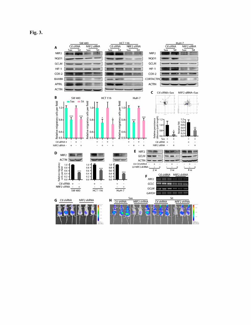
Fig. 3.
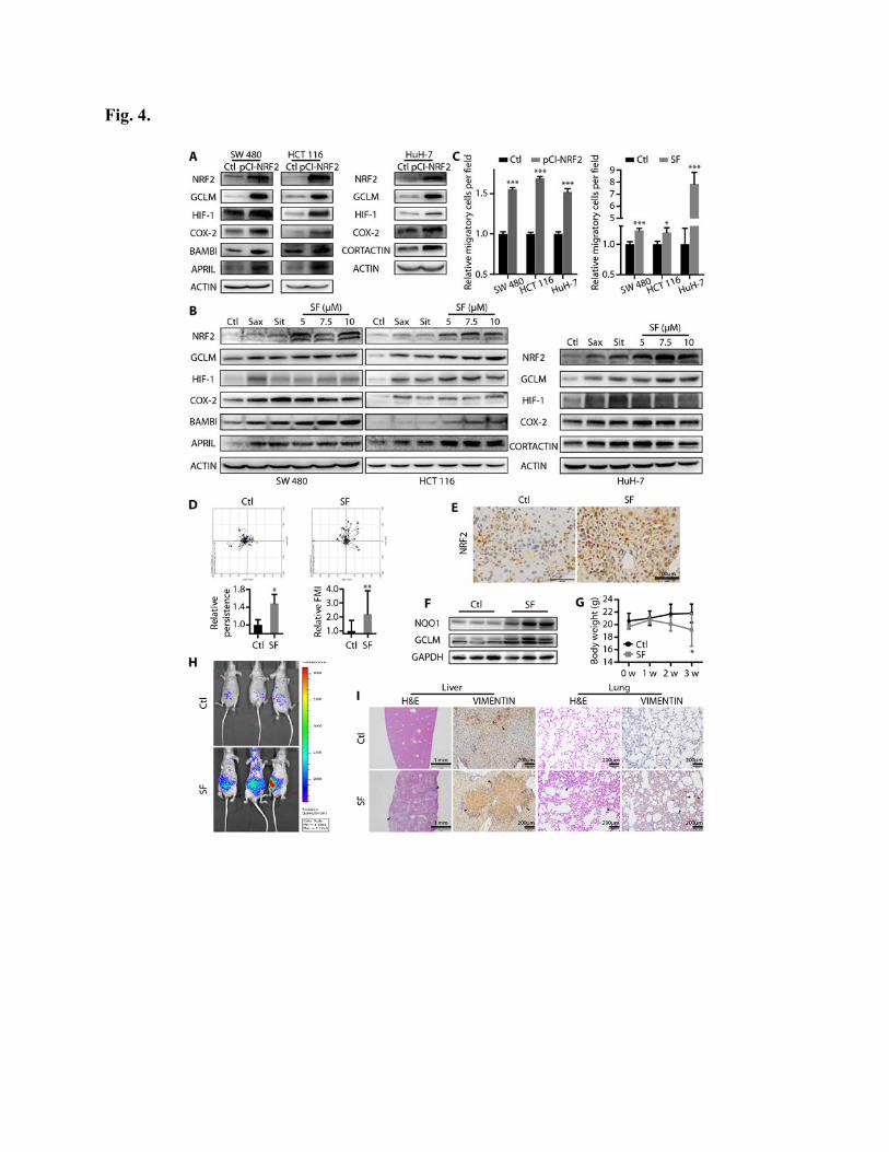
Fig. 4.

Fig. 5.

Fig. 6.