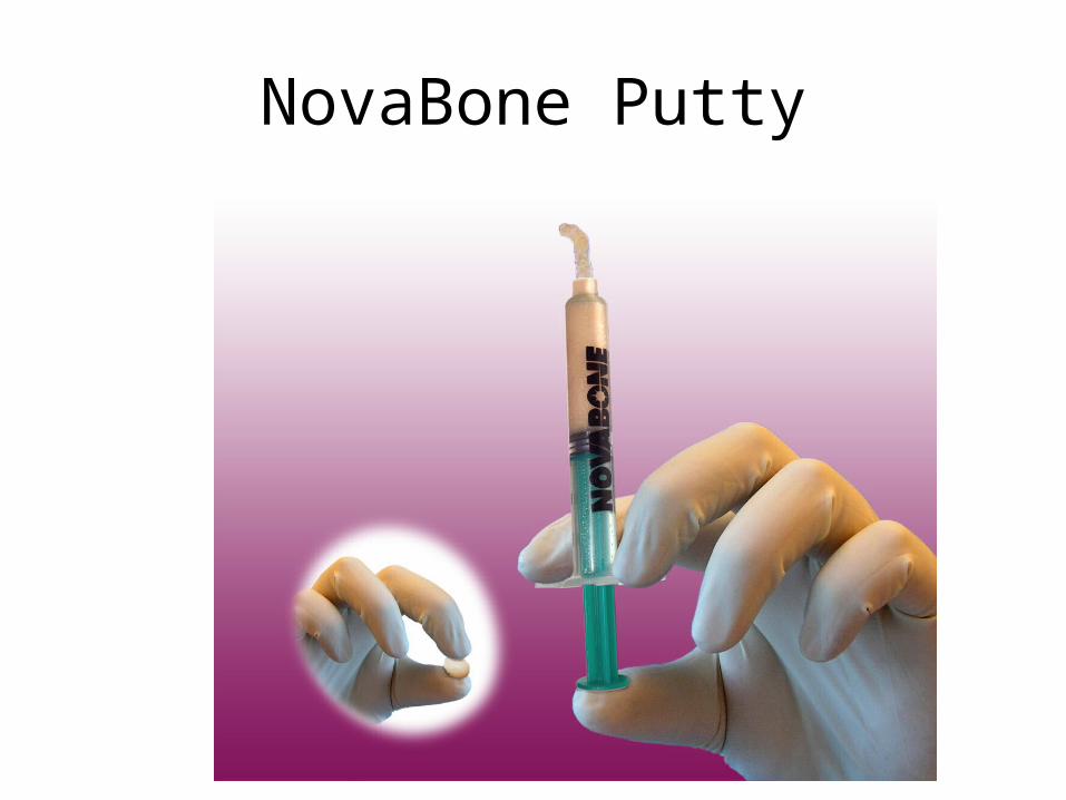
novabone putty
DESCRIPTION
NovaBone Putty. Osteostimulative Composite Putty is a new, next generation Calciumphosphosilicate bone graft material built from a bioactive glass platform with additives to improve handling and efficacy. Composition. Bioactive Glass – 69 % Glycerin – 19 % PEG – 12 %. Bioactive Phase. - PowerPoint PPT PresentationTRANSCRIPT

NovaBone Putty
Osteostimulative Composite
Putty is a new, next generation Calciumphosphosilicate bone graft material built from a bioactive glass
platform with additives to improve handling and efficacy
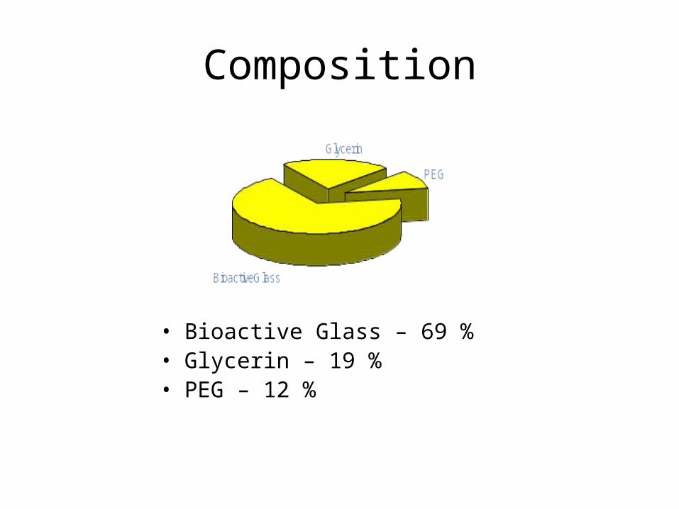
Composition
• Bioactive Glass – 69 %• Glycerin – 19 %• PEG – 12 %
Bioactive Phase
• Consists of 2 particle sizes– Phase 1: 90-710 µ Bioglass particles– Phase 2: 32-125 µ Calcium Phosphosilicate (CPS)
• Phase 2 particles provide the initial burst of calcium phosphate and also enhance the physical characteristics to improve handling
• Phase 2 resorbs at a much faster rate than the larger particle phase
Additive
• Polyethylene Glycol – 12.4%• Melts at low temperatures – 45o C• A nontoxic material that is used in food &
pharma industries• Water soluble - gets absorbed in 24-48 hrs• Additive fills spaces between the particles of
bioactive phases
– Enhances handling by giving a smooth surface to the putty
Binder - Glycerin
• Highly hydrophilic liquid• Gets absorbed into the blood stream within
48 hrs – 72 hrs• Enhances the pliability of the putty and keeps
it coherent• Used as softening agent and to keep all the
phases of putty together
Putty – Properties
• 100% Synthetic – Fully resorbable• Non toxic, non allergenic • Osteostimulative & Osteoconductive• No refrigeration required - 4 yr shelf life
(unlike other similar products)• May be combined with Autograft /Allograft• Extremely easy to use
Indications
• Immediate implant surgeries• Sinus elevation surgeries• Socket regeneration – Ridge preservation• Furcation defects & Periodontal pockets• Post apicoectomy bone regeneration• After third molar extractions• Maxillofacial surgeries – CLP, mastoid
obliteration, etc
Unique Properties
• Unique Presentation • No Mixing, thawing required• Excellent material retention at defect site• Adapts well to different shapes and surfaces
of defects• Eliminates concerns of device migration • Fool Proof
– No concerns of under/over condensation
Advantages• Overcomes the drawbacks of particulate grafts • No mixing required – package to placement• Excellent ease of handling • Great adaptability
– provides greater graft-implant surface area• Hydrophilic - blends with blood• Stays in place – no device migration
– Excellent for use in Furcation defects where retention is a problem
• Smooth surface texture– helps prevent damage to sinus membrane during sinus
elevation
Drawbacks
• Cannot be used in load bearing indications• May not be cohesive when mixed with
autograft / allograft – Overcome by “sandwich” /layer technique
• Temperature sensitive – Has to be stored & transported < 40oC– Do not leave the material in a car
Also Explore
• Composition & Properties• Mechanism of Action - Osteostimulation• Animal Studies• Surgical Video
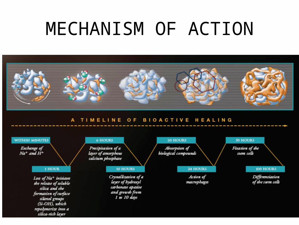
Putty – Mechanism of Action• After the clot forms – PEG, Glycerin & smaller CPS particles get
absorbed over 5-7 days
• A virtual porous network forms in the defect
• Smaller CPS particles provide the initial burst of calcium & phosphorous
• Spaces between particles permit rapid vascularization and bone in-growth
• Multiple foci of osseous regeneration areas appear throughout the defect thus enhancing the rate of bone formation
MECHANISM OF ACTION
Osteostimulation
• Osteoconduction - Passive mechanism by which bone formation occurs along the surfaces of an implant material
• Osteostimulation - “The stimulation of osteoblast proliferation and differentiation as evidenced during in vitro osteoblast cell culture studies by increased DNA content and elevated osteocalcin and alkaline phosphatase levels” FDA 2005
• Stimulates osteoblast recruitment, proliferation and differentiation at the defect site
• Increased rate of bone formation – New bone occurs throughout the defect
• Not just at the edges – osteoconduction– Multiple areas of bone formation
• Each particle reacts to generate bone• Bone formation seen within each individual particle
– Higher rate of particle resorption• Putty displays higher rate of bone regeneration than
particulate graft materials
• Does not generate bone in non-osseous sites
Osteostimulation

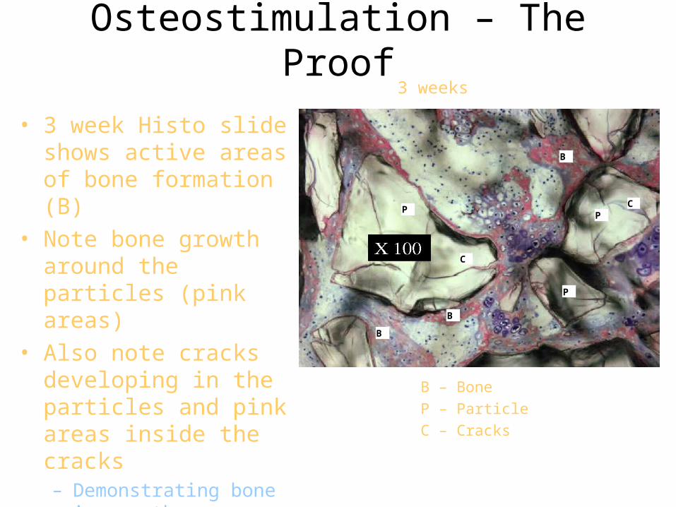
Osteostimulation – The Proof
• 3 week Histo slide shows active areas of bone formation (B)
• Note bone growth around the particles (pink areas)
• Also note cracks developing in the particles and pink areas inside the cracks– Demonstrating bone in-
growth
3 weeks
B – BoneP – ParticleC – Cracks
B
B
P
C
CP
B
P
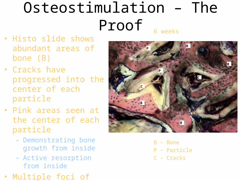
• Histo slide shows abundant areas of bone (B)
• Cracks have progressed into the center of each particle
• Pink areas seen at the center of each particle– Demonstrating bone growth
from inside– Active resorption from inside
• Multiple foci of bone formation
Osteostimulation – The Proof6 weeks
B
B
B
C
P
P
B – BoneP – ParticleC – Cracks
C
B
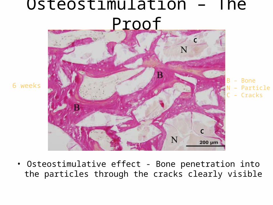
• Osteostimulative effect - Bone penetration into the particles through the cracks clearly visible
6 weeks
Osteostimulation – The Proof
C
C
B – BoneN – ParticleC – Cracks
Effect of NovaBone on bone marrow stromal cell differentiation
“.. The purpose of this study was to investigate the ability of three bioactive glasses (45S, 58S and 77S) to induce osteogenic differentiation and cell mineralization. A significant effect of the 45S and 77S bioactive materials was seen on early differentiation of the marrow stromal cells into osteoblast-like cells. 45S evidenced also the highest effect on cell mineralization …”
M Bosetti, M Cannas, Biomaterials, 26(18):3873-3879, 2005.
Osteostimulation – References
The effect of silica-containing calcium-phosphate particles on human osteoblasts in vitro.
“The results obtained demonstrated that the three new bioglasses enhanced the proliferation response of osteoblasts compared with osteoblasts alone….and did not induce stimulation of proinflammatory markers iNOS and IL-1B…”
Phan PV et al, J Biomed Mater Res A. 2003 Dec 1;67(3):1001-8
Osteostimulation – References
Bioglass 45S5 stimulates osteoblast turnover and enhances bone formation In vitro: implications and applications for bone tissue engineering.
“In conclusion, this study shows Bioglass 45S5 has the ability to stimulate the growth and osteogenic differentiation of human primary osteoblasts. These findings have the potential for tissue engineering where this bioactive glass substrate could be used as a template for the formation of bioengineered bone tissue.”
Xynos, ID, et al; Calcif Tissue Int. 2000 Oct;67(4):321-9
Osteostimulation – References
“The genetic basis for Osteogenesis stimulation by
controlled release of ionic dissolution products.”
“Gene array analysis confirmed genetic activation
controlled the osteoblast’s cell cycle to favor proliferation
and differentiation of only the cells that proceed towards
the creation of mineralized extra-cellular matrices,
osteocytes, and new bone”
Presented at the ORS 2008 Hench L, et al
Osteostimulation – References
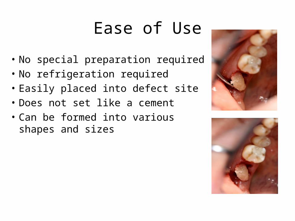
• No special preparation required• No refrigeration required• Easily placed into defect site • Does not set like a cement• Can be formed into various shapes and
sizes
Ease of Use
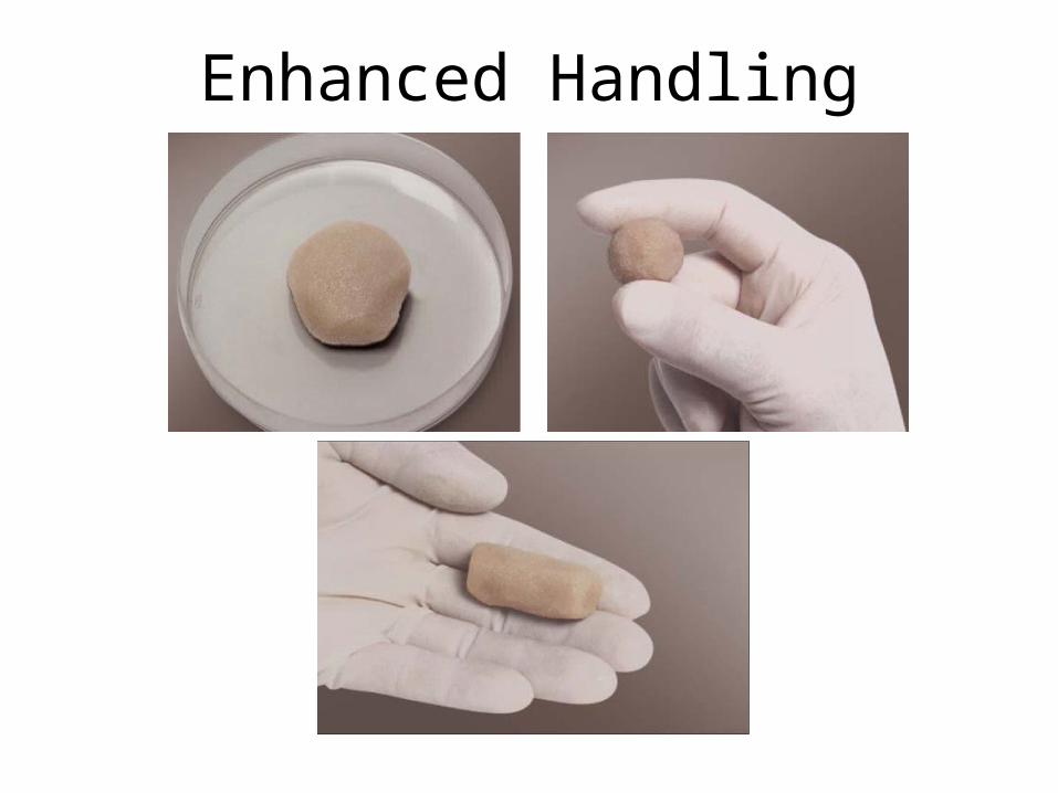
Enhanced Handling
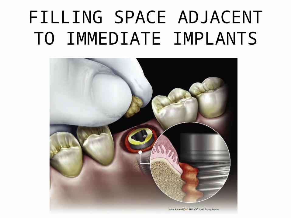
FILLING SPACE ADJACENT TO IMMEDIATE IMPLANTS
Animal Study - 1
• Rabbit study – distal femur• 5 mm diameter x 10 mm depth • Approx 0.3 cc of putty was used• Histological sections were obtained at 6 & 12
weeks
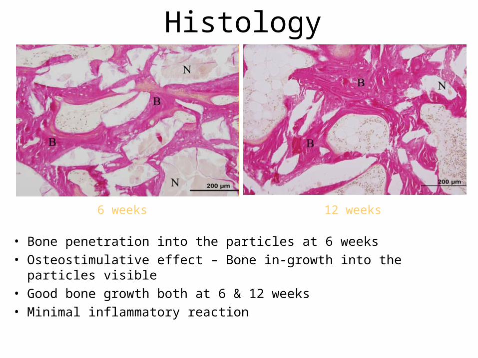
Histology
• Bone penetration into the particles at 6 weeks• Osteostimulative effect – Bone in-growth into the particles visible• Good bone growth both at 6 & 12 weeks• Minimal inflammatory reaction
6 weeks 12 weeks
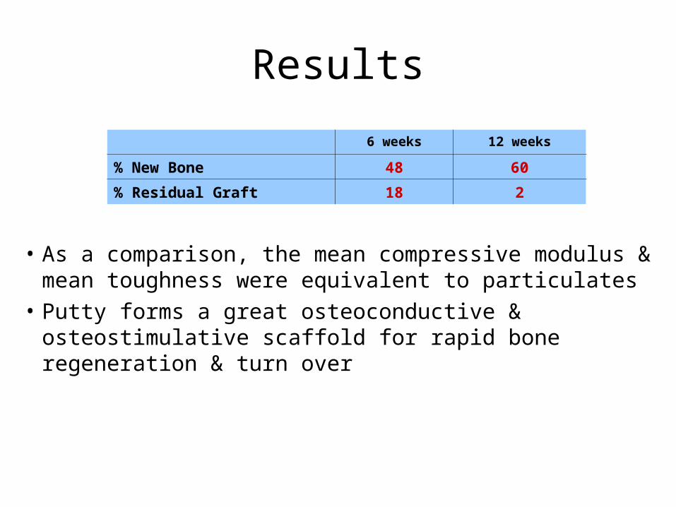
Results
• As a comparison, the mean compressive modulus & mean toughness were equivalent to particulates
• Putty forms a great osteoconductive & osteostimulative scaffold for rapid bone regeneration & turn over
6 weeks 12 weeks
% New Bone 48 60
% Residual Graft 18 2
Animal Study - 2
• Sheep study – Intravertebral Defects• 10 mm diameter x 15 mm depth • Particulate vs. Putty vs. Empty• Histological sections were obtained at 6 & 12
weeks
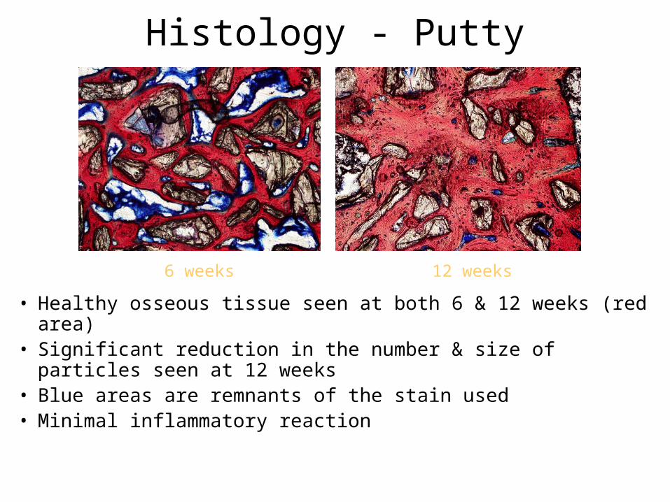
Histology - Putty
• Healthy osseous tissue seen at both 6 & 12 weeks (red area)• Significant reduction in the number & size of particles seen at
12 weeks• Blue areas are remnants of the stain used • Minimal inflammatory reaction
6 weeks 12 weeks
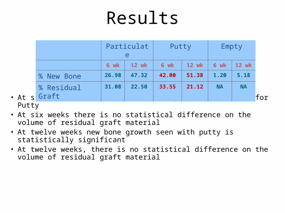
Results
• At six weeks % new bone growth is significantly higher for Putty • At six weeks there is no statistical difference on the volume of residual
graft material• At twelve weeks new bone growth seen with putty is statistically
significant • At twelve weeks, there is no statistical difference on the volume of
residual graft material
Particulate Putty Empty
6 wk 12 wk 6 wk 12 wk 6 wk 12 wk
% New Bone 26.98 47.32 42.00 51.38 1.20 5.18
% Residual Graft 31.08 22.50 33.55 21.12 NA NA
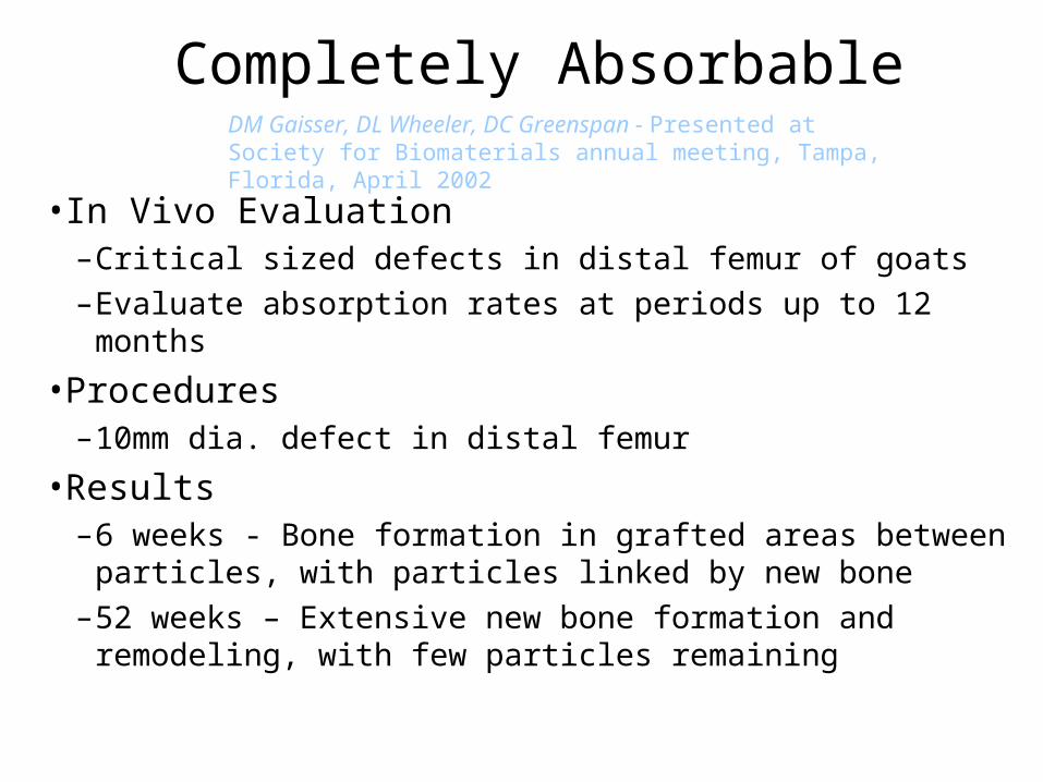
Completely Absorbable
• In Vivo Evaluation–Critical sized defects in distal femur of goats–Evaluate absorption rates at periods up to 12 months
• Procedures–10mm dia. defect in distal femur
• Results –6 weeks - Bone formation in grafted areas between particles,
with particles linked by new bone–52 weeks – Extensive new bone formation and remodeling,
with few particles remaining
DM Gaisser, DL Wheeler, DC Greenspan - Presented at Society for Biomaterials annual meeting, Tampa, Florida, April 2002
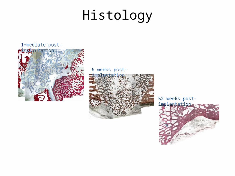
6 weeks post-implantation6 weeks post-implantation
52 weeks post-implantation52 weeks post-implantation
Histology
Immediate post-implantationImmediate post-implantation
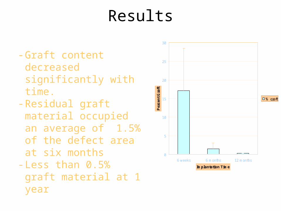
- Graft content decreased significantly with time.
- Residual graft material occupied an average of 1.5% of the defect area at six months
- Less than 0.5% graft material at 1 year
0
5
10
15
20
25
30
6 weeks 6 months 12 months
Implantation Time
Per
cen
t G
raft
% graft
Results
Human Clinical Cases
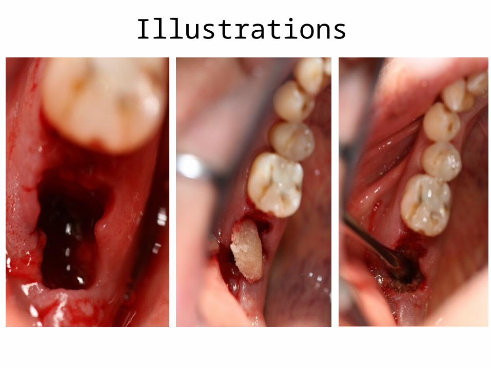
Illustrations
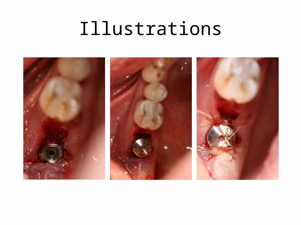
Illustrations
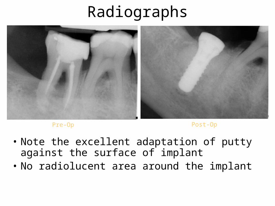
Radiographs
• Note the excellent adaptation of putty against the surface of implant
• No radiolucent area around the implant
Pre-Op Post-Op
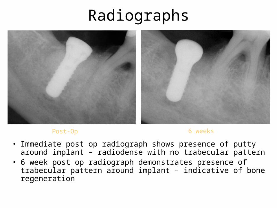
Radiographs
• Immediate post op radiograph shows presence of putty around implant – radiodense with no trabecular pattern
• 6 week post op radiograph demonstrates presence of trabecular pattern around implant – indicative of bone regeneration
6 weeksPost-Op
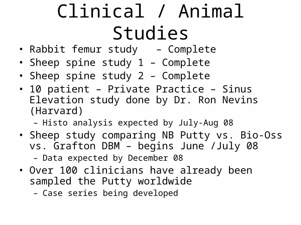
Clinical / Animal Studies• Rabbit femur study – Complete • Sheep spine study 1 – Complete• Sheep spine study 2 – Complete• 10 patient – Private Practice – Sinus Elevation study done by
Dr. Ron Nevins (Harvard)– Histo analysis expected by July-Aug 08
• Sheep study comparing NB Putty vs. Bio-Oss vs. Grafton DBM – begins June /July 08 – Data expected by December 08
• Over 100 clinicians have already been sampled the Putty worldwide– Case series being developed
Osteostimulation – The Proof
• 3 week Histo slide shows active areas of bone formation (B)
• Note bone growth around the particles (pink areas)
• Also note cracks developing in the particles and pink areas inside the cracks– Demonstrating bone in-
growth
3 weeks
B – BoneP – ParticleC – Cracks
B
B
P
C
CP
B
P
Case studies from Craniofacial and Orthopedic Surgeries
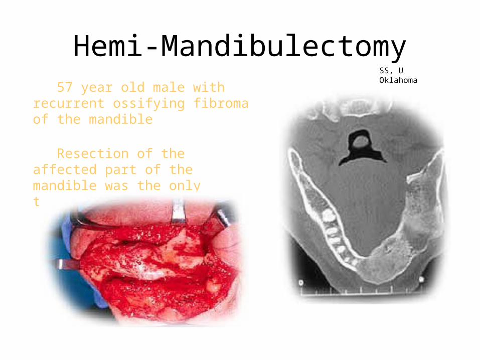
57 year old male with recurrent ossifying fibroma of the mandible
Resection of the affected part of the mandible was the only treatment
SS, U Oklahoma
Hemi-Mandibulectomy
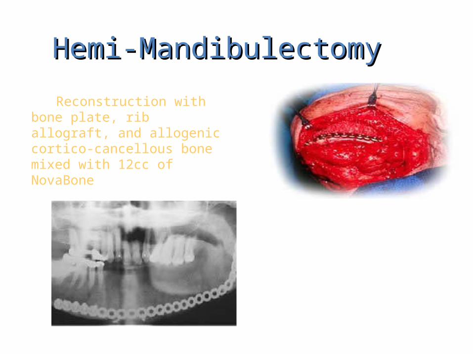
Reconstruction with bone plate, rib allograft, and allogenic cortico-cancellous bone mixed with 12cc of NovaBone
Hemi-MandibulectomyHemi-Mandibulectomy
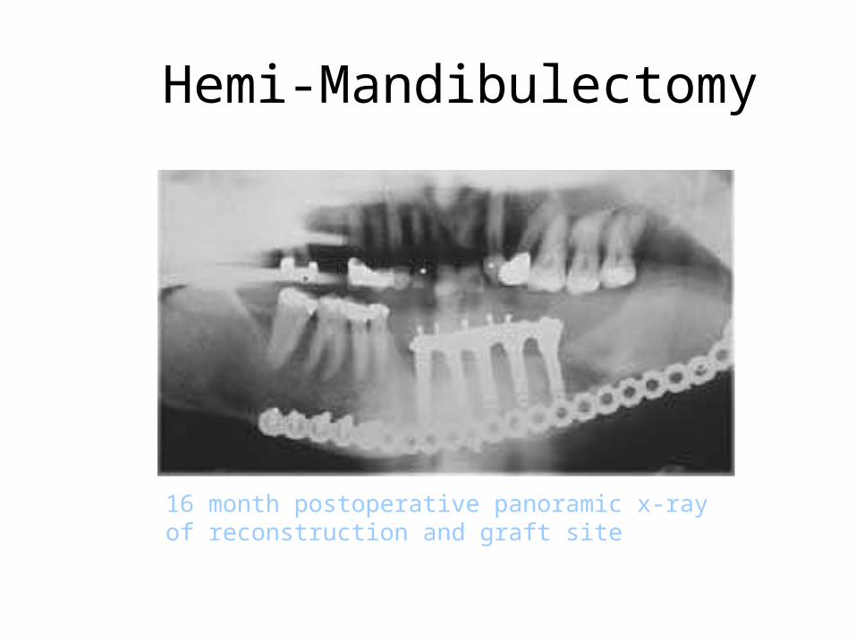
16 month postoperative panoramic x-ray of reconstruction and graft site
Hemi-Mandibulectomy
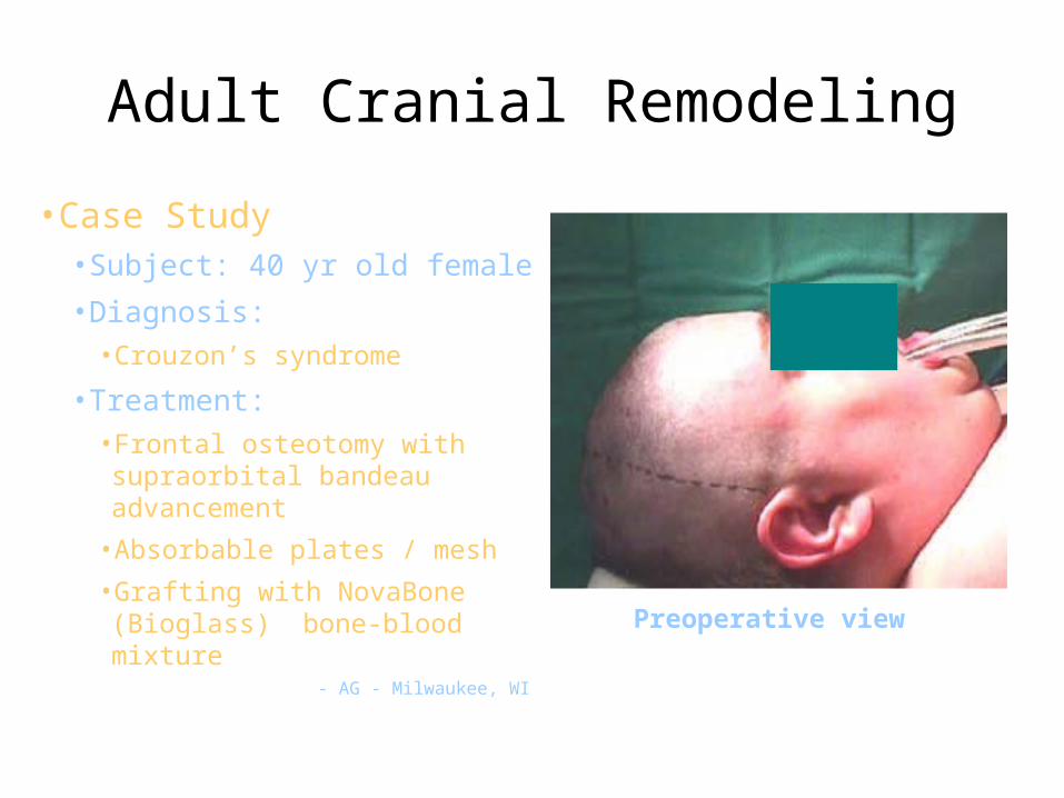
Adult Cranial Remodeling
Preoperative view
•Case Study•Subject: 40 yr old female•Diagnosis:
•Crouzon’s syndrome
•Treatment: •Frontal osteotomy with supraorbital
bandeau advancement•Absorbable plates / mesh•Grafting with NovaBone (Bioglass)
bone-blood mixture- AG - Milwaukee, WI
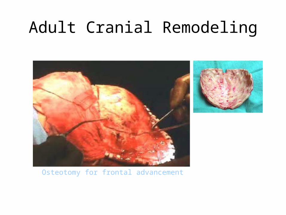
Adult Cranial Remodeling
Osteotomy for frontal advancement
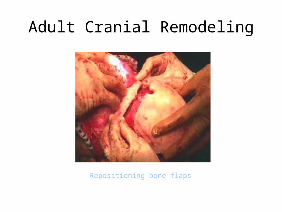
Adult Cranial Remodeling
Repositioning bone flaps
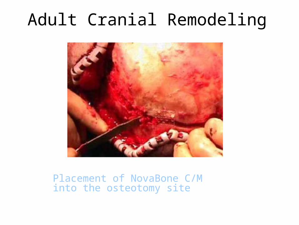
Adult Cranial Remodeling
Placement of NovaBone C/M into the osteotomy site
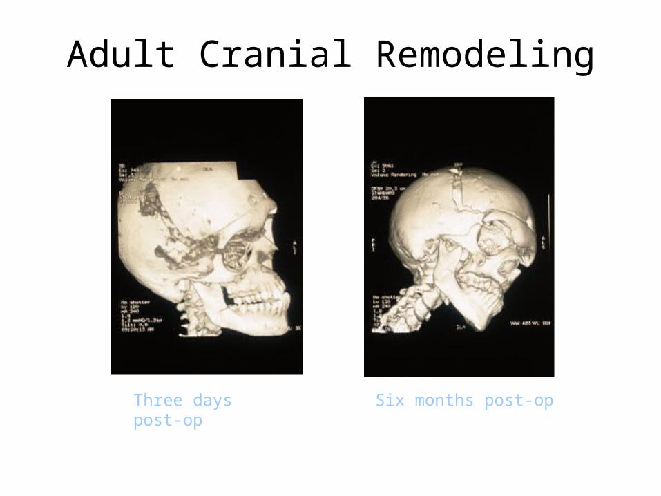
Adult Cranial Remodeling
Three days post-op Six months post-op
NovaBone Putty
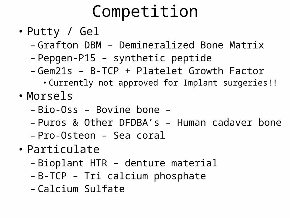
Competition• Putty / Gel
– Grafton DBM – Demineralized Bone Matrix – Pepgen-P15 – synthetic peptide– Gem21s – B-TCP + Platelet Growth Factor
• Currently not approved for Implant surgeries!!
• Morsels– Bio-Oss – Bovine bone – Biggest Competitor– Puros & Other DFDBA’s – Human cadaver bone– Pro-Osteon – Sea coral
• Particulate– Bioplant HTR – denture material– Β-TCP – Tri calcium phosphate– Calcium Sulfate
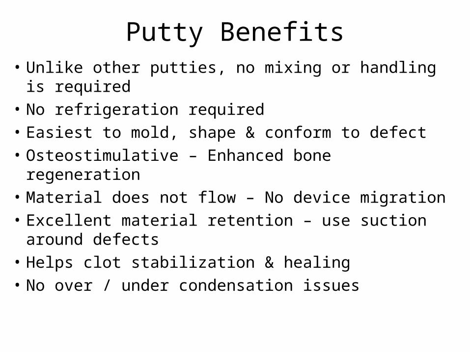
Putty Benefits• Unlike other putties, no mixing or handling is required• No refrigeration required• Easiest to mold, shape & conform to defect • Osteostimulative – Enhanced bone regeneration• Material does not flow – No device migration• Excellent material retention – use suction around
defects• Helps clot stabilization & healing• No over / under condensation issues
Putty vs. Bio-Oss
• No clinical comparison yet• Animal study in progress – sheep study• Bio-Oss – non resorbable & bovine derived• Putty has high resorption and enhanced bone
regeneration• Bio-Oss is only Osteoconductive while Putty is
Osteostimulative
Putty vs. Particulate• Putty can be used in unique ways and
indications• Putty is the easiest to use around implants
during immediate implant surgeries as it gives good adaptation against the implant
• Putty is ideal for furcation defects where most particulate materials have retention issues
• Also in ridge augmentation surgeries in the mandible, where putty can be molded to the desired shape