non-infectious complications of peritoneal dialysis ...€¦ · non-infectious complications of...
TRANSCRIPT
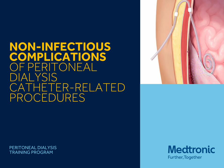
NON-INFECTIOUS COMPLICATIONS OF PERITONEAL DIALYSIS CATHETER-RELATED PROCEDURES
PERITONEAL DIALYSIS TRAINING PROGRAM
2The content of this program was developed for Medtronic to be distributed to customers for training purposes only. Do not further copy or distribute.
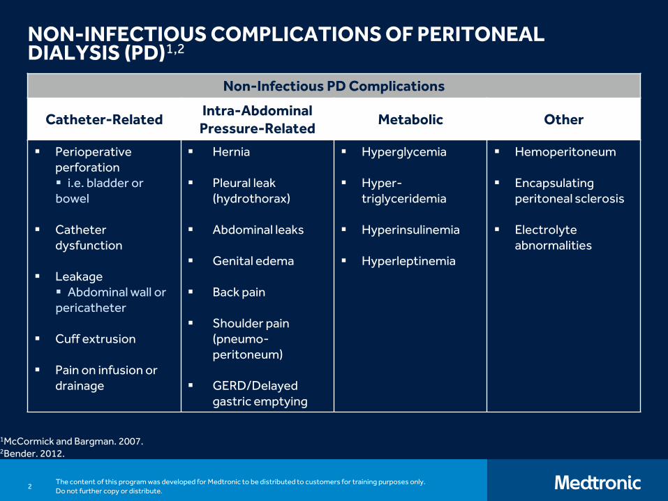
NON-INFECTIOUS COMPLICATIONS OF PERITONEAL DIALYSIS (PD)1,2
1McCormick and Bargman. 2007.2Bender. 2012.
Non-Infectious PD Complications
Catheter-RelatedIntra-Abdominal
Pressure-RelatedMetabolic Other
▪ Perioperative perforation ▪ i.e. bladder or bowel
▪ Catheter dysfunction
▪ Leakage▪ Abdominal wall or pericatheter
▪ Cuff extrusion
▪ Pain on infusion or drainage
▪ Hernia
▪ Pleural leak (hydrothorax)
▪ Abdominal leaks
▪ Genital edema
▪ Back pain
▪ Shoulder pain (pneumo-peritoneum)
▪ GERD/Delayed gastric emptying
▪ Hyperglycemia
▪ Hyper-triglyceridemia
▪ Hyperinsulinemia
▪ Hyperleptinemia
▪ Hemoperitoneum
▪ Encapsulating peritoneal sclerosis
▪ Electrolyte abnormalities
3The content of this program was developed for Medtronic to be distributed to customers for training purposes only. Do not further copy or distribute.
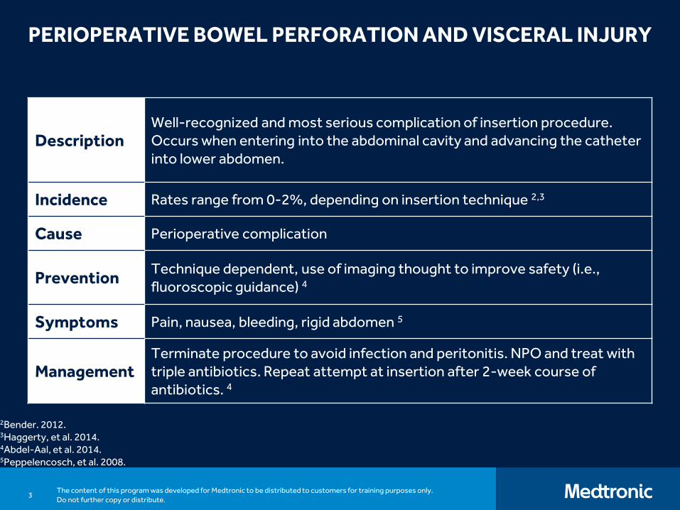
PERIOPERATIVE BOWEL PERFORATION AND VISCERAL INJURY
2Bender. 2012.3Haggerty, et al. 2014.4Abdel-Aal, et al. 2014.5Peppelencosch, et al. 2008.
DescriptionWell-recognized and most serious complication of insertion procedure. Occurs when entering into the abdominal cavity and advancing the catheter into lower abdomen.
Incidence Rates range from 0-2%, depending on insertion technique 2,3
Cause Perioperative complication
PreventionTechnique dependent, use of imaging thought to improve safety (i.e., fluoroscopic guidance) 4
Symptoms Pain, nausea, bleeding, rigid abdomen 5
ManagementTerminate procedure to avoid infection and peritonitis. NPO and treat with triple antibiotics. Repeat attempt at insertion after 2-week course of antibiotics. 4
4The content of this program was developed for Medtronic to be distributed to customers for training purposes only. Do not further copy or distribute.
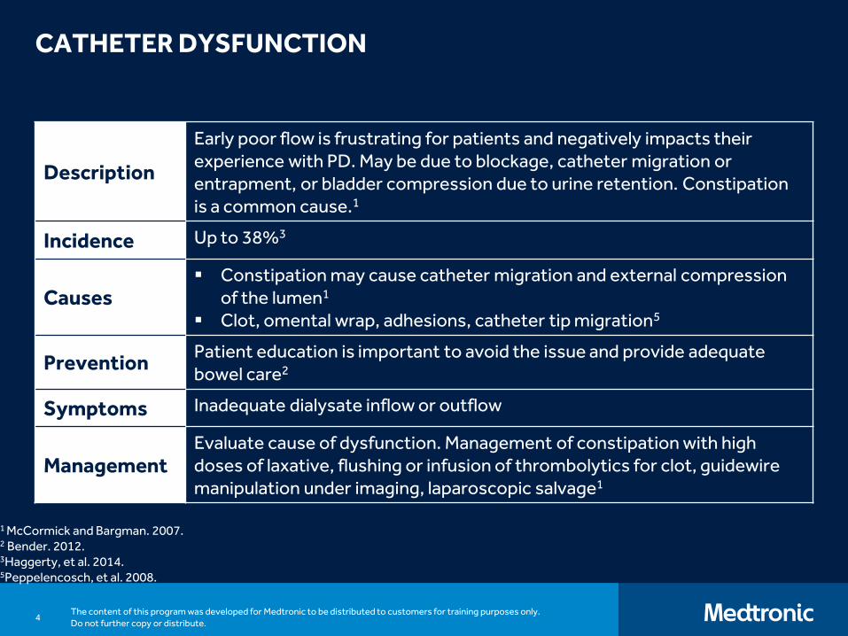
CATHETER DYSFUNCTION
1 McCormick and Bargman. 2007.2 Bender. 2012.3Haggerty, et al. 2014.5Peppelencosch, et al. 2008.
Description
Early poor flow is frustrating for patients and negatively impacts their experience with PD. May be due to blockage, catheter migration or entrapment, or bladder compression due to urine retention. Constipation is a common cause.1
Incidence Up to 38%3
Causes▪ Constipation may cause catheter migration and external compression
of the lumen1
▪ Clot, omental wrap, adhesions, catheter tip migration5
PreventionPatient education is important to avoid the issue and provide adequate bowel care2
Symptoms Inadequate dialysate inflow or outflow
ManagementEvaluate cause of dysfunction. Management of constipation with high doses of laxative, flushing or infusion of thrombolytics for clot, guidewire manipulation under imaging, laparoscopic salvage1
5The content of this program was developed for Medtronic to be distributed to customers for training purposes only. Do not further copy or distribute.
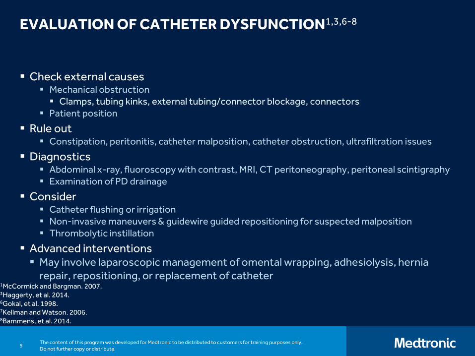
EVALUATION OF CATHETER DYSFUNCTION1,3,6-8
▪ Check external causes▪ Mechanical obstruction
▪ Clamps, tubing kinks, external tubing/connector blockage, connectors▪ Patient position
▪ Rule out▪ Constipation, peritonitis, catheter malposition, catheter obstruction, ultrafiltration issues
▪ Diagnostics▪ Abdominal x-ray, fluoroscopy with contrast, MRI, CT peritoneography, peritoneal scintigraphy▪ Examination of PD drainage
▪ Consider▪ Catheter flushing or irrigation▪ Non-invasive maneuvers & guidewire guided repositioning for suspected malposition▪ Thrombolytic instillation
▪ Advanced interventions▪ May involve laparoscopic management of omental wrapping, adhesiolysis, hernia
repair, repositioning, or replacement of catheter1McCormick and Bargman. 2007.3Haggerty, et al. 2014.6Gokal, et al. 1998.7Kellman and Watson. 2006.8Bammens, et al. 2014.

6The content of this program was developed for Medtronic to be distributed to customers for training purposes only. Do not further copy or distribute.
TYPES AND LOCATIONS OF LEAKAGE9
▪ Pericatheter
▪ Prior surgical incision site
▪ Patent processus vaginalis
▪ Non-specific areas of peritoneum
9Leblanc, et al. 2001.
7The content of this program was developed for Medtronic to be distributed to customers for training purposes only. Do not further copy or distribute.
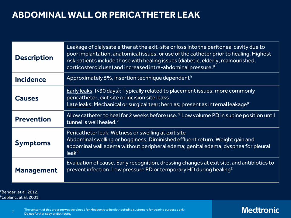
ABDOMINAL WALL OR PERICATHETER LEAK
2Bender, et al. 2012.9Leblanc, et al. 2001.
Description
Leakage of dialysate either at the exit-site or loss into the peritoneal cavity due to poor implantation, anatomical issues, or use of the catheter prior to healing. Highest risk patients include those with healing issues (diabetic, elderly, malnourished, corticosteroid use) and increased intra-abdominal pressure.9
Incidence Approximately 5%, insertion technique dependent9
CausesEarly leaks: (<30 days): Typically related to placement issues; more commonly pericatheter, exit site or incision site leaksLate leaks: Mechanical or surgical tear; hernias; present as internal leakage9
PreventionAllow catheter to heal for 2 weeks before use. 9 Low volume PD in supine position until tunnel is well healed.2
Symptoms
Pericatheter leak: Wetness or swelling at exit siteAbdominal swelling or bogginess, Diminished effluent return, Weight gain and abdominal wall edema without peripheral edema; genital edema, dyspnea for pleural leak9
ManagementEvaluation of cause. Early recognition, dressing changes at exit site, and antibiotics to prevent infection. Low pressure PD or temporary HD during healing2
8The content of this program was developed for Medtronic to be distributed to customers for training purposes only. Do not further copy or distribute.
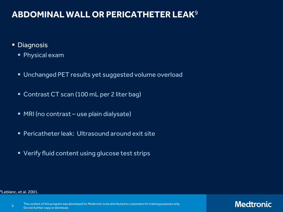
ABDOMINAL WALL OR PERICATHETER LEAK9
▪ Diagnosis
▪ Physical exam
▪ Unchanged PET results yet suggested volume overload
▪ Contrast CT scan (100 mL per 2 liter bag)
▪ MRI (no contrast – use plain dialysate)
▪ Pericatheter leak: Ultrasound around exit site
▪ Verify fluid content using glucose test strips
9Leblanc, et al. 2001.
9The content of this program was developed for Medtronic to be distributed to customers for training purposes only. Do not further copy or distribute.
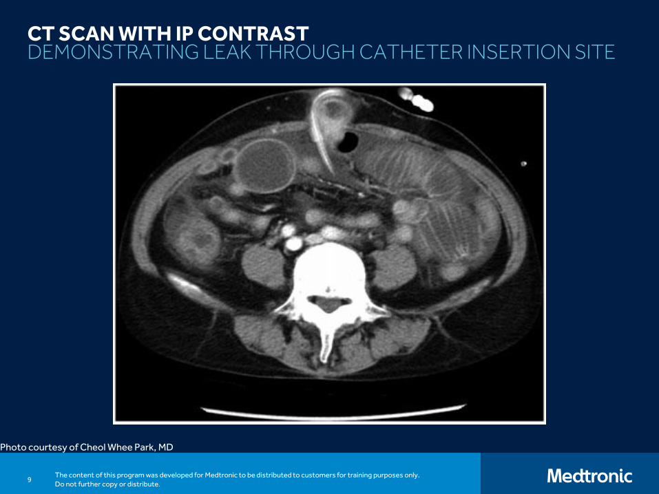
CT SCAN WITH IP CONTRASTDEMONSTRATING LEAK THROUGH CATHETER INSERTION SITE
Photo courtesy of Cheol Whee Park, MD
10The content of this program was developed for Medtronic to be distributed to customers for training purposes only. Do not further copy or distribute.
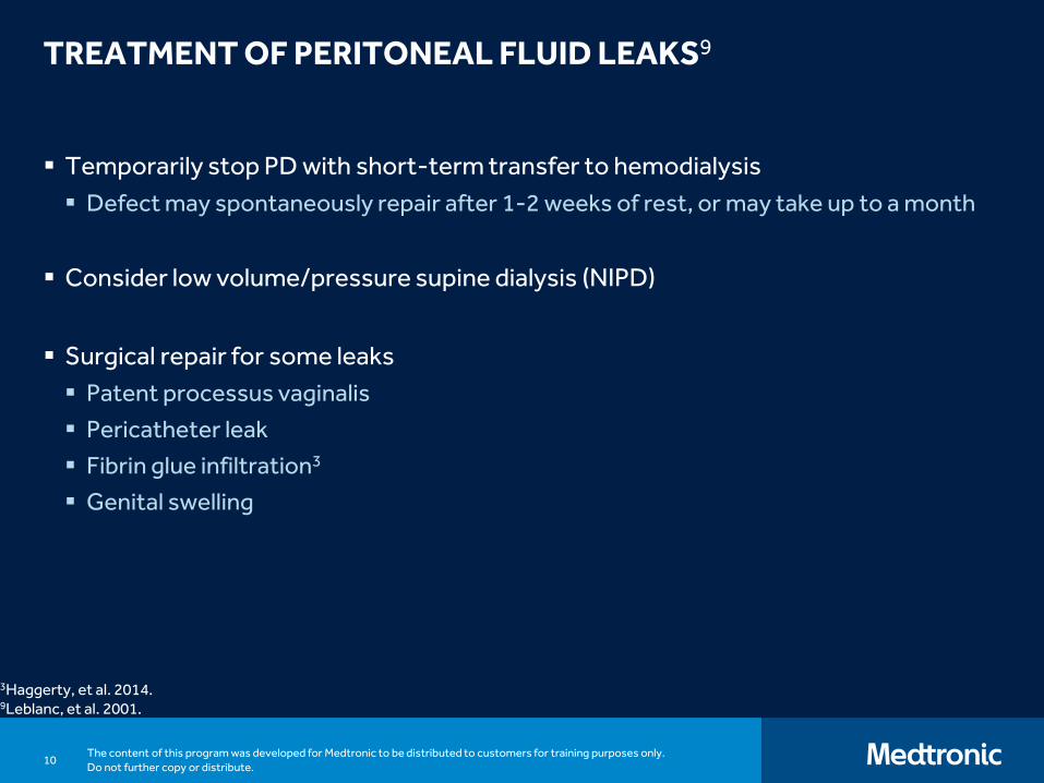
TREATMENT OF PERITONEAL FLUID LEAKS9
▪ Temporarily stop PD with short-term transfer to hemodialysis
▪ Defect may spontaneously repair after 1-2 weeks of rest, or may take up to a month
▪ Consider low volume/pressure supine dialysis (NIPD)
▪ Surgical repair for some leaks
▪ Patent processus vaginalis
▪ Pericatheter leak
▪ Fibrin glue infiltration3
▪ Genital swelling
3Haggerty, et al. 2014.9Leblanc, et al. 2001.
11The content of this program was developed for Medtronic to be distributed to customers for training purposes only. Do not further copy or distribute.
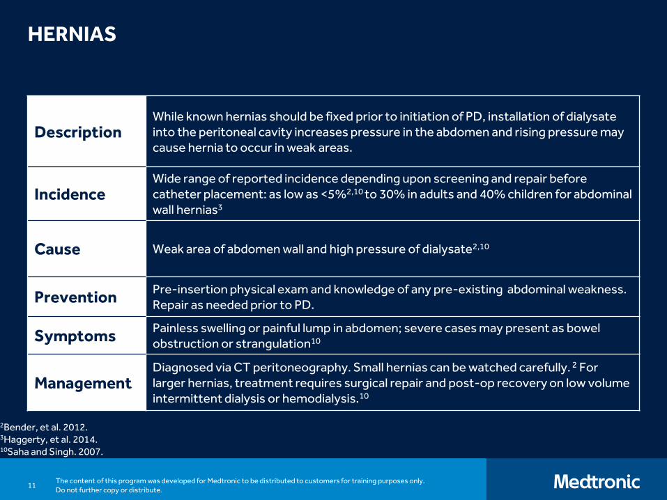
HERNIAS
2Bender, et al. 2012.3Haggerty, et al. 2014.10Saha and Singh. 2007.
DescriptionWhile known hernias should be fixed prior to initiation of PD, installation of dialysate into the peritoneal cavity increases pressure in the abdomen and rising pressure may cause hernia to occur in weak areas.
IncidenceWide range of reported incidence depending upon screening and repair before catheter placement: as low as <5%2,10 to 30% in adults and 40% children for abdominal wall hernias3
Cause Weak area of abdomen wall and high pressure of dialysate2,10
PreventionPre-insertion physical exam and knowledge of any pre-existing abdominal weakness. Repair as needed prior to PD.
SymptomsPainless swelling or painful lump in abdomen; severe cases may present as bowelobstruction or strangulation10
ManagementDiagnosed via CT peritoneography. Small hernias can be watched carefully. 2 For larger hernias, treatment requires surgical repair and post-op recovery on low volume intermittent dialysis or hemodialysis.10
12The content of this program was developed for Medtronic to be distributed to customers for training purposes only. Do not further copy or distribute.
TYPES OF HERNIAS10
▪ Incisional
▪ Inguinal
▪ Umbilical
▪ Epigastric
▪ Ventral
▪ Obturator
▪ Foramen of Morgagni
10Saha and Singh. 2007.

13The content of this program was developed for Medtronic to be distributed to customers for training purposes only. Do not further copy or distribute.
HERNIASRISK FACTORS9,10
▪ Multiple pregnancies
▪ Elderly males
▪ Previous hernia repair
▪ Previous abdominal surgery
▪ Abdominal obesity
▪ Midline incision for PD catheter placement
9Leblanc, et al. 2001.10Saha and Singh. 2007.

14The content of this program was developed for Medtronic to be distributed to customers for training purposes only. Do not further copy or distribute.
HERNIASCOMPLICATIONS OF HERNIAS10
Hernia
Bowel Incarceration and Strangulation
Transmural Leakage of Bacteria
Peritonitis
10Saha and Singh. 2007.
15The content of this program was developed for Medtronic to be distributed to customers for training purposes only. Do not further copy or distribute.
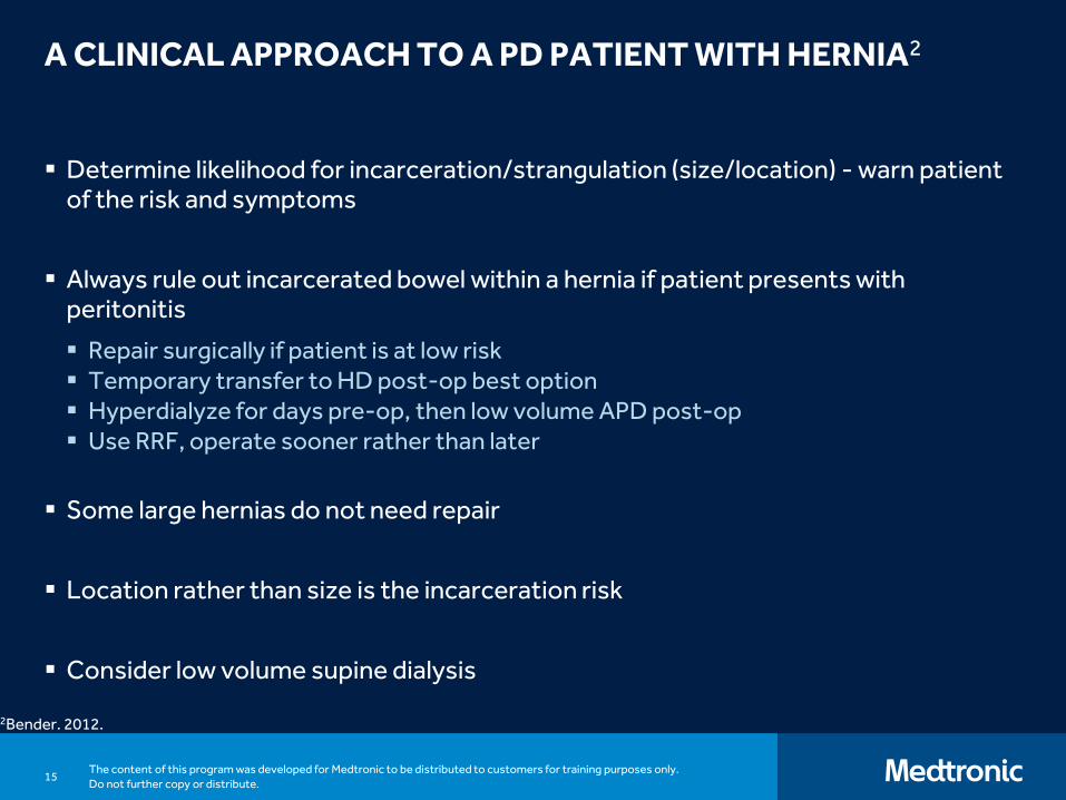
A CLINICAL APPROACH TO A PD PATIENT WITH HERNIA2
▪ Determine likelihood for incarceration/strangulation (size/location) - warn patient of the risk and symptoms
▪ Always rule out incarcerated bowel within a hernia if patient presents with peritonitis
▪ Repair surgically if patient is at low risk▪ Temporary transfer to HD post-op best option▪ Hyperdialyze for days pre-op, then low volume APD post-op ▪ Use RRF, operate sooner rather than later
▪ Some large hernias do not need repair
▪ Location rather than size is the incarceration risk
▪ Consider low volume supine dialysis
2Bender. 2012.
16The content of this program was developed for Medtronic to be distributed to customers for training purposes only. Do not further copy or distribute.
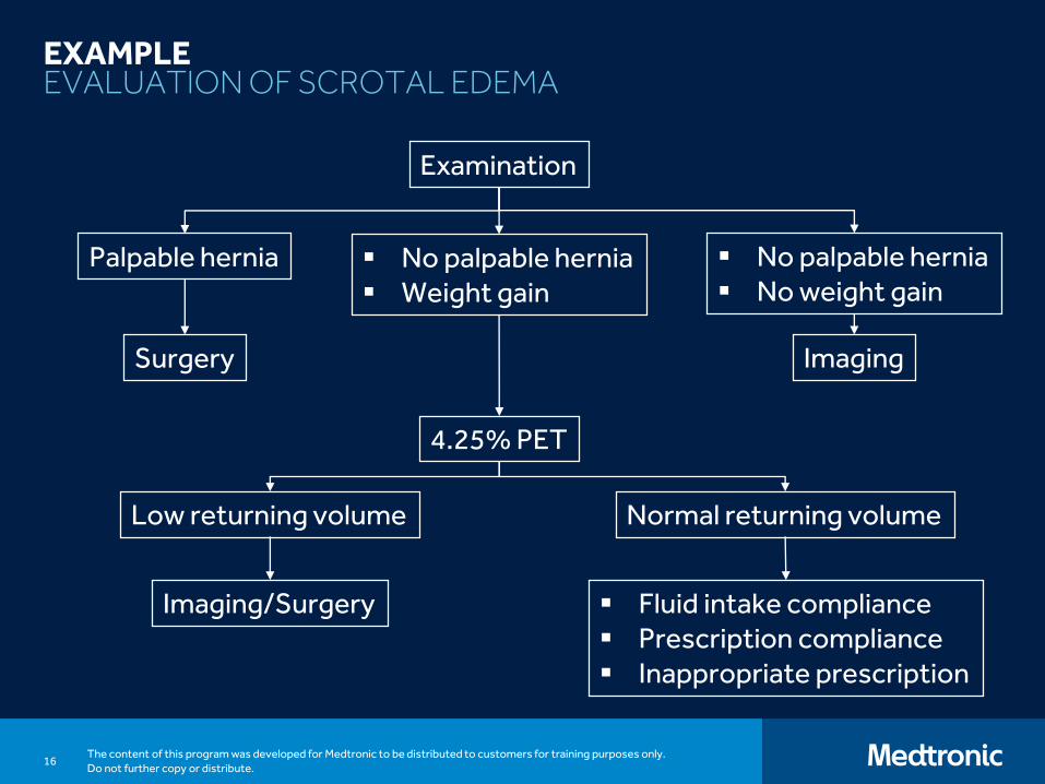
EXAMPLEEVALUATION OF SCROTAL EDEMA
Examination
Palpable hernia ▪ No palpable hernia▪ Weight gain
▪ No palpable hernia▪ No weight gain
4.25% PET
ImagingSurgery
Low returning volume Normal returning volume
Imaging/Surgery ▪ Fluid intake compliance▪ Prescription compliance▪ Inappropriate prescription
17The content of this program was developed for Medtronic to be distributed to customers for training purposes only. Do not further copy or distribute.
PAIN DURING INFUSION OR DRAINAGE
DescriptionInflow or outflow pain that occurs during exchange of dialysate, especially common during the early use of the catheter.2
Causes
▪ pH of dialysate: 5.0-5.5 infusion into the abdomen may cause discomfort. ▪ Solution too warm or too cold may increase discomfort.▪ May be due to placement of the catheter too deep in the abdomen or position of
the catheter tip against pelvic wall, bladder or rectum.4
PreventionPlacement is critical as catheters that are placed too deep in the pelvis might produce infusion or drain pain.4
Symptoms Discomfort during inflow or outflow of dialysate
Management
▪ Rule out peritonitis. ▪ Use of Tidal PD (75% to 90% tidal volume, cycler best) or allow time and
encourage patient to keep trying as pain may resolve on its own.2
▪ Consider addition of few mL of NaHCO3 to dialysate if persistent (can increase peritonitis rate) and ensure dialysate is body temperature.
▪ Extreme cases may require replacement of the catheter.4
2Bender, et al. 2006.4Abdel-Aal, et al. 2014.
18The content of this program was developed for Medtronic to be distributed to customers for training purposes only. Do not further copy or distribute.
HYDROTHORAX
DescriptionThe presence of peritoneal dialysis fluid in the pleural cavity, typically early in treatment as it is most frequently due to congenital defects of muscle fibers in the diaphragm1,10
Incidence▪ Range varies from 1.6 to 10%2,10
▪ More common in females10
CauseMovement of dialysate, under increased intra-abdominal pressure, from peritoneal to pleural cavity through congenital or acquired defects in the diaphragm2
Symptoms
▪ Shortness of breath, decrease in effluent return, pain; most common on the right side11
▪ Pleural fluid: transudate, high sugar concentration, D-lactate present, LDH level low
Management
Thoracentesis for dyspnea or minimally invasive techniques such as pleurodesis or more invasive approaches using video-assisted thoracoscopic surgery (VATS). The treatment goal is to seal the porous diaphragmatic vent and allow full separation of the peritoneal cavity from the pleural cavity. Low pressure PD or stop PD and transition to temporary hemodialysis. Resumption of PD after repair is possible.11
1McCormick and Bargman. 2007.2Bender. 2012.10Saha and Singh. 2007.11Guest. 2015.
19The content of this program was developed for Medtronic to be distributed to customers for training purposes only. Do not further copy or distribute.
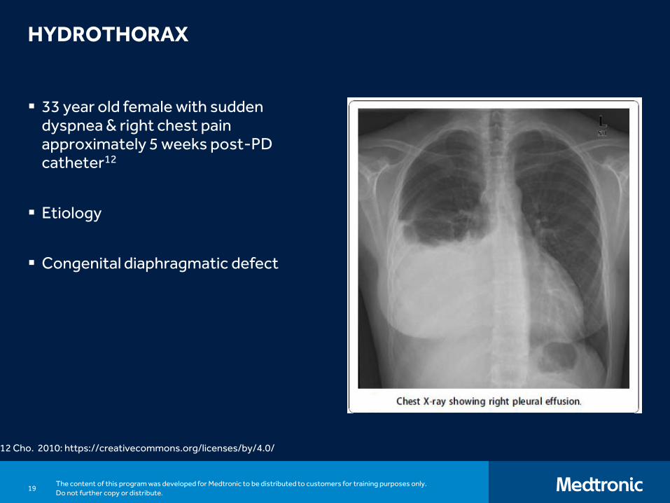
HYDROTHORAX
▪ 33 year old female with sudden dyspnea & right chest pain approximately 5 weeks post-PD catheter12
▪ Etiology
▪ Congenital diaphragmatic defect
12 Cho. 2010: https://creativecommons.org/licenses/by/4.0/
20The content of this program was developed for Medtronic to be distributed to customers for training purposes only. Do not further copy or distribute.
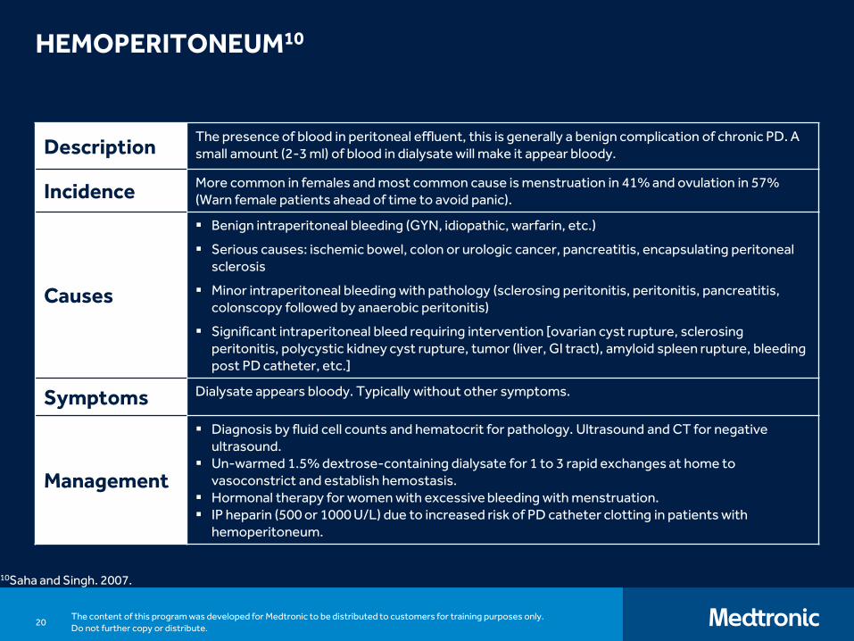
HEMOPERITONEUM10
10Saha and Singh. 2007.
DescriptionThe presence of blood in peritoneal effluent, this is generally a benign complication of chronic PD. A small amount (2-3 ml) of blood in dialysate will make it appear bloody.
IncidenceMore common in females and most common cause is menstruation in 41% and ovulation in 57% (Warn female patients ahead of time to avoid panic).
Causes
▪ Benign intraperitoneal bleeding (GYN, idiopathic, warfarin, etc.)
▪ Serious causes: ischemic bowel, colon or urologic cancer, pancreatitis, encapsulating peritoneal sclerosis
▪ Minor intraperitoneal bleeding with pathology (sclerosing peritonitis, peritonitis, pancreatitis, colonscopy followed by anaerobic peritonitis)
▪ Significant intraperitoneal bleed requiring intervention [ovarian cyst rupture, sclerosing peritonitis, polycystic kidney cyst rupture, tumor (liver, GI tract), amyloid spleen rupture, bleeding post PD catheter, etc.]
Symptoms Dialysate appears bloody. Typically without other symptoms.
Management
▪ Diagnosis by fluid cell counts and hematocrit for pathology. Ultrasound and CT for negative ultrasound.
▪ Un-warmed 1.5% dextrose-containing dialysate for 1 to 3 rapid exchanges at home to vasoconstrict and establish hemostasis.
▪ Hormonal therapy for women with excessive bleeding with menstruation. ▪ IP heparin (500 or 1000 U/L) due to increased risk of PD catheter clotting in patients with
hemoperitoneum.
21The content of this program was developed for Medtronic to be distributed to customers for training purposes only. Do not further copy or distribute.
REFERENCES
1. McCormick BB, Bargman JM. Noninfectious complications of peritoneal dialysis: implications for patient and technique survival. J Am Soc Nephrol. 2007;18(12):3023-5.
2. Bender FH. Avoiding harm in peritoneal dialysis patients. Advances in Chronic Kidney Disease. 2012;19(3):171-8.
3. Haggerty S, Roth S, Walsh D, et al. Guidelines for laparoscopic peritoneal dialysis access surgery. Society of American Gastrointestinal and Endoscopic Surgeons (SAGES) Guidelines Committee. 2014.
4. Abdel-Aal A, Dybbro P, Hathaway P, Guest S, Neuwirth M, Krishnamurthy V. Best practice consensus protocol for peritoneal dialysis catheter placement by interventional radiologists. Perit Dial Int. 2014;34(5):481–493.
5. Peppelenbosch A, van Kuijk WHM, Bouvy ND, van der Sande FM, Tordoir JHM. Peritoneal dialysis catheter placement technique and complications. NDT Plus. 2008;1(Suppl 4):iv23-i28.
6. Gokal R, Alexander S, Ash S, et al. Peritoneal catheters and exit-site practices toward optimum peritoneal access: 1998 Update. PeritDial Int. 1998;18:11-33.
7. Kelman E, Watson D. Preventing and managing complications of peritoneal dialysis. Molzahn A. Contemporary Nehprology Nursing: Principles and Practice. Second edition. New Jersey: American Nephrology Nurses’ Association; 2006:647-657.
8. Bammens B, Peeters D, Jaekers J, et al. Postimplantation X-ray parameters predict functional catheter problems in peritoneal dialysis. Kidney International. 2014;86:1001-1006.
9. Leblanc M, Ouimet D, Pichette V. Dialysate leaks in peritoneal dialysis. Semin Dial. 2001;14(1):50-54.
10. Saha TC, Singh H. Noninfectious complications of peritoneal dialysis. South Med J. 2007;100(1):54-58.
11. Guest S. The curious right-sided predominance of peritoneal dialysis-related hydrothorax. Clin Kidney J. 2015;8(2):212-214.
12. Cho et al. Acute hydrothorax complicating peritoneal dialysis: a case report. Journal of Medical Case Reports. 2010;4:355.
© 2018 Medtronic. All rights reserved. Medtronic, Medtronic logo and Further, Together are trademarks of Medtronic. All other brands are trademarks of a Medtronic company. TM* are trademarks of its respective owner. US180223