non-epithelial ovarian cancer - university of kentucky non epithelial... · non-epithelial ovarian...
TRANSCRIPT
S
Non-epithelial ovarian cancer
Rachel W. Miller Assistant Professor Gynecologic Oncology
University of Kentucky Markey Cancer Center
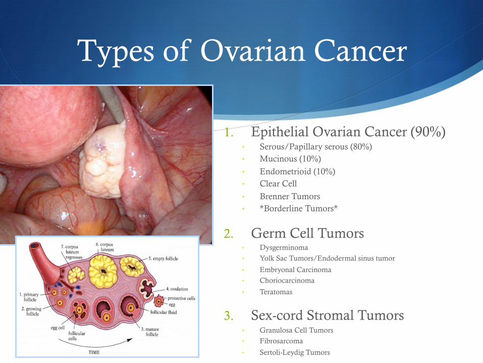
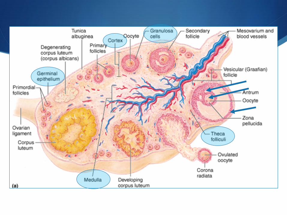
Types of Ovarian Cancer
1. Epithelial Ovarian Cancer (90%) • Serous/Papillary serous (80%) • Mucinous (10%)
• Endometrioid (10%) • Clear Cell
• Brenner Tumors • *Borderline Tumors*
2. Germ Cell Tumors • Dysgerminoma
• Yolk Sac Tumors/Endodermal sinus tumor
• Embryonal Carcinoma
• Choriocarcinoma
• Teratomas
3. Sex-cord Stromal Tumors • Granulosa Cell Tumors
• Fibrosarcoma
• Sertoli-Leydig Tumors
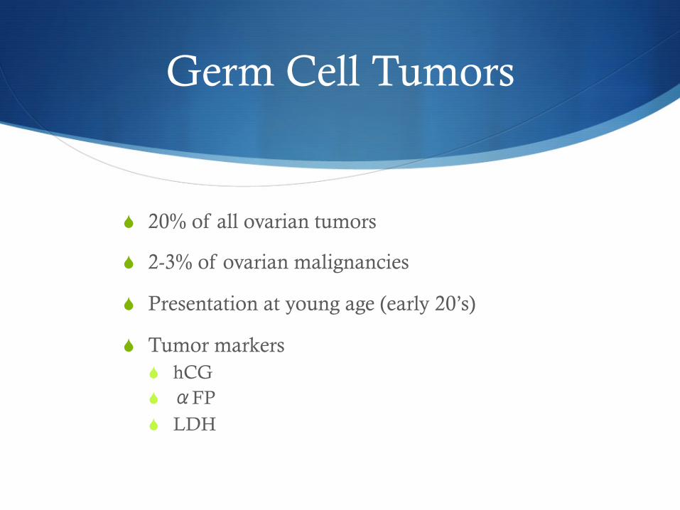
Germ Cell Tumors
S 20% of all ovarian tumors
S 2-3% of ovarian malignancies
S Presentation at young age (early 20’s)
S Tumor markers S hCG S αFP S LDH
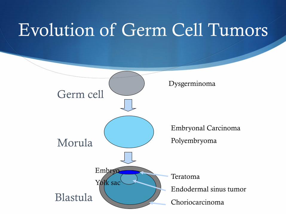
Dysgerminoma
Embryonal Carcinoma
Polyembryoma
Teratoma
Endodermal sinus tumor
Choriocarcinoma
Germ cell
Morula
Blastula
Embryo
Yolk sac
Evolution of Germ Cell Tumors
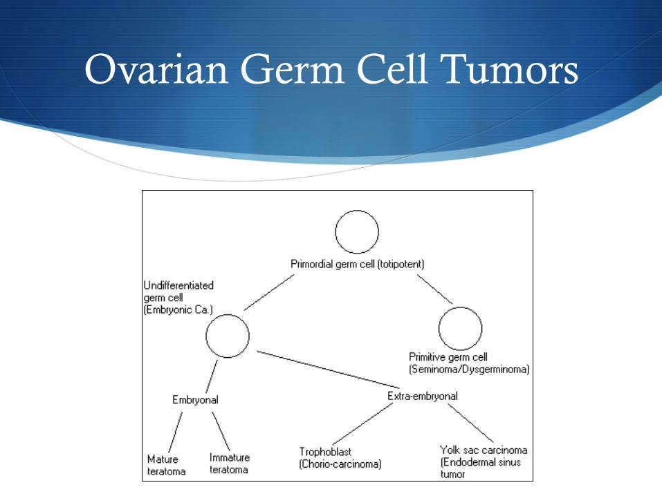
Ovarian Germ Cell Tumors
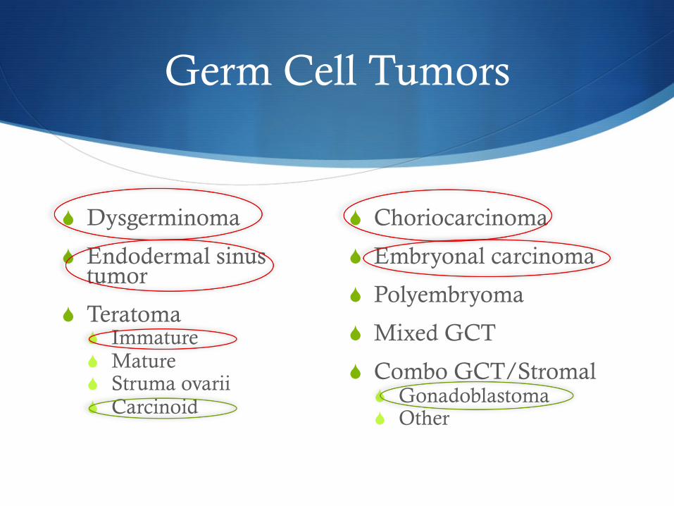
Germ Cell Tumors
S Dysgerminoma
S Endodermal sinus tumor
S Teratoma S Immature S Mature S Struma ovarii S Carcinoid
S Choriocarcinoma
S Embryonal carcinoma
S Polyembryoma
S Mixed GCT
S Combo GCT/Stromal S Gonadoblastoma S Other
Dysgerminoma
S Lance Armstrong
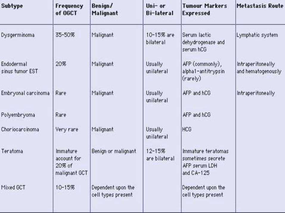
S Incidence S 1-2% of ovarian tumors S 3-5% of ovarian malignancies S 40% of all GCT S Peak incidence age 19 S 67% stage IA
S 10-15% bilaterality S 20% in “normal appearing” opposite ovary
Dysgerminoma
Dysgerminoma
Dysgerminoma
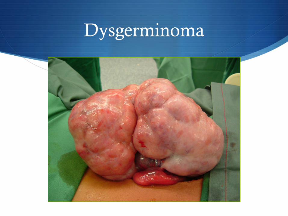
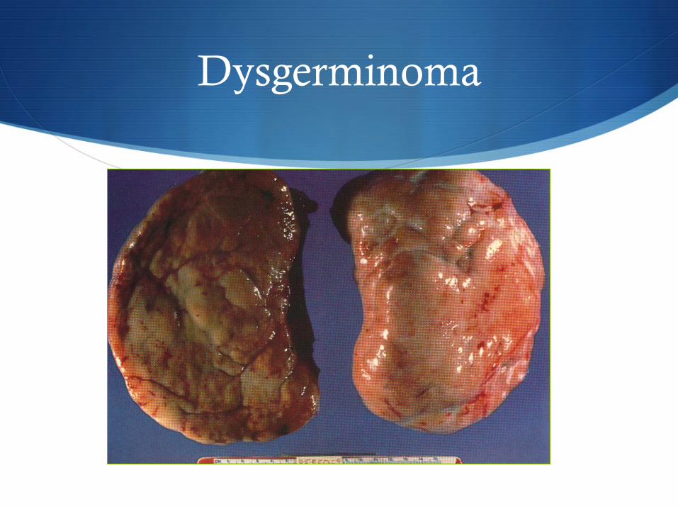
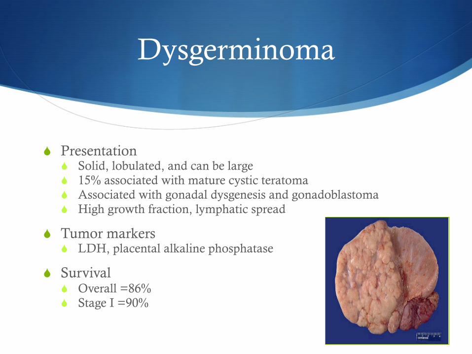
S Presentation S Solid, lobulated, and can be large S 15% associated with mature cystic teratoma S Associated with gonadal dysgenesis and gonadoblastoma S High growth fraction, lymphatic spread
S Tumor markers S LDH, placental alkaline phosphatase
S Survival S Overall =86% S Stage I =90%
Dysgerminoma
S Fertility-sparing surgery S 85% of patients are younger than 35 yo
S Consider uterine preservation (IVF)
S Radiosensitive
S Chemotherapy S Combination, dose-intense regimen
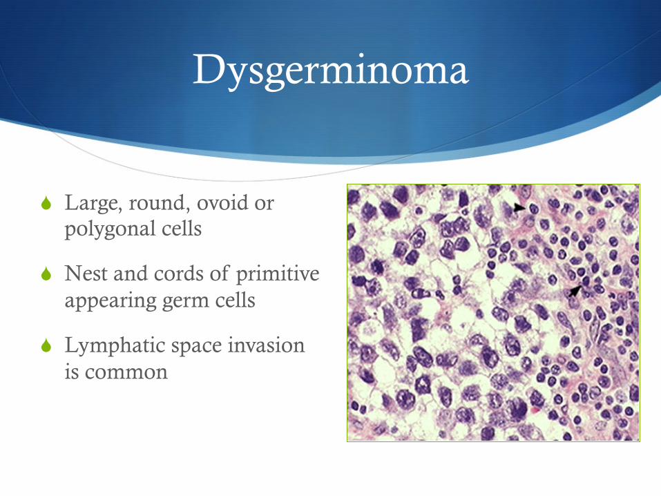
Dysgerminoma
S Large, round, ovoid or polygonal cells
S Nest and cords of primitive appearing germ cells
S Lymphatic space invasion is common
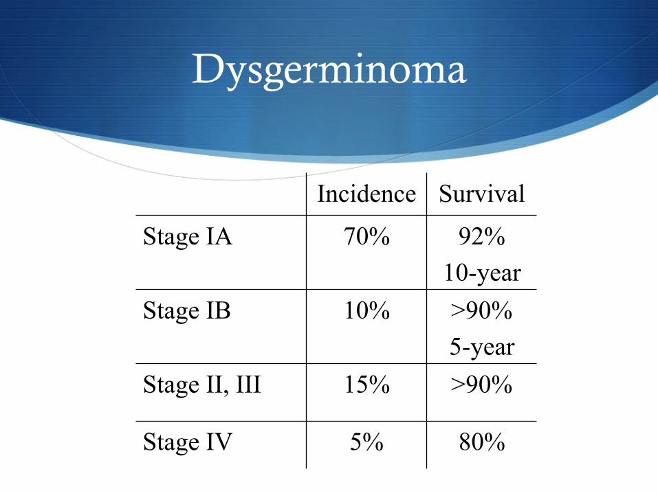
Dysgerminoma
Incidence Survival
Stage IA 70% 92% 10-year
Stage IB 10% >90% 5-year
Stage II, III 15% >90%
Stage IV 5% 80%
Endodermal Sinus Tumor
S Presentation S 20% of all GCT S Median age 19 yo S Abdominal pain, large mass S 10-30 cm common S Very rapid growth, intra-abdominal and hematological spread
S Tumor marker: AFP, α1 antitrypsin
S Synonyms S Yolk sac tumor
S Survival S Overall survival =70% S Stage I =90%
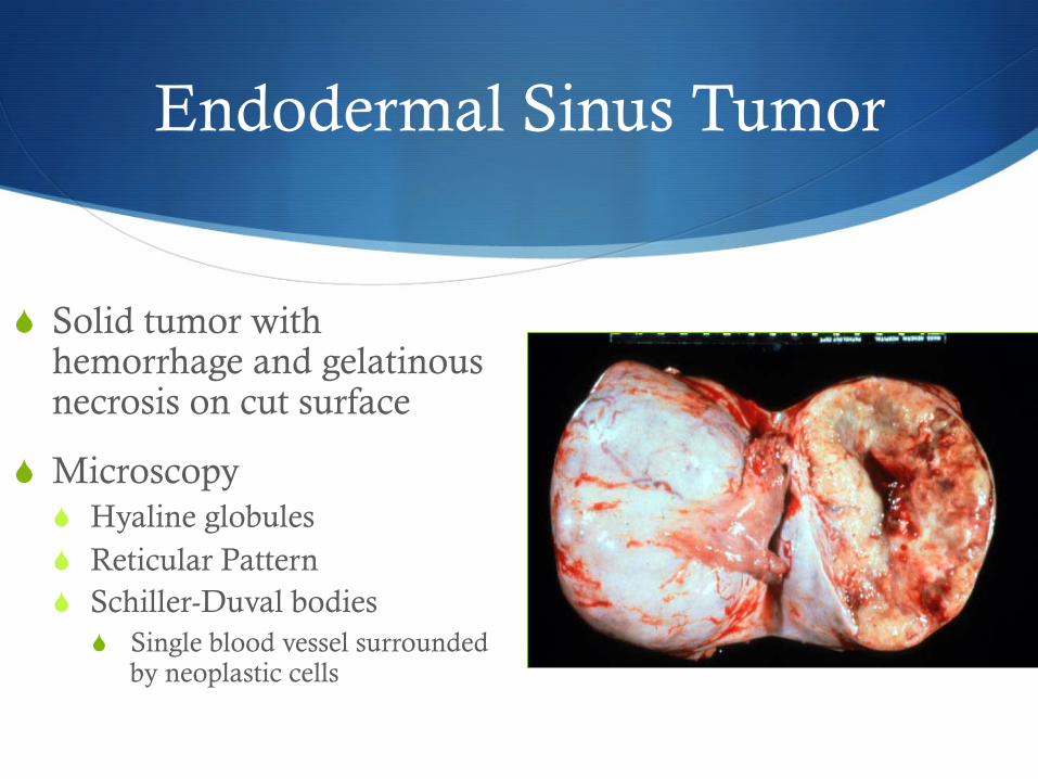
Endodermal Sinus Tumor
S Solid tumor with hemorrhage and gelatinous necrosis on cut surface
S Microscopy S Hyaline globules S Reticular Pattern S Schiller-Duval bodies
S Single blood vessel surrounded by neoplastic cells
Endodermal Sinus Tumor
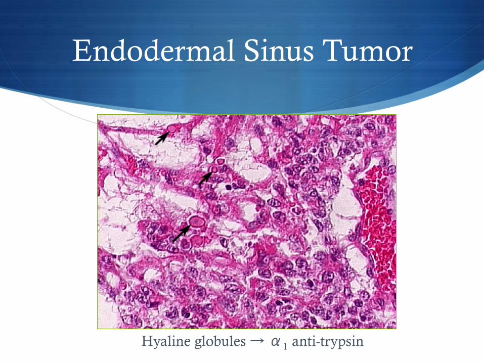
Hyaline globules → α1 anti-trypsin
Endodermal Sinus Tumor
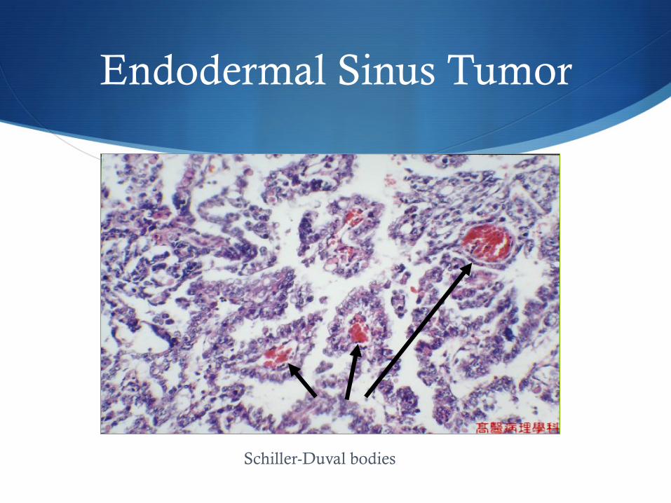
Schiller-Duval bodies
Teratomas
S Immature
S Mature
S Specialized S Struma ovarii
S Carcinoid
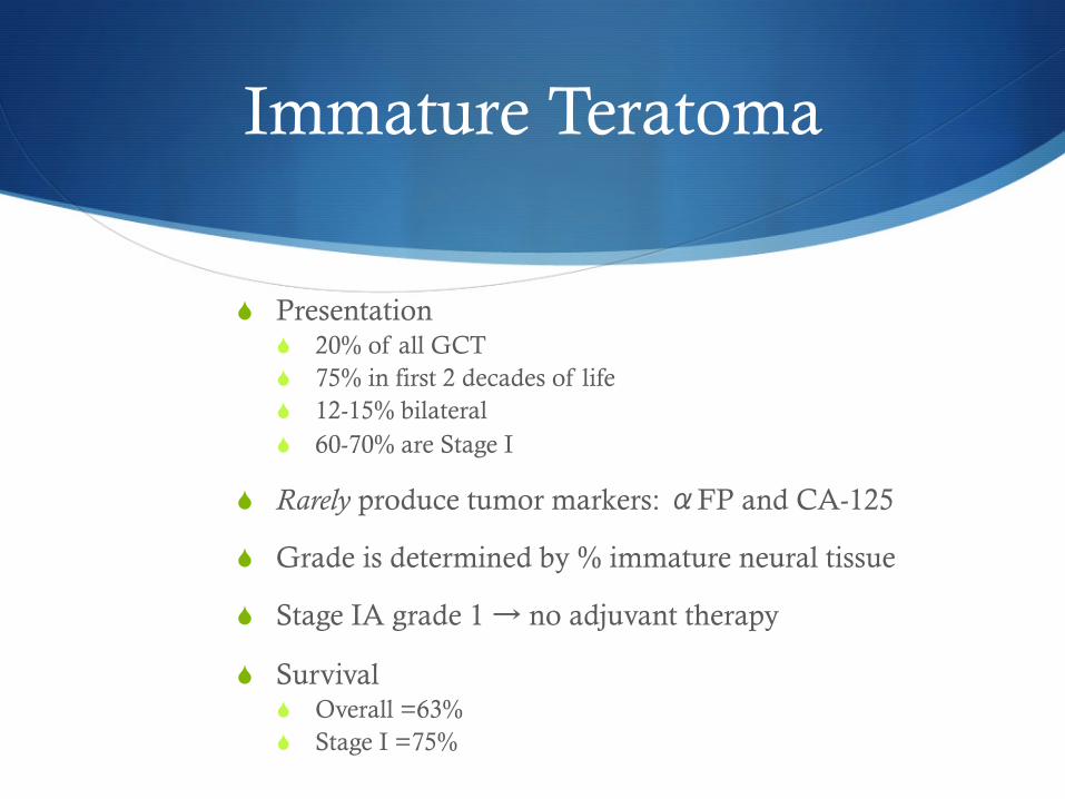
Immature Teratoma
S Presentation S 20% of all GCT S 75% in first 2 decades of life S 12-15% bilateral S 60-70% are Stage I
S Rarely produce tumor markers: αFP and CA-125
S Grade is determined by % immature neural tissue
S Stage IA grade 1 → no adjuvant therapy
S Survival S Overall =63% S Stage I =75%
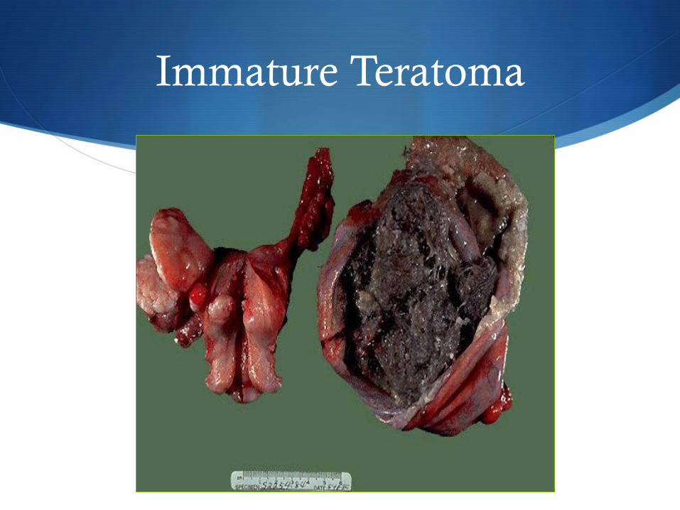
Immature Teratoma
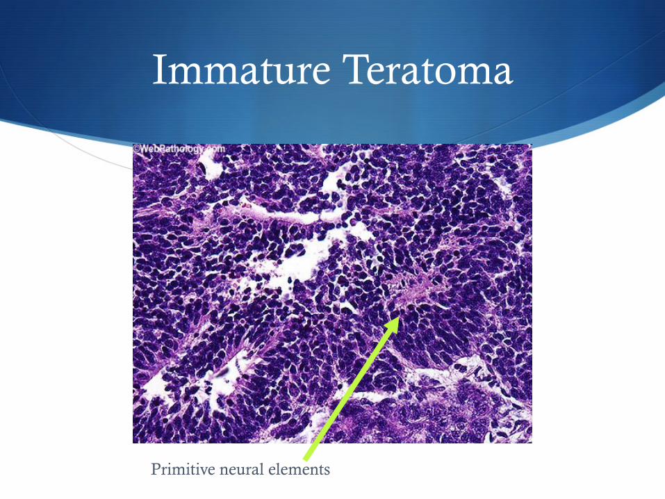
Immature Teratoma
Primitive neural elements
Immature Teratoma
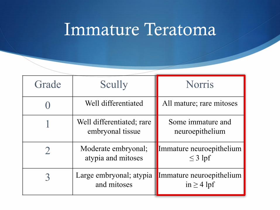
Grade Scully Norris
0 Well differentiated All mature; rare mitoses
1 Well differentiated; rare embryonal tissue
Some immature and neuroepithelium
2 Moderate embryonal; atypia and mitoses
Immature neuroepithelium ≤ 3 lpf
3 Large embryonal; atypia and mitoses
Immature neuroepithelium in ≥ 4 lpf
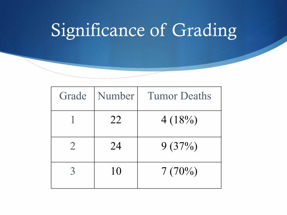
Significance of Grading
Grade Number Tumor Deaths
1 22 4 (18%)
2 24 9 (37%)
3 10 7 (70%)
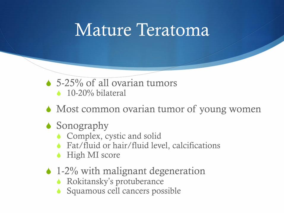
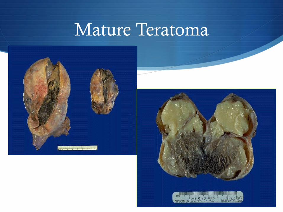
Mature Teratoma
S 5-25% of all ovarian tumors S 10-20% bilateral
S Most common ovarian tumor of young women
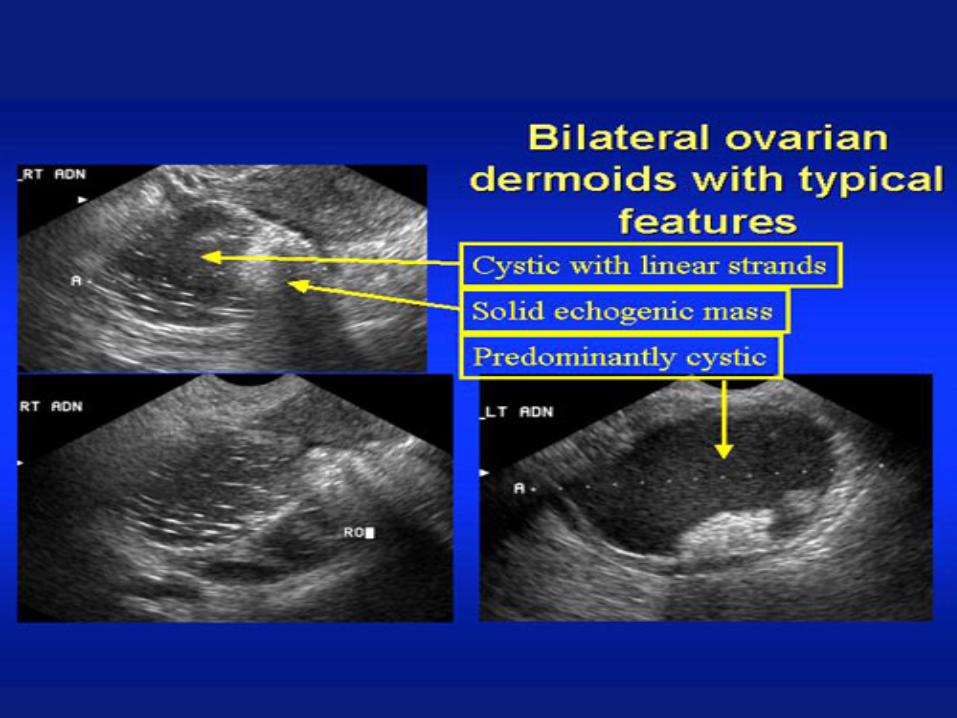
S Sonography S Complex, cystic and solid S Fat/fluid or hair/fluid level, calcifications S High MI score
S 1-2% with malignant degeneration S Rokitansky’s protuberance S Squamous cell cancers possible
Mature Teratoma
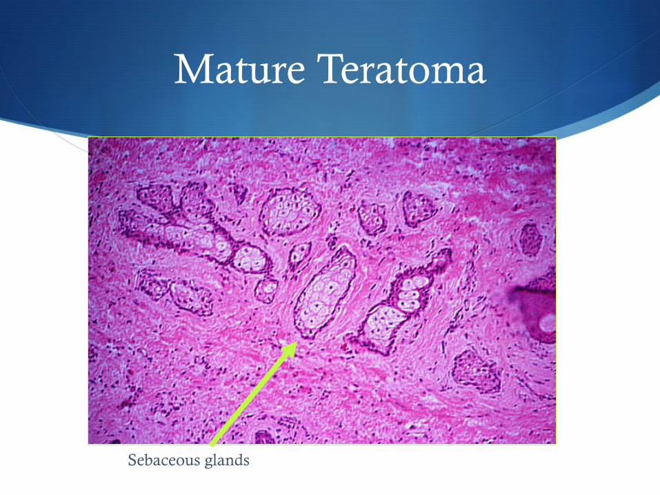
Mature Teratoma
Sebaceous glands
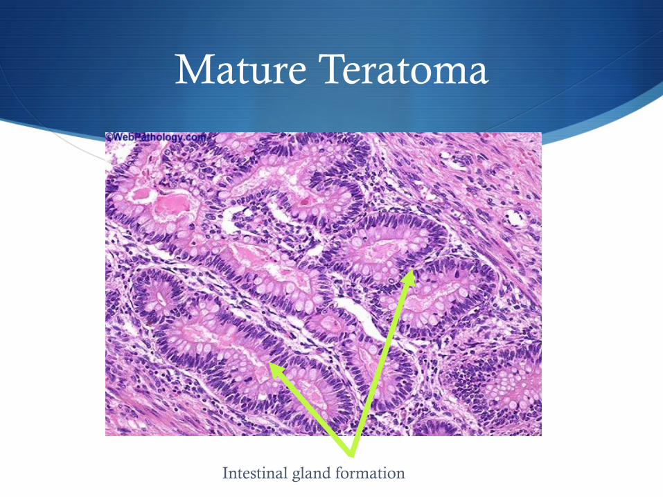
Mature Teratoma
Intestinal gland formation
Specialized Teratomas
S Struma ovarii S 2-3% of all teratomas S 25-35% have symptoms of hyperthyroidism S Usually benign, but may undergo malignant transformation
S Carcinoid tumors S Associated with GI or respiratory epithelium S Primary ovarian tumors are rare (N=50) S Often older postmenopausal women S 1/3 have carcinoid syndrome from serotonin S Symptoms resolve with excision S 5-hydroxyindoleacetic acid in urine
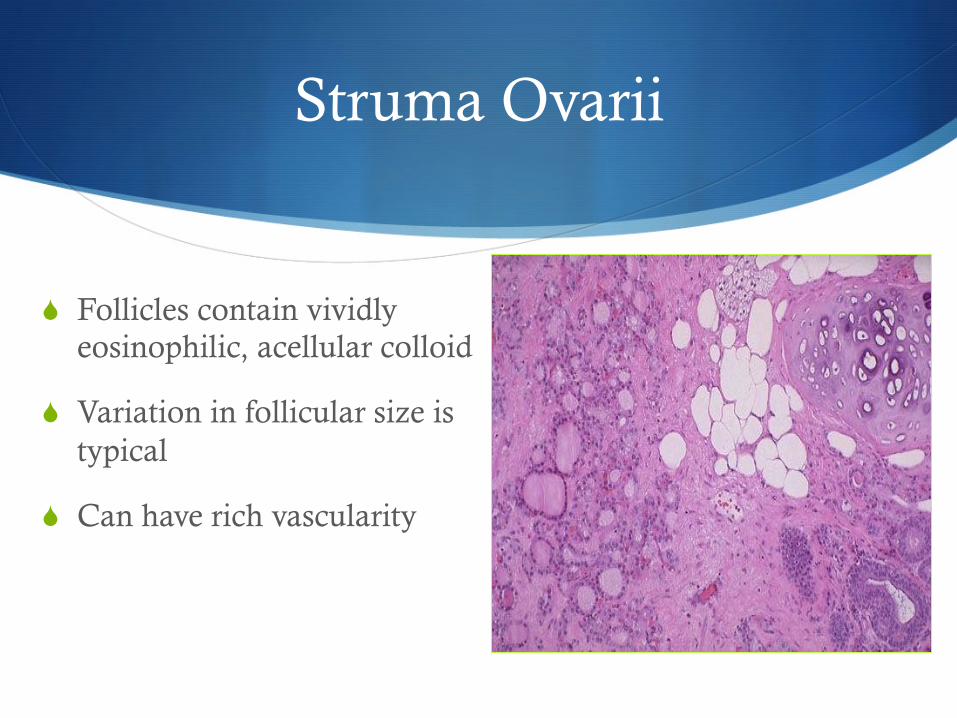
Struma Ovarii
S Follicles contain vividly eosinophilic, acellular colloid
S Variation in follicular size is typical
S Can have rich vascularity
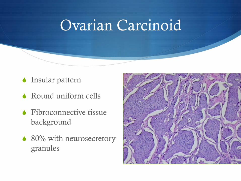
Ovarian Carcinoid
S Insular pattern
S Round uniform cells
S Fibroconnective tissue background
S 80% with neurosecretory granules
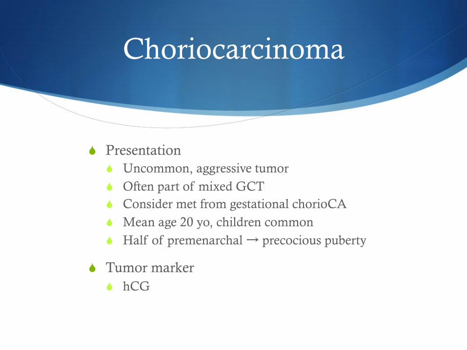
Choriocarcinoma
S Presentation S Uncommon, aggressive tumor S Often part of mixed GCT S Consider met from gestational chorioCA S Mean age 20 yo, children common S Half of premenarchal → precocious puberty
S Tumor marker S hCG
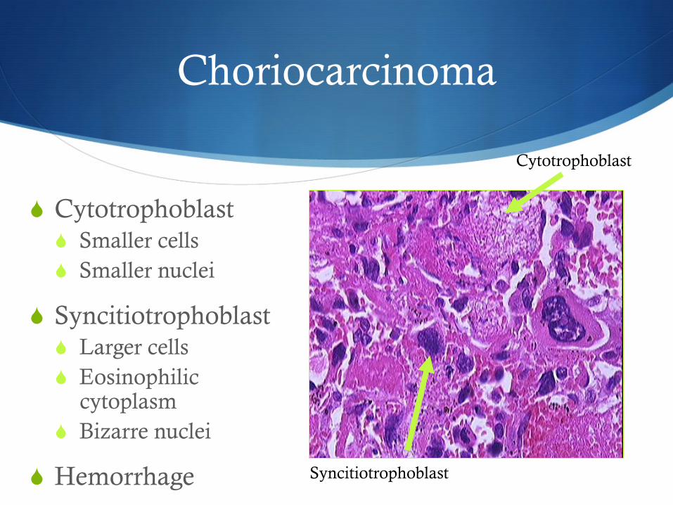
Choriocarcinoma
S Cytotrophoblast S Smaller cells S Smaller nuclei
S Syncitiotrophoblast S Larger cells S Eosinophilic
cytoplasm S Bizarre nuclei
S Hemorrhage
Cytotrophoblast
Syncitiotrophoblast
Embryonal Carcinoma
S Presentation S Mean age < 30 yo S Only 4% of GCT and often part of mixed tumor S 60% Stage IA S Poorly differentiated germ cell tumor S Aggressive, intra-abdominal spread and mets common
S Tumor markers: hCG, αFP
S Survival S Overall =40% S Stage I =75%
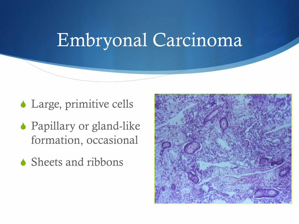
Embryonal Carcinoma
S Large, primitive cells
S Papillary or gland-like formation, occasional
S Sheets and ribbons
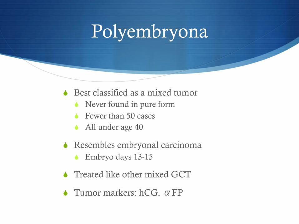
Polyembryona
S Best classified as a mixed tumor S Never found in pure form S Fewer than 50 cases S All under age 40
S Resembles embryonal carcinoma S Embryo days 13-15
S Treated like other mixed GCT
S Tumor markers: hCG, αFP
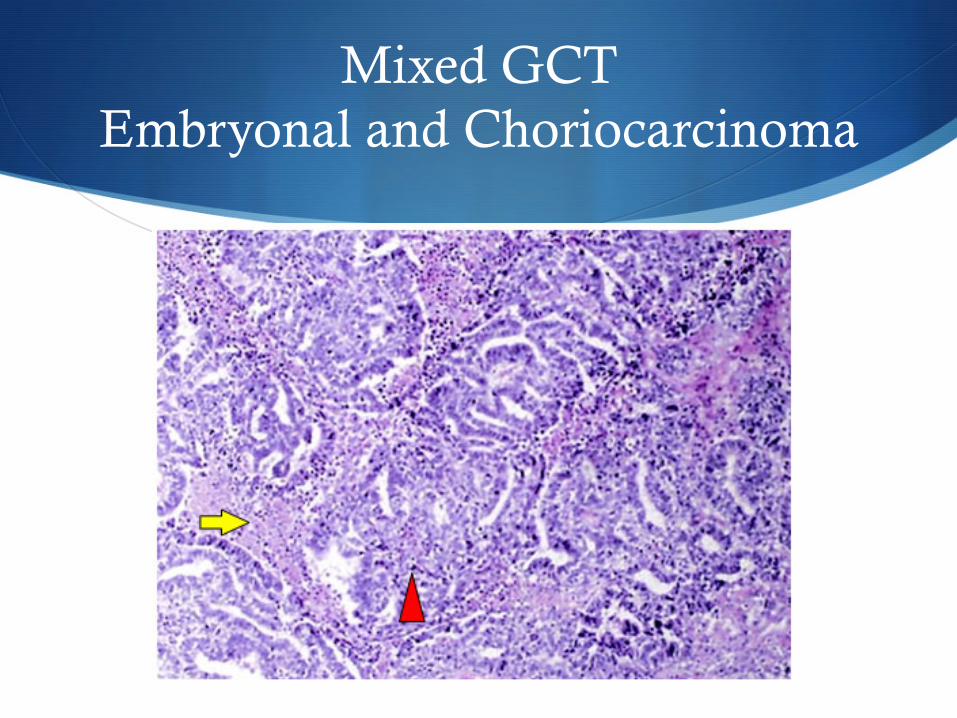
Mixed GCT Embryonal and Choriocarcinoma
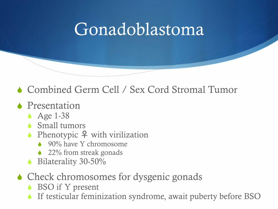
Gonadoblastoma
S Combined Germ Cell / Sex Cord Stromal Tumor
S Presentation S Age 1-38 S Small tumors S Phenotypic ♀ with virilization
S 90% have Y chromosome S 22% from streak gonads
S Bilaterality 30-50%
S Check chromosomes for dysgenic gonads S BSO if Y present S If testicular feminization syndrome, await puberty before BSO
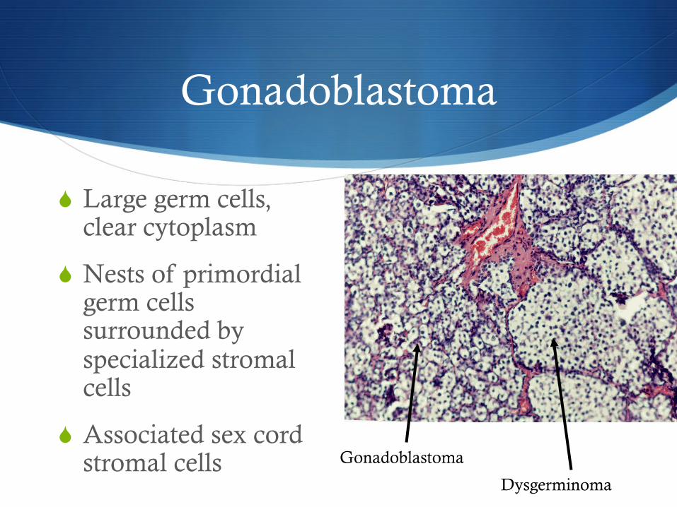
Gonadoblastoma
S Large germ cells, clear cytoplasm
S Nests of primordial germ cells surrounded by specialized stromal cells
S Associated sex cord stromal cells Gonadoblastoma
Dysgerminoma
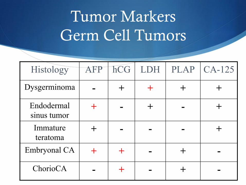
Tumor Markers Germ Cell Tumors
Histology AFP hCG LDH PLAP CA-125
Dysgerminoma - + + + +
Endodermal sinus tumor
+ - + - +
Immature teratoma
+ - - - +
Embryonal CA + + - + -
ChorioCA - + - + -
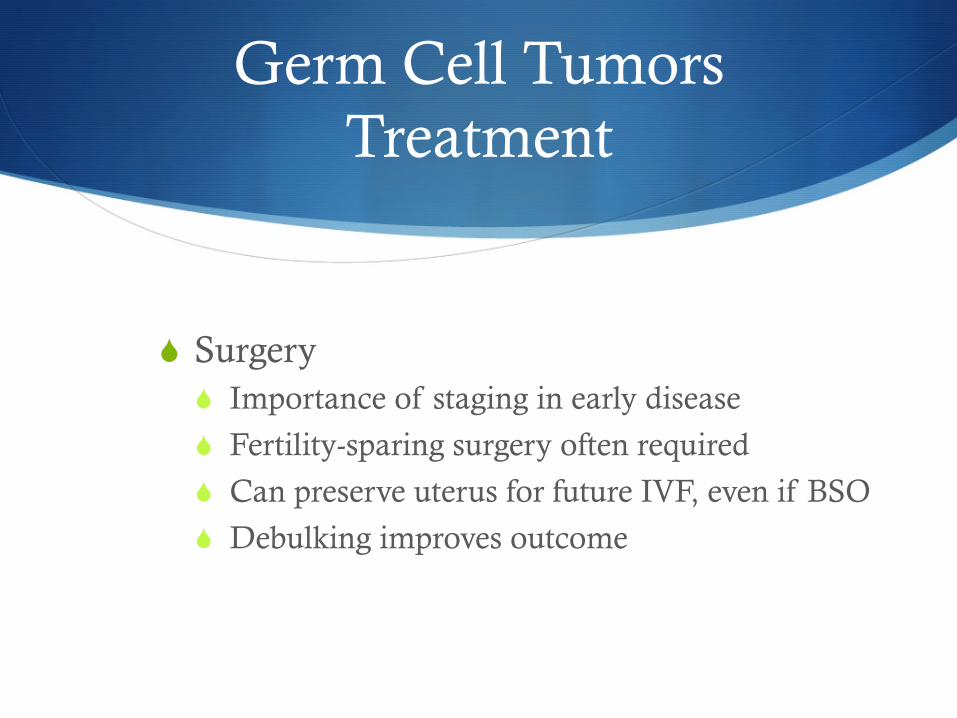
Germ Cell Tumors Treatment
S Surgery S Importance of staging in early disease
S Fertility-sparing surgery often required
S Can preserve uterus for future IVF, even if BSO
S Debulking improves outcome

Chemotherapy Germ Cell Tumors
S BEP S Bleomycin 20 U/m2 weekly x 9 S Etoposide 100 mg/m2 days 1-5 q 3 weeks x 3 S Cisplatin 20 mg/m2 days 1-5 q 3 weeks x 3
S VAC S Vincristine 105 mg/m2 weekly x 12 S Act D 0.5 mg days 1-5 q 4 weeks S Cytoxan 5-7 mg/kg days 1-5 q 4 weeks
S VBP S Vinblastine 12 mg/m2 q 3 weeks x 4 S Bleomycin 20 U/m2 weeks x 7, 8 on week 10 S Cisplatin 20 mg/m2 days 1-5 q 3 weeks x 3
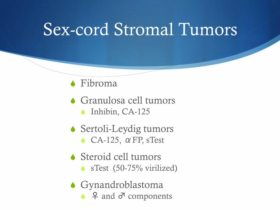
Sex-cord Stromal Tumors
S Fibroma
S Granulosa cell tumors S Inhibin, CA-125
S Sertoli-Leydig tumors S CA-125, αFP, sTest
S Steroid cell tumors S sTest (50-75% virilized)
S Gynandroblastoma S ♀ and ♂ components
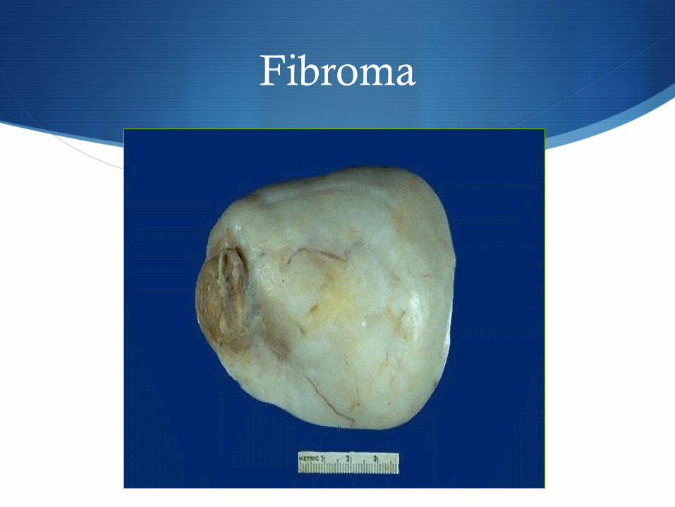
Fibroma
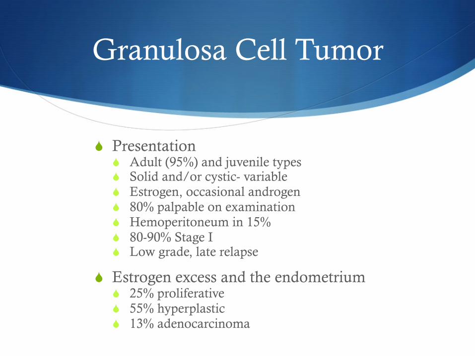
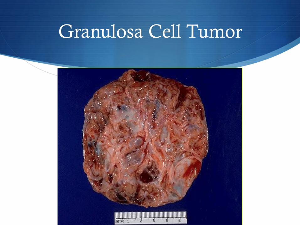
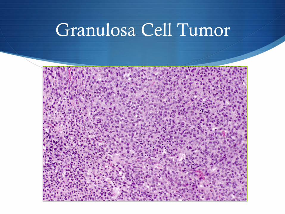
Granulosa Cell Tumor
S Presentation S Adult (95%) and juvenile types S Solid and/or cystic- variable S Estrogen, occasional androgen S 80% palpable on examination S Hemoperitoneum in 15% S 80-90% Stage I S Low grade, late relapse
S Estrogen excess and the endometrium S 25% proliferative S 55% hyperplastic S 13% adenocarcinoma
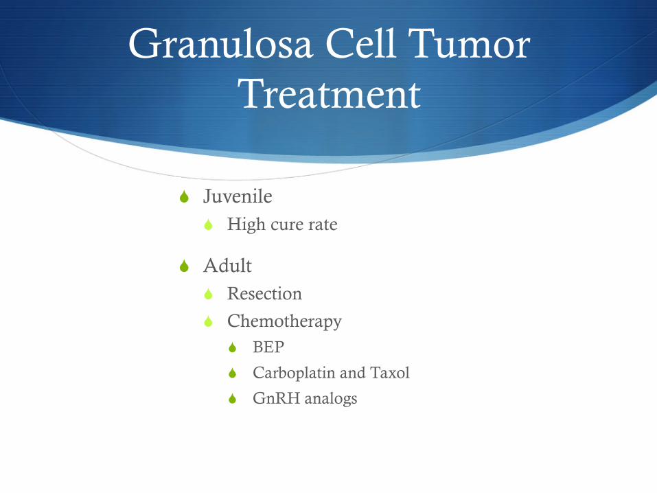
Granulosa Cell Tumor Treatment
S Juvenile S High cure rate
S Adult S Resection
S Chemotherapy S BEP
S Carboplatin and Taxol
S GnRH analogs
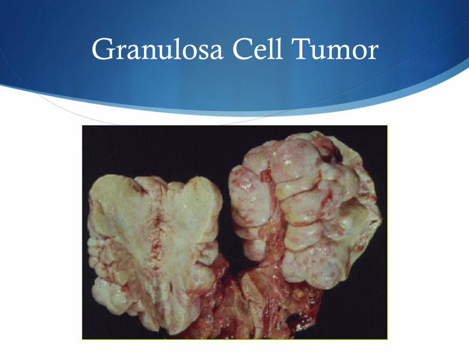
Granulosa Cell Tumor
Granulosa Cell Tumor
Granulosa Cell Tumor
Granulosa Cell Tumor
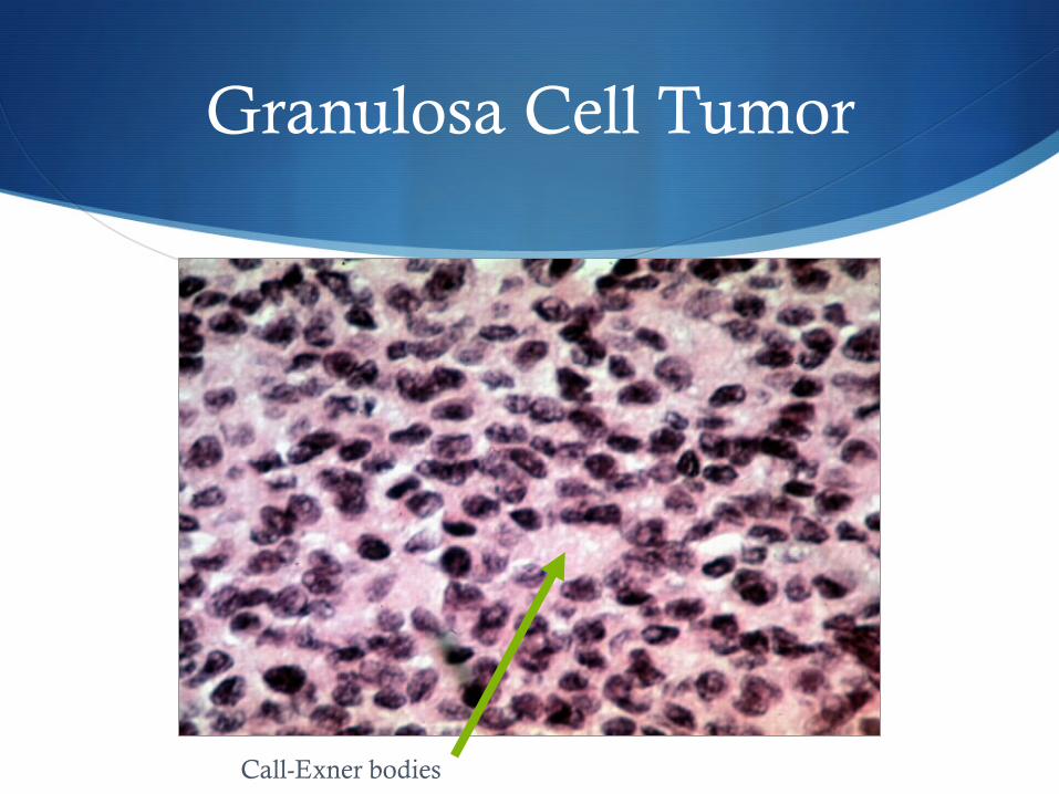
Call-Exner bodies

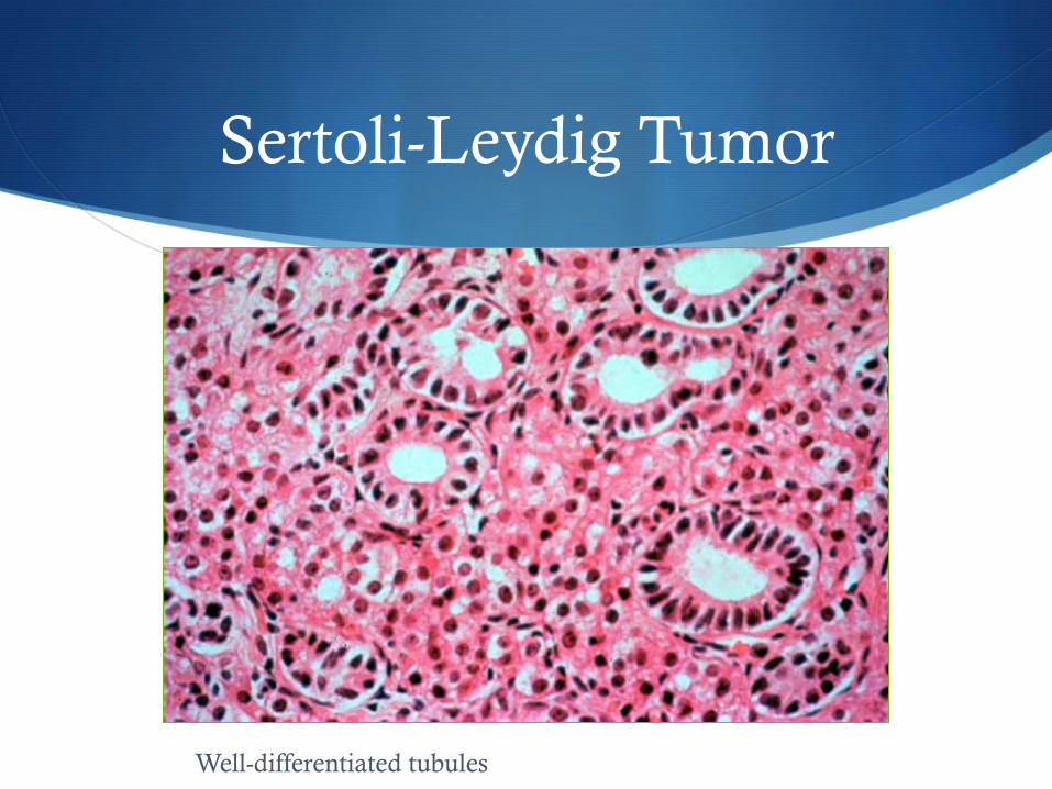
Sertoli-Leydig Tumors
S Benign S Sertoli cell tumors→ no hormones S Leydig tumors→ testosterone
S Potentially Malignant S Sertoli-Leydig tumors S Arrhenoblastoma, androblastoma S Grade 3
S 44% five-year survival
Grade % Cancer
1 0
2 10
3 60
Sertoli-Leydig Tumor
Well-differentiated tubules

S
Treatment Summary
Germ Cell and Stromal Tumors of the Ovary

Dysgerminoma USO staging if possible
BEP x 3 cycles if stage II-IV
Endodermal sinus tumor
Debulk but preserve fertility
BEP x 3-4 cycles
Embryonal carcinoma
As above BEP x 3-4 cycles
Malignant teratoma
As above BEP or VAC x 3-4 cycles
Granulosa cell tumor
USO if young o/w TAH/BSO
BEP x 3-4 cycles GnRH agonists for
advanced ds.
Sertoli-leydig cell
As above BEP or VAC x 3-4 cycles
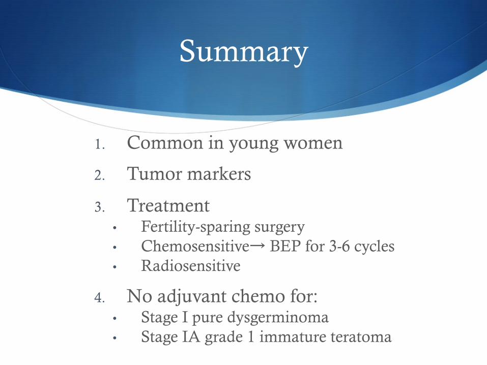
Summary
1. Common in young women
2. Tumor markers
3. Treatment • Fertility-sparing surgery • Chemosensitive→ BEP for 3-6 cycles • Radiosensitive
4. No adjuvant chemo for: • Stage I pure dysgerminoma • Stage IA grade 1 immature teratoma