nocardia brasiliensis cell wall lipids modulate …nocardia brasiliensis cell wall lipids modulate...
TRANSCRIPT

Nocardia brasiliensis Cell Wall Lipids Modulate Macrophage andDendritic Responses That Favor Development of ExperimentalActinomycetoma in BALB/c Mice
J. Humberto Trevino-Villarreal,a Lucio Vera-Cabrera,b Pedro L. Valero-Guillén,c and Mario C. Salinas-Carmonaa
Universidad Autonoma de Nuevo León, Faculty of Medicine and University Hospital, Department of Immunology, Monterrey, Méxicoa; Servicio de Dermatología, HospitalUniversitario “Dr. José E. González,” UANL, Monterrey, Méxicob; and Departamento de Genética y Microbiología, Facultad de Medicina, Campus Universitario deEspinardo, Universidad de Murcia, Murcia, Españac
Nocardia brasiliensis is a Gram-positive facultative intracellular bacterium frequently isolated from human actinomycetoma.However, the pathogenesis of this infection remains unknown. Here, we used a model of bacterial delipidation with benzine toinvestigate the role of N. brasiliensis cell wall-associated lipids in experimental actinomycetoma. Delipidation of N. brasiliensiswith benzine resulted in complete abolition of actinomycetoma without affecting bacterial viability. Chemical analyses revealedthat trehalose dimycolate and an unidentified hydrophobic compound were the principal compounds extracted from N. brasil-iensis with benzine. By electron microscopy, the extracted lipids were found to be located in the outermost membrane layer ofthe N. brasiliensis cell wall. They also appeared to confer acid-fastness. In vitro, the extractable lipids from the N. brasiliensis cellwall induced the production of the proinflammatory cytokines interleukin-1� (IL-1�), IL-6, and CCL-2 in macrophages. The N.brasiliensis cell wall extractable lipids inhibited important macrophage microbicidal effects, such as tumor necrosis factor alpha(TNF-�) and nitric oxide (NO) production, phagocytosis, bacterial killing, and major histocompatibility complex class II (MHC-II) expression in response to gamma interferon (IFN-�). In dendritic cells (DCs), N. brasiliensis cell wall-associated extractablelipids suppressed MHC-II, CD80, and CD40 expression while inducing tumor growth factor � (TGF-�) production. Immuniza-tion with delipidated N. brasiliensis induced partial protection preventing actinomycetoma. These findings suggest that N.brasiliensis cell wall-associated lipids are important for actinomycetoma development by inducing inflammation and modulat-ing the responses of macrophages and DCs to N. brasiliensis.
Actinomycetoma is a chronic, infectious disease that is charac-terized by excessive tumefaction and deformation of affected
tissues. Nocardia brasiliensis is the primary causative agent of ac-tinomycetoma in the Western Hemisphere. The highest incidenceof N. brasiliensis-induced actinomycetoma is observed in Mexicoand Venezuela (27, 46). Diagnosis of actinomycetoma is based onclinical findings such as multiple inflammatory granulomatousnodules and subcutaneous fistulas that drain a purulent exudate,containing granules consisting of microcolonies of the causalagent, N. brasiliensis (46), and confirmed by an enzyme-linkedimmunosorbent assay (ELISA) (39). This bacterium is a Gram-positive, acid-fast microorganism that belongs to the Corynebac-terineae suborder. Several microorganisms of clinical relevance,such as Mycobacterium, Corynebacterium, Actinomadura, andGordonia, among others, belong to the Corynebacterineae subor-der. The most distinctive characteristic that classifies specieswithin this suborder of microorganisms is the presence of a che-motype IV cell envelope (11, 26). This type of cell envelope ischaracterized by an abundance of lipids with unusual chemicalstructures that can constitute up to 40% of the dry weight of themicroorganism (11). These lipids are distributed in the followingthree major cell wall components: the cell membrane, the my-coloyl-arabinogalactan-peptidoglycan complex (MAPc), and theouter membrane layer (11, 12).
The cell membrane present in chemotype IV cell envelope-bearing Corynebacterineae spp. is chemically similar to the cellmembranes present in microorganisms of any other species. Incontrast, the MAPc is the true hallmark structure that defines andclassifies microorganisms into this group. The MAPc present in
chemotype IV cell envelopes is composed of a meso-diamin-opimelic acid-containing peptidoglycan skeleton covalentlylinked to a linear D-galactan chain (34). The galactan chain iscovalently bound to highly branched, D-arabinofuranosyl resi-dues that form penta-arabinofuranosyl terminal motifs. Arabi-nose residues are further sterified by tightly packed mycolic acidsthat are 40 to 60 carbons long and characteristically extend per-pendicularly to the plane of the cell envelope (1).
The mycolic acids contribute to the formation of the outermembrane layer of the cell envelope of Corynebacterineae, which isa zipper-like, asymmetric bilayer membrane. The inner leaflet iscomposed of the mycolic acids that extend from the MAPc. Theoutermost leaflet is composed of a variety of cell wall-associatedglycolipids that are noncovalently bound to the inner leaflet (11,20, 30, 32, 49). Because of the noncovalent association between theouter leaflet and the mycolic acids, it is possible to extract thewall-associated lipids from the outer membrane layer with or-ganic solvents, thus disrupting the integrity of the cell envelopewithout affecting bacterial viability (9, 10, 44).
Received 11 May 2012 Returned for modification 7 June 2012Accepted 24 July 2012
Published ahead of print 30 July 2012
Editor: J. L. Flynn
Address correspondence to Mario C. Salinas-Carmona, [email protected].
Copyright © 2012, American Society for Microbiology. All Rights Reserved.
doi:10.1128/IAI.00446-12
October 2012 Volume 80 Number 10 Infection and Immunity p. 3587–3601 iai.asm.org 3587
on February 21, 2020 by guest
http://iai.asm.org/
Dow
nloaded from

The nature and composition of the cell wall extractable lipidsthat are associated with the outer membrane layer vary betweenspecies. In general, for Mycobacterium spp. the main componentsare phthiocerol-containing lipids, phosphatidylinositol manno-sides (PIM), lipomannan (LM), lipoarabinomannan (LAM), tre-halose dimycolates (TDM or cord factor), trehalose monomyco-lates, glycopeptidolipids (GPL), and sulfolipids (11, 15, 16). ForNocardia spp., trehalose-containing lipids, glycolipids, diethylether-soluble lipids, tuberculostearic acid, nocobactin, and nocar-dones have been identified (2, 33, 35, 48).
Cell wall extractable lipids associated with the outer membranelayer (or simply cell wall-associated lipids) have important impli-cations in the pathogenesis of microorganisms of the Corynebac-terineae spp., including Nocardia spp. For instance, the diethylether-soluble lipids of Nocardia asteroides are highly toxic to mice.They induce a cachectic state that ultimately leads to death whensystemically administered (23). In addition to the diethyl ether-soluble lipids, other wall-associated lipids of N. asteroides areknown to induce inflammatory responses when systemically ad-ministered. The most notable of these compounds is 6,6=-treha-lose dimycolate, or TDM, which induces a strong and deadly ca-chectic state when systemically administered to mice (42). Whenadministered subdermally, TDM can also induce a strong, localinflammatory response that mimics important histopathologicalaspects of the disease caused by N. asteroides, including the induc-tion of granuloma formation (19). These findings suggest thatwall-associated lipids modulate important aspects of the immuneresponses elicited against the infecting microorganism and thatthese lipids are important determinants of N. asteroides virulence.
It is unknown whether the observations about the importanceof cell wall-associated lipids in bacterial pathogenesis from studieswith N. asteroides are also valid for other members of the Nocardiagenus, including N. brasiliensis. It is not known whether the cellwall-associated lipids are virulence factors implicated in N. brasil-iensis pathogenicity. It is also not known whether the wall-associ-ated lipids are implicated in the development of actinomycetomainduced by N. brasiliensis. Here, we report that N. brasiliensis cellwall-associated lipids are implicated in the development of exper-imental actinomycetoma and act principally by inducing a stronginflammatory response and inhibiting several of the microbicidaleffects of macrophages, including the inhibition of tumor necrosisfactor alpha (TNF-�) production, phagocytosis, production ofnitric oxide (NO), and bacterial killing. In addition, we demon-strate that the N. brasiliensis wall-associated lipids suppressed theexpression of major histocompatibility complex class II (MHC-II), CD80, and CD40 by dendritic cells (DCs) and strongly in-duced the production of tumor growth factor � (TGF-�) by thesecells.
MATERIALS AND METHODSMedia, cells, and culture conditions. The L929 cell line (designationnumber CCL-1, obtained from ATCC, Manassas, VA) was maintained inL929 medium, consisting of high-glucose Dulbecco’s modified Eagle me-dium (DMEM) (Gibco, Carlsbad, CA) supplemented with 10% fetal bo-vine serum (FBS) (HyClone; Thermo Fisher, Waltham, MA), GlutaMax(2 mM), penicillin (100 U/ml), streptomycin (100 �g/ml), and ampho-tericin (0.25 �g/ml) (all from Gibco). The cells were incubated at 37°C ina 5% CO2 atmosphere.
The L929 cell line was used to produce the L-conditioned medium.Briefly, after removing the medium from confluent L929 cell cultures, thecell monolayers were washed once with Hanks’ balanced salt solution
(HBSS) and incubated in fresh L929 medium for 7 days without changingthe medium. Subsequently, the conditioned medium was collected, fil-tered using a 0.2-�m syringe filter, and used to differentiate macrophagesfrom the bone marrow cell cultures as described below.
N. brasiliensis culture and delipidation with benzine. N. brasiliensis(ATCC 700358) was cultured statically at 37°C in brain heart infusionbroth (BHI) (Difco, BD Diagnostics, Sparks, MD) for 72 h before process-ing. Under these conditions, N. brasiliensis grows as a thick biofilm on thesurface of the media. To obtain a single cell suspension, the biomass wascollected in 50-ml centrifuge tubes, washed 3 times in sterile saline, anddissociated using a Dounce tissue grinder. After another wash step, theresidual cellular clumps were eliminated by centrifuging the suspensionsthree times at 200 � g. The supernatants containing the unicellular cells insuspension were collected after each centrifugation step. After the thirdcentrifugation, Kinyoun staining was performed to confirm the unicellu-larity of the cells in suspension. Only unicellular cell suspensions of N.brasiliensis were used throughout the study.
Unicellular cell suspensions were delipidated using benzine (boilingpoints, 35 to 60°C; Thermo Fisher Scientific, Inc., Waltham, MA). Briefly,after adding 30 ml of benzine per 500 mg of bacteria, the suspensions werevortexed vigorously for 5 min, and the bacteria were then allowed to settlefor 5 min. Then, the suspensions were centrifuged at 500 � g for 5 min.The supernatant, containing the extracted lipid material, was collected ina glass container for further chemical analysis (see below). The pellet,consisting of the bacteria, was subjected to delipidation for two or fouradditional delipidation processes to generate N. brasiliensis delipidatedthree times (Tx3) or five times (Tx5), respectively.
After the last extraction, the suspensions were washed extensively us-ing sterile saline, suspended in 20% glycerol in saline, and stored in 1-mlvials at �70°C. At least three vials were used in a plate-counting assay onBHI agar dishes to determine the number of CFU per vial.
Viability determination. The bacterial viability after the benzinetreatment was determined by two different approaches. First, 10 mg (wetweight) of delipidated or control bacteria was suspended in sterile saline.From this suspension, 100 �l, which contained 1 mg of bacteria, wasserially diluted and plated onto blood agar plates for CFU determination.Viability was determined by comparing the total CFU present in 1 mg ofthe delipidated bacteria to the CFU of untreated, control bacteria. Addi-tionally, the viability of the N. brasiliensis cells after benzine treatment wastested using the BacLight live/dead kit (Invitrogen, Molecular Probes,Eugene, OR) by following the manufacturer’s instructions.
Isolation and analysis of the lipids in benzine extract. Benzine ex-tracts were pooled, centrifuged at 20,000 � g for 15 min, and evaporatedusing a rotavapor. The sediment was suspended in 3 ml of benzine, cen-trifuged one more time, and evaporated using nitrogen. After beingweighed, the extracted material was redissolved in isopropanol (Sigma-Aldrich, St. Louis, MO) at a final concentration of 1 mg/ml and stored at�70°C.
The extracted lipids were initially examined by thin-layer chromatog-raphy (TLC) (10- by 10-cm plates, cut from an aluminum-backed 20- by20-cm silica gel F254 sheet; Merck & Co, Inc., Whitehouse Station, NJ)using chloroform-methanol (9:1, vol/vol) as the solvent. Plates were de-veloped using molybdophosphoric acid (10%, wt/vol, in 70% ethanol forall lipids) or orcinol (0.1%, wt/vol, in 40% H2SO4 for sugars) followed byheating at 140°C. Lipids were fractionated using column chromatography(silica gel, 0.040 to 0.063 mm; Merck & Co, Inc., Whitehouse Station, NJ)with an increasing concentration of methanol (0%, 2%, 5%, 10%, 15%,and 30%) in chloroform. The identity of some of the compounds wasinvestigated using spectroscopic techniques. Thus, the structures of theTDM and the monomycolate of trehalose (MMT) were obtained at 10%and 15% methanol in chloroform, respectively, and they were confirmedby 1H nuclear magnetic resonance (NMR) and 1H-1H homonuclear cor-related spectroscopy (1H-1H-COSY). The mycolic acids from TDM wereliberated by saponification (15% KOH in methanol, 90°C, overnight) andstudied by ion trap mass spectrometry in negative mode using an MSDI-
Trevino-Villarreal et al.
3588 iai.asm.org Infection and Immunity
on February 21, 2020 by guest
http://iai.asm.org/
Dow
nloaded from
onTrap-VL (Agilent) instrument. Acylglycerols and other apolar com-pounds, eluted with chloroform and chloroform-methanol (5%), wereidentified only tentatively. Among the lipids that were eluted with chlo-roform, a major, apolar compound was purified using silica gel 60 (70-230mesh; Merck & Co, Inc., Whitehouse Station, NJ), liquid chromatogra-phy, and benzine (boiling point, 60°C to 80°C) as the eluent. This com-pound was further examined by 1H NMR (400 MHz, 298 K; solvent,deuterochloroform) and by ESI-TOF-MS (electrospray ionization–timeof flight-mass spectrometry) using an ESI-TOF 6220 apparatus (AgilentTechnology, Santa Clara, CA).
Electron microscopy of N. brasiliensis. Unicellular suspensions of N.brasiliensis were fixed in 1.5% KMnO4 for 45 min at room temperature.After being washed two times with distilled water, the cells were sus-pended in 1% (wt/vol) uranyl nitrate in water and were incubated at roomtemperature for 1 h. The cells were then incubated with acetone followedby an overnight incubation in Epon-acetone. After the incubation, thecells were incubated in Epon for an additional 72 h at 60°C. The resultingblocks were sectioned and visualized by transmission electron micros-copy.
Infection of mice with N. brasiliensis. Ten- to 12-week-old BALB/cmice were infected with 10 mg (wet weight) of N. brasiliensis suspended in100 �l of sterile saline solution in the rear footpad, as previously reported(38). Using the ellipsoid formula, inflammation was measured with ascientific caliper and recorded as cm3 of inflammation. All animal exper-iments were performed according to approved animal protocols by fol-lowing regulations on experimental animal use as specified by our insti-tution.
Isolation of bone marrow-derived macrophages (BMDM). Six- toeight-week-old naïve BALB/c mice were euthanized by cervical disloca-tion. The limbs were removed, and after dissection, the femurs and tibiaswere perfused with macrophage-differentiating medium consisting ofhigh-glucose DMEM (Gibco) supplemented with 20% fetal calf serum(FCS), 30% L-conditioned medium, glutamine (2 mM), and penicillin/gentamicin. Bone marrow cells were washed in HBSS, adjusted to 5 � 106
cells per ml, and cultured in macrophage-differentiating medium in non-tissue-culture-treated petri dishes at 37°C in a 5% CO2 atmosphere. After2 days of culture, the nonadherent cells were removed by gently washingthe plates with HBSS, and fresh macrophage differentiation medium wasadded to the adherent cell population. After an additional 4 days of incu-bation, the cells were harvested, analyzed for macrophage differentiation,and used for experiments. Over 95% of the cells that were differentiatedwith this protocol were CD11b�/F4/80� macrophages as determined byflow cytometry (data not shown).
Isolation of bone marrow-derived myeloid DCs. Bone marrow cellswere harvested as previously described. The bone marrow cells were thenadjusted to a concentration of 5 � 106 per ml and seeded in polystyrenetissue culture-treated petri dishes in RPMI 1640 (Gibco) that was supple-mented with 10% FBS, glutamine (2 mM), antibiotics, and 40 ng/ml ofrecombinant granulocyte-macrophage colony-stimulating factor (GM-CSF) (Peprotech, Rocky Hill, NJ). The medium was changed and replacedwith fresh medium after 2 and 4 days. At day 6, the cells were harvested,analyzed by flow cytometry, and used for experiments. Over 90% of thecells that were obtained using this protocol were CD11c�/CD11b� DCs(data not shown).
In vitro macrophage and DC infection with wild-type or delipidatedN. brasiliensis. BMDM were cultured in macrophage medium consistingof high-glucose DMEM (Gibco) supplemented with 10% FBS and L-glu-tamine (2 mM), without antibiotics. DCs were cultured in DC mediumconsisting of RPMI 1640 (Gibco) supplemented with 10% FBS and glu-tamine (2 mM) and without antibiotics. Prior to infection, both theBMDM and DCs were seeded at a final concentration of 5 � 104 cells perwell and cultured for 18 h at 37°C in a 5% CO2 atmosphere. Before theincubation was complete, a frozen vial of wild-type, Tx3, and Tx5 N.brasiliensis was thawed, and the cells were washed, suspended, and ad-justed to 1.2 � 106 CFU/ml in either macrophage medium (containing
2% FBS for the infection of macrophages) or DC medium (containing 2%FBS for the infection of DCs). After 18 h of incubation, the medium fromthe macrophage or DC cultures was removed and changed with freshmedium containing wild-type, Tx3, or Tx5 N. brasiliensis. The bacteriawere suspended in culture to yield a final multiplicity of infection (MOI)of 3:1 (3 bacteria per macrophage or DC). After an initial 2-h infection,the medium that contained the bacteria in suspension was removed. Afterwashing the cellular monolayer with HBSS to removed nonphagocytosedbacteria, fresh medium was added (this moment was considered 0 h ofinfection). Cells were then incubated for an additional 12, 24, or 48 hbefore being processed further for different experiments such as measure-ment of cytokine levels, NO detection, and determination of MHC mol-ecule expression or macrophage intracellular killing.
Determination of the macrophage phagocytic index. BMDM weresuspended in macrophage medium (supplemented with 2% FBS), seededon glass chamber slides (Lab-Tek; Nalge Nunc, Naperville, IL) at a con-centration of 4 � 104 cells per chamber, and incubated for 18 h at 37°C ina 5% CO2 atmosphere. Then, the monolayers were washed with HBSS.Fresh medium containing wild-type, Tx3, or Tx5 N. brasiliensis at a finalconcentration of 2.5 � 105 cells per ml was then added. After 60 min ofincubation at 37°C, the macrophages were extensively washed with HBSSto remove the nonphagocytosed bacteria, fixed with 10% buffered forma-lin, Kinyoun stained, and then analyzed using oil immersion microscopy(100�). At least 200 cells per well in triplicates were examined by countingthe number of internalized bacteria per cell. The phagocytic index wascalculated by multiplying the percentage of cells that contained at leastone bacterium by the mean number of bacteria per positive cell.
Stimulation of macrophages and DCs with N. brasiliensis wall-as-sociated extractable lipids. Lipids (stored in isopropanol at a concentra-tion of 1 mg/ml) were thawed, heated at 60°C for 10 min to completelysuspend the lipidic extract, sonicated, and adjusted to 100 �g, 10 �g, or 1�g per ml in isopropanol. Lipidic suspensions were layered onto 24-welltissue culture plates (Corning Inc., Corning, NY) and were incubated at37°C under sterile conditions until the solvent was completely evaporated(usually after an overnight incubation). A well containing the solventwithout lipids was evaporated and used as a control. Once the solvent hadevaporated completely, the plates were seeded with BMDM or DCs aspreviously described (see above).
Macrophage intracellular killing assay with alamarBlue. Macro-phages were infected with wild-type, Tx3, or Tx5 N. brasiliensis as previ-ously described. After 12, 24, and 48 h of infection, the media from themacrophage cultures that contained extracellular bacteria in suspensionwere collected and saved. The macrophage monolayers were lysed with0.05% SDS to release the intracellular bacteria. After extensive washingwith HBSS to remove the SDS, the intracellular and extracellular bacteriawere pooled to determine the total CFU of N. brasiliensis using an alamar-Blue assay (13, 40). Briefly, a standard curve was generated by incubating0.2 � 106, 0.4 � 106, 0.8 � 106, 1.0 � 106, 1.2 � 106, 1.4 � 106, 1.6 � 106,1.8 � 106, and 2 � 106 CFU of N. brasiliensis in LB medium (Difco, BDDiagnostics, Sparks, MD) containing 10% alamarBlue (Invitrogen, Carls-bad, CA). In parallel, the samples that contained the total bacteria fromthe infected macrophage cultures were also incubated in 10% LB alamar-Blue solution. After 24 h of incubation at 37°C in constant rotation, thereduction of the alamarBlue by the bacteria was measured using a fluo-rometer (excitation, 530 nm; emission, 590 nm). The fluorescence valuesfor each group of infected macrophages were extrapolated against thefluorescence values of the standard curve to determine the number ofCFU that were present in each macrophage sample.
Macrophage activation in response to IFN-� stimulation. In someexperiments, the effects of gamma interferon (IFN-�) on modulating themicrobicidal effects of macrophages against N. brasiliensis were investi-gated. Macrophages were seeded for 18 h (as previously described) inmacrophage medium containing 2% FBS with 100 U/ml of IFN-� (eBio-science, San Diego, CA). After incubation, macrophages were washed toremove IFN-� and infected with wild-type, Tx3, or Tx5 N. brasiliensis as
Nocardia brasiliensis Lipids in Actinomycetoma
October 2012 Volume 80 Number 10 iai.asm.org 3589
on February 21, 2020 by guest
http://iai.asm.org/
Dow
nloaded from

described above. After 12, 24, and 48 h, macrophages were either analyzedfor MHC-II or T cell costimulatory molecule expression by flow cytom-etry or assessed for the number of N. brasiliensis CFU using the alamar-Blue assay. In addition, NO levels in culture supernatant were determinedusing the Griess reaction (Invitrogen, Carlsbad, CA) by following themanufacturer’s instructions.
Cytokine determination by ELISA. Macrophages and DCs were in-fected with wild-type, Tx3, or Tx5 N. brasiliensis or stimulated with N.brasiliensis wall-associated extractable lipids as described above. After 12,24, or 48 h of infection or stimulation with lipids, the supernatant wasrecovered, filtered through a 0.22-�m syringe filter, and stored at �80°Cuntil further analysis. Analysis of interleukin-1� (IL-1�), IL-6, CCL-2,TNF-�, and TGF-� from the supernatants was performed using the com-mercial eBioscience ready-set ELISA kit according to the manufacturer’sinstructions.
Antibodies and flow-cytometric analysis. The following antibodies(eBioscience) and concentrations were used: fluorescein isothiocyanate(FITC) rat anti-mouse MHC-II (clone NIMR-4, 1:200), PE rat anti-mouse CD80 (clone 16-10A1, 1:200), FITC rat anti-mouse CD86 (cloneGL-1, 1:200), PE rat anti-mouse PDL-2 (clone 122, 1:100), PE rat anti-mouse CD40 (clone 1C10, 1:100), FITC rat anti-mouse F4/80 (cloneBM8, 1:100), PE rat anti-mouse CD11b (clone M1/70, 1:250), and FITCrat anti-mouse CD11c (clone N418, 1:200).
For flow-cytometric analysis, BMDM and DCs were infected withwild-type, Tx3, or Tx5 N. brasiliensis or were stimulated with N. brasilien-sis wall-associated extractable lipids as described above. After 12, 24, or 48h, the macrophages were harvested by trypsinization (0.25% trypsin inphosphate-buffered saline [PBS] [Gibco]) and washed in PBS withoutCa2� or Mg2� (DPBS). The cells were suspended in 100 �l of antibodybinding buffer (2 mM EDTA, 0.3% bovine serum albumin [BSA] inDPBS), and 10% normal rat serum was added to block nonspecific Fcbinding. After 10 min of incubation in the blocking buffer, the antibodydiluted in antibody binding buffer was added. After 30 min of incubation
at 4°C, the cells were washed in PBS, fixed in 10% buffered formalin, andprocessed using FACSCalibur (BD Biosciences) and Cellquest Pro version5.1 for data analysis.
Protection experiment. To investigate the protective effect of the de-lipidated N. brasiliensis, we used three groups of 10 mice. One group wasimmunized with 1 mg of heat-killed N. brasiliensis suspended in 100 �l ofPBS. Another group received 1 mg of live Tx5 N. brasiliensis suspended in100 �l of PBS for immunization. The third group was injected with thevehicle alone (100 �l of PBS) as a control.
Fifteen days after immunization, the mice were challenged with 1 mgof wild-type N. brasiliensis. This day was considered day 1 in our experi-ments. Inflammation was recorded until day 120 after challenge as previ-ously described.
Statistics. The individual significance value per group was calculatedusing a two-way analysis of variance (ANOVA) and a Bonferroni multipletest to ascertain significance. A P value of less than 0.05 was consideredsignificant.
RESULTSBenzine treatment does not affect the viability of N. brasiliensis,but it does abolish its ability to induce experimental actinomy-cetoma. The effect of benzine treatment on N. brasiliensis viabilitywas investigated, and no significant difference in the number ofCFU obtained after each treatment was observed (Fig. 1A). Inaddition, we observed 92.5 � 5.1%, 94.4 � 4.8%, and 91.2 � 6.7%viability in wild-type, Tx3, and Tx5 N. brasiliensis, respectively,when the BacLight live/dead viability kit was used. No statisticalsignificant differences were found between groups, suggestingthat benzine treatment does not affect N. brasiliensis viability.
We next investigated the induction of actinomycetoma by ben-zine-delipidated N. brasiliensis. As previously reported (37, 38),
FIG 1 (A) Viability of wild-type, Tx3, and Tx5 N. brasiliensis by plate counting. (B) Inflammation area (in cm3) in the footpad of mice infected with wild-type,Tx3, or Tx5 N. brasiliensis. (C) Kinyoun staining of wild-type and Tx5 N. brasiliensis. N. brasiliensis loses its acid-fastness after benzine treatment. (D)Transmission electron microscopy images of wild-type and Tx5 N. brasiliensis. A disruption in the integrity of the cell envelope is observed after benzinetreatment. The outmost electrodense layer is missing from the Tx5 N. brasiliensis cell wall.
Trevino-Villarreal et al.
3590 iai.asm.org Infection and Immunity
on February 21, 2020 by guest
http://iai.asm.org/
Dow
nloaded from

wild-type N. brasiliensis induced an inflammatory process charac-terized by an initial inflammatory peak approximately 7 dayspostinfection; the inflammation persisted and became more se-vere for the duration of the actinomycetoma lesion (Fig. 1B). Clin-ically, mice inoculated with control N. brasiliensis presented a typ-ical actinomycetoma characterized by tumefaction, deformation,multiple nodules, and fistulas that drained a purulent exudateonto the skin surface by day 45 postinfection. Mice inoculatedwith Tx3 and Tx5 N. brasiliensis failed to induce inflammationafter day 30 postinfection. Consequently, no actinomycetoma wasobserved in these groups of mice (Fig. 1B). These results demon-strate that delipidation with benzine abolished the ability of N.brasiliensis to induce actinomycetoma. Because similar resultswere obtained using the Tx3 and Tx5 N. brasiliensis, we decided tocompare the effects of the wild-type N. brasiliensis to those of theTx5 N. brasiliensis group.
Loss of acid-fastness and bacterial aggregation after treat-ment with benzine are associated with modifications in the out-ermost, electrodense layer of the N. brasiliensis cell envelope.Several cytochemical and morphological changes were observedin N. brasiliensis following benzine treatment. These included re-duced bacterial aggregation in suspension and loss of bacterialacid-fastness after benzine treatment. In addition, transmissionelectron microscopy revealed a disruption of N. brasiliensis cellwall integrity, which was characterized by a lack of the outermostelectrodense layer (Fig. 1D, OL), in N. brasiliensis with lipids ex-tracted five times with benzine. These results indicate that thelipids extracted with benzine locate in the outermost layer of theN. brasiliensis cell wall envelope.
Analysis of the N. brasiliensis lipids in the benzine extract.1H-1H-COSY and 1H NMR analyses of the different fractionsobtained by column chromatography (chloroform-methanol)of the benzine extract revealed the presence of apolar lipids,free mycolic acids, and other compounds, such as TDM andMMT (Fig. 2). Among the different lipids identified in ourchemical analysis, the predominant lipids were TDM and ahighly apolar compound.
The structure of the N. brasiliensis TDM was resolved by cor-relating the compound structure to that previously reported forother Corynebacterineae TDM, in particular the Rhodococcusopacus TDM (33). Notably, the anomeric H-1/H-1= protons res-onated at 4.9 ppm (J1, 2 4 Hz), and the H-6s resonated at 4.7ppm and 3.9 ppm. This indicated an �, �= configuration for treh-alose and that the two C-6-hydroxyl groups were esterified. Thetwo anomeric protons of MMT (6-mono-mycoloyl trehalose) res-onated at 5.3 ppm and at 4.9 ppm, and the H-6 protons resonatedat 4.5 ppm, 4.0 ppm, 3.6 ppm (two of them), suggesting that onlyone C-6 hydroxyl was linked to a molecule of mycolic acid.
The mycolic acids of TDM varied from 52 to 62 carbon atoms,and the majority of the mycolic acids were C56 and C58 with twoand three unsaturated carbons, respectively.
Among the apolar lipids, which according to 1H NMR weremostly triacylglycerols, a very apolar compound was purified (notshown). This apolar compound showed an Rf of approximately0.9 in silica gel plates when benzine was used as the solvent. Thecompound showed an apparent monoisotopic mass (M�H)� of683.45, and in the 1H NMR analysis a resonance between 0.8 and1.5 ppm was detected. The complete structure of this apolar com-pound could not be further resolved. It was identified as a possibleisoprenoid-like compound.
Induction of proinflammatory cytokines and inhibition ofTNF-� production by N. brasiliensis cell wall-associated lipids.We investigated the induction of proinflammatory cytokines bydelipidated and wild-type N. brasiliensis. Briefly, bone marrow-derived macrophages were infected with wild-type or Tx5 N.brasiliensis, and the concentrations of the proinflammatory cyto-kines IL-1�, IL-6, CCL-2, and TNF-� were analyzed at severaltime points.
BMDM infected with wild-type N. brasiliensis produced higherconcentrations of IL-1� than did macrophages infected with Tx5N. brasiliensis (P 0.0001) (Fig. 3A).
Wild-type N. brasiliensis induced a progressive increase in theproduction of IL-6 by BMDM. The maximal production of thiscytokine was observed after 48 h of infection (767.7 � 29.4 pg/ml[Fig. 3B]). Although a similar pattern of IL-6 production was ob-served in macrophages infected with Tx5 N. brasiliensis, the con-centrations of IL-6 were always lower than the levels induced bywild-type N. brasiliensis (P 0.0001 in comparison with 48-hwild-type N. brasiliensis [Fig. 3B]).
Induction of CCL-2 expression by wild-type N. brasiliensispeaked after 12 h of infection (265.7 � 63.9 pg/ml), and the ex-pression decreased gradually afterwards (86.7 � 28.6 pg/ml after48 h postinfection [Fig. 3C]). In macrophages infected with Tx5N. brasiliensis, a similar pattern of CCL-2 induction was observed(a higher peak of 91.9 � 34.6 pg/ml at 12 h, decreasing gradually to33.5 � 18.9 pg/ml after 48 h). However, CCL-2 induction by Tx5N. brasiliensis infection was always significantly lower than thelevels observed in infection with wild-type N. brasiliensis (Fig. 3C).
After 24 h of infection, we observed a reduction in the pro-duction of TNF-� by macrophages infected with wild-type N.brasiliensis compared to Tx5 N. brasiliensis (489.2 � 65.7 pg/mlin the wild type versus 597.7 � 54.5 pg/ml in Tx5; P 0.01 [Fig.3D]).
The effects of the isolated N. brasiliensis cell wall-associatedlipids on the induction of the proinflammatory cytokines byBMDM were also investigated. BMDM were incubated with in-creasing concentrations of the N. brasiliensis extracted lipids overseveral time points, and the production of IL-1�, IL-6, CCL-2, andTNF-� was analyzed. IL-1� production was maximal after 48 h oflipid stimulation, and the higher production of this cytokine wasinduced by the 10- and 100-�g concentrations (335 � 33.5 pg/mlversus 272.1 � 54.7 pg/ml, respectively [Fig. 3E]).
The N. brasiliensis extracted lipids induced a dose- and time-dependent production of IL-6 (Fig. 3F) that was similar to thetime-dependent increment observed after infection with the wild-type microorganism (Fig. 3B and F). In addition, the induction ofCCL-2 by the N. brasiliensis extracted lipids also resembled thepattern of production induced by the wild-type microorganism.Induction of CCL-2 by the N. brasiliensis lipids resulted in a max-imal peak after 12 h of stimulation that decreased gradually after-ward (Fig. 3C and G). However, the production of CCL-2 byBMDM was elevated at all time points when stimulated with 1 �gof extracted lipids (176.8 � 59.8 pg/ml after 12 h, 121.3 � 34.4pg/ml after 24 h, and 109.8 � 31.2 pg/ml after 48 h of stimulation[Fig. 3G]).
Production of TNF-� by BMDM was inversely proportional tothe concentration of lipids used to stimulate the macrophages(Fig. 3H). After 48 h of lipid stimulation, we observed the produc-tion of TNF-� to be 864.3 � 78.9 pg/ml, 705.7 � 76.4 pg/ml, and231.7 � 45.5 pg/ml with 1 �g, 10 �g, and 100 �g of N. brasiliensis
Nocardia brasiliensis Lipids in Actinomycetoma
October 2012 Volume 80 Number 10 iai.asm.org 3591
on February 21, 2020 by guest
http://iai.asm.org/
Dow
nloaded from

lipids, respectively (P 0.001 between 1 �g and 100 �g and be-tween 10 �g and 100 �g [Fig. 3H]). Importantly, we did not ob-serve lipid-induced macrophage toxicity at any lipid concentra-tion as determined by trypan blue viability staining (data notshown).
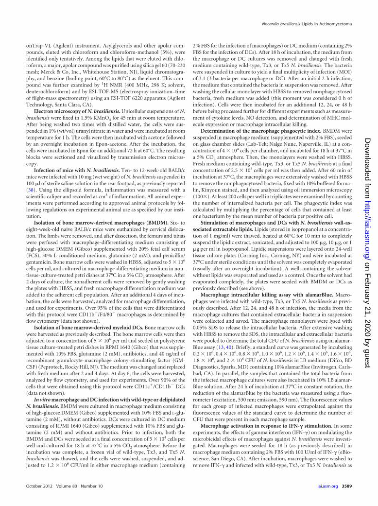
N. brasiliensis lipids induce the production of TGF-� inBMDM and dendritic cells. To further understand the responseof macrophages to the delipidated N. brasiliensis, we analyzed theproduction of TGF-� in BMDM that were infected with wild-typeor Tx5 N. brasiliensis. Macrophages infected with wild-type N.brasiliensis produced significantly higher levels of TGF-� after 24and 48 h of infection than did macrophages infected with Tx5 N.brasiliensis (after 24 h, 322.5 � 39.8 pg/ml with the wild type and187.9 � 27.6 pg/ml with Tx5 N. brasiliensis; P 0.001; after 48 h,341.3 � 76.3 pg/ml with the wild type and 222.9 � 31.4 pg/ml withTx5 N. brasiliensis; P 0.01 [Fig. 4A]).
We next investigated the production of TGF-� by macro-phages in response to different concentrations of the isolated N.brasiliensis wall-associated lipids. We observed a dose-dependentincrease in the production of TGF-� in response to N. brasiliensislipids (223.4 � 21.6 pg/ml, 234.5 � 17.9 pg/ml, and 412.8 � 39.9pg/ml with 1 �g, 10 �g, and 100 �g of N. brasiliensis lipids, respec-tively; P 0.0001 [Fig. 4B]).
We also investigated the production of TGF-� by DCs infectedwith wild-type or Tx5 N. brasiliensis. We observed an increasedproduction of TGF-� in response to wild-type N. brasiliensis at alltime points tested compared to infection with Tx5 N. brasiliensis(P 0.0001 between wild-type and Tx5 N. brasiliensis at all ana-lyzed times [Fig. 4C]). The effect of the N. brasiliensis lipids on theproduction of TGF-� by DCs was also tested. We found that therewas a dose-dependent increase in the production of TGF-� byDCs in response to N. brasiliensis lipids at all of the time points
FIG 2 Chemical analysis of lipids extracted from N. brasiliensis cell wall with benzine. (A) MMT, monomycolate of trehalose; TDM, 6,6=di-mycoloyl trehalose;FMA � others, free mycolic acid and other lipids. Apolar lipid refers to the main apolar lipid isolated, purified, and analyzed. (B) 1H NMR spectrum of the apolarcompound (400 MHz, 298 K, deuterochloroform). (C) 1H NMR spectrum of cord factor, 400 MHz, 298 K, deuterochloroform-deuteromethanol (6:1, vol/vol).The resonances due to the protons of trehalose and mycoloyl substituents are indicated. C2H and C3H, correspond, respectively, to the protons located atpositions C2 and C3 of the mycoloyl substituents. MeOD, deuteromethanol.
Trevino-Villarreal et al.
3592 iai.asm.org Infection and Immunity
on February 21, 2020 by guest
http://iai.asm.org/
Dow
nloaded from

analyzed (after 48 h, 154.3 � 23.4 pg/ml with 10 �g of lipids versus209.7 � 43.5 pg/ml with 100 �g of N. brasiliensis lipids; P 0.0001[Fig. 4D]). These results suggest that the N. brasiliensis surfacelipids are important inducers of the production of TGF-� in bothmacrophages and DCs.
Delipidated N. brasiliensis has a lower survival rate inBMDM than does wild-type N. brasiliensis. An important obser-vation from our in vitro experiments was that after 48 h of infec-tion a higher number of N. brasiliensis colonies were observed inthe BMDM cultures infected with wild-type N. brasiliensis than in
FIG 3 Production of cytokines at different time points by bone marrow-derived macrophages in response to wild-type and Tx5 N. brasiliensis (A throughD) or to increasing concentrations of isolated wall-associated lipids from N. brasiliensis (E through H). Control BMDM were uninfected or unstimulatedwith lipids (0 �g in panels E through H). Data expressed as means � standard deviations. *, P 0.05; **, P 0.001; ***, P 0.0001 as assessed using aBonferroni posttest.
Nocardia brasiliensis Lipids in Actinomycetoma
October 2012 Volume 80 Number 10 iai.asm.org 3593
on February 21, 2020 by guest
http://iai.asm.org/
Dow
nloaded from
the macrophage cultures infected with Tx5 N. brasiliensis (Fig.5B). In addition, when the BMDM cultures were fixed and Kiny-oun stained, we observed that the wild-type N. brasiliensis organ-isms were found predominantly in the extracellular space whereasthe Tx5 N. brasiliensis organisms were found mainly in the cyto-plasm of the BMDM (Fig. 5A). These findings suggest that differ-ences existed in the phagocytosis and the intracellular survivalbetween wild-type and delipidated N. brasiliensis. Therefore, weinvestigated the differences in the phagocytic index for BMDMinfected with wild-type and delipidated N. brasiliensis. We foundthat there was a higher phagocytic index in the BMDM infectedwith the Tx5 N. brasiliensis than in the macrophages infected withwild-type N. brasiliensis (Fig. 5A). The increase in the BMDMphagocytic index was directly proportional to the number of ben-zine extractions that the bacteria were subjected to.
Next, we investigated the effects of the benzine treatment onthe intracellular survival of N. brasiliensis in BMDM. BMDM wereinfected with wild-type or Tx5 N. brasiliensis, and after infectionthe numbers of CFU were assessed at several time points using analamarBlue CFU determination assay. We observed fewer CFU inthe BMDM cultures infected with Tx5 N. brasiliensis than in thoseinfected with wild-type N. brasiliensis (after 48 h, 1.35 � 106 and0.9 � 106 for wild type and Tx5, respectively; P 0.01 [Fig. 5B]).This finding suggests that lipids interfere with the ability of mac-rophages to eliminate N. brasiliensis.
We analyzed the production of IFN-� by BMDM infected withdelipidated and with wild-type N. brasiliensis. It was determinedthat BMDM infected with Tx5 N. brasiliensis underwent an im-portant peak in IFN-� production after 48 h of infection, and thispeak was absent in macrophages infected with wild-type N. brasil-
iensis (234.5 � 37.6 pg/ml, not shown). This finding suggestedthat IFN-� is an important mediator that regulates the response ofmacrophages against the delipidated N. brasiliensis. Next, we in-vestigated the survival of the delipidated N. brasiliensis in IFN-�-activated BMDM. We observed a lower number of N. brasiliensisCFU in the BMDM cultures infected with Tx5 N. brasiliensis thanin the macrophages infected with wild-type N. brasiliensis. In thisexperiment, the reduction in the CFU was higher than the reduc-tion that was observed in the non-IFN-�-activated macrophages(after 48 h, the wild type had 1.1 � 106 and Tx5 had 0.6 � 106
CFU; P 0.001 between wild type and Tx5 [Fig. 5C]).N. brasiliensis wall-associated lipids suppress the IFN-�-me-
diated production of NO by BMDM. Compared to wild-type N.brasiliensis, our in vitro experiments suggested that delipidated N.brasiliensis exhibited a reduced intracellular survival in IFN-�-activated macrophages. This suggested that the wall-associatedlipids affected the microbicidal response of macrophages in re-sponse to IFN-� stimulation. Therefore, we investigated the pro-duction of NO by IFN-�-activated macrophages in response toinfection with wild-type or Tx5 N. brasiliensis. BMDM were acti-vated with IFN-�, infected with wild-type or Tx5 N. brasiliensis,and subsequently assessed for the production of NO at severaltime points after infection. No difference in the production of NObetween the groups was observed after 12 h of infection. However,after 24 h of infection, we observed a statistically significant reduc-tion in the IFN-�-dependent NO production by macrophages in-fected with wild-type N. brasiliensis compared to macrophagesinfected with Tx5 N. brasiliensis (wild type, 12.6 � 5.1 �g/ml; Tx5,50.5 � 13.1 �g/ml; P 0.0001 between the wild type and Tx5 [Fig.5E]). This difference was no longer evident after 48 h of infection
FIG 4 Production of TGF-� at different time points by bone marrow-derived macrophages (BMDM) in response to wild-type and Tx5 N. brasiliensis (A) or toincreasing concentrations of isolated wall-associated lipids from N. brasiliensis (B). Production of TGF-� at different time points by dendritic cells in responseto wild-type and Tx5 N. brasiliensis (C) or to increasing concentrations of isolated wall-associated lipids from N. brasiliensis (D). As controls, cells were uninfectedor unstimulated with lipids (0 �g in panels B and D). Data expressed as means � standard deviations. *, P 0.05; **, P 0.001; ***, P 0.0001 as assessed usinga Bonferroni posttest.
Trevino-Villarreal et al.
3594 iai.asm.org Infection and Immunity
on February 21, 2020 by guest
http://iai.asm.org/
Dow
nloaded from

(Fig. 5E). This suggests that the N. brasiliensis lipids partially in-hibited the production of NO by macrophages in response toIFN-�. Next, we assessed the effect of the isolated N. brasiliensislipids on the production of NO by macrophages in response toIFN-�. We observed a dose-dependent reduction in the produc-tion of NO by BMDM in response to IFN-� stimulation after 24and 48 h of lipid stimulation (Fig. 5F). At these time points, mac-rophages incubated with 100 �g and 10 �g of lipids and stimu-
lated with IFN-� exhibited a statistically significant reduction inthe production of NO compared to macrophages stimulated with1 �g or 0 �g of lipids. (In both cases, the P value was lower than0.0001 [Fig. 5F]).
N. brasiliensis cell-associated lipids interfere with the IFN-�-mediated microbicidal activity of macrophages. Stimulationwith the N. brasiliensis lipids affects the production of NO bymacrophages in response to IFN-�. Because NO is essential to
FIG 5 (A) Phagocytic index of BMDM infected with wild-type or Tx5 N. brasiliensis. (B) Number of CFU obtained in BMDM cultures infected with wild-typeand Tx5 N. brasiliensis after several time intervals. (C) Number of CFU in IFN-�-activated BMDM, infected with wild-type or Tx5 N. brasiliensis. (D) NOproduction in IFN-�-activated BMDM, in response to infection with wild-type or Tx5 N. brasiliensis. (E) NO production by IFN-�-activated BMDM stimulatedwith several concentrations of isolated N. brasiliensis wall-associated lipids. (F) Total number of CFU in cultures of BMDM stimulated with several concentra-tions of wall-associated lipids, activated with IFN-�, and then infected with wild-type or Tx5 N. brasiliensis. Data are expressed as means � standard deviations.*, P 0.05; ***, P 0.0001, as assessed using a Bonferroni posttest.
Nocardia brasiliensis Lipids in Actinomycetoma
October 2012 Volume 80 Number 10 iai.asm.org 3595
on February 21, 2020 by guest
http://iai.asm.org/
Dow
nloaded from

mediate effective killing of intracellular microorganisms by mac-rophages, we hypothesized that stimulation with the N. brasiliensislipids would interfere with the macrophages’ capability to respondto IFN-� and kill N. brasiliensis. To test this hypothesis, BMDMwere stimulated with N. brasiliensis lipids in the presence or ab-sence of IFN-�. The BMDM were then infected with wild-type orTx5 N. brasiliensis. After 48 h of infection, we observed 1.2 �106 � 0.18 � 106 CFU for the N. brasiliensis-infected cultures ofmacrophages that were not stimulated with lipids and were in-fected with wild-type N. brasiliensis (Fig. 5F). We observed signif-icantly fewer CFU of N. brasiliensis in the macrophage culturesthat were not stimulated with lipids and were infected with theTx5 N. brasiliensis (0.85 � 106 � 0.22 � 106; P 0.001). Lipidstimulation decreased the ability of the macrophages to respondto IFN-� stimulation and to kill the Tx5 N. brasiliensis in a dose-dependent fashion. We observed more bacterial CFU in the mac-rophage cultures stimulated with 100 �g of lipids and infectedwith Tx5 N. brasiliensis than in the macrophage cultures that werenot stimulated with lipids prior to infection with wild-type N.brasiliensis (1.31 � 106 � 0.28 � 106 in macrophages stimulatedwith lipids and infected with Tx5 N. brasiliensis compared to 1.2 �106 � 0.18 � 106 CFU in macrophages not stimulated with lipidsand infected with wild-type N. brasiliensis). N. brasiliensis lipids
are not toxic for macrophages even at the 100-�g concentration asdetermined by the trypan blue viability staining (data not shown).Altogether, these results suggest that the N. brasiliensis lipids im-pair the capacity of the macrophages to eliminate N. brasiliensis byinhibiting the macrophages’ response to IFN-� stimulation.
N. brasiliensis wall-associated lipids inhibit the expressionof the MHC-II molecule in BMDM and dendritic cells. In addi-tion to induction of NO production, IFN-� activates antigen pre-sentation and induces MHC-II molecule expression on macro-phages. We analyzed, by flow cytometry, MHC-II moleculeexpression in IFN-�-activated BMDM infected with wild-type orTx5 N. brasiliensis. We observed higher MHC-II expression inIFN-�-activated macrophages infected with Tx5 N. brasiliensisthan in IFN-�-stimulated macrophages infected with wild-type N.brasiliensis (621.3 � 46.7 versus 576.6 � 34.5 fluorescence meanintensity [FMI]; P 0.05 [Fig. 6A]).Compared with macrophagesstimulated with IFN-� alone, we also observed a reduced expres-sion of MHC-II on IFN-�-activated macrophages stimulated with100 �g of purified N. brasiliensis wall-associated lipids (no lipids,571.3 � 48.3 FMI; 10 �g lipids, 581.8 � 15.6 FMI; 100 �g, 487.7FMI; P 0.01 [Fig. 6B]), suggesting that at a high concentration,N. brasiliensis wall-associated lipids can inhibit macrophage ex-pression of MHC-II molecules in response to IFN-�.
FIG 6 (A) Bar graphs showing the expression of MHC-II molecules in IFN-�-activated bone marrow-derived macrophages (BMDM) infected with wild-type or Tx5N. brasiliensis. As controls, BMDM were incubated in medium with (medium IFN-��) or without (medium IFN-��) IFN-�, and expression of MHC-II molecules wasassessed by flow cytometry. (B) Expression of MHC-II molecules in IFN-�-activated BMDM stimulated with several concentrations of N. brasiliensis wall-associatedlipids. As controls, BMDM were incubated in medium with (medium IFN-��) or without (medium IFN-��) IFN-�, and expression of MHC-II molecules was assessedby flow cytometry. (C) MHC-II molecules in dendritic cells infected with wild-type or Tx5 N. brasiliensis. As controls, DCs were incubated in medium alone, andexpression of MHC-II molecules was assessed by flow cytometry. (D) Expression of MHC-II molecules in DCs stimulated with several concentrations of N. brasiliensiswall-associated lipids. As controls, DCs were incubated in medium alone, and expression of MHC-II molecules was assessed by flow cytometry. Data expressed asmeans � standard deviations of the fluorescence mean intensity (FMI) of MHC-II expression. *, P 0.05; ***, P 0.0001 as assessed using a Bonferroni posttest.
Trevino-Villarreal et al.
3596 iai.asm.org Infection and Immunity
on February 21, 2020 by guest
http://iai.asm.org/
Dow
nloaded from

We also investigated MHC-II expression on DCs infected withwild-type or Tx5 N. brasiliensis, finding a higher MHC-II expres-sion on DCs infected with Tx5 N. brasiliensis than on DCs infectedwith wild-type N. brasiliensis (P 0.01 [Fig. 6C]). When the effectof isolated lipids on MHC-II expression was investigated, we alsoobserved lower MHC-II expression on DCs stimulated withhigher concentrations (100 �g) of N. brasiliensis lipids, althoughthis effect was not statistically significant compared to stimulationwith lower lipid concentration (10 �g) or basal MHC-II expres-sion in DCs (Fig. 6D).
N. brasiliensis lipids regulate the expression of T cell co-stimulatory molecules in macrophages and dendritic cells. Inaddition to the presentation of peptide antigens on MHC-II mol-ecules, another requirement for T cell activation is the expressionof T cell costimulatory molecules by antigen-presenting cells.Therefore, we investigated the expression of the costimulatorymolecules CD80, CD86, and CD40 and the negative costimulatorymolecule PDL-2 in macrophages stimulated with IFN-� and in-fected with wild-type or Tx5 N. brasiliensis using flow cytometry.We observed no difference in the expression of CD80 or PDL-2between the groups (Fig. 7A, subpanels a and b). CD86 expressionwas found to be significantly increased in the wild-type and theTx5 groups compared to the positive and negative controls. How-ever, these differences were not statistically significant. The ex-pression of CD40 was higher in the macrophages infected with theTx5 N. brasiliensis than in those infected with wild-type N. brasil-iensis and controls (478.6 � 86.5 FMI in Tx5 versus 324.7 � 27.2FMI in the wild type and 349.8 � 35.9 FMI in the positive control;P 0.01 [Fig. 7A, subpanel c]). These data suggest that N. brasil-iensis lipids decrease the expression of CD40 by IFN-�-activatedmacrophages.
The expression of the costimulatory molecules CD80, CD86,and CD40 and the inhibitory costimulatory molecule PDL-2 inuninfected DCs (exposed to medium alone) and in DCs infectedwith wild-type or with Tx5 N. brasiliensis was also determined. Wedid not observe a difference in the expression of CD86 or PDL-2between the groups (Fig. 7B, subpanels a and b). However, weobserved a statistically significant increase in the expression ofCD80 (524.4 � 19.5 FMI in the Tx5 group versus 410.1 � 34.5FMI in the wild-type group and 358.9 � 39.7 FMI in the medium-alone group; P 0.01 [Fig. 7B, subpanel a]) and CD40 (5,878.5 �195 FMI in the Tx5 group versus 5,010.1 � 345.7 FMI in thewild-type group and 4,589 � 739.7 FMI in the medium-alonegroup; P 0.01 [Fig. 7B, subpanel c]) in DCs infected with theTx5 N. brasiliensis compared to the uninfected DCs and the DCsinfected with the wild-type N. brasiliensis.
Delipidated N. brasiliensis induces a partial protective im-munity against N. brasiliensis. Compared to wild-type N. brasil-iensis, macrophages and DCs infected with delipidated N. brasil-iensis expressed higher levels of MHC-II and the T cellcostimulatory molecules CD80, CD86, and CD40. This suggeststhat the delipidation of N. brasiliensis may result in a more effi-cient presentation of bacterial antigens and could lead to the in-duction of protective immunity against N. brasiliensis. To test thishypothesis, we immunized mice with live Tx5 N. brasiliensis, heat-killed N. brasiliensis (HKB), or only vehicle (PBS) as a control.Fifteen days after immunization, mice where challenged with 10mg of wild-type N. brasiliensis (wet weight) and analyzed for thedevelopment of mycetoma for 120 days. As previously presentedin Fig. 1, mice immunized with the Tx5 N. brasiliensis did not
develop mycetoma. When challenged with the wild-type N. brasil-iensis after 15 days, mice immunized with Tx5 N. brasiliensis diddevelop mycetoma. Importantly, differences were observed in thepatterns of the mycetoma development of the mice immunizedwith the Tx5 N. brasiliensis and the controls. First, mice immu-nized with the Tx5 N. brasiliensis developed significantly less in-flammation than did the mice immunized with HKB or PBS (Fig.7C). Second, there was a delay in the development of the mycet-oma in the mice immunized with the Tx5 N. brasiliensis. Mycet-oma was observed around day 30 or 40 postinfection in mice thatwere immunized with vehicle or HKB, respectively. In mice im-munized with Tx5 N. brasiliensis, mycetoma was observed after 60days of infection (Fig. 7C). Clinically, the mycetoma that devel-oped in mice immunized with Tx5 N. brasiliensis was significantlysmaller, caused a reduced deformation of the footpad, and pre-sented less severe clinical features of actinomycetoma, such as thefistulas and nodules that were present in the mice immunized withvehicle or HKB. These results indicate that immunization with thedelipidated Tx5 N. brasiliensis conferred partial protection againstactinomycetoma development in mice experimentally infectedwith wild-type N. brasiliensis.
DISCUSSION
The cellular and molecular mechanisms involved in the N. brasil-iensis-induced actinomycetoma remain largely unknown. Severalprotein molecules, including proteases (28), catalases (3), and su-peroxide dismutases (3, 36), have been implicated as determinantsof the virulence of Nocardia spp. However, these factors seem toplay a role in the context of macrophage infection and do notaccount for the extensive inflammatory response and the tissuedestruction that characterize the disease induced by the wholemicroorganism. Here, we report that the N. brasiliensis wall-asso-ciated lipids are implicated in the development of actinomyce-toma primarily by inducing a strong inflammatory response, byinhibiting important microbicidal effects by macrophages, and bysuppressing the expression of MHC-II and T cell costimulatorymolecules by macrophages and DCs.
The effect of the N. brasiliensis wall-associated lipids in thedevelopment of actinomycetoma was investigated using chemicaldelipidation with benzine as originally described by Bloch (9, 10).Delipidation with benzine has been extensively used to investigatethe role of the wall-associated lipids in the pathogenesis of micro-organisms of the Mycobacterium genus because it removes the walllipids and glycolipids important for bacterial pathogenesis with-out affecting the viability of the microorganism (9, 10, 21, 44). Inagreement with previous reports on the Mycobacterium spp., wefound that treatment with benzine removed lipids from the out-ermost, electron-dense layer of N. brasiliensis (Fig. 1D) withoutaffecting the viability of the bacteria (Fig. 1A). In addition, asobserved in the Mycobacterium spp., our chemical analyses re-vealed the presence of TDM as one of the predominant lipidsextracted from N. brasiliensis by benzine, but in Mycobacteriumspp., removing TDM does not affect bacterial acid-fastness (9, 10,21, 41) in contrast to our finding showing its modification. Anexplanation for this discrepancy could be related to our observa-tion that, in addition to the extraction of TDM, benzine extractionresulted in the extraction of a highly apolar lipid as the secondmost abundant compound with an isoprenoid structure. Thesetypes of apolar compounds, which also include free mycolates,have been reported to be important constituents of the Coryne-
Nocardia brasiliensis Lipids in Actinomycetoma
October 2012 Volume 80 Number 10 iai.asm.org 3597
on February 21, 2020 by guest
http://iai.asm.org/
Dow
nloaded from

FIG 7 (A) Bar graphs showing the expression of several T cell costimulatory molecules in IFN-�-activated BMDM infected with wild-type or Tx5 N. brasiliensis.As controls, BMDM were incubated in medium with (medium IFN-��) or without (medium IFN-��) IFN-�, and expression of MHC-II molecules was assessedby flow cytometry. (B) Expression of several T cell costimulatory molecules in dendritic cells infected with wild-type or Tx5 N. brasiliensis. As controls, DCs wereincubated in medium alone, and expression of MHC-II molecules was assessed by flow cytometry. Data expressed as means � standard deviations of thefluorescence mean intensity (FMI) of MHC-II expression. *, P 0.05; ***, P 0.0001 as assessed using a Bonferroni posttest. (C) Dot plot showing the level offootpad inflammation (in cm3) in mice immunized with heat-killed N. brasiliensis (HKB), PBS, or Tx5 N. brasiliensis and challenged with wild-type N. brasiliensis15 days after immunization.
Trevino-Villarreal et al.
3598 iai.asm.org Infection and Immunity
on February 21, 2020 by guest
http://iai.asm.org/
Dow
nloaded from

bacterineae cell envelope. Particularly in Mycobacterium tubercu-losis, the loss of mycolates by genetic mutations, such as those thatoccur in the phoP and kasB mutant strains, resulted in the loss ofacid-fastness. These findings are similar to what we observed afterthe benzine treatment of N. brasiliensis (5, 6).
An important finding of our study was that delipidated N.brasiliensis did not induce actinomycetoma, in contrast to the se-vere lesion induced by wild-type N. brasiliensis. Delipidated N.brasiliensis induced a weak, transitory inflammatory process thatwas resolved after 30 days of infection (Fig. 1B). Considering theinflammatory nature of actinomycetoma, our finding stronglysuggests that one of the most important mechanisms by which N.brasiliensis mediates the development of actinomycetoma isthrough the induction of inflammation.
Our laboratory has previously demonstrated that during thecourse of infection with N. brasiliensis there is a high production ofproinflammatory cytokines, particularly IL-1� and IL-6 (43).Based on our in vivo data showing that delipidated N. brasiliensisdoes not induce actinomycetoma and considering the inflamma-tory nature of the disease, we hypothesized that the wall-associ-ated lipids were implicated in the induction of the inflammatoryresponse and found that macrophages infected with delipidatedN. brasiliensis induced lower levels of IL-1�, IL-6, and CCL-2 thandid wild-type N. brasiliensis. These findings mirror what has beenpreviously reported with benzine-delipidated M. tuberculosis.Similarly, a reduction in the production of IL-1� and IL-6 in mac-rophages infected with the benzine-delipidated M. tuberculosiscompared to macrophages infected with wild-type bacteria wasobserved (21). We also showed that macrophages stimulated withN. brasiliensis wall-associated lipids induced the production ofIL-1�, IL-6, and CCL-2 to levels similar to those observed whenthe cells were infected with the complete microorganisms. Thisconfirms the role of the N. brasiliensis wall-associated lipids in thestimulation of the production of the proinflammatory cytokines.
We previously reported that N. brasiliensis does not induce theproduction of TNF-� in experimental actinomycetoma (43).TNF-� mediates important aspects of the immune responseagainst intracellular microorganisms as suggested by the increasedprevalence of tuberculosis in patients receiving anti-TNF-� treat-ment and higher mortality rate in TNF-� knockout (KO) miceinfected with M. tuberculosis (4, 18, 31). It has been reported thathypervirulent M. tuberculosis strains suppress the induction ofTNF-�, and interestingly, a wall-associated phenolic glycolipidhas been implicated in this process (17). Here, we report that theN. brasiliensis wall-associated lipids suppress the production ofTNF-� by BMDM (Fig. 3D and H). Although this was not inves-tigated further in our study, we think that the wall-associated lip-ids might be implicated in the suppression of the production ofTNF-� that we observed in vivo during the course of infectionwith N. brasiliensis (43).
In addition to modulation of cytokines, wall-associated lipidsaffected macrophage phagocytosis of N. brasiliensis, suggestingtheir role as inhibitors of phagocytosis. The most intriguing dif-ference found in our in vitro infection experiments was that thedelipidated N. brasiliensis cells exhibited a reduced survival inmacrophages compared to the wild-type bacteria (Fig. 5B). This issimilar to what has been observed in macrophages that are in-fected with delipidated M. tuberculosis (21). In M. tuberculosis, ithas been found that some of the wall-associated lipids that areremoved after the benzine treatment arrest the maturation of the
phagosome within macrophages by inhibiting the fusion of thephagosome with the lysosome (22). Consequently, delipidated M.tuberculosis has a lower survival within macrophages. Similar ob-servations have been made for N. asteroides (7, 8). Interestingly,the N. asteroides TDM, which is readily removed from the cellenvelope by benzine extraction, has been implicated in this pro-cess (45). Therefore, it seems likely that some of the lipids re-moved from the N. brasiliensis cell envelope after treatment withbenzine inhibit phagosome-lysosome fusion. Thus, it is possiblethat TDM present in our preparations from N. brasiliensis benzineextracts might block phagosome-lysosome fusion in macro-phages. However, due to contamination with other lipids, we can-not exclude the participation of the apolar lipid or the MMT inthis process. Further research with purified lipids must be con-ducted to determine which compound is responsible for the ob-served results with macrophages in vitro.
Reduced survival of the delipidated N. brasiliensis compared tothe wild-type bacteria was more evident when the macrophageswere stimulated with IFN-�. This suggests that, in addition to thepossible arrest in the maturation of the phagosome, the N. brasil-iensis wall-associated lipids interfere with the IFN-�-dependentactivation of macrophages. Our findings support the notion thatthe N. brasiliensis lipids mediate an early suppression of the IFN-�-mediated production of NO by macrophages in response to N.brasiliensis infection. Although the production of NO was restoredlater during the infection, it is possible that the early NO suppres-sion allows bacterial growth to the extent that the macrophages areno longer able to control the infection when NO levels are restored(Fig. 5E).
Suppression of NO production in IFN-�-activated macro-phages by the wall-associated lipids was also demonstrated in ex-periments with purified N. brasiliensis lipids in which the ability ofthe IFN-�-activated macrophages to eliminate delipidated N.brasiliensis was abolished in a dose-dependent manner after stim-ulation with lipids. This strongly suggests that the N. brasiliensiswall-associated lipids impair the ability of the macrophages torespond to IFN-�.
The wall-associated lipids suppressed the expression ofMHC-II molecules on macrophages activated with IFN-�. More-over, stimulation with the wall-associated lipids inhibited the ex-pression of the MHC-II molecules on macrophages in a dose-dependent manner (Fig. 6A and B). Furthermore, the N.brasiliensis wall-associated lipids inhibited the expression of the Tcell costimulatory molecules CD86 and CD40 in macrophagesactivated with IFN-�. A similar observation has been reported formacrophages infected with benzine-delipidated M. tuberculosis,demonstrating higher expression of MHC-II, CD40, and CD86than macrophages infected with wild-type bacteria (24), suggest-ing TDM as the main compound involved in the inhibition of theexpression of MHC-II and T cell costimulatory molecules by mac-rophages. Because of the presence of TDM in our lipid extractpreparation, it is also possible that the effects observed in vitro inthe macrophages stimulated with the N. brasiliensis wall-associ-ated lipids were caused by TDM.
It is clear from the infection experiments that the N. brasiliensiswall-associated lipids regulate the expression of the MHC-II and Tcell costimulatory molecules in DCs and other functions, such asthe induction of the expression of TGF-�. Importantly, the stim-ulation of DCs with wall-associated lipids induced the productionof TGF-� in a dose-dependent fashion. This finding is important
Nocardia brasiliensis Lipids in Actinomycetoma
October 2012 Volume 80 Number 10 iai.asm.org 3599
on February 21, 2020 by guest
http://iai.asm.org/
Dow
nloaded from

because the production of TGF-� by the DCs at the time of anti-gen presentation may skew the response toward one that is Tregmediated, instead of a T-CD4 Th1 and a T-CD8 cytotoxicresponse (47), which is necessary to control infection by intracel-lular microorganisms such as N. brasiliensis. Evidence of this canbe seen in the increased incidence of nocardiosis in immunocom-promised individuals (29). Together with the reduced expressionof antigen-presenting molecules, the N. brasiliensis wall-associ-ated lipids may impair the development of an effective, long-last-ing, protective immune response, which is required to eliminatethe microorganism from the host. We were able to induce partialprotection against the development of actinomycetoma after im-munization with delipidated N. brasiliensis. The reason why onlypartial protection was induced by delipidated N. brasiliensis is notcompletely understood. A possible explanation is that, because ofthe chemical and not the genetic delipidation of the bacteria usedin our study, N. brasiliensis could relipidize in vivo and restore theinhibition of the antigen presentation and the inhibition of T cellactivation. For this reason, we suggest that a genetically delipi-dated strain of N. brasiliensis could be a promising candidate as avaccine for actinomycetoma. Similar approaches to prevent M.tuberculosis infections have led to production of attenuated strainsas candidates for vaccines against pulmonary tuberculosis (5,14, 25).
In conclusion, we demonstrate the implications of the cell wall-associated lipids in the pathogenesis of another member of theCorynebacterineae suborder, N. brasiliensis. Our results suggestthat either decreasing or eliminating the production of certainapolar lipids of the N. brasiliensis cell wall will help to understandpathogenesis and to design a vaccine.
ACKNOWLEDGMENTS
This work fulfills in part the requirements for a Doctorate degree for J.Humberto Trevino-Villarreal.
We thank Julio Sepulveda for his assistance with the electron micros-copy and Adrian Rosas for his technical assistance. We also thankReynaldo Pecina Rodriguez for his skilled assistance in the animal facility.
This work was partially supported by grants from CONACYT2009.2010 Mexico and PAICYT UANL.
REFERENCES1. Beaman BL. 1975. Structural and biochemical alterations of Nocardia
asteroides cell walls during its growth cycle. J. Bacteriol. 123:1235–1253.2. Beaman BL, Beaman L. 1994. Nocardia species: host-parasite relation-
ships. Clin. Microbiol. Rev. 7:213–264.3. Beaman BL, Black CM, Doughty F, Beaman L. 1985. Role of superoxide
dismutase and catalase as determinants of pathogenicity of Nocardia as-teroides: importance in resistance to microbicidal activities of humanpolymorphonuclear neutrophils. Infect. Immun. 47:135–141.
4. Bean AG, et al. 1999. Structural deficiencies in granuloma formation inTNF gene-targeted mice underlie the heightened susceptibility to aerosolMycobacterium tuberculosis infection, which is not compensated for bylymphotoxin. J. Immunol. 162:3504 –3511.
5. Bhatt A, et al. 2007. Deletion of kasB in Mycobacterium tuberculosiscauses loss of acid-fastness and subclinical latent tuberculosis in immuno-competent mice. Proc. Natl. Acad. Sci. U. S. A. 104:5157–5162.
6. Bhatt A, Molle V, Besra GS, Jacobs WR, Kremer L. 2007. The Myco-bacterium tuberculosis FAS-II condensing enzymes: their role in mycolicacid biosynthesis, acid-fastness, pathogenesis and in future drug develop-ment. Mol. Microbiol. 64:1442–1454.
7. Black CM, Paliescheskey M, Beaman BL, Donovan RM, Goldstein E.1986. Acidification of phagosomes in murine macrophages: blockage byNocardia asteroides. J. Infect. Dis. 154:952–958.
8. Black CM, Paliescheskey M, Beaman BL, Donovan RM, Goldstein E.1986. Modulation of lysosomal protease-esterase and lysozyme in Kupffer
cells and peritoneal macrophages infected with Nocardia asteroides. In-fect. Immun. 54:917–919.
9. Bloch H. 1950. Studies on the virulence of tubercle bacilli; isolation andbiological properties of a constituent of virulent organisms. J. Exp. Med.91:197–218.
10. Bloch H. 1950. Studies on the virulence of tubercle bacilli: the relationshipof the physiological state of the organisms to their pathogenicity. J. Exp.Med. 92:507–526.
11. Brennan PJ, Nikaido H. 1995. The envelope of mycobacteria. Annu. Rev.Biochem. 64:29 – 63.
12. Brennan PJ. 2003. Structure, function, and biogenesis of the cell wall ofMycobacterium tuberculosis. Tuberculosis (Edinb.) 83:91–97.
13. Chacon-Moreno BE, et al. 2009. Efficacy of ciprofloxacin and moxifloxa-cin against Nocardia brasiliensis in vitro and in an experimental model ofactinomycetoma in BALB/c mice. Antimicrob. Agents Chemother. 53:295–297.
14. Copenhaver RH, et al. 2004. A mutant of Mycobacterium tuberculosisH37Rv that lacks expression of antigen 85A is attenuated in mice butretains vaccinogenic potential. Infect. Immun. 72:7084 –7095.
15. Daffe M, Laneelle MA, Puzo G. 1983. Structural elucidation by fielddesorption and electron-impact mass spectrometry of the c-mycosidesisolated from Mycobacterium smegmatis. Biochim. Biophys. Acta 751:439 – 443.
16. Daffe M, Draper P. 1998. The envelope layers of mycobacteria withreference to their pathogenicity. Adv. Microb. Physiol. 39:131–203.
17. De Lorimier R, Hellinga HW, Spicer LD. 1996. NMR studies of struc-ture, hydrogen exchange, and main-chain dynamics in a disrupted-coremutant of thioredoxin. Protein Sci. 5:2552–2565.
18. Flynn JL, et al. 1995. Tumor necrosis factor-alpha is required in theprotective immune response against Mycobacterium tuberculosis in mice.Immunity 2:561–572.
19. Han Y, Kawada N, Yano I. 1998. Granuloma formation and in vitromacrophage activation in mice by mycoloyl glycolipids from Nocardiaasteroides and related taxa. Osaka City Med. J. 44:201–217.
20. Hoffmann C, Leis A, Niederweis M, Plitzko JM, Engelhardt H. 2008.Disclosure of the mycobacterial outer membrane: cryo-electron tomogra-phy and vitreous sections reveal the lipid bilayer structure. Proc. Natl.Acad. Sci. U. S. A. 105:3963–3967.
21. Indrigo J, Hunter RLJ, Actor JK. 2002. Influence of trehalose 6,6=-dimycolate (TDM) during mycobacterial infection of bone marrow mac-rophages. Microbiology 148:1991–1998.
22. Indrigo J, Hunter RLJ, Actor JK. 2003. Cord factor trehalose 6,6=-dimycolate (TDM) mediates trafficking events during mycobacterial in-fection of murine macrophages. Microbiology 149:2049 –2059.
23. Ioneda T, Beaman BL, Viscaya L, Almeida ET. 1993. Composition andtoxicity of diethyl ether soluble lipids from Nocardia asteroides GUH-2and Nocardia asteroides 10905. Chem. Phys. Lipids 65:171–178.
24. Kan-Sutton C, Jagannath C, Hunter R, Jr. 2009. Trehalose 6,6=-dimycolate on the surface of Mycobacterium tuberculosis modulates sur-face marker expression for antigen presentation and costimulation in mu-rine macrophages. Microbes Infect. 11:40 – 48.
25. Katti MK, et al. 2008. The fbpA mutant derived from Mycobacteriumtuberculosis H37Rv has an enhanced susceptibility to intracellular antimi-crobial oxidative mechanisms, undergoes limited phagosome maturationand activates macrophages and dendritic cells. Cell. Microbiol. 10:1286 –1303.
26. Klein CE, Steinmayer T, Kaufmann D, Weber L, Brocker E-B. 1991.Identification of a melanoma progression antigen as integrin VLA-2. J.Invest. Dermatol. 96:281–284.
27. Lichon V, Khachemoune A. 2006. Mycetoma: a review. Am. J. Clin.Dermatol. 7:315–321.
28. Licón-Trillo Á, Castro-Corona MÁ, Salinas-Carmona MC. 2003. Im-munogenicity and biophysical properties of a protease involved in patho-genesis of mycetoma. FEMS Immunol. Med. Microbiol. 37:37– 44.
29. Martínez R, Reyes S, Menéndez R. 2008. Pulmonary nocardiosis: riskfactors, clinical features, diagnosis and prognosis. Curr. Opin. Pulm. Med.14:219 –227.
30. Minnikin DE. 1982. Lipids: complex lipids, their chemistry, biosyn-thesis and roles, p 95–184. In Ratledge C (ed), The biology of themycobacteria: physiology, identification and classification. AcademicPress, San Diego, CA.
31. Nacci F, Matucci-Cerinic M. 2011. Tuberculosis and other infections in
Trevino-Villarreal et al.
3600 iai.asm.org Infection and Immunity
on February 21, 2020 by guest
http://iai.asm.org/
Dow
nloaded from
the anti-tumour necrosis factor-alpha (anti-TNF-�) era. Best Pract. Res.Clin. Rheumatol. 25:375–388.
32. Niederweis M, Danilchanka O, Huff J, Hoffmann C, Engelhardt H.2010. Mycobacterial outer membranes: in search of proteins. Trends Mi-crobiol. 18:109 –116.
33. Niescher S, Wray V, Lang S, Kaschabek SR, Schlömann M. 2006.Identification and structural characterisation of novel trehalose dinocar-diomycolates from n-alkane-grown Rhodococcus opacus 1CP. Appl. Mi-crobiol. Biotechnol. 70:605– 611.
34. Puech V, et al. 2001. Structure of the cell envelope of corynebacteria:importance of the non-covalently bound lipids in the formation of the cellwall permeability barrier and fracture plane. Microbiology 147:1365–1382.
35. Ratledge C, Patel PV. 1976. Isolation, properties and taxonomic rele-vance of lipid-soluble, iron-binding compounds (the nocobactins) fromNocardia. J. Gen. Microbiol. 93:141–152.
36. Revol A, Espinoza-Ruiz M, Medina-Villanueva I, Salinas-Carmona MC.2006. Expression of superoxide dismutase during the early infection ofmurine peritoneal macrophages. Can. J. Microbiol. 52:1255–1260.
37. Salinas-Carmona MC. 2000. Nocardia brasiliensis: from microbe to hu-man and experimental infections. Microbes Infect. 2:1373–1381.
38. Salinas-Carmona MC, Torres-Lopez E, Ramos AI, Licon-Trillo A, Gon-zalez-Spencer D. 1999. Immune response to Nocardia brasiliensis anti-gens in an experimental model of actinomycetoma in BALB/c mice. Infect.Immun. 67:2428 –2432.
39. Salinas-Carmona MC, Welsh O, Casillas SM. 1993. Enzyme-linkedimmunosorbent assay for serological diagnosis of Nocardia brasiliensisand clinical correlation with mycetoma infections. J. Clin. Microbiol. 31:2901–2906.
40. Shiloh MU, Ruan J, Nathan C. 1997. Evaluation of bacterial survival andphagocyte function with a fluorescence-based microplate assay. Infect.Immun. 65:3193–3198.
41. Silva CL, Ekizlerian SM, Fazioli RA. 1985. Role of cord factor in themodulation of infection caused by mycobacteria. Am. J. Pathol. 118:238 –247.
42. Silva CL, Tincani I, Brandao Filho SL, Faccioli LH. 1988. Mouse ca-chexia induced by trehalose dimycolate from Nocardia asteroides. J. Gen.Microbiol. 134:1629 –1633.
43. Solis-Soto JM, et al. 2008. In situ detection and distribution of inflam-matory cytokines during the course of infection with Nocardia brasilien-sis. Histol. Histopathol. 23:573–581.
44. Sorkin E, Erlenmeyer H, Bloch H. 1952. Purification of a lipid material(‘cord factor’) obtained from young cultures of tubercle bacilli. Nature170:124.
45. Spargo BJ, Crowe LM, Ioneda T, Beaman BL, Crowe JH. 1991. Cordfactor (alpha,alpha-trehalose 6,6=-dimycolate) inhibits fusion betweenphospholipid vesicles. Proc. Natl. Acad. Sci. U. S. A. 88:737–740.
46. Welsh O, Vera-Cabrera L, Salinas-Carmona MC. 2007. Mycetoma. Clin.Dermatol. 25:195–202.
47. Yamazaki S, Steinman RM. 2009. Dendritic cells as controllers of anti-gen-specific Foxp3� regulatory T cells. J. Dermatol. Sci. 54:69 –75.
48. Yano I, et al. 1987. Isolation of mycolic acid-containing glycolipids inNocardia rubra and their granuloma forming activity in mice. J. Pharma-cobiodyn. 10:113–123.
49. Zuber B, et al. 2008. Direct visualization of the outer membrane ofmycobacteria and corynebacteria in their native state. J. Bacteriol. 190:5672–5680.
Nocardia brasiliensis Lipids in Actinomycetoma
October 2012 Volume 80 Number 10 iai.asm.org 3601
on February 21, 2020 by guest
http://iai.asm.org/
Dow
nloaded from