newsletter for optometrists - centre for eye health · newsletter for optometrists. ... figure 5:...
TRANSCRIPT
cfeh.com.au
Thirty-year-old George presented to his optometrist with blurry vision in his left eye. His last eye examination was approximately 18 months ago.
George has had glasses on a number of occasions in the past. However, he has always found it difficult to adapt to new glasses and so hasn’t ever persisted with wearing them.
General health was good, with no significant history of systemic disease or medication use.
Fundoscopy in each of George’s eyes was unremarkable. What could be causing the issue with George’s left eye?
Case RepoRT
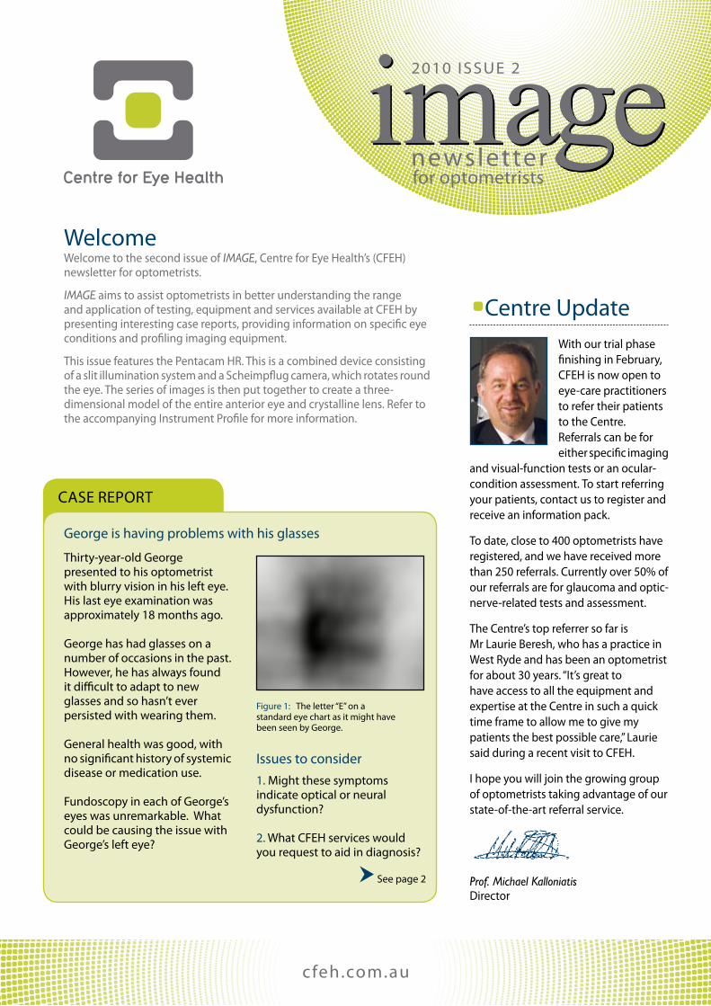
Figure 1: The letter “e” on a standard eye chart as it might have been seen by George.
George is having problems with his glasses
Welcome to the second issue of IMAGE, Centre for eye Health’s (CFeH) newsletter for optometrists.
IMAGE aims to assist optometrists in better understanding the range and application of testing, equipment and services available at CFeH by presenting interesting case reports, providing information on specific eye conditions and profiling imaging equipment.
This issue features the pentacam HR. This is a combined device consisting of a slit illumination system and a scheimpflug camera, which rotates round the eye. The series of images is then put together to create a three-dimensional model of the entire anterior eye and crystalline lens. Refer to the accompanying Instrument profile for more information.
Issues to consider1. Might these symptoms indicate optical or neural dysfunction?
2. What CFeH services would you request to aid in diagnosis?
see page 2
Disclaimer: This newsletter is not intended to provide or substitute advice through the appropriate health service providers. Although every care is taken by CFEH to ensure that this newsletter is free from any error or inaccuracy, CFEH does not make any representation or warranty regarding the currency, accuracy or completeness of this newsletter.
Copyright: © 2010, Centre for Eye Health Limited. All images and content in this letter are the property of Centre for Eye Health Limited and cannot be reproduced without prior written permission of the Director, Centre for Eye Health Limited.
Centre UpdateWith our trial phase finishing in February, CFeH is now open to eye-care practitioners to refer their patients to the Centre. Referrals can be for either specific imaging
and visual-function tests or an ocular-condition assessment. To start referring your patients, contact us to register and receive an information pack.
To date, close to 400 optometrists have registered, and we have received more than 250 referrals. Currently over 50% of our referrals are for glaucoma and optic-nerve-related tests and assessment.
The Centre’s top referrer so far is Mr Laurie Beresh, who has a practice in West Ryde and has been an optometrist for about 30 years. “It’s great to have access to all the equipment and expertise at the Centre in such a quick time frame to allow me to give my patients the best possible care,” Laurie said during a recent visit to CFeH.
I hope you will join the growing group of optometrists taking advantage of our state-of-the-art referral service.
Prof. Michael Kalloniatis Director
image2010 IssUe 2
newsletter
Welcome
for optometrists

Figure 2: pentacam assessment of the right eye showing sagittal curvature of the anterior corneal surface.
Figure 4: pentacam assessment of the left eye showing sagittal curvature of the anterior corneal surface.
Figure 5: Belin/ambrosio enhanced ectasia analysis of the left eye cornea. Results show clear ectasia of the central anterior and posterior surfaces.
Case RepoRT (continued)
cfeh.com.aupage 2
Results and DiscussionDifferential diagnoses for George include media abnormalities involving the cornea, crystalline lens or vitreous, as well as neural abnormalities involving the retina and/or optic nerve.
George’s spectacle refraction and visual acuities were right eye -0.75/ -1.00 x 7 6/6, and left eye +0.50/ -2.50 x 130 6/7.5. His optometrist suspected keratoconus based upon early clinical signs of the condition, including a recent change in refraction and steeper than average keratometry readings in the left eye.
The referring optometrist requested that CFeH perform corneal topography and cornea and anterior-segment assessment using the pentacam HR. output from this instrument includes anterior and posterior surface corneal topography, antero-posterior thickness across the cornea, and anterior chamber depth measures.
elevation maps for the right eye showed little or no elevation (compared to a reference sphere) of the central anterior corneal surface and mild elevation of the inferior central region of the posterior corneal surface. The pachymetry map in this eye revealed the inferior central cornea to be mildly thinner than the corneal apex, while the sagittal curvature map (Figure 2) showed asymmetric corneal astigmatism (see arrows) with the inferior central cornea steeper (7.65 mm) than the curvature at the corneal apex (8.12 mm).
imagenewsletter
Belin/ambrosio analysis also indicated significant protrusion of the central corneal region (see arrows in Figure 5) on both the anterior and posterior corneal surfaces, with the effect more marked on the posterior surface. These findings indicate developing keratoconus in the right eye and established keratoconus in the left eye, which account for George’s blurry vision in the left eye and poor acceptance of spectacle lens correction.
This case report indicates a frequently-observed pathway from patient symptoms to disease diagnosis for keratoconus. It also highlights the diagnostic value provided by assessment of corneal topography, and how referral for this service to another practitioner with a corneal topographer – or, in this instance, CFeH – can establish a diagnosis in individuals suspected of having keratoconus.
George’s left eye showed mild elevation of the
anterior corneal surface and moderate elevation
of the posterior corneal surface.
Prepared by: Dr Andrew Whatham, CFEH Principal Optometrist.
Belin/ambrosio analysis (Figure 3) determines the elevation of the central cornea relative to a reference sphere fitted to the peripheral cornea only (i.e., excluding the central cornea). This analysis for George revealed no significant elevation of the right anterior corneal surface (left map). However, it did show mild central elevation of the posterior corneal surface (right map and indicated by the arrow). In contrast to the right eye, the left eye showed mild elevation of the anterior corneal surface and moderate elevation of the posterior corneal surface.
The pachymetry map showed little difference between corneal thickness at the thinnest location and that at the corneal apex, while the sagittal curvature map (Figure 4) showed increased curvature (see arrow) at the inferior central location (6.78 mm) compared to the corneal apex (7.46 mm).
Figure 3: Belin/ambrosio enhanced ectasia analysis of the right eye cornea. Results show mild ectasia of the central posterior surface of the cornea.

eye-Condition spotlight
sTaFF pRoFILe
1300 421 960
KeratoconusKeratoconus occurs when the cornea becomes progressively more conical in shape secondary to central or paracentral stromal thinning and protrusion1. The area of thinning and protrusion most commonly occurs inferiorly, but may also develop laterally or superior to the central cornea. It is considered to be a bilateral condition, although onset and presentation can be quite asymmetrical.
The affected region of the cornea increases in curvature, and becomes relatively steeper than normal. This typically produces irregular myopic astigmatism that frequently reduces best-corrected separate visual acuity. Corneal refractive aberrations are also affected by the disease, with coma being the largest contributor2.
Measurement of corneal surface topography is the standard of care for evaluating individuals with suspected corneal irregularities such as keratoconus. one instrument used for this purpose is the pentacam HR (see the accompanying Instrument profile).
a common management modality for individuals who have keratoconic eyes is to fit the conically-shaped cornea with rigid contact lenses. The aim is to reduce irregular astigmatism and higher-order aberrations.
More recently, soft contact lenses have been manufactured that try to correct the exaggerated higher-order aberrations in keratoconic eyes3,4.
If corneal thinning and protrusion progress to the point at which vision and/or ocular comfort is significantly poor with rigid contact lenses, or globe integrity is threatened, then corneal grafting is considered.
a recent strategy to minimise the progression of keratoconus is collagen
page 3
image
andrew Whatham joined Centre for eye Health (CFeH) from its establishment in June 2009, and subsequently took up a position as a principal optometrist.
andrew brings to CFeH both a clinical and vision-research background. He has worked in private optometric practice in australia, and completed a Dphil in physiology through the University of oxford in the UK. This was followed by stints in
clinical research at the ophthalmology Clinic, Geneva University Hospitals in switzerland; the school of optometry at The University of NsW (UNsW); and the Institute for eye Research (now the Brien Holden Vision Institute) on the UNsW campus.
“Working at CFeH, I see how the available ocular diagnostic instrumentation makes a tangible difference to the community,” andrew says. “It is exciting to be part of this new model of eye care.”
crosslinking. The aim is to strengthen the bonds between collagen fibres in the anterior corneal stroma. Recent reports indicate that crosslinking slows progression of the corneal ectasia and even improves vision to some extent5.
Corneal topographic assessment is an indispensable tool in tracking the progress of keratoconus and evaluating treatments. The pentacam HR is particularly useful, because in addition to the universally-measured anterior corneal surface topography, it measures posterior corneal topography, topographic pachymetry and corneal refractive aberrations. as such, the pentacam can detect posterior corneal changes that may occur earlier, or to a greater extent, than anterior surface changes in keratoconic eyes and possibly lead to earlier diagnosis of the condition.
Figure 6: scheimpflug image showing a vertical cross-section (left is superior cornea) through a keratoconic cornea.
Dr Andrew WhathamPrincipal Optometrist
Corneal topographicassessment is an
indispensable tool in tracking the progress
of keratoconus.

References
page 4
Centre for eye HealthThe University of New south Wales,Rupert Myers Building (south wing), Kensington NsW 2052
ph: (02) 8115 0700/1300 421 960Fax: (02) 8115 0799 email: [email protected]
Contact Details our Clinical staffprofessor Michael Kalloniatis, Director Bsc optom, Msc optom, phD, GradCertocTher
associate professor David pye, Deputy Director Boptom (Hons), Moptom, FCLsa
Consultant ophthalmologists, through south eastern sydney Illawarra area Health service
anna Delmadoros, principal optometrist Boptom (Hons), Moptom
paula Katalinic, principal optometrist Boptom (Hons), Moptom, GradCertocTher
Dr andrew Whatham, principal optometrist Boptom (Hons), Dphil, GradCertocTher
Michael Yapp, principal optometrist Boptom (Hons), Moptom, GradCertocTher
agnes Choi, optometrist Boptom, GradCertocTher
George Rennie, optometrist Boptom (Hons)
cfeh.com.au
Centre for eye Health is an initiative of Guide Dogs NsW/aCT and The University of New south Wales
Case Report – Claude is having trouble seeing in low lightat his first eye examination three years ago, Claude, aged 50, complained of having trouble reading small print in the evening. He was prescribed reading glasses and told to return if he noticed his vision worsening. In February 2010, Claude’s optometrist referred him to CFeH after noticing attenuated arterioles and some mild pigment clumping on direct ophthalmoscopy.
What CFeH services would you request to aid in managing Claude’s condition? Instrument to be profiled optomap noninvasively generates an ultra-widefield digital image of the retina. It is extremely useful for documenting retinal pathology affecting the central and mid-peripheral retina.
Centre For eye HealthCFeH is a free referral service assisting eye-care practitioners to optimally manage their patients. With 24 state-of-the-art instruments in one location, the Centre offers a range of advanced eye imaging and visual assessment services.
1. Kanski JJ (2007) Clinical ophthalmology, 6th edition. Butterworth-Heinemann elsevier, edinburgh, UK.
2. Maeda N, Fujikado T, Kuroda T, Mihashi T, Hirohara Y, Nishida K, Watanabe H, Tano Y (2002). Wavefront aberrations measured with Hartmann-shack sensor in patients with keratoconus. ophthalmology 109(11):1996-2003.
3. Katsoulos C, Karageorgiadis L, Vasileiou N, Mousafeiropoulos T, asimellis G (2009). Customized hydrogel contact lenses for keratoconus incorporating correction for vertical coma aberration. ophthalmic physiol opt. 29(3):321-329.
4. Marsack JD, parker Ke, applegate Ra (2008). performance of wavefront-guided soft lenses in three keratoconus subjects. optom Vis sci. 85(12):e1172-8.
5. Vinciguerra p, albè e, Trazza s, seiler T, epstein D (2009). Intra- operative and postoperative effects of corneal collagen cross-linking on progressive keratoconus. arch ophthalmol. 127(10):1258-65.
Disclaimer: This newsletter is not intended to provide or substitute advice through the appropriate health service providers. Although every care is taken by CFEH to ensure that this newsletter is free from any error or inaccuracy, CFEH does not make any representation or warranty regarding the currency, accuracy or completeness of this newsletter.
Copyright: © 2010, Centre for Eye Health Limited. All images and content in this letter are the property of Centre for Eye Health Limited and cannot be reproduced without prior written permission of the Director, Centre for Eye Health Limited.
Next Issue
printed in March 2010 on recycled paper.