new refractive surgery procedures and their implications ... · new refractive surgery procedures...
TRANSCRIPT

New Refractive Surgery Procedures and Their Implications for Aviation Safety
Van B. NakagawaraKathryn J. WoodRon W. MontgomeryCivil Aerospace Medical InstituteFederal Aviation AdministrationOklahoma City, OK 73125
April 2006
Final Report
DOT/FAA/AM-06/9Office of Aerospace MedicineWashington, DC 20591

2
NOTICE
This document is disseminated under the sponsorship of the U.S. Department of Transportation in the interest
of information exchange. The United States Government assumes no liability for the contents thereof.
___________
This publication and all Office of Aerospace Medicine technical reports are available in full-text from the Civil Aerospace Medical Institute’s publications Web site:
www.faa.gov/library/reports/medical/oamtechreports/index.cfm

i
Technical Report Documentation Page 1. Report No. 2. Government Accession No. 3. Recipient's Catalog No.
DOT/FAA/AM-06/9 4. Title and Subtitle 5. Report Date
April 2006 New Refractive Surgery Procedures and Their Implicationsfor Aviation Safety 6. Performing Organization Code
7. Author(s) 8. Performing Organization Report No. Nakagawa VB, Wood KJ, Montgomery RW
9. Performing Organization Name and Address 10. Work Unit No. (TRAIS) FAA Civil Aerospace Medical Institute P.O. Box 25082 11. Contract or Grant No. Oklahoma City, OK 73125
12. Sponsoring Agency name and Address 13. Type of Report and Period Covered Office of Aerospace Medicine Federal Aviation Administration 800 Independence Ave., S.W. Washington, DC 20591 14. Sponsoring Agency Code
15. Supplemental Notes
16. Abstract Since the early 1980s, civil airmen have been allowed to correct refractive error (i.e., myopia, hyperopia, astigmatism) with corrective surgery. Prior Federal Aviation Administration research studies have shown that the number of civil airmen with refractive surgery continues to increase. A study that reviewed refractive surgery use in civil airmen for the years 1994-96, reported that the largest percentage had radial keratotomy (RK). A similar study that reported on the years 1996-2001, however, showed that there had been a substantial increase in the percentage of airmen with laser refractive surgery, i.e., photorefractive keratectomy (PRK) and laser in situ keratomileusis (LASIK). A reference guide on refractive surgery was published in September of 1998 (DOT/FAA/AM-98/25); however, at that time long-term clinical data on PRK and LASIK were not available. The introduction of new refractive surgical techniques (e.g., laser epithelial keratomileusis [LASEK], laser thermal keratoplasty [LTK], conductive keratoplasty [CK], Intacs™, phakic IOLs, and presbyopia surgeries) and technology (e.g., wavefront-guided systems, Femtosecond Lasers, inlays, and onlays) has further added to concerns regarding the use of refractive surgical procedures by aviators. In order to provide the aviation community with information to formulate administrative decisions and policies associated with existing and emerging refractive surgical procedures, this paper reviews current procedures and discusses their applicability in the civil aviation environment.
17. Key Words 18. Distribution Statement
Aviation Vision, Aeromedical Certification,Refractive Surgery
Document is available to the public through the Defense Technical Information Center, Ft. Belvior, VA 22060; and the National Technical Information Service, Springfield, VA 22161
19. Security Classif. (of this report) 20. Security Classif. (of this page) 21. No. of Pages 22. Price Unclassified Unclassified 45
Form DOT F 1700.7 (8-72) Reproduction of completed page authorized

iii
TABLE OF CONTENTS
INTRODUCTION . . . . . . . . . . . . . . . . . . . . . . . . . . . . . . . . . . . . . . . . . . . . . . . . . . . . . . . . . . . . . . . . . . . . . 1
REFRACTIVE CONDITIONS . . . . . . . . . . . . . . . . . . . . . . . . . . . . . . . . . . . . . . . . . . . . . . . . . . . . . . . . . . . . 1
Myopia . . . . . . . . . . . . . . . . . . . . . . . . . . . . . . . . . . . . . . . . . . . . . . . . . . . . . . . . . . . . . . . . . . . . . . . . . . 1
Hyperopia . . . . . . . . . . . . . . . . . . . . . . . . . . . . . . . . . . . . . . . . . . . . . . . . . . . . . . . . . . . . . . . . . . . . . . . . 2
Astigmatism . . . . . . . . . . . . . . . . . . . . . . . . . . . . . . . . . . . . . . . . . . . . . . . . . . . . . . . . . . . . . . . . . . . . . . . 2
Presbyopia . . . . . . . . . . . . . . . . . . . . . . . . . . . . . . . . . . . . . . . . . . . . . . . . . . . . . . . . . . . . . . . . . . . . . . . . 2
REFRACTIVE ERROR MODIFICATION TECHNIQUES . . . . . . . . . . . . . . . . . . . . . . . . . . . . . . . . . . . . . 2
Radial Keratotomy . . . . . . . . . . . . . . . . . . . . . . . . . . . . . . . . . . . . . . . . . . . . . . . . . . . . . . . . . . . . . . . . . . 3
Photorefractive Keratectomy . . . . . . . . . . . . . . . . . . . . . . . . . . . . . . . . . . . . . . . . . . . . . . . . . . . . . . . . . . 3
Laser in situ Keratomileusis . . . . . . . . . . . . . . . . . . . . . . . . . . . . . . . . . . . . . . . . . . . . . . . . . . . . . . . . . . . 6
Laser Epithelial Keratomileusis . . . . . . . . . . . . . . . . . . . . . . . . . . . . . . . . . . . . . . . . . . . . . . . . . . . . . . . . . 9
Thermokeratoplasty . . . . . . . . . . . . . . . . . . . . . . . . . . . . . . . . . . . . . . . . . . . . . . . . . . . . . . . . . . . . . . . . 10
Conductive Keratoplasty . . . . . . . . . . . . . . . . . . . . . . . . . . . . . . . . . . . . . . . . . . . . . . . . . . . . . . . . . . . . 11
Intacs™ . . . . . . . . . . . . . . . . . . . . . . . . . . . . . . . . . . . . . . . . . . . . . . . . . . . . . . . . . . . . . . . . . . . . . . . . . 12
Clear Lens Extraction . . . . . . . . . . . . . . . . . . . . . . . . . . . . . . . . . . . . . . . . . . . . . . . . . . . . . . . . . . . . . . . 13
NEW TECHNOLOGY . . . . . . . . . . . . . . . . . . . . . . . . . . . . . . . . . . . . . . . . . . . . . . . . . . . . . . . . . . . . . . . . . 14
Phakic Intraocular Lenses . . . . . . . . . . . . . . . . . . . . . . . . . . . . . . . . . . . . . . . . . . . . . . . . . . . . . . . . . . . . 15
PRESBYOPIA SURGERY . . . . . . . . . . . . . . . . . . . . . . . . . . . . . . . . . . . . . . . . . . . . . . . . . . . . . . . . . . . . . . . 15
Anterior Ciliary Sclerotomy . . . . . . . . . . . . . . . . . . . . . . . . . . . . . . . . . . . . . . . . . . . . . . . . . . . . . . . . . . 16
Laser Presbyopia Reversal . . . . . . . . . . . . . . . . . . . . . . . . . . . . . . . . . . . . . . . . . . . . . . . . . . . . . . . . . . . . 16
Surgical Reverse Presbyopia . . . . . . . . . . . . . . . . . . . . . . . . . . . . . . . . . . . . . . . . . . . . . . . . . . . . . . . . . . 17
WAVEFRONT TECHNOLOGY . . . . . . . . . . . . . . . . . . . . . . . . . . . . . . . . . . . . . . . . . . . . . . . . . . . . . . . . . . 18
FEMTOSECOND LASER . . . . . . . . . . . . . . . . . . . . . . . . . . . . . . . . . . . . . . . . . . . . . . . . . . . . . . . . . . . . . . . 19
Inlays And Onlays . . . . . . . . . . . . . . . . . . . . . . . . . . . . . . . . . . . . . . . . . . . . . . . . . . . . . . . . . . . . . . . . . 20
AEROMEDICAL ISSUES . . . . . . . . . . . . . . . . . . . . . . . . . . . . . . . . . . . . . . . . . . . . . . . . . . . . . . . . . . . . . . . 21
SUMMARY AND DISCUSSION . . . . . . . . . . . . . . . . . . . . . . . . . . . . . . . . . . . . . . . . . . . . . . . . . . . . . . . . . 21
REFERENCES . . . . . . . . . . . . . . . . . . . . . . . . . . . . . . . . . . . . . . . . . . . . . . . . . . . . . . . . . . . . . . . . . . . . . . . . 23

iv
FIGURES AND TABLES
Figure 1 . Myopia . . . . . . . . . . . . . . . . . . . . . . . . . . . . . . . . . . . . . . . . . . . . . . . . . . . . . . . . . . . . . . . . . 1
Figure 2 . Hyperopia . . . . . . . . . . . . . . . . . . . . . . . . . . . . . . . . . . . . . . . . . . . . . . . . . . . . . . . . . . . . . . . 2
Figure 3 . Astigmatism . . . . . . . . . . . . . . . . . . . . . . . . . . . . . . . . . . . . . . . . . . . . . . . . . . . . . . . . . . . . . . 2
Figure 4 . Accommodation . . . . . . . . . . . . . . . . . . . . . . . . . . . . . . . . . . . . . . . . . . . . . . . . . . . . . . . . . . 2
Figure 5 . Radial Keratotomy (RK) . . . . . . . . . . . . . . . . . . . . . . . . . . . . . . . . . . . . . . . . . . . . . . . . . . . . 3
Figure 6 . Photorefractive Keratectomy (PRK) . . . . . . . . . . . . . . . . . . . . . . . . . . . . . . . . . . . . . . . . . . . . 3
Figure 7 . Microkeratome cutting a corneal flap . . . . . . . . . . . . . . . . . . . . . . . . . . . . . . . . . . . . . . . . . . 7
Figure 8 . Excimer laser ablating tissue from corneal stroma during LASIK . . . . . . . . . . . . . . . . . . . . . . 7
Figure 9 . Creation of the LASIK flap . . . . . . . . . . . . . . . . . . . . . . . . . . . . . . . . . . . . . . . . . . . . . . . . . . 7
Figure 10 . Laser epithelial keratomileusis . . . . . . . . . . . . . . . . . . . . . . . . . . . . . . . . . . . . . . . . . . . . . . . 10
Figure 11 . Laser Thermal Keratoplasty . . . . . . . . . . . . . . . . . . . . . . . . . . . . . . . . . . . . . . . . . . . . . . . . . 11
Figure 12 . Conductive Keratoplasty . . . . . . . . . . . . . . . . . . . . . . . . . . . . . . . . . . . . . . . . . . . . . . . . . . . 11
Figure 13 . Steepening of the cornea to correct for hyperopia . . . . . . . . . . . . . . . . . . . . . . . . . . . . . . . . 12
Figure 14 . Intacs semi-circular plastic inserts . . . . . . . . . . . . . . . . . . . . . . . . . . . . . . . . . . . . . . . . . . 12
Figure 15 . Array® Multifocal IOL . . . . . . . . . . . . . . . . . . . . . . . . . . . . . . . . . . . . . . . . . . . . . . . . . . . . 14
Figure 16 . CrystaLens® Accommodating IOL . . . . . . . . . . . . . . . . . . . . . . . . . . . . . . . . . . . . . . . . . . . 14
Figure 17 . An anterior chamber phakic IOL . . . . . . . . . . . . . . . . . . . . . . . . . . . . . . . . . . . . . . . . . . . . 15
Figure 18 . Anterior sclerotomy involves making radial incisions . . . . . . . . . . . . . . . . . . . . . . . . . . . . . . 16
Figure 19 . SurgiLight IR-3000 infrared laser . . . . . . . . . . . . . . . . . . . . . . . . . . . . . . . . . . . . . . . . . . . . 17
Figure 20 . Surgical reversal of presbyopia . . . . . . . . . . . . . . . . . . . . . . . . . . . . . . . . . . . . . . . . . . . . . . . 17
Figure 21 . Insertion of scleral expansion bands . . . . . . . . . . . . . . . . . . . . . . . . . . . . . . . . . . . . . . . . . . . 17
Figure 22 . IntraLase FS Laser Keratome procedure . . . . . . . . . . . . . . . . . . . . . . . . . . . . . . . . . . . . . . . . 20
Figure 23 . Placement of corneal onlay . . . . . . . . . . . . . . . . . . . . . . . . . . . . . . . . . . . . . . . . . . . . . . . . . 20
Figure 24 . Inlay procedure . . . . . . . . . . . . . . . . . . . . . . . . . . . . . . . . . . . . . . . . . . . . . . . . . . . . . . . . . . 21
Figure 25 . Refractive surgery, by type, in civil airmen . . . . . . . . . . . . . . . . . . . . . . . . . . . . . . . . . . . . . . 22
Figure 26 . Refractive surgery, by type, in civil airmen . . . . . . . . . . . . . . . . . . . . . . . . . . . . . . . . . . . . . . 22
Table 1 . PRK percentages . . . . . . . . . . . . . . . . . . . . . . . . . . . . . . . . . . . . . . . . . . . . . . . . . . . . . . . . . . 5
Table 2 Patient-reported satisfaction and complications after PRK . . . . . . . . . . . . . . . . . . . . . . . . . . 5
Table 3 . Percentages of refractive procedures . . . . . . . . . . . . . . . . . . . . . . . . . . . . . . . . . . . . . . . . . . . . 8
Table 4 . Patient reports of satisfaction and complications after myopic LASIK . . . . . . . . . . . . . . . . . . 9
APPENDIX A. Eye diagram . . . . . . . . . . . . . . . . . . . . . . . . . . . . . . . . . . . . . . . . . . . . . . . . . . . . . . . . . . . . A1
APPENDIX B. Refractive surgery options . . . . . . . . . . . . . . . . . . . . . . . . . . . . . . . . . . . . . . . . . . . . . . . . . . B1
1
New RefRactive SuRgeRy PRoceduReS aNd theiR imPlicatioNS foR aviatioN Safety
INTRODUCTION
Refractive error is a defect of the eye that prevents light rays from being brought to a single focus on the retina . To see clearly, refractive errors are normally corrected with ophthalmic lenses (e .g ., glasses, and contact lenses) or refractive surgery . There are three principal types of refractive conditions: myopia (nearsightedness), hypero-pia (farsightedness), and astigmatism (light rays from a single point object are not focused at a single point on the retina) . Although not a refractive condition, presbyopia (a reduction of accommodative ability occurring with age), which normally occurs about 40 years of age, results in blurred vision at near requiring the use of multifocal or reading glasses .
More than 155 million Americans are dependent on spectacles or contact lenses to achieve a quality of vision satisfactory for their daily needs (1) . About 16% of these individuals wear contact lenses (2) . In the last 25 years, there has been increasing marketing and advertising pres-sure on those with refractive error advocating a lifestyle free of glasses or contact lenses . The perception of lifestyle improvement is a major factor that influences a patient to seek refractive surgery as an alternative method of refractive correction .
In September of 1998, the Office of Aviation Medicine Report “The Aeromedical Certification of Photorefractive Keratectomy in Civil Aviation: A Reference Guide” (3) was published to provide information to those responsible for making certification decisions regarding two new re-fractive surgery procedures (Photorefractive Keratectomy [PRK], Laser1 in situ Keratomileusis [LASIK]) and to assist Aviation Medical Examiners in counseling civil airmen who were interested in having refractive surgery . These procedures have continued to evolve and grow in popularity since that time . In addition, there has been an influx of new refractive surgery procedures . This docu-ment reviews long-term effects and visual performance issues of patients with PRK and LASIK that were not available when the original reference guide was published and discusses the benefits and risks associated with the use of new refractive procedures .
REFRACTIVE CONDITIONS
MyopiaA myopic or nearsighted person has difficulty seeing
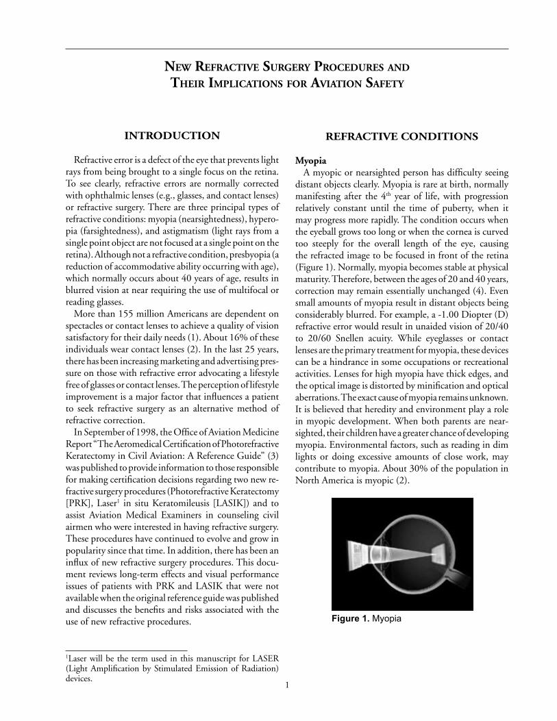
distant objects clearly . Myopia is rare at birth, normally manifesting after the 4th year of life, with progression relatively constant until the time of puberty, when it may progress more rapidly . The condition occurs when the eyeball grows too long or when the cornea is curved too steeply for the overall length of the eye, causing the refracted image to be focused in front of the retina (Figure 1) . Normally, myopia becomes stable at physical maturity . Therefore, between the ages of 20 and 40 years, correction may remain essentially unchanged (4) . Even small amounts of myopia result in distant objects being considerably blurred . For example, a -1 .00 Diopter (D) refractive error would result in unaided vision of 20/40 to 20/60 Snellen acuity . While eyeglasses or contact lenses are the primary treatment for myopia, these devices can be a hindrance in some occupations or recreational activities . Lenses for high myopia have thick edges, and the optical image is distorted by minification and optical aberrations . The exact cause of myopia remains unknown . It is believed that heredity and environment play a role in myopic development . When both parents are near-sighted, their children have a greater chance of developing myopia . Environmental factors, such as reading in dim lights or doing excessive amounts of close work, may contribute to myopia . About 30% of the population in North America is myopic (2) .
Figure 1. Myopia
1Laser will be the term used in this manuscript for LASER (Light Amplification by Stimulated Emission of Radiation) devices .

2
HyperopiaHyperopia, or farsightedness, is a disorder in which
vision is more blurred at near . This occurs when the eye is too short in length or the cornea is too flat, causing an image to be focused behind the retina (Figure 2) . At birth, eyes are normally hyperopic (about +2 .00 or +3 .00 D) . With aging, the eye normally lengthens . (Note: Each millimeter that the eye is too short is equal to 3 .00 D of hyperopic refractive change [4] .) Statistics are vague on the prevalence of hyperopia . Some epidemiological studies incorrectly incorporate presbyopia, which also requires plus power lenses, as part of the total percentage of hyperopia in the population . An estimated 40% of Americans are hyperopic (1) . Many of these hyperopes are children who are able to overcome their farsighted-ness due to their ability to accommodate . It is not until most hyperopes are in their late thirties or early forties that they experience clinical symptoms with hyperopia they have had their entire life .
AstigmatismAstigmatism causes blurred vision when looking at
objects both near and distant . The cornea is normally smooth and uniformly curved on all sides; however, with astigmatism the cornea is irregularly curved (steeper in one meridian) (Appendix A) . This irregular shape causes light to bend, or refract, causing light rays to become fo-cused at multiple points, which results in distorted vision at any distance . Astigmatism may occur in addition to myopic or hyperopic conditions (Figure 3) . At birth, the cornea is usually spherical; however, by 4 years of age the cornea shape changes . With-the-rule astigmatism occurs as the vertical corneal meridian steepens with age, while against-the-rule astigmatism occurs as the horizontal corneal meridian steepens with age (4) . Irregular astigma-tism most often occurs if the cornea has been damaged by trauma, inflammation, scar tissue, or developmental anomalies . This type of astigmatism normally cannot be completely corrected by ophthalmic spectacle lenses due to the lack of any geometric form from the irregular corneal surface .
PresbyopiaPresbyopia (the inability to focus at near) occurs when
the crystalline lens loses its ability to accommodate or change in shape (Figure 4) . Before developing presby-opia, the crystalline lens becomes flatter or thinner when focusing on objects at distance and becomes rounder or thicker when focusing on objects at near . Virtually everyone experiences some degree of presbyopia by early to mid-forty years of age . The ability to accommodate continues to decrease until about 55 years of age . Pres-byopia can occur in combination with any other type of refractive error and can complicate these visual condi-tions . For example, mildly farsighted individuals may find that they need reading glasses to see at near, while nearsighted people may need bifocals so that they can see comfortably at all distances .
REFRACTIVE ERROR MODIFICATION TECHNIQUES
Eye care physicians have used many different techniques in an attempt to alter or reduce refractive error . Due to the limited success and complications, refractive surgeons no longer use many of these techniques (e .g ., cycloplegia, clear lens extraction, and scleral reinforcement) (5) . Ad-ditionally, there were several refractive procedures that concentrated on modifying the anterior surface of the cornea, which supplies 44 of the 66 D (2/3) of the eye’s total refractive power (Appendix A) . These refractive surgical procedures (e .g ., keratomileusis, keratopha-kia, epikeratophakia, stromal thermokeratoplasty, and
Figure 2. Hyperopia
Figure 3. Astigmatism
Figure 4. Accommodation

3
intrastromal corneal ring) were more complex and had many complications, which led to their being discon-tinued (5) . This paper will review refractive procedures that are currently being used by refractive surgeons and new procedures that are still in the investigational phase or being performed on a limited basis .
Radial KeratotomySato of Japan first used radial keratotomy (RK) on a
wide scale in 1953 . In the early 1970s, Dr . Fyodorov of Russia considerably refined this technique . Since the late 1970s when it was first introduced in the United States, RK has been performed on more than a million Ameri-cans . The RK procedure involved making radial incisions on the peripheral cornea . These incisions weakened the cornea and allowed intraocular pressure to push the pe-ripheral cornea out and flatten the apex, which reduces myopia (Figure 5) . (Note: See Appendix B for surgical criteria table .)
In March 1982, a multi-center trial with 10 participat-ing surgeons was designed to determine the outcome of a single, standardized technique for myopia (Prospective Evaluation of Radial Keratotomy or PERK Study), which evaluated 757 eyes with a mandated eight-incision proce-dure . PERK study data at 5 years reported that two-thirds of patients no longer wore any correction, most of the other one-third only wore a correction part time, 60% were within ± 1 .00 D of correction, and 88% had uncor-rected visual acuity of 20/40 or better (6) . At 5 years, only 13% of eyes reported progressive hyperopic shift of 1 .00 D or more, but by 10 years, 43% of eyes had changed in the hyperopic direction by 1 .00 D or more (6-9) . Of the 374 patients (88%) who returned for the 10-year examination, 70% reported not wearing spectacles or contact lenses for distance vision, 60% were still within ± 1 .00 D of correction, and 85% had uncorrected visual acuity of 20/40 or better (7) . In addition, other long-term studies reported further complications such as reduced corneal strength (10-13), fluctuation of vision (14-19), glare (20-23), poor refractive predictability (7,24,25), and altitude-induced corneal changes (26-29) . With the advent of new laser procedures and many reports on both short- and long-term complications from RK, this procedure is rarely used today .
Photorefractive KeratectomyThe excimer laser has been used in ophthalmic and
refractive applications since the early 1980s . The laser employs a 193 nanometer (nm) ultraviolet-C light, which is emitted as an excited dimer of the argon fluoride gas mixture . This high-energy laser light causes an almost instantaneous vaporization of small amounts of the cornea by direct photochemical disruption of molecular bonds, with minimal impact on neighboring ocular tissue (30,31) . The excimer laser was initially approved by the Food and Drug Administration (FDA) to be used for photothera-peutic keratectomy (32) to reduce corneal scaring .
During the photorefractive keratectomy (PRK) pro-cedure, the corneal epithelium is first removed either mechanically or with the excimer laser . After program-ming the amount of intended refractive change required and baseline eye examination data, a computer-assisted algorithm determines the excimer treatment parameters . The laser is then used to reshape the anterior curvature by removing basement membrane, Bowman’s membrane, and portions of the corneal stroma (33) (Figure 6) . In October 1995, the FDA approved the use of the excimer laser to perform PRK (34) . Initial approval was granted for the correction of low-to-moderate levels of myopia (34) . As more information became available, approval was also given to correct higher levels of myopia (35), astigmatism (36), and low-to-moderate levels of hyperopia (37,38) (Appendix B) .
PRK is an outpatient procedure, requires only topical anesthesia, and takes about 10 minutes . The laser beam exposure time is dependent upon the amount of refractive error to be treated (average 30 seconds) . After treatment, bandage contact lens(es) are placed on the eye(s) to assist in the healing process and to reduce pain . Treated eye(s) are often painful 1 to 2 hours postoperative and become increasingly painful during the first 8 to 12 hours . By the following day, the pain is reduced considerably, and the cornea is reepithelialized in most patients within 48 Figure 5. Radial Keratotomy (RK)
Figure 6. Photorefractive Keratectomy (PRK)
4
hours . Vision is usually considerably improved within 3 to 4 days, and most patients become slightly overcorrected for a few weeks before stabilizing . Refractive corrections stabilize within 3 to 6 months for lower amounts of cor-rection but may take 6 to 18 months for higher amounts of correction (39,40) .
During the evolution of PRK, the ablation zone diam-eter progressively increased from 3 .5 millimeters (mm) to 6 .5 mm or more (41) . Patients with smaller ablation zones reported a higher incidence of symptomatic halos under night-driving conditions (42,43) . The larger opti-cal zone reduced the effect of optical irregularities at the junction of the ablation zone and the untreated cornea, which is thought to cause symptomatic halos (44) . (Note: 78% of patients with ablation zones of ≤ 5 .00 mm re-ported seeing halos at night (45,46) .) Larger ablation zones (≥ 6 mm) had less critical centering requirements when used for mild-to-moderate refractive corrections (45,47) . However, when larger ablation zones are used for higher corrections, centering is critical due to the steep and deep transition zone between the treated and untreated portions of the cornea (48) . Precise centration of the laser over the pupil entrance is important, as clear, crisp vision depends upon the regularity and centration of the ablated optical zone . Initially, self-fixation by the patient was used, and there were reports of decentration occurring in about 20% of PRK treatments (49) . Patients with decentered ablations have problems with monocular diplopia, glare, and irregular astigmatism (50,51) . When this occurs, the most common option for correcting ir-regular astigmatism is a rigid contact lens . Thus, a patient who had previously been unable to wear contact lenses is at a greater risk for a decrease in best spectacle corrected visual acuity (BSCVA) (40,52) .
While the epithelium has completely healed within 4 to 5 days after surgery in most cases (53,54), epithelial wound healing has occurred as late as several months after surgery (48) . Studies found that a smooth corneal surface at 1 to 3 months after surgery did not guarantee a smooth surface at 6 months, as the extent of deposition of new corneal tissue was unpredictable (55,56) .
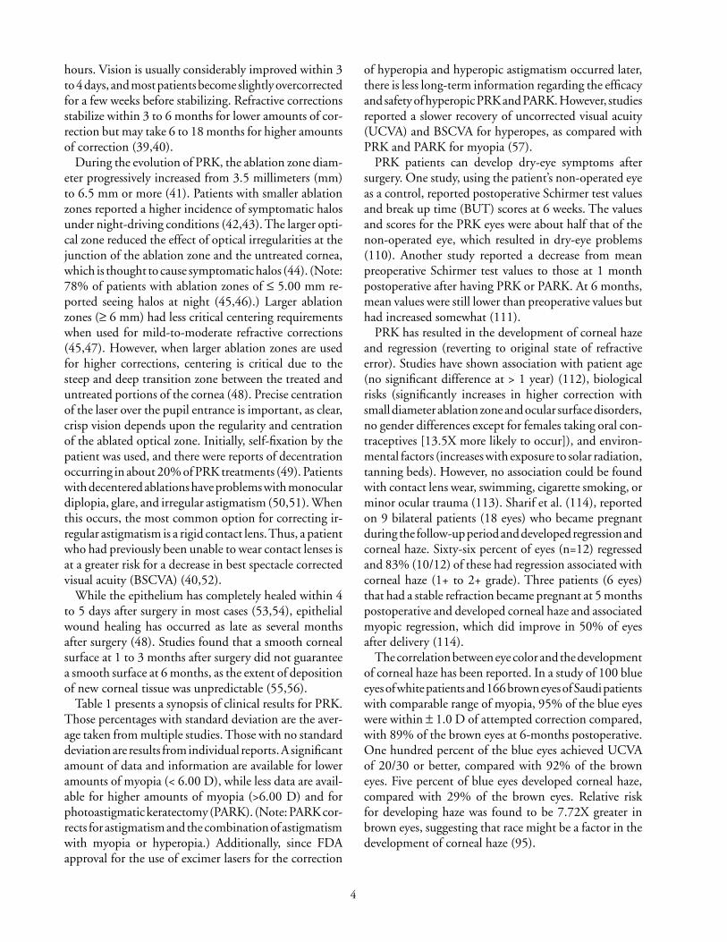
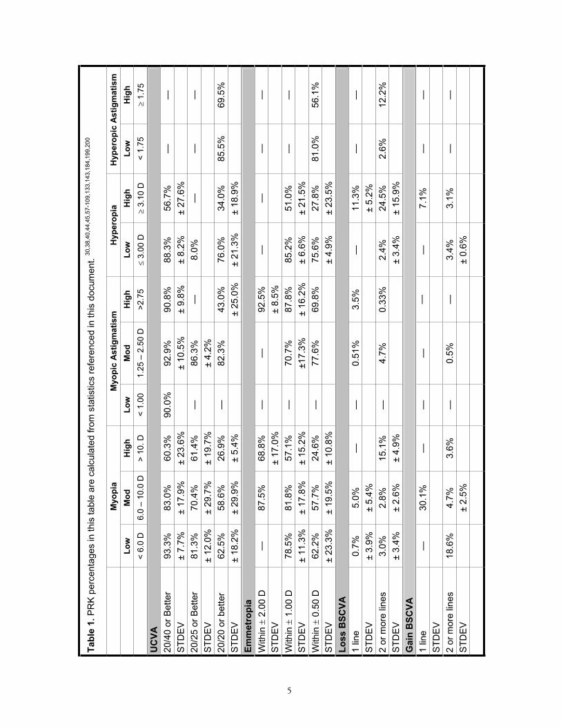
Table 1 presents a synopsis of clinical results for PRK . Those percentages with standard deviation are the aver-age taken from multiple studies . Those with no standard deviation are results from individual reports . A significant amount of data and information are available for lower amounts of myopia (< 6 .00 D), while less data are avail-able for higher amounts of myopia (>6 .00 D) and for photoastigmatic keratectomy (PARK) . (Note: PARK cor-rects for astigmatism and the combination of astigmatism with myopia or hyperopia .) Additionally, since FDA approval for the use of excimer lasers for the correction
of hyperopia and hyperopic astigmatism occurred later, there is less long-term information regarding the efficacy and safety of hyperopic PRK and PARK . However, studies reported a slower recovery of uncorrected visual acuity (UCVA) and BSCVA for hyperopes, as compared with PRK and PARK for myopia (57) .
PRK patients can develop dry-eye symptoms after surgery . One study, using the patient’s non-operated eye as a control, reported postoperative Schirmer test values and break up time (BUT) scores at 6 weeks . The values and scores for the PRK eyes were about half that of the non-operated eye, which resulted in dry-eye problems (110) . Another study reported a decrease from mean preoperative Schirmer test values to those at 1 month postoperative after having PRK or PARK . At 6 months, mean values were still lower than preoperative values but had increased somewhat (111) .
PRK has resulted in the development of corneal haze and regression (reverting to original state of refractive error) . Studies have shown association with patient age (no significant difference at > 1 year) (112), biological risks (significantly increases in higher correction with small diameter ablation zone and ocular surface disorders, no gender differences except for females taking oral con-traceptives [13 .5X more likely to occur]), and environ-mental factors (increases with exposure to solar radiation, tanning beds) . However, no association could be found with contact lens wear, swimming, cigarette smoking, or minor ocular trauma (113) . Sharif et al . (114), reported on 9 bilateral patients (18 eyes) who became pregnant during the follow-up period and developed regression and corneal haze . Sixty-six percent of eyes (n=12) regressed and 83% (10/12) of these had regression associated with corneal haze (1+ to 2+ grade) . Three patients (6 eyes) that had a stable refraction became pregnant at 5 months postoperative and developed corneal haze and associated myopic regression, which did improve in 50% of eyes after delivery (114) .
The correlation between eye color and the development of corneal haze has been reported . In a study of 100 blue eyes of white patients and 166 brown eyes of Saudi patients with comparable range of myopia, 95% of the blue eyes were within ± 1 .0 D of attempted correction compared, with 89% of the brown eyes at 6-months postoperative . One hundred percent of the blue eyes achieved UCVA of 20/30 or better, compared with 92% of the brown eyes . Five percent of blue eyes developed corneal haze, compared with 29% of the brown eyes . Relative risk for developing haze was found to be 7 .72X greater in brown eyes, suggesting that race might be a factor in the development of corneal haze (95) .
5
Tabl
e 1.
PR
K p
erce
ntag
es in
this
tabl
e ar
e ca
lcul
ated
from
sta
tistic
s re
fere
nced
in th
is d
ocum
ent.
30,3
8,40
,44,
45,5
7-10
9,13
3,14
3,18
4,19
9,20
0
Myo
pia
Myo
pic
Ast
igm
atis
m
Hyp
erop
ia
Hyp
erop
ic A
stig
mat
ism
Lo
w
Mod
H
igh
Low
M
od
Hig
h Lo
w
Hig
h Lo
w
Hig
h <
6.0
D
6.0
– 10
.0 D
> 10
. D
< 1.
00
1.25
– 2
.50
D
>2.7
5
3.0
0 D
3
.10
D
< 1.
75
1.7
5 U
CVA
20
/40
or B
ette
r 93
.3%
83
.0%
60
.3%
90
.0%
92
.9%
90
.8%
88
.3%
56
.7%
—
—
S
TDE
V
± 7.
7%
± 17
.9%
±
23.6
%
±
10.5
%
± 9.
8%
± 8.
2%
± 27
.6%
20
/25
or B
ette
r 81
.3%
70
.4%
61
.4%
—
86
.3%
—
8.
0%
—
—
—
STD
EV
±
12.0
%
± 29
.7%
±
19.7
%
±
4.2%
20/2
0 or
bet
ter
62.5
%
58.6
%
26.9
%
—
82.3
%
43.0
%
76.0
%
34.0
%
85.5
%
69.5
%
STD
EV
±
18.2
%
± 29
.9%
±
5.4%
±
25.0
%
± 21
.3%
±
18.9
%
Emm
etro
pia
With
in 2
.00
D
—
87.5
%
68.8
%
—
—
92.5
%
—
—
—
—
STD
EV
±
17.0
%
± 8.
5%
With
in 1
.00
D
78.5
%
81.8
%
57.1
%
—
70.7
%
87.8
%
85.2
%
51.0
%
—
—
STD
EV
±
11.3
%
± 17
.8%
±
15.2
%
±1
7.3%
±
16.2
%
± 6.
6%
± 21
.5%
W
ithin
0.5
0 D
62
.2%
57
.7%
24
.6%
—
77
.6%
69
.8%
75
.6%
27
.8%
81
.0%
56
.1%
S
TDE
V
± 23
.3%
±
19.5
%
± 10
.8%
± 4.
9%
± 23
.5%
Lo
ss B
SCVA
1
line
0.
7%
5.0%
—
—
0.
51%
3.
5%
—
11.3
%
—
—
STD
EV
±
3.9%
±
5.4%
± 5.
2%
2 or
mor
e lin
es
3.0%
2.
8%
15.1
%
—
4.7%
0.
33%
2.
4%
24.5
%
2.6%
12
.2%
S
TDE
V
± 3.
4%
± 2.
6%
± 4.
9%
±
3.4%
±
15.9
%
Gai
n B
SCVA
1
line
—
30
.1%
—
—
—
—
—
7.1%
—
—
S
TDE
V
2
or m
ore
lines
18
.6%
4.
7%
3.6%
—
0.
5%
—
3.4%
3.
1%
—
—
STD
EV
± 2.
5%
± 0.
6%

6
There have been reported problems with late-onset corneal haze (LOCH) . Diagnostic criterion for LOCH is acute haze of grade ≥ 2 occurring between 4 and 12 months postoperative . One study, which followed 314 eyes for 12 to 41 months, reported that 11 eyes developed LOCH after exposure to high levels of environmental UV-radiation (115) . The study suggests that the use of UV-protective eyewear should be encouraged during the first year after PRK . In another study that followed 1,000 patients for 12 months, all corneas were clear at 4 months, after 4 months, however, 18 eyes of 17 patients developed LOCH, resulting in decreased visual acuity and regression . Treatment with topical steroids resulted in partial reversal of haze and regression . The study suggests that corneal healing and remodeling may continue for at least 1 year after PRK (116) .
Compared with reports about RK, initial trials for PRK did not report significant problems with diurnal fluctuation of vision (121), progressive hyperopic shifts (37), reduced corneal strength (122), poor refractive predictability (74), and fluctuation of vision (121) . How-ever, long-term PRK studies have reported problems with glare (94,108,119,123), halos (108,124,125), regression (81,126), haze (44,65,98,108,109,112-116,127,128), decreased visual performance at night (119,129,130), undercorrection (81,131), overcorrection (130,132), loss of BSCVA (40,63,66,68,70,76,94,103,109,133,134), and severe dry eye problems (110,111,135-137) . As a result of these reported problems and advances in laser refractive
procedures, PRK has been all but replaced . A survey of refractive surgeons in the United States reported that the percentage performing PRK had decreased from 26% in 1997 to less than 1% in 2002 (138) . A list of patient- reported satisfaction and complications after PRK surgery is summarized in Table 2 .
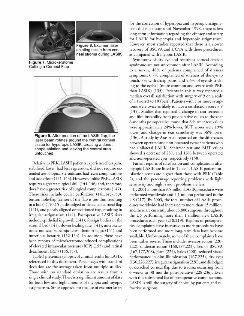
Laser in situ KeratomileusisLaser in situ Keratomileusis (LASIK) is a technique that
uses the excimer laser and a specially designed knife blade called a microkeratome, which slices a thin, horizontal flap (100 to 200 µm in depth) off the top of the cornea leaving it connected by a small hinge of tissue (139) (Figure 7) . The corneal flap is folded aside, and the excimer laser is used to remove tissue from the corneal stroma (Figure 8) . The flap is then replaced . Many patients report seeing clearly im-mediately after surgery and have little or no discomfort .
LASIK was initially used to treat higher amounts of myopia . As surgeons increased their surgical skill, LASIK has become the surgery of choice and is now used to treat even low-to-moderate amounts of myopia, astigmatism, and hyperopia (Figure 9) . Initially, refractive surgeons felt that there was no need for clinical trials and FDA approval, since LASIK is a procedure that uses 2 FDA-approved devices . In July 1997, the FDA approved the use of the excimer laser and the microkeratome for the LASIK procedure (140) . (Appendix B)
Table 2. Patient reported satisfaction and complications after PRK.
Myopic PRK Hyperopic PRK Reported Patient SatisfactionSatisfied - very satisfied 70-92% 44,117-120 96-100% 90,91
Dissatisfied 3-20% 117,120
Quality of Life Improvement 78% 117
Decrease 17% 117
Correction Required After Surgery None 66-100% 117, 119
Sometimes worn 30% 119
Always worn 10% 119
Glare Daytime 55% 62,108,118 14-16% 90,91
Halos At night 7% 100
Always 34% 62,119 Sometimes 26% 119
Never 40-79% 119
Night Vision Problems Always 32-40% 119
Sometimes 30% 119
Never 30% 119
Decreased night vision 32% 100,118
Increased difficulty driving at night 30-31%118 27-29% 90,91
7
Relative to PRK, LASIK patients experienced less pain, stabilized faster, had less regression, did not require ex-tended use of topical steroids, and had fewer complications and side effects (141-143) . However, unlike PRK, LASIK requires a greater surgical skill (144-146) and, therefore, does have a greater risk of surgical complications (147) . These risks include ocular perforation (141,148-150), button hole-flap (center of the flap is too thin resulting in a hole) (150,151), dislodged or detached corneal flap (141), and poorly aligned or positioned flap, resulting in irregular astigmatism (141) . Postoperative LASIK risks include epithelial ingrowth (141), foreign bodies in the stromal bed (141), slower healing rate (141), microkera-tome-induced subconjunctival hemorrhages (141) and infectious keratitis (152-154) . In addition, there have been reports of microkeratome-induced complications of elevated intraocular pressure (IOP) (155) and retinal detachment (RD) (156,157) .
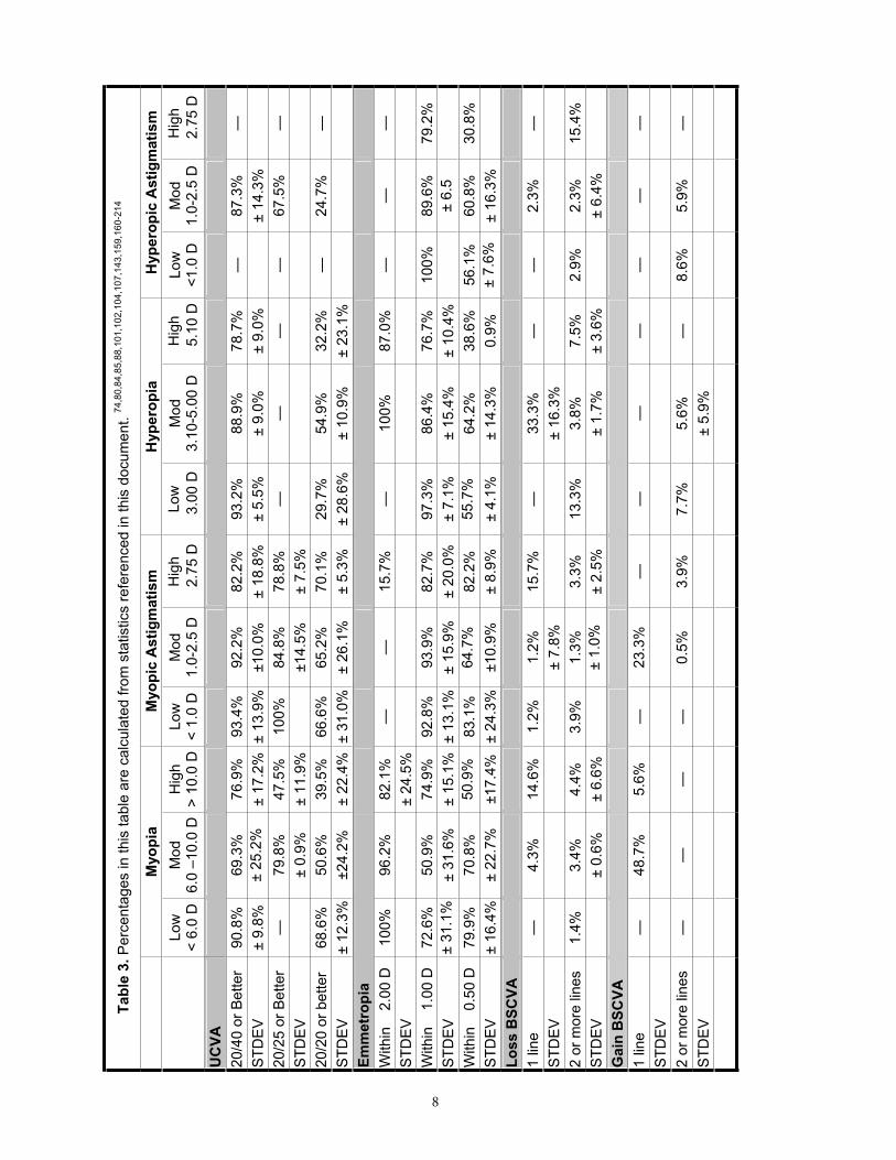
Table 3 presents a synopsis of clinical results for LASIK referenced in this document . Percentages with standard deviation are the average taken from multiple studies . Those with no standard deviation are results from a single clinical study . There is a significant amount of data for both low and high amounts of myopia and myopic astigmatism . Since approval for the use of excimer lasers
for the correction of hyperopia and hyperopic astigma-tism did not occur until November 1998, there is less long-term information regarding the efficacy and safety for LASIK for hyperopia and hyperopic astigmatism . However, most studies reported that there is a slower recovery of BSCVA and UCVA with these procedures, as compared with myopic LASIK .
Symptoms of dry eye and recurrent corneal erosion syndrome are not uncommon after LASIK . According to a survey, 48% of patients complained of dryness symptoms, 6 .7% complained of soreness of the eye to touch, 8% with sharp pains, and 5 .6% of eyelids stick-ing to the eyeball (more common and severe with PRK than LASIK) (135) . Patients in this survey reported a median overall satisfaction with surgery of 9 on a scale of 1 (worst) to 10 (best) . Patients with 1 or more symp-toms were twice as likely to have a satisfaction score < 8 (135) . Studies that reported a change in tear secretion and film instability from preoperative values to those at 6-months postoperative found that Schirmer test values were approximately 24% lower, BUT scores were 19% lower, and change in tear osmolarity was 36% lower (136) . A study by Aras et al . reported on the differences between operated and non-operated eyes of patients who had unilateral LASIK . Schirmer test and BUT values showed a decrease of 23% and 13% between operated and non-operated eyes, respectively (158) .
Patient reports of satisfaction and complications after myopic LASIK are listed in Table 4 . LASIK patient sat-isfaction scores are higher than those with PRK (Table 2), and the percentage reporting problems with light sensitivity and night vision problems are less .
By 2001, more than 9 .5 million LASIK procedures were performed worldwide and 5 .1 million performed in the US (217) . By 2003, the total number of LASIK proce-dures worldwide had increased to more than 15 million, and there are currently about 3,800 surgeons throughout the US performing more than 1 million new LASIK procedures each year (218,219) . Reports of postopera-tive complaints have increased as more procedures have been performed and more long-term data have become available . Unfortunately, some of these complaints have been rather severe . These include: overcorrection (220-222), undercorrection (160,187,223), loss of BSCVA (167,177,208), glare (224), halos (200), reduced visual performance in dim illumination (167,225), dry eyes (136,226,227), irregular astigmatism (226), and dislodged or detached corneal flap due to trauma occurring from 6 weeks to 38 months postoperative (228-236) . Even with this substantial list of postoperative complications, LASIK is still the surgery of choice by patients and re-fractive surgeons .
Figure 7. Microkeratome Cutting a Corneal Flap
Figure 8. Excimer laser ablating tissue from cor-neal stroma during LASIK
Figure 9. After creation of the LASIK flap, the laser beam rotates around the central corneal tissue for hyperopic LASIK, creating a donut shape ablation and leaving the central area untouched
8
Tabl
e 3.
Per
cent
ages
in th
is ta
ble
are
calc
ulat
ed fr
om s
tatis
tics
refe
renc
ed in
this
doc
umen
t.74
,80,
84,8
5,88
,101
,102
,104
,107
,143
,159
,160
-214
Myo
pia
Myo
pic
Ast
igm
atis
m
Hyp
erop
ia
Hyp
erop
ic A
stig
mat
ism
Lo
w<
6.0
D
Mod
6.0
–10.
0 D
H
igh
> 10
.0 D
Low
< 1.
0 D
Mod
1.0-
2.5
D
Hig
h
2.75
D
Low
3.
00 D
M
od3.
10-5
.00
D
Hig
h
5.10
DLo
w<1
.0 D
M
od1.
0-2.
5 D
Hig
h
2.75
DU
CVA
20
/40
or B
ette
r 90
.8%
69
.3%
76
.9%
93
.4%
92
.2%
82
.2%
93
.2%
88
.9%
78
.7%
—
87
.3%
—
S
TDE
V
± 9.
8%
± 25
.2%
±
17.2
%±
13.9
%±1
0.0%
±
18.8
%
± 5.
5%
± 9.
0%
± 9.
0%
±
14.3
%
20
/25
or B
ette
r —
79
.8%
47
.5%
10
0%
84.8
%
78.8
%
—
—
—
—
67.5
%
—
STD
EV
± 0.
9%
± 11
.9%
±1
4.5%
±
7.5%
20
/20
or b
ette
r 68
.6%
50
.6%
39
.5%
66
.6%
65
.2%
70
.1%
29
.7%
54
.9%
32
.2%
—
24
.7%
—
S
TDE
V
± 12
.3%
±2
4.2%
±
22.4
%±
31.0
%±
26.1
%
± 5.
3%
± 28
.6%
±
10.9
%
± 23
.1%
Em
met
ropi
a W
ithin
2.
00 D
10
0%
96.2
%
82.1
%
—
—
15.7
%
—
100%
87
.0%
—
—
—
S
TDE
V
± 24
.5%
W
ithin
1.
00 D
72
.6%
50
.9%
74
.9%
92
.8%
93
.9%
82
.7%
97
.3%
86
.4%
76
.7%
10
0%
89.6
%
79.2
%
STD
EV
± 31
.1%
±
31.6
%
± 15
.1%
± 13
.1%
± 15
.9%
±
20.0
%
± 7.
1%
± 15
.4%
±
10.4
%
± 6.
5 W
ithin
0.
50 D
79
.9%
70
.8%
50
.9%
83.1
%64
.7%
82.2
%55
.7%
64.2
%
38.6
%
56.1
%
60.8
%
30.8
%
STD
EV
±
16.4
%
± 22
.7%
±1
7.4%
± 24
.3%
±10.
9%
± 8.
9%
± 4.
1%
± 14
.3%
0.
9%
± 7.
6%
± 16
.3%
Loss
BSC
VA
1 lin
e
—
4.3%
14
.6%
1.
2%
1.2%
15
.7%
—
33
.3%
—
—
2.
3%
—
STD
EV
±
7.8%
±
16.3
%
2 or
mor
e lin
es
1.4%
3.
4%
4.4%
3.
9%
1.3%
3.
3%
13.3
%
3.8%
7.
5%
2.9%
2.
3%
15.4
%
STD
EV
± 0.
6%
± 6.
6%
±
1.0%
±
2.5%
± 1.
7%
± 3.
6%
±
6.4%
Gai
n B
SCVA
1
line
—
48
.7%
5.
6%
—
23.3
%
—
—
—
—
—
—
—
STD
EV
2
or m
ore
lines
—
—
—
—
0.
5%
3.9%
7.
7%
5.6%
—
8.
6%
5.9%
—
S
TDE
V
±
5.9%

9
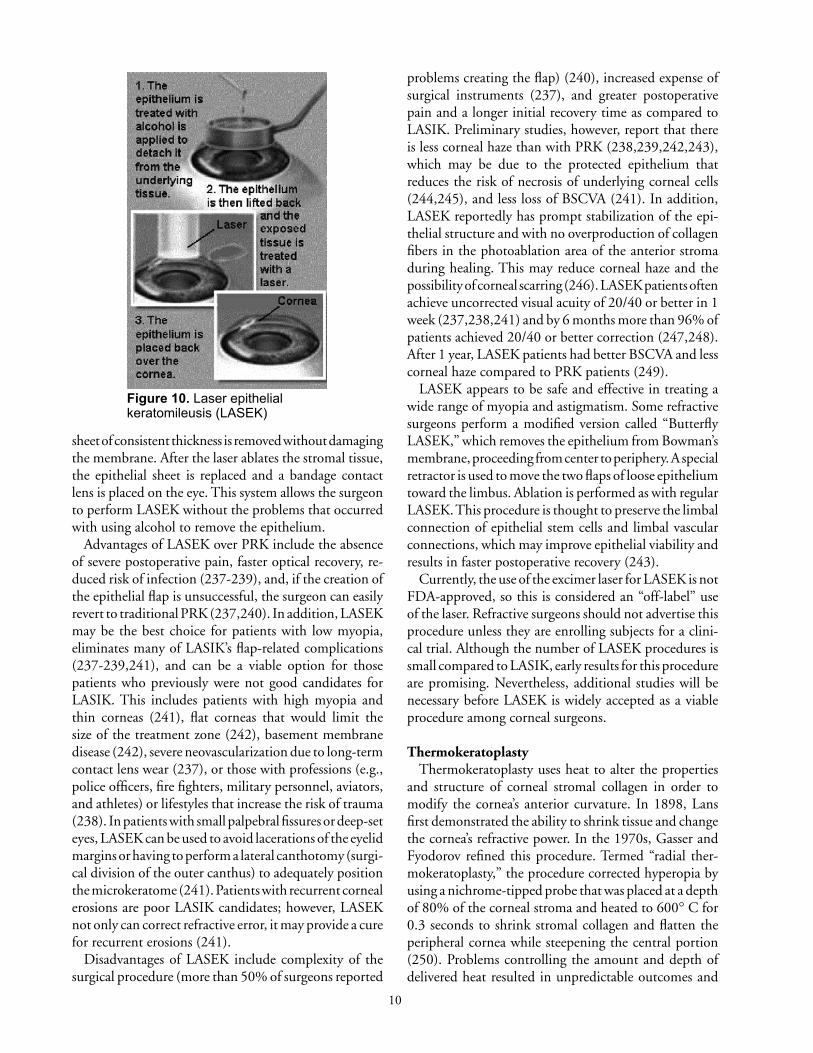
Laser Epithelial KeratomileusisLaser epithelial keratomileusis (LASEK), which is also
called Epithelial-LASIK, E-LASIK, and Epi-LASEK, was introduced in 1999 and is reported to combine the advantages and reduce the disadvantages of PRK and LASIK (237) . During the LASEK procedure (Figure 10), a pre-incision of the corneal epithelium is performed with a special microtrephine (an instrument used to make a circular cut on the epithelium) with a 70-µm depth-cali-brated blade . The microtrephine is designed to leave a hinge at the cornea’s 12-o’clock position . Two to 3 drops of 18 to 20% alcohol solution are instilled for 30 seconds to loosen the corneal epithelium from the basement mem-brane . The area is then dried, thoroughly washed with water, and dried again . (Note: Alcohol solution is applied for 30 to 40 seconds in younger men, postmenopausal women, and long-time contact lens users . Applications of longer than 40 seconds can decrease the elasticity of the epithelial flap .) The corneal epithelium is lifted at the
pre-incision, and the epithelial flap is gently detached and folded up at the 12-o’clock position, leaving a smooth surface . After the ablation, the epithelial flap is carefully repositioned with a small spatula over the stroma . A soft contact lens is applied for 3 to 4 days to keep the flap in place . Postoperative antibiotic and cortisone treatments are administered for a few days . In 3-4 days, the epithelial layer is usually regenerated and intact (238) . It is postu-lated that the epithelial flap protects the bare surface of the stroma and prevents the influx of inflammatory cells from the tears, thus reducing the inflammatory damage to the corneal stroma (237) .
Many surgeons have experienced difficulties removing the corneal epithelium when performing LASEK and have had to revert to performing PRK . VisiJet Inc ., of Irvine, CA, received FDA approval in September 2004, for the “EpiLift System,” which is used to separate corneal tissue during the LASEK procedure . The system separates the epithelium from Bowman’s membrane, and an epithelial
Table 4. Patient reports of satisfaction and complications after myopic LASIK.
Myopic LASIK Reasons for Having Surgery Improve UCVA 88% 167
Improve cosmesis 21% 167
Achieved goals for surgery 94% 167
Reported Patient Satisfaction With resultsSatisfied - very satisfied 50-98% 162167,215,216
Dissatisfied Would have procedure again 97-99% 159,215,216
Improvement of Functional Vision Improvement 81-100% 167
Decrease — Correction Required After Surgery None 95-96% 215,216
Sometimes (for presbyopia) 25% 215
Always — Light Sensitivity Unchanged 73% 215
Worse 27% 215
Better 6% 215
Night Vision Problems Unchanged 76% 215
Worse 24% 215
Better 17% 215
Never — Decreased night vision 14-39 % 170,216
Increased difficulty driving at night 9% 167
10
sheet of consistent thickness is removed without damaging the membrane . After the laser ablates the stromal tissue, the epithelial sheet is replaced and a bandage contact lens is placed on the eye . This system allows the surgeon to perform LASEK without the problems that occurred with using alcohol to remove the epithelium .
Advantages of LASEK over PRK include the absence of severe postoperative pain, faster optical recovery, re-duced risk of infection (237-239), and, if the creation of the epithelial flap is unsuccessful, the surgeon can easily revert to traditional PRK (237,240) . In addition, LASEK may be the best choice for patients with low myopia, eliminates many of LASIK’s flap-related complications (237-239,241), and can be a viable option for those patients who previously were not good candidates for LASIK . This includes patients with high myopia and thin corneas (241), flat corneas that would limit the size of the treatment zone (242), basement membrane disease (242), severe neovascularization due to long-term contact lens wear (237), or those with professions (e .g ., police officers, fire fighters, military personnel, aviators, and athletes) or lifestyles that increase the risk of trauma (238) . In patients with small palpebral fissures or deep-set eyes, LASEK can be used to avoid lacerations of the eyelid margins or having to perform a lateral canthotomy (surgi-cal division of the outer canthus) to adequately position the microkeratome (241) . Patients with recurrent corneal erosions are poor LASIK candidates; however, LASEK not only can correct refractive error, it may provide a cure for recurrent erosions (241) .
Disadvantages of LASEK include complexity of the surgical procedure (more than 50% of surgeons reported
problems creating the flap) (240), increased expense of surgical instruments (237), and greater postoperative pain and a longer initial recovery time as compared to LASIK . Preliminary studies, however, report that there is less corneal haze than with PRK (238,239,242,243), which may be due to the protected epithelium that reduces the risk of necrosis of underlying corneal cells (244,245), and less loss of BSCVA (241) . In addition, LASEK reportedly has prompt stabilization of the epi-thelial structure and with no overproduction of collagen fibers in the photoablation area of the anterior stroma during healing . This may reduce corneal haze and the possibility of corneal scarring (246) . LASEK patients often achieve uncorrected visual acuity of 20/40 or better in 1 week (237,238,241) and by 6 months more than 96% of patients achieved 20/40 or better correction (247,248) . After 1 year, LASEK patients had better BSCVA and less corneal haze compared to PRK patients (249) .
LASEK appears to be safe and effective in treating a wide range of myopia and astigmatism . Some refractive surgeons perform a modified version called “Butterfly LASEK,” which removes the epithelium from Bowman’s membrane, proceeding from center to periphery . A special retractor is used to move the two flaps of loose epithelium toward the limbus . Ablation is performed as with regular LASEK . This procedure is thought to preserve the limbal connection of epithelial stem cells and limbal vascular connections, which may improve epithelial viability and results in faster postoperative recovery (243) .
Currently, the use of the excimer laser for LASEK is not FDA-approved, so this is considered an “off-label” use of the laser . Refractive surgeons should not advertise this procedure unless they are enrolling subjects for a clini-cal trial . Although the number of LASEK procedures is small compared to LASIK, early results for this procedure are promising . Nevertheless, additional studies will be necessary before LASEK is widely accepted as a viable procedure among corneal surgeons .
ThermokeratoplastyThermokeratoplasty uses heat to alter the properties
and structure of corneal stromal collagen in order to modify the cornea’s anterior curvature . In 1898, Lans first demonstrated the ability to shrink tissue and change the cornea’s refractive power . In the 1970s, Gasser and Fyodorov refined this procedure . Termed “radial ther-mokeratoplasty,” the procedure corrected hyperopia by using a nichrome-tipped probe that was placed at a depth of 80% of the corneal stroma and heated to 600° C for 0 .3 seconds to shrink stromal collagen and flatten the peripheral cornea while steepening the central portion (250) . Problems controlling the amount and depth of delivered heat resulted in unpredictable outcomes and
Figure 10. Laser epithelial keratomileusis (LASEK)
11
refractive regression . Tissue necrosis resulted from high temperatures . There were also problems with epithelial thinning, recurrent corneal erosions, iritis, stromal melt-ing, and loss of BSCVA (5) .
Laser Thermal Keratoplasty (LTK) is a noninvasive procedure that uses laser light, which is absorbed by the cornea to produce the desired temperature elevations (251) (Figure 11) . New advances in laser technology, a better understanding of corneal physiology, and the introduction of corneal topography analysis have led to the development of a safer and more effective LTK procedure . The patient’s preoperative refraction dictates the optical zone diameter and the number of laser spots used . A smaller optical zone (6mm) will result in a steeper cornea than a larger zone (7mm) . Additionally, 8 laser spots are applied for corrections of +0 .75 to +0 .875 D, 15 spots for +1 .0 to +1 .67 D, 24 spots for +1 .75 to 2 .25 D, and 32 spots for +2 .375 to +3 .0 D (252) .
In June 2000, the FDA approved the Hyperion LTK System, an infrared holmium-YAG laser, to treat patients who are farsighted (between +0 .75 to +2 .25 D, with or without astigmatism < 0 .75 D), at least 40 years of age, and with a stable refraction (253) . Contraindications for LTK include pregnant or nursing women, patients with an abnormal shape or thinning of the cornea, central corneal scarring, history of herpetic eye infection, an autoimmune or collagen vascular disease, clinically significant atopic syndrome (allergic), insulin-dependent diabetes, or a compromised immune system (253) .
The Hyperion LTK System produces a non-visible laser beam that heats corneal tissue, causing it to shrink and tighten at the periphery, thus steepening the central area (254) . Short-term studies have shown that, for most patients, LTK is temporary, since at 24 months postop-erative a majority of patients have regressed to one-half of the correction observed at 6 months postoperative (253) . While there seems to be less surgical complications
with LTK, results and stability depend on the amount of correction attempted and the patient’s age (i .e ., younger patients may have more regression) (253,255) .
LTK is a relatively new procedure and, at this time, only short-term clinical study data have been published regarding its effectiveness and possible complications (256) . This procedure is marketed as a treatment for older patients (≥ 40 years of age) who are mildly hyperopic (or early presbyopes) and are reluctant to have LASIK or PRK . The procedure takes only a few seconds and is performed in the surgeon’s office . The major advantage for older presbyopic airmen is that LTK will reportedly allow them to see clearly at near and intermediate distances again . With data that are currently available, it is impossible to determine how popular this procedure will become with airmen and what long-term complications may occur . The FAA is currently approving on a case-by-case basis airmen who have had the LTK procedure . Airmen who have had the LTK procedure may be required to take a medical flight test . Upon passing the test, their medical certificate is issued under the “special issuance” section of part 67 (14 CFR § 67 .401) .
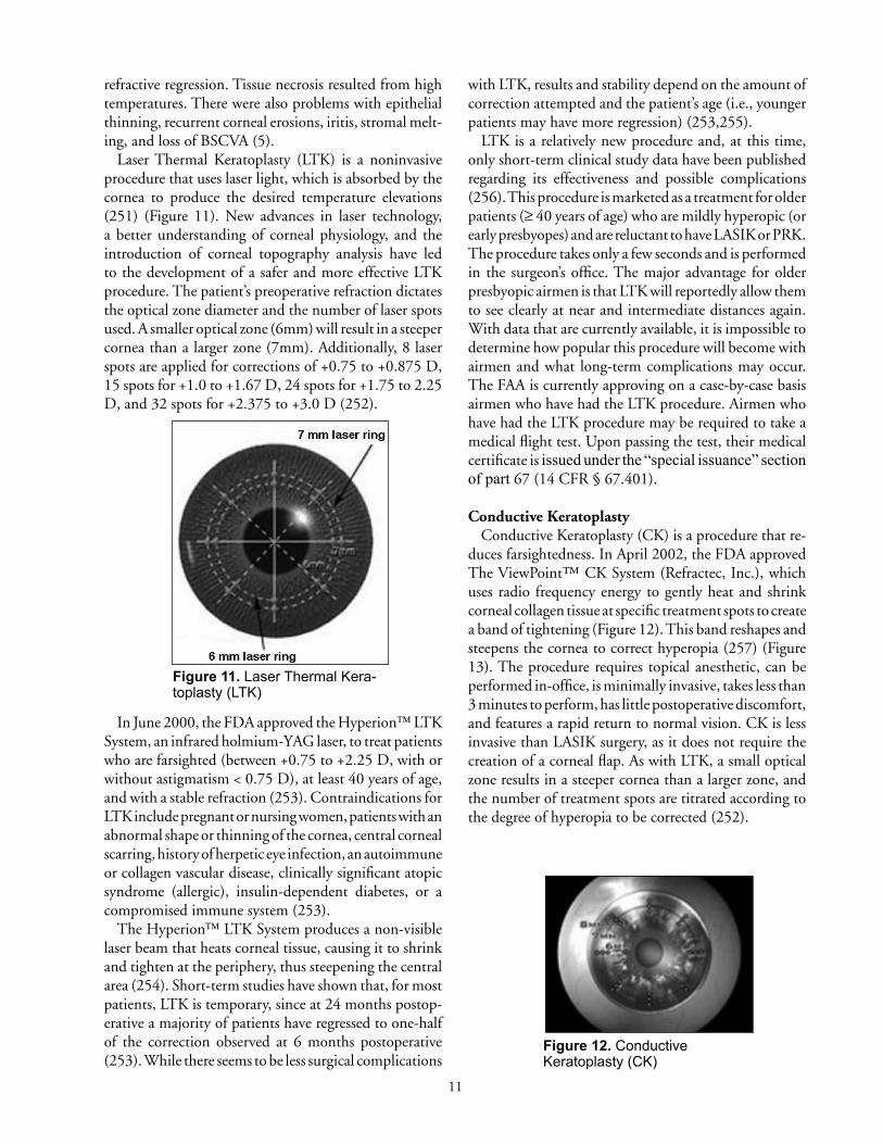
Conductive KeratoplastyConductive Keratoplasty (CK) is a procedure that re-
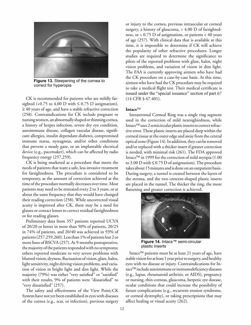
duces farsightedness . In April 2002, the FDA approved The ViewPoint™ CK System (Refractec, Inc .), which uses radio frequency energy to gently heat and shrink corneal collagen tissue at specific treatment spots to create a band of tightening (Figure 12) . This band reshapes and steepens the cornea to correct hyperopia (257) (Figure 13) . The procedure requires topical anesthetic, can be performed in-office, is minimally invasive, takes less than 3 minutes to perform, has little postoperative discomfort, and features a rapid return to normal vision . CK is less invasive than LASIK surgery, as it does not require the creation of a corneal flap . As with LTK, a small optical zone results in a steeper cornea than a larger zone, and the number of treatment spots are titrated according to the degree of hyperopia to be corrected (252) .
Figure 11. Laser Thermal Kera-toplasty (LTK)
Figure 12. Conductive Keratoplasty (CK)
12
CK is recommended for patients who are mildly far-sighted (+0 .75 to 4 .00 D with ≤ 0 .75 D astigmatism), ≥ 40 years of age, and have a stable refractive correction (258) . Contraindications for CK include pregnant or nursing women, an abnormally shaped or thinning cornea, a history of herpes infection, severe dry eye condition, autoimmune disease, collagen vascular disease, signifi-cant allergies, insulin dependant diabetes, compromised immune status, nystagmus, and/or other conditions that prevent a steady gaze, or an implantable electrical device (e .g ., pacemaker), which can be affected by radio frequency energy (257,259) .
CK is being marketed as a procedure that meets the needs of patients that want a safe, less-invasive treatment for farsightedness . The procedure is considered to be temporary, as the amount of correction achieved at the time of the procedure normally decreases over time . Most patients may need to be retreated every 2 to 3 years, or at about the same frequency that they would have changed their reading correction (258) . While uncorrected visual acuity is improved after CK, there may be a need for glasses or contact lenses to correct residual farsightedness or for reading glasses .
Preliminary data from 357 patients reported UCVA of 20/20 or better in more than 50% of patients, 20/25 in 74% of patients, and 20/40 was achieved in 93% of patients (257,259,260) . Less than 1% of patients lost 2 or more lines of BSCVA (257) . At 9-months postoperative, the majority of the patients responded with no symptoms; others reported moderate to very severe problems with blurred vision, dryness, fluctuation of vision, glare, halos, light sensitivity, night driving vision problems, and varia-tion of vision in bright light and dim light . While the majority (79%) was either “very satisfied” or “satisfied” with their results, 9% of patients were “dissatisfied” to “very dissatisfied” (257) .
The safety and effectiveness of the View Point- CK System have not yet been established in eyes with diseases of the cornea (e .g ., scar, or infection), previous surgery
or injury to the cornea, previous intraocular or corneal surgery, a history of glaucoma, > 4 .00 D of farsighted-ness, or > 0 .75 D of astigmatism, or patients < 40 years of age (257) . With clinical data that is available at this time, it is impossible to determine if CK will achieve the popularity of other refractive procedures . Longer studies are required to determine the significance to pilots of the reported problems with glare, halos, night vision problems, and variation of vision in dim light . The FAA is currently approving airmen who have had the CK procedure on a case-by-case basis . At this time, airmen who have had the CK procedure may be required to take a medical flight test . Their medical certificate is issued under the “special issuance” section of part 67 (14 CFR § 67 .401) .
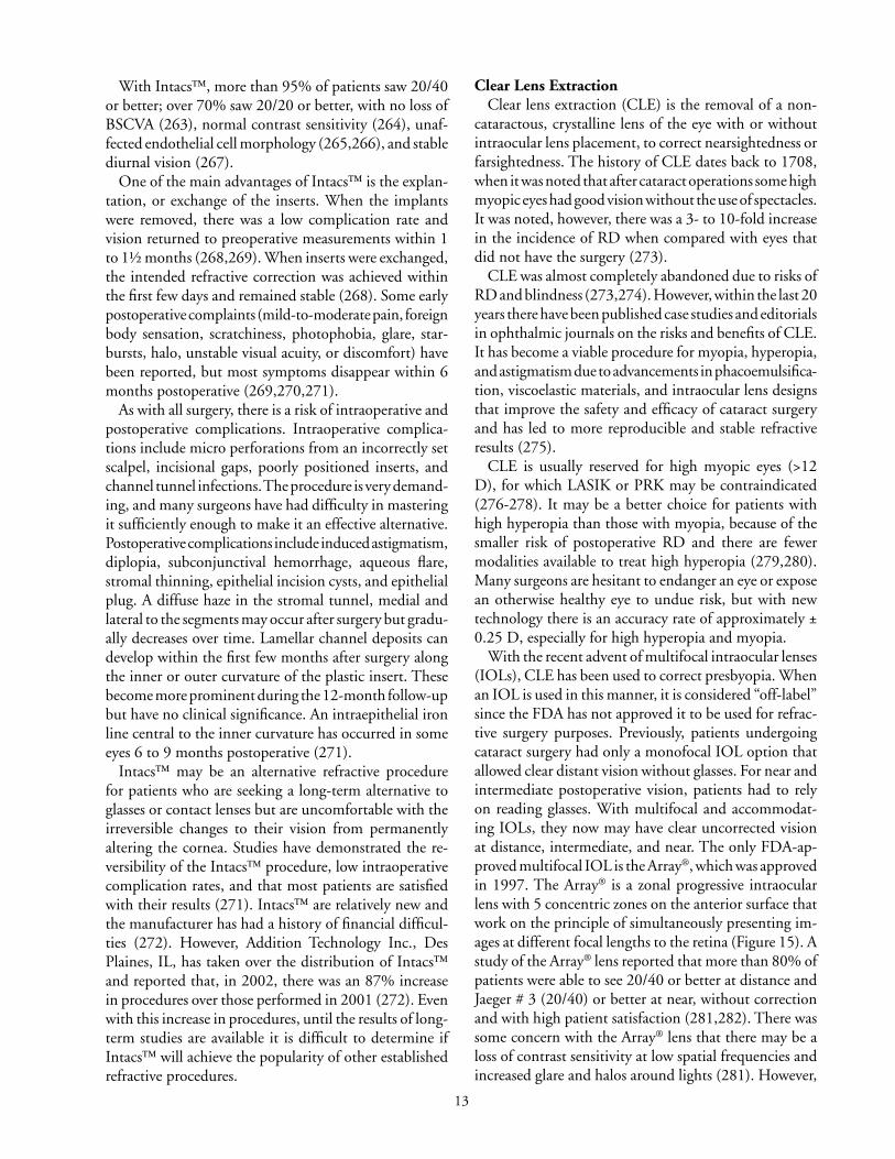
IntacsIntrastromal Corneal Ring was a single ring segment
used in the correction of mild nearsightedness, while Intacs uses 2 semicircular plastic inserts to correct refrac-tive error . These plastic inserts are placed deep within the corneal tissue at the outer edge and away from the central optical zone (Figure 14) . In addition, they can be removed and/or replaced with a thicker insert if greater correction is needed, with minimal risk (261) . The FDA approved Intacs in 1999 for the correction of mild myopia (1 .00 to 3 .00 D with ≤ 0 .75 D of astigmatism) . The procedure takes about 15 minutes and is done on an outpatient basis . During surgery, a tunnel is created between the layers of the stroma, and the two crescent-shaped plastic inserts are placed in the tunnel . The thicker the ring, the more flattening and greater correction is achieved .
Intacs patients must be at least 21 years of age, have stable vision for at least 1 year prior to surgery, and healthy eyes with no disease or injury . Contraindications for In-tacs include autoimmune or immunodeficiency diseases (e .g ., lupus, rheumatoid arthritis, or AIDS), pregnancy or nursing, thin corneas, glaucoma, herpetic eye disease, ocular conditions that could increase the possibility of future complications (e .g ., recurrent erosion syndrome, or corneal dystrophy), or taking prescriptions that may affect healing or visual acuity (262) .
Figure 13. Steepening of the cornea to correct for hyperopia
Figure 14. Intacs™ semi-circular plastic inserts
13
With Intacs, more than 95% of patients saw 20/40 or better; over 70% saw 20/20 or better, with no loss of BSCVA (263), normal contrast sensitivity (264), unaf-fected endothelial cell morphology (265,266), and stable diurnal vision (267) .
One of the main advantages of Intacs is the explan-tation, or exchange of the inserts . When the implants were removed, there was a low complication rate and vision returned to preoperative measurements within 1 to 1½ months (268,269) . When inserts were exchanged, the intended refractive correction was achieved within the first few days and remained stable (268) . Some early postoperative complaints (mild-to-moderate pain, foreign body sensation, scratchiness, photophobia, glare, star-bursts, halo, unstable visual acuity, or discomfort) have been reported, but most symptoms disappear within 6 months postoperative (269,270,271) .
As with all surgery, there is a risk of intraoperative and postoperative complications . Intraoperative complica-tions include micro perforations from an incorrectly set scalpel, incisional gaps, poorly positioned inserts, and channel tunnel infections . The procedure is very demand-ing, and many surgeons have had difficulty in mastering it sufficiently enough to make it an effective alternative . Postoperative complications include induced astigmatism, diplopia, subconjunctival hemorrhage, aqueous flare, stromal thinning, epithelial incision cysts, and epithelial plug . A diffuse haze in the stromal tunnel, medial and lateral to the segments may occur after surgery but gradu-ally decreases over time . Lamellar channel deposits can develop within the first few months after surgery along the inner or outer curvature of the plastic insert . These become more prominent during the 12-month follow-up but have no clinical significance . An intraepithelial iron line central to the inner curvature has occurred in some eyes 6 to 9 months postoperative (271) .
Intacs may be an alternative refractive procedure for patients who are seeking a long-term alternative to glasses or contact lenses but are uncomfortable with the irreversible changes to their vision from permanently altering the cornea . Studies have demonstrated the re-versibility of the Intacs procedure, low intraoperative complication rates, and that most patients are satisfied with their results (271) . Intacs are relatively new and the manufacturer has had a history of financial difficul-ties (272) . However, Addition Technology Inc ., Des Plaines, IL, has taken over the distribution of Intacs and reported that, in 2002, there was an 87% increase in procedures over those performed in 2001 (272) . Even with this increase in procedures, until the results of long-term studies are available it is difficult to determine if Intacs will achieve the popularity of other established refractive procedures .
Clear Lens ExtractionClear lens extraction (CLE) is the removal of a non-
cataractous, crystalline lens of the eye with or without intraocular lens placement, to correct nearsightedness or farsightedness . The history of CLE dates back to 1708, when it was noted that after cataract operations some high myopic eyes had good vision without the use of spectacles . It was noted, however, there was a 3- to 10-fold increase in the incidence of RD when compared with eyes that did not have the surgery (273) .
CLE was almost completely abandoned due to risks of RD and blindness (273,274) . However, within the last 20 years there have been published case studies and editorials in ophthalmic journals on the risks and benefits of CLE . It has become a viable procedure for myopia, hyperopia, and astigmatism due to advancements in phacoemulsifica-tion, viscoelastic materials, and intraocular lens designs that improve the safety and efficacy of cataract surgery and has led to more reproducible and stable refractive results (275) .
CLE is usually reserved for high myopic eyes (>12 D), for which LASIK or PRK may be contraindicated (276-278) . It may be a better choice for patients with high hyperopia than those with myopia, because of the smaller risk of postoperative RD and there are fewer modalities available to treat high hyperopia (279,280) . Many surgeons are hesitant to endanger an eye or expose an otherwise healthy eye to undue risk, but with new technology there is an accuracy rate of approximately ± 0 .25 D, especially for high hyperopia and myopia .
With the recent advent of multifocal intraocular lenses (IOLs), CLE has been used to correct presbyopia . When an IOL is used in this manner, it is considered “off-label” since the FDA has not approved it to be used for refrac-tive surgery purposes . Previously, patients undergoing cataract surgery had only a monofocal IOL option that allowed clear distant vision without glasses . For near and intermediate postoperative vision, patients had to rely on reading glasses . With multifocal and accommodat-ing IOLs, they now may have clear uncorrected vision at distance, intermediate, and near . The only FDA-ap-proved multifocal IOL is the Array®, which was approved in 1997 . The Array® is a zonal progressive intraocular lens with 5 concentric zones on the anterior surface that work on the principle of simultaneously presenting im-ages at different focal lengths to the retina (Figure 15) . A study of the Array® lens reported that more than 80% of patients were able to see 20/40 or better at distance and Jaeger # 3 (20/40) or better at near, without correction and with high patient satisfaction (281,282) . There was some concern with the Array® lens that there may be a loss of contrast sensitivity at low spatial frequencies and increased glare and halos around lights (281) . However,

14
studies showed no difference in low-contrast sensitivity between binocular Array® patients and binocular mono-focal patients; the occurrence of ghosting and glare was similar in both the Array® and monofocal IOL groups (283-285) . Additionally, when stereoacuity was compared in 31 patients with unilateral Array® lens implants to 29 patients with bilateral Array® lens implants, results were similar for both groups (286) . Another study, which measured night driving performance of patients with Ar-ray® IOLs to those with monofocal IOLs, under 3 poor visibility conditions, found that there was no difference in recognition rates and recognition distances for signs and hazards (287) .
An accommodating IOL has a monofocal optic and special hinges at the optic-haptic junction, which move by contraction and relaxation of the ciliary muscle within the eye, resulting in a form of accommodation so that patients can see clearer at far, near, and intermediate dis-tances (Figure 16) . The CrystaLens®, which received FDA approval in 2003, is currently the only FDA-approved accommodating IOL . Clinical trial data reported, at 1-year postoperative, that 98 .4% of patients had 20/40 or better UCVA; 100% had BSCVA of 20/20; 100% had 20/32 or better UCVA intermediate vision; 98 .4% had J3 or better UCVA near vision; and 100% had BSCVA
of 20/20 (288,289) . It was also reported that the Crysta-Lens® performed equally compared to a monofocal lens in contrast sensitivity both with and without a glare source . Currently, the FAA does not issue airmen a certificate if they have either a multifocal or accommodating IOL with CLE, since it does not have FDA approval and is considered an “off-label” use .
Clear lens extraction, however, appears to be one of the more stable refractive procedures available, with ± 0 .02 D/year refractive change reported over a 9-year observation period . Several studies have shown that, for quality of vision, an unaltered cornea is optically superior to an operated cornea . Optical aberrations can occur after any corneal procedure that creates abnormal contours and, with larger amounts of refractive correc-tion, there is a decrease in the quality of vision in low-contrast situations (e .g ., driving at night) . CLE, however, is not without complications, which can range from posterior capsular opacification (279,280), intraocular lens dislocation (291), macular edema (more common in hyperopes) (292), glaucoma (280,290,293), uveitis (273), endothelial loss (292,294), postoperative hypotony (295), to more serious ones that can result in blindness (e .g ., RD, [275,276,278,290,291] or endophthalmitis [273,275]) . Colin et al . (296), reported in an extended study that followed patients with high myopia and CLE for 4 years found that 2% of the patients had RD, but by 7 years that number had increased to 8 .1% . Ramos et al . (297), in their review of literature, reported that there are several variables that have a significant affect on the rate of RD after cataract surgery, which included age, gender, refractive state, status of the fellow eye, and the condition of the posterior vitreous . Age was the most significant variable for RD (298-300) . Patients whose age was less than 60 years had a RD rate of 2 .43%, while those ages 60 to 69 had a rate of 1 .51%, those ages 70 to 79 had a rate of 0 .82%, and those 80 and over had a rate of 0 .47% (298) . Results of CLE are encouraging; however, patients must be aware of the increased risk for severe vision loss (274) .
NEW TECHNOLOGY
The knowledge gained from past and current refractive surgery techniques has led to new alternatives or consid-erations concerning the visual system . One important lesson learned is that 20/20 acuity is not the only means to measure good vision . Snellen 20/20 measures about 80% of the total visual system of the human eye and is a low-order aberration (prism, sphere, and cylinder) or quantitative measures of central vision . The remain-ing 20% involves higher-order aberrations (qualitative measures) .
Figure 16. CrystaLens® Accommo-dating IOL
Figure 15. Array® Multifocal IOL

15
Phakic Intraocular LensesPhakic intraocular lenses (IOLs) are synthetic lenses
that are implanted into the eye as an addition to the eye’s crystalline lens . They can be used to correct ex-treme myopia or hyperopia and can be combined with LASIK to correct even greater refractive errors . During the procedure, a small incision (approximately 3-5 mm) is placed at the periphery of the cornea, and the IOL is inserted through the incision into the anterior chamber (Figure 17) or the posterior chamber just in front of the crystalline lens (301) . An anterior chamber phakic IOL is secured into place on the iris with two small flexible hooks . A posterior chamber phakic IOL is placed behind the iris, in front of the crystalline lens, and centered with the pupil . Phakic IOLs do not interfere with accommoda-tion of the crystalline lens (302) .
Aphakic IOLs were first introduced in the 1950s, and the designs and materials have changed dramatically over the years . Whether used to correct aphakia or a high refractive error, IOLs are known for exact predictability, reproducibility of the surgical success, and reversibility of the procedure . One of the factors contributing to the safety of this procedure is that the intraocular anatomy or physiology of the eye is not changed (i .e ., the central cornea is not touched during surgery) . The lens can be removed or exchanged with no long-term effects (302) .
There are only two phakic IOLs that have received FDA approval to be used for refractive surgery . They are the “Verisyse” (formally Ophtec’s Artisan™) from Optec/Advanced Medical Optics, which is an anterior chamber lens used to treat severe myopia and hyperopia, and the “Visian Implantable Contact Lens” (ICL)” from STAAR, which is a posterior chamber aphakic IOL now being marketed as a phakic IOL or ICL . Both lenses are intended for people with healthy eyes, stable vision, and with no more than 2 .5 D of astigmatism .
Clinical studies with phakic IOLs have reported an in-crease in BSCVA (303,304), stable vision (305,306), and an improvement in the quality of vision (i .e ., no change in vision when illumination varies; 307,308) . As with all types of surgery, there is the possibility of complications, which include loss of endothelial cells (301,309-312), decentration of lens (121), pupillary block (313), RD (313,314), cataract formation (301,313,315,316), iris retraction or pupil ovalization (310,317,318), halo/glare (310,318,319), and a decrease in crystalline lens trans-mittance (311) .
There are several other phakic IOLs seeking FDA approval . They include: “Veriflex Phakic IOL” from Optec/AMO, a foldable version of the Artisan/Verisyse phakic IOL; “AcrySof Phakic ACL” from Alcon; “Icare” from Cornéal, a one-piece foldable and injectable IOL made of a soft hydrophilic acrylic material; “Phakic Re-fractive Lens (PRL)” a posterior chamber lens made of a silicone material; and “GBR/Vivarte” from IOLTECH . In Europe, these and other similar phakic IOLs have been in the hands of surgeons since as early as 1989 . While this is a promising procedure for the correction of higher refractive errors, studies are needed to determine if the results will have long-term refractive stability without sight-threatening complications .
PRESBYOPIA SURGERY
Presbyopia eventually affects everyone . People often see presbyopia as a sign of advancing age and bifocals as a handicap . The presbyopic population in the United States is expected to double every 5 years until the year 2010 (320,321) . Presbyopia usually occurs around 40 years of age . Aviators with presbyopia can experience problems reading maps, charts, or seeing the instrument panel . Approximately 56% of the civil airman population is ≥ 40 years of age . Many of these airmen are presbyopic and need correction for near and/or intermediate vision (322) .
The long-established belief that presbyopia is caused by an age-related loss of elasticity or hardening of the crystalline lens has recently been challenged . Hardening of the lens and lens capsule does not appear to start in all individuals at the same time, nor does it progress at the same rate . Presbyopia, however, occurs at the same age, ± 1½ years, in 100% of the population (323) . The ciliary muscles, crystalline lens, and zonules are the 3 primary components of the eye that provide accommodation . A new theory for the onset of presbyopia is that the lens continues to grow throughout life, while the eye, itself, stops growing after puberty . At approximately 40 years of age, the size of the crystalline lens begins to compromise
Figure 17. An anterior chamber phakic IOL
16
the space of the lens-zonule-ciliary muscle complex . As the lens encroaches this area, the zonules become lax, and the force of the ciliary muscle contraction cannot be transmitted to the crystalline lens to allow the lens to change shape (321,323,324) . This theory regarding the etiology of presbyopia has prompted refractive surgeons to conduct clinical studies on different methods for the surgical reversal of presbyopia, including Anterior Ciliary Sclerotomy (ACS), Laser Presbyopia Reversal (LAPR), and Surgical Reverse Presbyopia (SRP) .
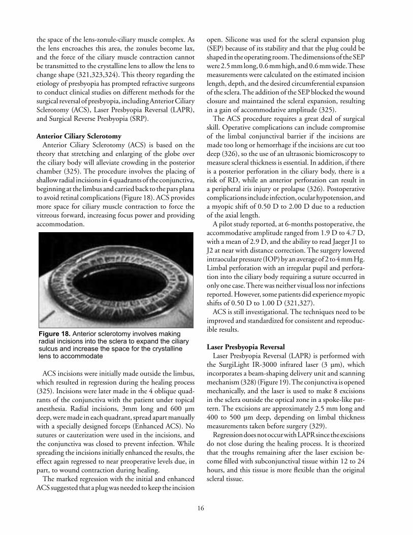
Anterior Ciliary SclerotomyAnterior Ciliary Sclerotomy (ACS) is based on the
theory that stretching and enlarging of the globe over the ciliary body will alleviate crowding in the posterior chamber (325) . The procedure involves the placing of shallow radial incisions in 4 quadrants of the conjunctiva, beginning at the limbus and carried back to the pars plana to avoid retinal complications (Figure 18) . ACS provides more space for ciliary muscle contraction to force the vitreous forward, increasing focus power and providing accommodation .
ACS incisions were initially made outside the limbus, which resulted in regression during the healing process (325) . Incisions were later made in the 4 oblique quad-rants of the conjunctiva with the patient under topical anesthesia . Radial incisions, 3mm long and 600 µm deep, were made in each quadrant, spread apart manually with a specially designed forceps (Enhanced ACS) . No sutures or cauterization were used in the incisions, and the conjunctiva was closed to prevent infection . While spreading the incisions initially enhanced the results, the effect again regressed to near preoperative levels due, in part, to wound contraction during healing .
The marked regression with the initial and enhanced ACS suggested that a plug was needed to keep the incision
open . Silicone was used for the scleral expansion plug (SEP) because of its stability and that the plug could be shaped in the operating room . The dimensions of the SEP were 2 .5 mm long, 0 .6 mm high, and 0 .6 mm wide . These measurements were calculated on the estimated incision length, depth, and the desired circumferential expansion of the sclera . The addition of the SEP blocked the wound closure and maintained the scleral expansion, resulting in a gain of accommodative amplitude (325) .
The ACS procedure requires a great deal of surgical skill . Operative complications can include compromise of the limbal conjunctival barrier if the incisions are made too long or hemorrhage if the incisions are cut too deep (326), so the use of an ultrasonic biomicroscopy to measure scleral thickness is essential . In addition, if there is a posterior perforation in the ciliary body, there is a risk of RD, while an anterior perforation can result in a peripheral iris injury or prolapse (326) . Postoperative complications include infection, ocular hypotension, and a myopic shift of 0 .50 D to 2 .00 D due to a reduction of the axial length .
A pilot study reported, at 6-months postoperative, the accommodative amplitude ranged from 1 .9 D to 4 .7 D, with a mean of 2 .9 D, and the ability to read Jaeger J1 to J2 at near with distance correction . The surgery lowered intraocular pressure (IOP) by an average of 2 to 4 mm Hg . Limbal perforation with an irregular pupil and perfora-tion into the ciliary body requiring a suture occurred in only one case . There was neither visual loss nor infections reported . However, some patients did experience myopic shifts of 0 .50 D to 1 .00 D (321,327) .
ACS is still investigational . The techniques need to be improved and standardized for consistent and reproduc-ible results .
Laser Presbyopia ReversalLaser Presbyopia Reversal (LAPR) is performed with
the SurgiLight IR-3000 infrared laser (3 µm), which incorporates a beam-shaping delivery unit and scanning mechanism (328) (Figure 19) . The conjunctiva is opened mechanically, and the laser is used to make 8 excisions in the sclera outside the optical zone in a spoke-like pat-tern . The excisions are approximately 2 .5 mm long and 400 to 500 µm deep, depending on limbal thickness measurements taken before surgery (329) .
Regression does not occur with LAPR since the excisions do not close during the healing process . It is theorized that the troughs remaining after the laser excision be-come filled with subconjunctival tissue within 12 to 24 hours, and this tissue is more flexible than the original scleral tissue .
Figure 18. Anterior sclerotomy involves making radial incisions into the sclera to expand the ciliary sulcus and increase the space for the crystalline lens to accommodate

17
The LAPR procedure offers a number of advantages . Unlike other surgical presbyopia treatments, with LAPR there is no need for suturing any type of implants to the sclera, thus avoiding their potential complications or side effects . In addition, because the infrared laser partially coagulates blood vessels as it removes tissue, it makes a very clean excision of tissue .
Possible risks from LAPR include ocular hypotony, infection, and scleral perforation . Caution must be ap-plied in cases of thin sclera . Since the conjunctival pe-ritomy may cause subconjunctival hemorrhages, clinical researchers are evaluating a transconjunctival approach to minimize bleeding and shorten the procedure, which now takes approximately 30 minutes . In addition, since the sclera is weakened, studies will be required to deter-mine whether this procedure places eyes at greater risk to rupture following blunt trauma . Furthermore, because the excisions are transparent and expose the bluish hue from the uvea, some patients may have a long-term cosmetic problem . The LAPR procedure may be contraindicated for patients with glaucoma, cataracts, uveitis, dry eyes, collagen vascular disease, and diabetes .
Patients (n=70 eyes) between the ages of 42 and 65 were treated in clinical trials in Venezuela and Argentina . LAPR was performed in 1 eye and repeated 1 to 4 months later in the second eye . Patients had near vision of J6 to J7 before surgery, but within 2 weeks after surgery, accommodation increased by 1 to 2 .5 D, with further improvement within 2 weeks to 2 months . Distance vision remained the same . Although postoperative near vision ranged from J3 to J1, monocularly, when vision was tested binoculary visual acuity increased . Almost no visual regression occurred up to 18 months after surgery . There was reported foreign-body sensation and an increase in dryness for a few days after surgery . One patient described minimal distortion of vision on day 1 postoperative, but this cleared by day 2 . Some eyes remain red for about a month .
To date, most of the LAPR clinical trials have been obtained through small, private clinics outside the United
States . Larger, controlled clinical trials are beginning in Europe . SurgiLight has received permission to patent this technology, and the first trials in the U .S . were cleared in January 2003 by the FDA under an Investigational Device Exemption (330) .
Surgical Reverse PresbyopiaSurgical Reverse Presbyopia (SRP) is a new technique
that allows the ciliary body to expand and stretch out-wardly, increasing the distance between the lens equator and the ciliary muscle, which appears to restore accom-modation (323) (Figure 20) . The procedure is based on Schachar’s theory that accommodation decreases with age because the crystalline lens grows concentrically, decreas-ing the space between the ciliary body and the lens .
After several design changes, a technique was de-veloped for the placement of 4 separate PMMA scleral expansion bands (SEB) into partial-thickness scleral tunnels (Figure 21). Each tunnel is located 3.50 mm posterior to the limbus in the oblique quadrants (323). A lamellar diamond knife creates the scleral tunnel 5.0 mm in length and 150 µm in thickness . The SEB (4 .0 mm in length, 900 µm in width, 0 .3 mm in depth) is inserted into the tunnel, with the 2 ends outside of the tunnel on the scleral surface .
Selection criteria for candidates for SRP are patients between the ages of 40 and 70 years, no refractive error
Figure 19. SurgiLight IR-3000 Infrared Laser
Figure 20. Surgical Reversal of Presbyopia (SRP)
Figure 21. Insertion of scleral expansion bands (SEB)
18
at distance, and with binocular vision (i .e ., vision in both eyes) . Patients < 40 are considered if they are slightly hyperopia (< +1 .00 D) and are having difficulty with near-work requirements (323) .
Contraindications for SRP include prior cataract extrac-tion, scleromalacia (degeneration or thinning of sclera), previous trabeculotomy, blood coagulation abnormalities (widespread blood clotting and/or profuse hemorrhaging), collagen-vascular diseases, developing cataract, and poorly controlled glaucoma . Relative contraindications include, monocularity, > 70 years of age, severe keratoconjunctivitis sicca, insulin dependant diabetes, and hyperopia > +1 .00 D (323) . (Note: Candidates with these relative contrain-dications are evaluated on a case-by-case basis .)
In an initial study, preoperative eyes had an average of 2 .1 D of accommodation . One month after surgery, 9 patients had amplitude of accommodation ranging from 3 .3 D to 5 .5 D, with an average of 4 .3 D . (Note: Interestingly, the fellow eye, which did not have the implantation, also gained amplitude after surgery, averaging 3 .5 D .) It was postulated that this phenomenon was probably due to a central nervous system mechanism (321) . Complications from the procedure have been minimal . One patient had conjunctival suture dehiscence (separation) that required a conjunctival graft and 5 patients had segment rotation . Surgery with SEB appears to restore accommodation . However, the procedure is still evolving and predictability needs improvement (321) .
Currently, the FAA does not allow procedures such as ACS, LAPR, and SRP for the correction of presbyopia in civil airmen . These procedures are still in their investi-gational stages, and it is impossible to predict how these procedures might perform in an aviation environment . Airmen should be cautioned against having any investiga-tional procedure performed without understanding how it may affect their ability to receive a medical certificate .
WAVEFRONT TECHNOLOGY
Lasers used for refractive surgery have gone through several generations of upgrades . Wavefront technology is a term used to describe laser systems that can customize the IntraLASIK or LASIK procedures . While results have been good, and in some cases better than non-wavefront treatments for specific patients, not everyone qualifies for this approach because of FDA recommendations and the limitations of the technology .
A wavefront-guided system consists of 2 parts: a wave-front sensor and a programmed excimer laser, based on information from the sensor (331) . The wavefront sensor system includes a fixation target, an input laser beam that generates a point light source, a wavefront sensor that measures the slope of the exiting wavefront, and
software that will determine the characteristics of the ablation for the excimer laser . A visual fixation target assists the patient in maintaining view, direction, and accommodation during the wavefront measurement . As the patient fixates, a laser beam is directed into the eye that generates a point light source onto the retina . This light is reflected from the retina back through the pupil, and the wavefront of the light leaving the pupil is relayed to the wavefront sensor . The wavefront is distorted by the refractive properties in the ocular media . When the beam enters the eye, it has a flat wavefront, but after traveling through the cornea, crystalline lens, and the other ocular media, the flat wavefront becomes irregular and analysis reveals the aberrations of the optical system (331) .
Conventional refractive surgery corrects 2 types of refractive error, spherical and cylindrical . Wavefront-guided refractive surgery creates treatments tailored to the individual eye based on the total wavefront error in patients’ eyes, rather than the conventional manifest refraction (331,332,333) . It is thought that more than 40% of eyes have higher order aberrations that are 0 .25 D or higher . If the pupil size is increased from 3 to 6 mm in diameter, this aberration can alter the spherocylindrical data by several diopters .
Once the wavefront map is known for low-order aber-rations, how much tissue to ablate can then be calculated . However, conversion of higher-order aberrations is more difficult . Based on the beam characteristics, energy per pulse, repetition rate, pulse size and pulse shape, and other factors, special algorithms can be generated for a treatment table that defines the location, size, and sequence of each laser .
There are several companies that produce lasers capable of performing wavefront guided treatments . These include VISX® CustomVue®, Bausch and Lomb® Zyoptix®, Alcon® CustomCornea®, and WaveLight Laser Tech-nologie, Allegretto Wave™ . All of these systems have FDA approval .
The VISX® CustomVue® laser uses SmartBeam™ technology, which adjusts beam size and shapes ranging from 0 .065 to 6 .5 mm to allow for personalized refractive treatments . It also uses the ActiveTrak™ 3-D Active Eye Tracking that can capture all 3 dimensions of intra-opera-tive eye movements without the requirement of pupillary dilation . The Variable Spot Scanning (VSS™) technology adds a blend zone that increases the ablation zone by 51%, increasing the overall diameter of the ablation to 8 millimeters . This system is reported to remove minimal corneal tissue, have a shorter treatment time, and greater flexibility to personalize the laser treatment (334) .
The Bausch and Lomb® Zyoptix® laser uses a 2 mm “flying spot beam .” Each spot ablates the corneal tissue in a pattern that allows the debris to clear in one place

19
before contacting that area again . This allows each pulse to polish the corneal surface for a smoother result, and the scanner feedback mechanism ensures that every pulse is placed exactly as prescribed, with no laser beams fired at consecutive locations or overlap . No dilation is required, so there is quicker recovery time and less discomfort during the surgery with a maximum treatment area of up to 14 mm (335) .
The Alcon® CustomCornea® uses the LADARVi-sion® tracking system and the LADARVision 4000 laser system . The tracking device uses a closed-loop technology, which compensates for eye movements through continu-ous, constant feedback on the location of the eye (more than 4,000 times per second), telling the laser where the eye will be at the precise moment the laser is applied . This system reshapes the cornea by correcting for both lower- and higher-order aberrations (336) .
The Allegretto Wave™ laser (WaveLight Laser Tech-nologie, Erlangen, Germany) features a spot 200Hz, Gaussian-shaped 0 .95 mm flying spot, an active tracker with a response time of less than 6 .0 milliseconds, does not require dilation of the eyes, and can be used for the treatment of both hyperopia and myopia . At 1-year postoperative, results from the FDA Allegretto myopia study treated for up to -12 D of myopia and 6 .0 D of astigmatism reported that 85% of eyes were within ± 0 .5 D of target range, and more than half of the eyes (56%) achieved UCVA of 20/16 . Even the high myopes did well with 80% of those eyes, with > - 7 .0 D within ± 0 .5 D of target range, compared to 86% of those with < - 7 .0 D . Of those with > - 7 .0 D of myopia, 80% achieved UCVA of 20/20 or better, while 89% of those with < -7 .0 D attained the same results . Only 0 .6% lost 2 or more lines of BSCVA, while more than 58% gained 1 or more lines . Patients in the trials were asked how much glare they experienced from bright lights (e .g ., headlights, and/or streetlights) before and after LASIK . There was an overall improvement in nighttime visual function and night driving (337) . Investigators believe that this technology delivers excellent refractive results, with no spherical aberrations and a larger optical zone . By preserving the optical zone size and better shaping of the ablation profile, nighttime visual function improved (338,339) . The Allegretto laser has been used worldwide for several years and has been FDA approved in the USA since late 2003 .
Wavefront-guided technology, along with improved microkeratomes, better eye tracking with the laser, and more precise information from the aberrometer to the laser is reported to improve outcomes and decrease the need for enhancement procedures . Some optical aberrations, however, will still be present because laser ablation always induces new errors . Currently, wavefront
technology is recommended for those patients with pupil diameter greater than 6 .6 mm, require optimum night vision, have higher prescriptions, and a large number of higher-order aberrations . This new technology comes at higher cost . Wavefront technology can add $100 to $400 per eye to the cost . Patients for which custom wavefront LASIK is either unnecessary or unsuitable include those with refractive error outside the FDA-approval range, thin corneas, pupil diameters less than 5 mm, and a large disparity between the manifest refraction and the wavefront aberrometer-derived refraction (340) . There is limited data from wavefront technology that can be compared with standard LASIK data (340) . One compara-tive study of 12 patients with standard LASIK in 1 eye and wavefront LASIK in the contralateral eye reported at 6 months postoperative an UCVA of 20/40 or better in 100% of eyes in both groups and 20/20 UCVA in 67% (8 eyes) with wavefront and 83% (10 eyes) with standard LASIK (341) . Kaiserman et al . (342), reported that at 1 month after surgery UCVA of 20/20 or better was achieved by 72% of those with wavefront LASIK and 70% of those with standard LASIK . However, 88% of those with wavefront LASIK and only 40% of those with standard LASIK had improved contrast sensitivity measurements at one-month postoperative (342) . It will be interesting to see how this technology performs after more long-term clinical data become available .
FEMTOSECOND LASER
Complications associated with LASIK are often related to the use of mechanical microkeratomes (343,344) . Mi-crokeratomes have a cutting accuracy of approximately ± 60µm . A microkeratome set to cut an 180µm flap could produce a flap of 120µm to 240µm, which could prove unacceptable if large amounts of tissue are to be removed to correct high myopia . In addition, mechanical micro-keratomes do not cut a perfectly planar flap . Often a flap is cut that varies in thickness between the peripheral and central areas . This can result in thin or irregular flap and induce astigmatism .
The FDA has approved the IntraLase femtosecond (FS) laser keratome, which has several advantages over a mechanical microkeratome . The laser keratome uses a spot size of about 3 µm creating the flap by placing tiny bursts of energy at a prescribed depth in the stroma (Figure 22) . Because this laser is computer-controlled, the surgeon can cut flaps as thin as 100µm with an accuracy of ± 5µm, leaving more tissue to be photoablated for higher correc-tions (345-347) . There have been no reported partial or buttonhole flaps, epithelial sloughs, or free caps to date (345,347,348) . Additionally, it has been reported that no procedures have had to be aborted due to loss of suction,
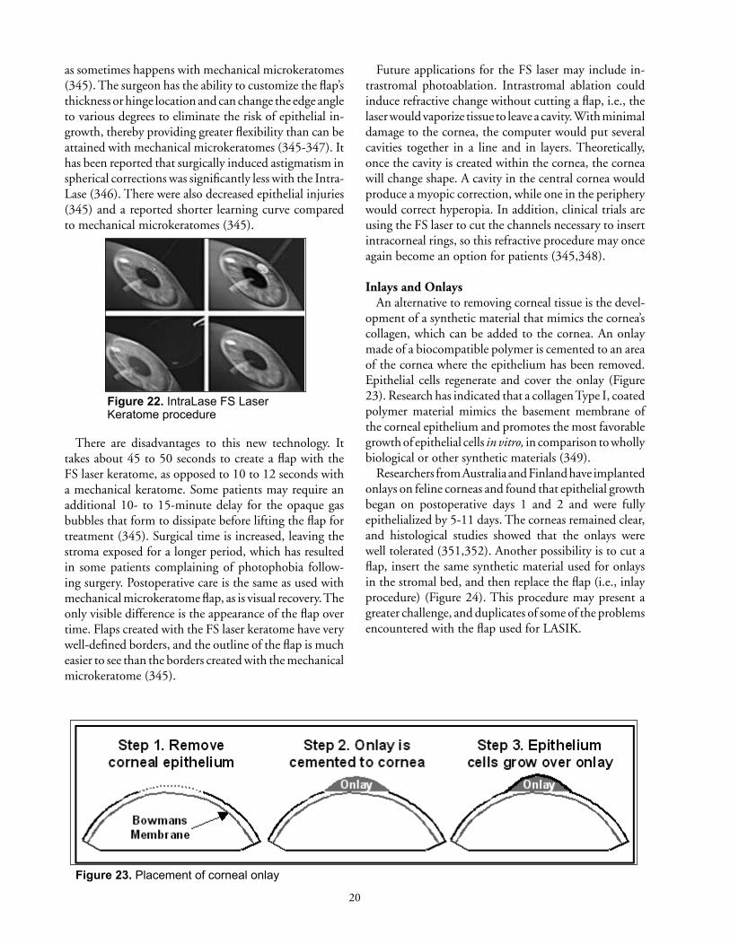
20
as sometimes happens with mechanical microkeratomes (345) . The surgeon has the ability to customize the flap’s thickness or hinge location and can change the edge angle to various degrees to eliminate the risk of epithelial in-growth, thereby providing greater flexibility than can be attained with mechanical microkeratomes (345-347) . It has been reported that surgically induced astigmatism in spherical corrections was significantly less with the Intra-Lase (346) . There were also decreased epithelial injuries (345) and a reported shorter learning curve compared to mechanical microkeratomes (345) .
There are disadvantages to this new technology . It takes about 45 to 50 seconds to create a flap with the FS laser keratome, as opposed to 10 to 12 seconds with a mechanical keratome . Some patients may require an additional 10- to 15-minute delay for the opaque gas bubbles that form to dissipate before lifting the flap for treatment (345) . Surgical time is increased, leaving the stroma exposed for a longer period, which has resulted in some patients complaining of photophobia follow-ing surgery . Postoperative care is the same as used with mechanical microkeratome flap, as is visual recovery . The only visible difference is the appearance of the flap over time . Flaps created with the FS laser keratome have very well-defined borders, and the outline of the flap is much easier to see than the borders created with the mechanical microkeratome (345) .
Future applications for the FS laser may include in-trastromal photoablation . Intrastromal ablation could induce refractive change without cutting a flap, i .e ., the laser would vaporize tissue to leave a cavity . With minimal damage to the cornea, the computer would put several cavities together in a line and in layers . Theoretically, once the cavity is created within the cornea, the cornea will change shape . A cavity in the central cornea would produce a myopic correction, while one in the periphery would correct hyperopia . In addition, clinical trials are using the FS laser to cut the channels necessary to insert intracorneal rings, so this refractive procedure may once again become an option for patients (345,348) .
Inlays and OnlaysAn alternative to removing corneal tissue is the devel-
opment of a synthetic material that mimics the cornea’s collagen, which can be added to the cornea . An onlay made of a biocompatible polymer is cemented to an area of the cornea where the epithelium has been removed . Epithelial cells regenerate and cover the onlay (Figure 23) . Research has indicated that a collagen Type I, coated polymer material mimics the basement membrane of the corneal epithelium and promotes the most favorable growth of epithelial cells in vitro, in comparison to wholly biological or other synthetic materials (349) .
Researchers from Australia and Finland have implanted onlays on feline corneas and found that epithelial growth began on postoperative days 1 and 2 and were fully epithelialized by 5-11 days . The corneas remained clear, and histological studies showed that the onlays were well tolerated (351,352) . Another possibility is to cut a flap, insert the same synthetic material used for onlays in the stromal bed, and then replace the flap (i .e ., inlay procedure) (Figure 24) . This procedure may present a greater challenge, and duplicates of some of the problems encountered with the flap used for LASIK .
Figure 22. IntraLase FS Laser Keratome procedure
Figure 23. Placement of corneal onlay

21
AEROMEDICAL ISSUES
Civil airmen with RK procedures were initially al-lowed to obtain FAA medical certificates through a waiver process . Airmen were to discontinue flying for 6 months after having RK and then were required to apply for a new certificate . At the time of their examination, they were required to submit a “Report of Eye Evalua-tion” (FAA Form 8500-7), completed by their eyecare specialist verifying that healing was complete, vision was stable, and they met the vision standards for the applied certificate class . A follow-up eye evaluation report was required 6 months after an airman medical certificate was issued . To expedite tracking of this procedure, in 1983 the FAA’s Aerospace Medical Certification Division as-signed RK its own pathology code (130) . This code was placed on the medical records of all airmen with RK . By the early 1990s, applicants with RK could obtain a medical certificate without a waiver by passing the visual acuity standards for their class of medical certificate . Air-men who had a current medical certificate and had RK performed were to discontinue flying until an eye care specialist verified that their vision was stable and they had been released (352) . An eye evaluation report (FAA Form 8500-7), completed by the surgeon who performed the procedure or another eye care specialist familiar with the applicant’s ophthalmological history, was submitted to the FAA verifying that the airman could meet these conditions and return to flying . If an airman with RK had not submitted a previous eye evaluation report, one was required at the time of certification renewal .
Although the FDA did not approve the first excimer laser for use in the US until October 1995, applicants with PRK applied for and received FAA medical certificates prior to that date . These airmen had gone to foreign countries for this procedure or were in the original investigational studies . Additionally, when surgeons changed to primarily performing LASIK, applicants applied for and routinely received their medical certificates . No waiver process is currently required for airmen with PRK or LASIK for them
to receive a medical certificate . The present regulation for airman applicants with refractive surgery states:
An applicant who has been treated with refractive sur-gery may be issued a medical certificate by the Aviation Medical Examiner (AME) if the applicant meets the visual acuity standard and the “Report of Eye Evaluation” indicates that healing is complete, visual acuity remains stable, and the applicant does not suffer from significant glare intolerance. This state of recovery is usually reached within 6- to 12-weeks after surgery (352).
A study of civil airmen with refractive surgery reported an increase of approximately 37% in the number of such airmen between the periods of 1994-1996 to 1997-1998 . In addition, there was a change in the distribution of the types of refractive surgery procedures . While the largest number of civil airmen had RK, that percentage had decreased and the percentage of airmen with LASIK procedures had more than tripled (Figure 25) (353) .
A later study of civil airmen with refractive surgery looked at the total civil airman population for the years 1996 to 2001 (Figure 26) . The results of this study reaf-firmed those of the previous study . A greater number of civil airmen had LASIK (61%); while the prevalence of RK decreased to 24% . Interestingly, PRK has decreased from 10% to 5% . This information confirms that civil airmen are continuing to have refractive surgery performed and suggests that they do not see it as a hindrance to flying or in obtaining an airman medical certificate .
SUMMARY AND DISCUSSION
During the last decade, there have been several new procedures introduced to reduce refractive errors, mak-ing it possible to see without contact lenses or glasses . Procedures for hyperopia, myopia, astigmatism, and now, presbyopia, are being performed in increasing numbers . RK, which was introduced in the late 1970s and gained widespread popularity, has been replaced by other pro-cedures due to reported patient dissatisfaction and many short- and long-term complications . Long-term prospec-tive evaluations of RK reported that there is a continuation
Figure 24. Inlay procedure

22
of progressive hyperopic shifts and reduction of corneal strength (Note: Ruptures at the incision sites have been reported more than 10 years after surgery [10-13]) . Even more worrisome in the aviation environment are diurnal fluctuations of vision of more than 1 .50 D and altitude-induced corneal changes (14-19) .
After extensive investigational trials, in 1995 the FDA approved the excimer laser for PRK refractive surgery . There was, however, reported patient dissatisfaction with postoperative pain, long recovery time, and refractive re-gression . In addition, early treatments had problems with haze, halos/glare, decreased visual performance at night, and dry eye problems, which raised concerns regarding PRK in the aviation environment . Due to these problems with PRK, refractive surgeons developed LASIK, which quickly became the surgery of choice . LASIK requires more surgical skill with the addition of the microkeratome to cut the corneal flap . Since there were no investigational studies prior to surgeons routinely performing LASIK, there were no clinical data regarding the possibility of operative and postoperative complications . Some sur-geons, remembering the long-term, postoperative prob-lems with RK, cautioned against the use of LASIK until more long-term clinical data became available . However,
as of June 2002, there have been more than 9 .5 million LASIK procedures performed and, of those, more than 5 .1 million were performed in the U .S . Information is now being published with increasing frequency, some reporting long-term and rather severe complications . Many of the complications (e .g ., under/over correction, loss of BSCVA, glare/halos, reduced visual performance in dim illumination, severe dry eyes, dislodged, or detached corneal flap up to 38 months postoperative) have raised concerns regarding the use of LASIK in the aviation environment .
The scientific evolution in adaptive optics shows that the human eye can achieve visual acuity of better than 20/15 with improved image quality . The application of adaptive optics has added to our knowledge about vision and has led to advances in diagnostic technology . New refractive alternatives will need to effectively correct lower-order aberrations while inducing fewer higher-order ones . New refractive techniques will need to have 4 critical ingredients: be safe and effective, maintain an aspheric cornea, minimize insult to the visual axis, and minimize tissue loss .
Optimum vision is essential for pilots who must detect and identify airborne traffic as well as hazards that may be on the runways or taxi lanes . Cockpit instrumentation and printed materials (e .g ., charts, maps, and flight manifests) must be clearly visible to ensure that proper flight pro-cedures and safety are maintained . It is unknown at this time how the long-term effects of refractive surgery may affect the performance of civil airman and if the known refractive surgery complications summarized in this paper may be exacerbated by age . It is important that pilots be aware of possible problems that may result from having refractive surgery that may affect their ability to safely perform aviation tasks . The news media and advertis-ers continues to promote the benefits of laser refractive surgery and often fail to report on patients who have
Figure 25. Refractive surgery, by type, in civil airmen
Figure 26. Refractive surgery by type in civil airmen

23
less than ideal outcomes . There are sites on the Internet where refractive surgery patients whose procedure resulted in less-than-perfect results can voice their concerns and organize support groups in their area (354) . To provide the aviation community with information to formulate administrative decisions and policies associated with both current and new refractive surgical procedures and techniques, continued monitoring of current refractive surgical procedures and clinical results of new refractive surgery techniques are required .
REFERENCES
1 . Vision Channel, Refractive Errors . At URL: www .vi-sionchannel .net/refractiveerrors/ (Accessed 1/03) .
2 . American Optometric Association . At URL: www .aoanet .org/conditions/facts_stats .asp . (Accessed 1/03) .
3 . Nakagawara VB, Wood KJ . The Aeromedical certification of photorefractive keratectomy in civil aviation: A reference guide . Washington DC: Department of Transportation/Federal Aviation Administration . 1998 . Report No . DOT/FAA/AM-98/25 .
4 . Stein HA, Slatt BJ, Stein RM . Refractive errors and how to correct them . In: Stein HA, Slatt BJ, Stein RM, eds . The Ophthalmic Assistant . St . Louis, MO: The CV Mosby Company; 1988:211-35 .
5 . Allard R . Corneal refractive surgery: Part I - past and present . Contact Lens Spectr . Aug 1991; 5(8):44-50 .
6 . Waring GO 3rd, Lynn MJ, Nizam A, Kutner MH, et al . Results of the prospective evaluation of radial keratotomy (PERK) Study five years after surgery . The Perk Study Group . Ophthalmol . Aug 1991; 98(8):1164-76 .
7 . Waring GO 3rd, Lynn MJ, McDonnell PJ, PERK Study Group . Results of the prospective evaluation of radial keratotomy (PERK) study 10 years after surgery . Arch Ophthalmol. Oct 1994; 112(10):1298-1308 .
8 . Werblin TP, Stafford GM . Hyperopic shift after refractive keratotomy using the Casebeer System . J Cataract Refract Surg. Oct 1996; 22(8):1030-6 .
9 . Deitz MR, Sanders DR, Raanan MG, LeLuca M . Long-term (5- to 12-year) follow-up of metal-blade radial keratotomy procedures . Arch Ophthalmol. May 1994; 112(5):614-20 .
10 . Panda A, Sharma N, Kumar A . Ruptured globe 10 years after radial keratotomy . J Refract Surg. Jan-Feb 1999; 15(1):64-5 .
11 . Glasgow BJ, Brown HH, Aizuss DH, Mondino BJ, et al . Traumatic dehiscence of incisions seven years after radial keratotomy . Am J Ophthalmol. Dec 1988; 106(6):703-7 .
12 . Lee BL, Manche EE, Glasgow BJ . Rupture of radial and arcuate keratotomy scars by blunt trauma 91 months after incisional keratotomy . Am J Ophthal-mol. Jul 1995; 121(1):108-10 .
13 . Goldberg MA, Valluri S, Pepose JS . Air bag-related corneal rupture after radial keratotomy . Am J Oph-thalmol. Dec 1995; 120(6):800-2 .
14 . Anonymous . Radial keratotomy for myopia: Ameri-can Academy of Ophthalmology . [see comments] [Review] Ophthalmol. Jul 1993; 100(7):1103-15 .
15 . Wyzinski P . Daily refractive changes persisting after radial keratotomy . Am J Ophthalmol. Aug 1989; 108(2):205-6 .
16 . Wyzinski P, O’Dell LW . Diurnal cycle of refraction after radial keratotomy . Ophthalmol. Feb 1987; 94(2):120-4 .
17 . Santos VR, Waring GO 3d, Lynn MJ, Schanzlin DJ, et al . Morning-to-evening change in refraction, corneal curvature, and visual acuity 2 to 4 years after radial keratotomy in the PERK Study . [see com-ments] Ophthalmol. Nov 1988; 95(11):1487-93 .
18 . McDonnell PJ, Nizam A, Lynn MJ, Waring GO 3rd . Morning-to-evening change in refraction, corneal curvature, and visual acuity 11 years after radial keratotomy in the prospective evaluation of radial keratotomy study . Ophthalmol. Feb 1996; 103(2):233-9 .
19 . Bullimore MA, Sheedy JE, Owen D, Refractive Surgery Study Group . Diurnal visual changes in radial keratotomy: Implications for visual standards . Optom Vis Sci . 1994; 71(8):516-21 .
20 . Waring GO 3rd, Casebeer JC, Dru RM . One-year results of a prospective multicenter study of the Casebeer system of refractive keratotomy . Case-beer Chiron Study Group . Ophthalmol. Sep 1996; 103(9):1337-47 .
21 . Grimmett MR, Holland EJ . Complications of small clear-zone radial keratotomy . Ophthalmol. Sep 1996; 103(9):1348-56 .
24
22 . Tinning S . Contrast sensitivity and glare following keratotomy . AGARD Conference Proceedings No 492 . 1991; AD-A237 593 .
23 . Diamond S . Present status of radial keratotomy myopia surgery: aerospace considerations . Aviat Space Environ Med . Aug 1990; 61(8):732-4 .
24 . Waring GO 3d, Lynn MJ, Fielding B, Asbell PA, et al . Results of the prospective evaluation of radial keratotomy (PERK) Study 4 years after surgery for myopia . [see comments] JAMA . Feb 23 1990; 263(8):1083-91 .
25 . Stulting AA . Radial keratotomy in perspective . [Review] [Afrikaans] S Afr Med J . Apr 1988; 73(7):417-21 .
26 . Creel DJ, Crandall AS, Swartz M . Hyperopic shift induced by high altitude after radial keratotomy . J Refract Surg. Jul-Aug 1997; 13(4):398-400 .
27 . Mader TH, White LJ . Refractive changes at extreme altitude after radial keratotomy . Am J Ophthalmol. Jun 1995; 119(6):733-7 .
28 . Mader TH, Shallhorn SC, Blanton CL, Bilber B, et al . Refractive changes during prolonged exposure to altitude following refractive surgery . Oct 1994; MIPR NO: 94MM4576 .
29 . White LJ, Mader TH . Refractive changes with increasing altitude after radial keratotomy . [letter] Am J Ophthalmol. Jun 1993; 115(6):821-3 .
30 . Aron-Rosa DS, Boulnoy JL, Carre F, Delacour J, et al . Excimer laser surgery of the cornea: Qualitative and quantitative aspects of photoablation according to the energy density . J Cataract Refract Surg. Jan 1986; 12(1):27-33 .
31 . Trokel S . Evolution of excimer laser corneal surgery [see comments] . J Cataract Refract Surg. Jul 1989; 15(4):373-83 .
32 . Hersh PS, Spinak A, Garrana R, Mayers M . Pho-totherapeutic keratectomy: strategies and results in 12 eyes . Refract Corneal Surg . Mar-Apr 1993; 9(2 Suppl):S90-S95 .
33 . Fantes FE, Waring GO 3d . Effect of excimer laser radiant exposure on uniformity of ablated corneal surface . Lasers Surg Med . 1989; 9(6):533-42 .
34 . Editor . Summit PRK laser conditionally okayed by FDA panel . AOA News . Nov 1, 1994 . 33(9):1&12-13 .
35 . Dausch D, Klein R, Schroder E, Dausch B . Excimer laser photorefractive keratectomy with tapered transition zone for high myopia . A preliminary report of six cases . J Cataract Refract Surg. Sep 1993; 19(5):590-4 .
36 . Editor . Lasers proving useful in treating astigmatism . AOA News . Jun 1, 1992; 30(23):5&8 .
37 . O’Brart DP, Stephenson CG, Oliver K, Marshall J . Excimer laser photorefractive keratectomy for the correction of hyperopia using an erodible mask and axicon system . Ophthalmol. Nov 1997; 104(11):1959-70 .
38 . Jackson WB, Mintsioulis G, Agapitos PJ, Casson EJ . Excimer laser photorefractive keratectomy for low hyperopia: Safety and efficacy . J Cataract Refract Surg. May 1997; 23(4):480-7 .
39 . Sher NA, Hardten DR, Fundingsland B, DeMarch J, et al . 193-nm excimer photorefractive keratec-tomy in high myopia . Ophthalmol. Sep 1994; 101(9):1575-82 .
40 . Epstein D, Faterholm P, Hamberg-Nyström H, Tengroth B . Twenty-four-month follow-up of ex-cimer laser photorefractive keratectomy for myopia, refractive and visual acuity results . Ophthalmol. Sep 1994; 101(9):1558-64 .
41 . Seiler T, Kahle G, Kriegerowski M . Excimer laser (193-nm) myopic keratomileusis in sighted and blind human eyes . Refract Corneal Surg. Sep-Oct 1990; 6(5):165-73 .
42 . Fichte CM, Bell AM . Ongoing results of excimer laser photorefractive keratectomy for myopia: sub-jective patient impressions . J Cataract Refract Surg. Mar 1994; 20(2): 268-70 .
43 . Pallikaris IG, Lambropoulos IE, Kolydas PK, Nicolopoulos NS, et al . Excimer laser photorefrac-tive keratectomy for myopia: clinical results in 96 eyes . Refract Corneal Surg. Mar-Apr 1993; 9(Suppl):S101-02 .
44 . Shah SI, Hersh PS . Photorefractive keratectomy for myopia with a 6-mm beam diameter . J Refract Surg. Mar-Apr 1996; 12(2): 341-6 .
45 . Tengroth B, Epstein D, Fagerholm P, Fagerholm P, et al . Excimer laser photorefractive keratectomy for myopia: clinical results in sighted eyes . Ophthalmol. May 1993; 100(5):739-45 .

25
46 . Klyce SD, Smolek MK . Corneal topography of ex-cimer laser photorefractive keratectomy . J Cataract Refract Surg. Feb 1993; 19(Suppl):S122-30 .
47 . Krueger RR, Binder PS, McDonnell PJ . The effects of excimer laser photoablation on the cornea . In: Salz JJ, McDonnell PJ, McDonald MB, eds . Corneal Laser Surgery . St . Louis, MO: Mosby Year Book Inc; 1995:11-44 .
48 . Aktunç R, Aktunç T . Centration of excimer laser photorefractive keratectomy and changes in astig-matism . J Refract Surg. Jan-Feb 1996; 12(Suppl):S268-71 .
49 . Yavitz E . RK vs . PRK: Efficacy and outcome . Optom Manage. Dec 1994; 29(12):13 .
50 . Maguen E, Machat JJ . Complications of pho-torefractive keratectomy, primarily with the VISX excimer laser . In: Salz JJ, McDonnell PJ, McDonald MB, eds . Corneal Laser Surgery. St . Louis, MO: Mosby Year Book Inc; 1995:143-58 .
51 . Diamond S . Excimer laser photorefractive keratec-tomy (PRK) for myopia present status: Aeromedical considerations . Aviat Space Environ Med. Jul 1995; 66(7):690-3 .
52 . Seiler T, Reckmann W, Maloney RK . Effective spherical aberration of the cornea as a quantitative descriptor in corneal topography . J Cataract Refract Surg. Feb 1993; 19(Suppl):S155-65 .
53 . Gartry DS . Treating myopia with the excimer laser: the present position . BMJ. Apr 15, 1995; 310(6985):979-85 .
54 . Seiler T, McDonnell PJ . Excimer laser photorefrac-tive keratectomy . Surv Ophthalmol. Sep-Oct 1995; 40(2):89-118 .
55 . Citron C . Unpredictable corneal cictrization . Arch Ophthalmol. Nov 1990; 108(11):1540-1 .
56 . Maguire LJ, Bechara S . Epithelial distortions at the ablation zone margin after excimer laser photore-fractive keratectomy for myopia . Am J Ophthalmol. Jun 1994; 117(6):809-10 .
57 . Pacella E, Abdolrahimzadeh S, Gabrieli CB . Excimer laser photorefractive keratectomy for hyperopia . Ophthalmic Surg Lasers . Jan-Feb 2001; 32(1):30-4 .
58 . Wee TL, Chan WK, Tseng P, Tan D, et al . Excimer laser photorefractive keratectomy for the correction of myopia . Singapore Med J . Apr 1999; 40(4):246-50 .
59 . Lipshitz I, Fisher L, Lazar M, Loewenstein A . Bi-lateral comparison of photorefractive keratectomy for myopia using two excimer lasers . J Refract Surg . May-Jun 1999; 15(3):334-7 .
60 . Siganos DS, Pallikaris IG, Margaritis VN . Pho-torefractive keratectomy with a transition zone for myopia from -7 to -14 diopters . J Refract Surg . Feb 1996; 12(2):S261-3 .
61 . Balakrishnan V, Lim AS, Low CH, Lee CP, et al . Excimer laser photorefractive keratectomy in Sin-gapore—a new treatment modality for myopia . Singapore Med J . Aug 1993; 34(4):309-12 .
62 . Hersh PS, Stulting RD, Steinert RF, Waring GO 3rd, et al . Results of phase III excimer laser pho-torefractive keratectomy for myopia . The Sum-mit PRK Study Group . Ophthalmol . Oct 1997; 104(10):1535-53 .
63 . Johnson DG, Kezirian GM, George SP, Casebeer JC, et al . Removal of corneal epithelium with phototherapeutic technique during multizone, multipass photorefractive keratectomy . J Refract Surg . Jan-Feb 1998; 14(1):38-48 .
64 . Kwitko ML, Gow J, Bellavance F, Wu J . Excimer laser photorefractive keratectomy: one year follow-up . Ophthalmic Surg Lasers . May 1996; 27(5 Suppl):S454-7 .
65 . Machat JJ, Tayfour F . Photorefractive keratectomy for myopia: preliminary results in 147 eyes . Refract Corneal Surg . Mar-Apr 1993; 9(2 Suppl):S16-9 .
66 . Nagy ZZ, Fekete O, Suveges I . Photorefractive keratectomy for myopia with the Meditec MEL 70G-Scan flying spot laser . J Refract Surg . May-Jun 2001; 17(3):319-26 .
67 . Nagy ZZ, Suveges I, Nemeth J, Fust A . Refractive results after photorefractive excimer laser treatment in mild myopic and in mild hyperopic eyes . Acta Chir Hung . 1995-96; 35(3-4):315-24 .
68 . Ozdamar A, Aras C, Sener B, Bahcecioglu H . Two-year results of photorefractive keratectomy with scanning spot ablation for myopia of less than -6 .0 diopters . Ophthalmic Surg Lasers . Nov 1998; 29(11):904-8 .
69 . Pallikaris I, McDonald MB, Siganos D, Klonos G, et al . Tracker-assisted photorefractive keratectomy for myopia of -1 to -6 diopters . J Refract Surg . Feb 1996; 12(2):240-7 .
26
70 . Pietila J, Makinen P, Pajari S, Uusitalo H . Photore-fractive keratectomy for -1 .25 to -25 .00 diopters of myopia . J Refract Surg . Nov-Dec 1998; 14(6):615-22 .
71 . Shah S, Chatterjee A, Smith RJ . Predictability of spherical photorefractive keratectomy for myopia . Ophthalmol . Dec 1998; 105(12):2178-84; discus-sion 2184-5 .
72 . Shen EP, Yang CN, Hu FR . Corneal astigmatic change after photorefractive keratectomy and photoastigmatic refractive keratectomy . J Cataract Refract Surg . Mar 2002; 28(3):491-8 .
73 . Tuunanen TH, Tervo TT . Results of photorefractive keratectomy for low, moderate, and high myopia . J Refract Surg . Jul-Aug 1998; 14(4):437-46 .
74 . Fernandez AP, Jaramillo J, Jaramillo M . Comparison of photorefractive keratectomy and laser in situ keratomileusis for myopia of -6 D or less using the Nidek EC-5000 laser . J Refract Surg . Nov-Dec 2000; 16(6):711-5 .
75 . Hamberg-Nystrom H, Fagerholm P, Tengroth B, Sjoholm C . Thirty-six month follow-up of excimer laser photorefractive keratectomy for myopia . Ophthalmic Surg Lasers . May 1996; 27(5 Suppl):S418-20 .
76 . Kapadia MS, Wilson SE . One-year results of PRK in low and moderate myopia: Fewer than 0 .5% of eyes lose two or more lines of vision . Cornea . Mar 2000; 19(2):180-4 .
77 . Piebenga LW, Matta CS, Deitz MR, Tauber J . Two-zone excimer photorefractive keratectomy for moderate myopia . Six- to 18-month follow-up . Ophthalmol . Jul 1995; 102(7):1054-9 .
78 . Talamo JH, Siebert K, Wagoner MD, Yeh E, et al . Multicenter study of photorefractive keratectomy for myopia of 6 .00 to 8 .00 diopters . VISX Moderate Myopia Study Group . J Refract Surg . Jul-Aug 1995; 11(4):238-47 .
79 . Williams DK . Multizone photorefractive kera-tectomy for high and very high myopia: long-term results . J Cataract Refract Surg . Sep 1997; 23(7):1034-41 .
80 . Hersh PS, Brint SF, Maloney RK, Durrie DS, et al . Photorefractive keratectomy versus laser in situ keratomileusis for moderate to high myopia . A randomized prospective study . Ophthalmol . Aug 1998; 105(8):1512-22, discussion 1522-3 .
81 . Keskinbora HK . Long-term results of multizone photorefractive keratectomy for myopia of -6 .0 to -10 .0 diopters . J Cataract Refract Surg . Oct 2000; 26(10):1484-91 .
82 . Kim JH, Hahn TW, Lee YC, Sah WJ . Clinical experience of two-step photorefractive keratectomy in 19 eyes with high myopia . Refract Corneal Surg . Mar-Apr 1993; 9(2 Suppl):S44-7 .
83 . Pop M, Payette Y . Multipass versus single pass photorefractive keratectomy for high myopia us-ing a scanning laser . J Refract Surg . Jul-Aug 1999; 15(4):444-50 .
84 . Spadea L, Colucci S, Bianco G, Balestrazzi E . Long-term results of excimer laser photorefractive keratectomy in high myopia: A preliminary report . Ophthalmic Surg Lasers . Jun 1998; 29(6):490-6 .
85 . Taylor HR, Kelly P, Alpins N . Excimer laser cor-rection of myopic astigmatism . J Cataract Refract Surg . Mar 1994; 20(Suppl):243-51 .
86 . Danjoux JP, Fraenkel G, Lawless MA, Rogers C . Treatment of myopic astigmatism with the Summit Apex Plus excimer laser . J Cataract Refract Surg . Dec 1997; 23(10):1472-9 .
87 . Stevens J, Giubilei M, Ficker L, Rosen P . Prospective study of photorefractive keratectomy for myopia us-ing the VISX StarS2 excimer laser system . J Refract Surg . Sep-Oct 2002; 18(5):502-8 .
88 . Stojanovic A, Nitter TA . 200 Hz flying-spot technology of the LaserSight LSX excimer laser in the treatment of myopic astigmatism: six and 12 month outcomes of laser in situ keratomileusis and photorefractive keratectomy . J Cataract Refract Surg . Aug 2001; 27(8):1263-77 .
89 . Shah S, Chatterjee A, Smith RJ . Predictability and outcomes of photoastigmatic keratectomy using the Nidek EC-5000 excimer laser . J Cataract Refract Surg . Apr 2002; 28(4):682-8 .
90 . Nagy ZZ, Krueger RR, Hamberg-Nystrom H, Fust A, et al . Photorefractive keratectomy for hyperopia in 800 eyes with the Meditec MEL 60 laser . J Refract Surg . Sep-Oct 2001; 17(5):525-33 .
91 . Pietila J, Makinen P, Pajari S, Uusitalo H . Excimer laser photorefractive keratectomy for hyperopia . J Refract Surg . Sep-Oct 1997; 13(6):504-10 .

27
92 . Dausch D, Smecka Z, Klein R, Schroder E, et al . Excimer laser photorefractive keratectomy for hyperopia . J Cataract Refract Surg . Mar 1997; 23(2):169-76 .
93 Kremer I, Gabbay U, Blumenthal M . One-year follow-up results of photorefractive keratectomy for low, moderate, and high primary astigmatism . Ophthalmol . May 1996; 103(5):741-8 .
94 . Fisher EM, Ginsberg NE, Scher KS, Hersh PS . Photorefractive keratectomy for myopia with a 15 Hz repetition rate . J Cataract Refract Surg . Mar 2000; 26(3):363-8 .
95 . Tabbara KF, El-Sheikh HF, Sharara NA, Aabed B . Corneal haze among blue eyes and brown eyes after photorefractive keratectomy . Ophthalmol . Nov 1999; 106(11):2210-5 .
96 . Kim JH, Kim MS, Hahn TW, Lee YC, et al . Five years results of photorefractive keratectomy for myopia . J Cataract Refract Surg . Jun 1997; 23(5):731-5 .
97 . Kim JH, Hahn TW, Lee YC, Joo CK, et al . Pho-torefractive keratectomy in 202 myopic eyes: One year results . Refract Corneal Surg . Mar-Apr 1993; 9(2 Suppl):S11-6 .
98 . Kim JH, Sah WJ, Kim MS, Lee YC, et al . Three-year results of photorefractive keratectomy for myopia . J Refract Surg . May-Jun 1995; 11(3 Suppl):S248-52 .
99 . Kim JH, Hahn TW, Lee YC, Sah WJ . Excimer laser photorefractive keratectomy for myopia: two-year follow-up . J Cataract Refract Surg . Mar 1994; 20(Suppl):229-33 .
100 . Schallhorn SC, Blanton CL, Kaupp SE, Sutphin J, et al . Preliminary results of photorefractive keratec-tomy in active-duty United States Navy personnel . Ophthalmol . Jan 1996; 103(1):5-22 .
101 . Tole DM, McCarty DJ, Couper T, Taylor HR . Comparison of laser in situ keratomileusis and photorefractive keratectomy for the correction of myopia of -6 .00 diopters or less . Melbourne Ex-cimer Laser Group . J Refract Surg . Jan-Feb 2001; 17(1):46-54 .
102 . Lee JB, Kim JS, Choe C, Seong GJ, et al . Com-parison of two procedures: photorefractive kera-tectomy versus laser in situ keratomileusis for low to moderate myopia . Jpn J Ophthalmol . Sep-Oct 2001; 45(5):487-91 .
103 . Wang Z, Pang G, Zheng W, Wang W, et al . [An analysis on follow-up results of photorefractive keratectomy for treatment of myopia .] [Article in Chinese, English] Zhonghua Yan Ke Za Zhi . May 1995; 31(3):172-80 .
104 . El-Maghraby A, Salah T, Waring GO 3rd, Klyce S, et al . Randomized bilateral comparison of excimer laser in situ keratomileusis and photorefractive keratectomy for 2 .50 to 8 .00 diopters of myopia . Ophthalmol . Mar 1999; 106(3):447-57 .
105 . Nagy ZZ, Munkacsy G, Popper M . Photorefractive keratectomy using the meditec MEL 70 G-scan laser for hyperopia and hyperopic astigmatism . J Refract Surg . Sep-Oct 2002; 18(5):542-50 .
106 . Williams DK . Excimer laser photorefractive kera-tectomy for extreme myopia . J Cataract Refract Surg . Sep 1996; 22(7):910-4 .
107 . Wang Z, Chen J, Yang B . Comparison of laser in situ keratomileusis and photorefractive keratectomy to correct myopia from -1 .25 to -6 .00 diopters . J Refract Surg . Sep-Oct 1997; 13(6):528-34 .
108 . Loewenstein A, Lipshitz I, Varssano D, Lazar M . Complications of excimer laser photorefractive keratectomy for myopia . J Cataract Refract Surg . Oct 1997; 23(8):1174-6 .
109 . Alio JL, Artola A, Claramonte PJ, Ayala MJ, et al . Complications of photorefractive keratectomy for myopia: two year follow-up of 3000 cases . J Cataract Refract Surg . May 1998; 24(5):619-26 .
110 . Ozdamar A, Aras C, Karakas N, Sener B, et al . Changes in tear flow and tear film stability after photorefractive keratectomy . Cornea . Jul 1999; 18(4):437-9 .
111 . Siganos DS, Popescu CN, Siganos CS, Pistola G . Tear secretion following spherical and astigmatic excimer laser photorefractive keratectomy . J Cata-ract Refract Surg . Nov 2000; 26(11):1585-9 .
112 . Hefetz L, Domnitz Y, Haviv D, Krakowsky D, et al . Influence of patient age on refraction and corneal haze after photorefractive keratectomy . Br J Ophthalmol. Aug 1997; 81(8):637-8 .
113 . Corbett MC, O’Brart DP, Warburton FG, Marshall J . Biologic and environmental risk factors for regres-sion after photorefractive keratectomy . Ophthalmol . Sep 1996; 103(9):1381-91 .
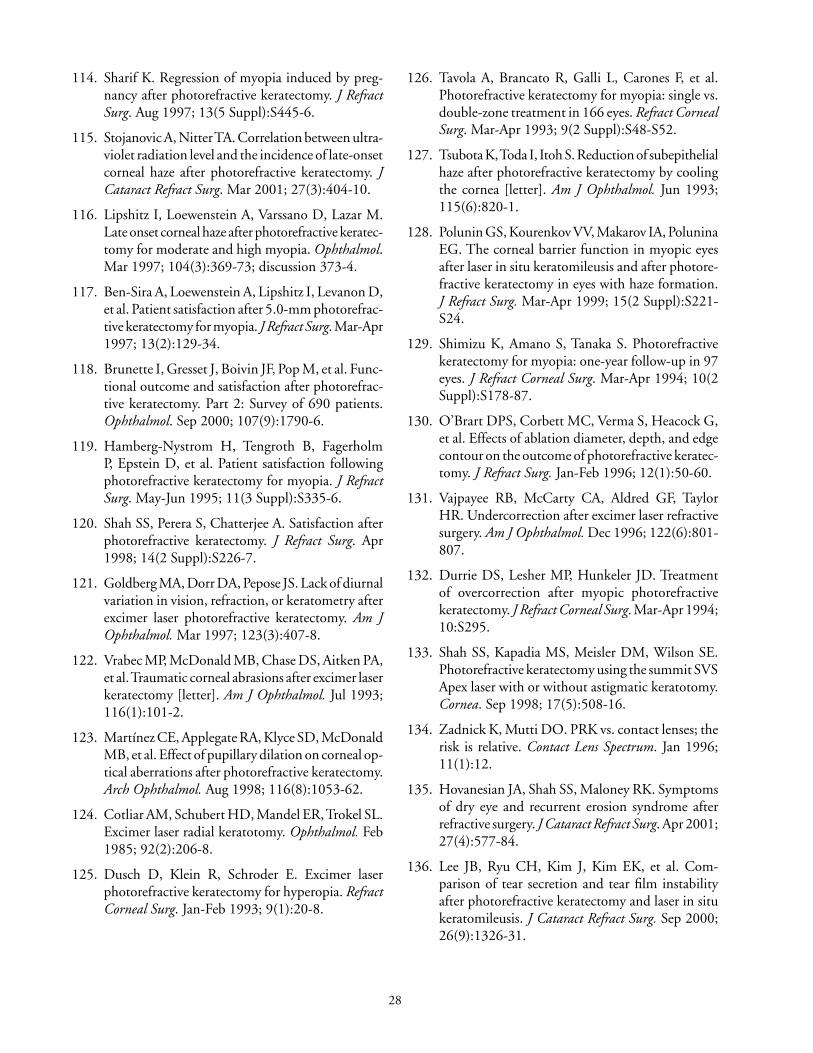
28
114 . Sharif K . Regression of myopia induced by preg-nancy after photorefractive keratectomy . J Refract Surg . Aug 1997; 13(5 Suppl):S445-6 .
115 . Stojanovic A, Nitter TA . Correlation between ultra-violet radiation level and the incidence of late-onset corneal haze after photorefractive keratectomy . J Cataract Refract Surg . Mar 2001; 27(3):404-10 .
116 . Lipshitz I, Loewenstein A, Varssano D, Lazar M . Late onset corneal haze after photorefractive keratec-tomy for moderate and high myopia . Ophthalmol . Mar 1997; 104(3):369-73; discussion 373-4 .
117 . Ben-Sira A, Loewenstein A, Lipshitz I, Levanon D, et al . Patient satisfaction after 5 .0-mm photorefrac-tive keratectomy for myopia . J Refract Surg . Mar-Apr 1997; 13(2):129-34 .
118 . Brunette I, Gresset J, Boivin JF, Pop M, et al . Func-tional outcome and satisfaction after photorefrac-tive keratectomy . Part 2: Survey of 690 patients . Ophthalmol . Sep 2000; 107(9):1790-6 .
119 . Hamberg-Nystrom H, Tengroth B, Fagerholm P, Epstein D, et al . Patient satisfaction following photorefractive keratectomy for myopia . J Refract Surg . May-Jun 1995; 11(3 Suppl):S335-6 .
120 . Shah SS, Perera S, Chatterjee A . Satisfaction after photorefractive keratectomy . J Refract Surg . Apr 1998; 14(2 Suppl):S226-7 .
121 . Goldberg MA, Dorr DA, Pepose JS . Lack of diurnal variation in vision, refraction, or keratometry after excimer laser photorefractive keratectomy . Am J Ophthalmol. Mar 1997; 123(3):407-8 .
122 . Vrabec MP, McDonald MB, Chase DS, Aitken PA, et al . Traumatic corneal abrasions after excimer laser keratectomy [letter] . Am J Ophthalmol. Jul 1993; 116(1):101-2 .
123 . Martínez CE, Applegate RA, Klyce SD, McDonald MB, et al . Effect of pupillary dilation on corneal op-tical aberrations after photorefractive keratectomy . Arch Ophthalmol. Aug 1998; 116(8):1053-62 .
124 . Cotliar AM, Schubert HD, Mandel ER, Trokel SL . Excimer laser radial keratotomy . Ophthalmol. Feb 1985; 92(2):206-8 .
125 . Dusch D, Klein R, Schroder E . Excimer laser photorefractive keratectomy for hyperopia . Refract Corneal Surg . Jan-Feb 1993; 9(1):20-8 .
126 . Tavola A, Brancato R, Galli L, Carones F, et al . Photorefractive keratectomy for myopia: single vs . double-zone treatment in 166 eyes . Refract Corneal Surg . Mar-Apr 1993; 9(2 Suppl):S48-S52 .
127 . Tsubota K, Toda I, Itoh S . Reduction of subepithelial haze after photorefractive keratectomy by cooling the cornea [letter] . Am J Ophthalmol. Jun 1993; 115(6):820-1 .
128 . Polunin GS, Kourenkov VV, Makarov IA, Polunina EG . The corneal barrier function in myopic eyes after laser in situ keratomileusis and after photore-fractive keratectomy in eyes with haze formation . J Refract Surg. Mar-Apr 1999; 15(2 Suppl):S221-S24 .
129 . Shimizu K, Amano S, Tanaka S . Photorefractive keratectomy for myopia: one-year follow-up in 97 eyes . J Refract Corneal Surg . Mar-Apr 1994; 10(2 Suppl):S178-87 .
130 . O’Brart DPS, Corbett MC, Verma S, Heacock G, et al . Effects of ablation diameter, depth, and edge contour on the outcome of photorefractive keratec-tomy . J Refract Surg. Jan-Feb 1996; 12(1):50-60 .
131 . Vajpayee RB, McCarty CA, Aldred GF, Taylor HR . Undercorrection after excimer laser refractive surgery . Am J Ophthalmol. Dec 1996; 122(6):801-807 .
132 . Durrie DS, Lesher MP, Hunkeler JD . Treatment of overcorrection after myopic photorefractive keratectomy . J Refract Corneal Surg . Mar-Apr 1994; 10:S295 .
133 . Shah SS, Kapadia MS, Meisler DM, Wilson SE . Photorefractive keratectomy using the summit SVS Apex laser with or without astigmatic keratotomy . Cornea . Sep 1998; 17(5):508-16 .
134 . Zadnick K, Mutti DO . PRK vs . contact lenses; the risk is relative . Contact Lens Spectrum . Jan 1996; 11(1):12 .
135 . Hovanesian JA, Shah SS, Maloney RK . Symptoms of dry eye and recurrent erosion syndrome after refractive surgery . J Cataract Refract Surg . Apr 2001; 27(4):577-84 .
136 . Lee JB, Ryu CH, Kim J, Kim EK, et al . Com-parison of tear secretion and tear film instability after photorefractive keratectomy and laser in situ keratomileusis . J Cataract Refract Surg. Sep 2000; 26(9):1326-31 .
29
137 . Timo T, Mustonen R, Tarkkanen A . Management of dry eye may reduce haze after excimer laser photorefractive keratectomy . [letter] Refract Corneal Surg . Jul-Aug 1993; 9:306 .
138 . Duffey RJ, Leaming D . U .S . trends in refractive surgery: 2001 International Society of Refractive Surgery survey . J Refract Surg. Mar-Apr 2002; 18(2):185-188 .
139 . Gilbert ML . LASIK Beneath the surface . Eyecare Technol . Jan 1996; 6(1):47-9 .
140 . Editor . LASIK wins FDA panel approval nod . AOA News . Jul 28, 1997; 36(2):1,6 .
141 . Augustine JM . Post-operative advantages of LASIK outweigh intraoperative risks . Primary Care Optom News . Mar 1996; 1(2):32 .
142 . Kremer FB, Gdovin GR . Update on LASIK: clini-cal results from 202 eyes . Rev Optom . Dec 1995; 132(12):39-43 .
143 . Helmy SA, Salah A, Badawy TT, Sidky AN . Pho-torefractive keratectomy and laser in situ keratomi-leusis for myopia between 6 .00 and 10 .00 diopters . J Refract Surg. Mar-Apr 1996;12(2):417-21 .
144 . Karpecki PM, Smith BJ, Smith JM . Excimer laser technology explores new horizons . Rev Optom . Oct 1996; 133(10):63-4, 66 .
145 . Gimbel HV, Basti S, Kaye GB, Ferensowicz M . Experience during the learning curve of laser in situ keratomileusis . J Cataract Refract Surg. Jun 1996; 22(5):542-50 .
146 . Farah SG, Azar DT, Gurdal C, Wong J . Laser in situ keratomileusis: Literature review of a develop-ing technique . J Cataract Refract Surg. Jul 1998; 24(7):989-1006 .
147 . Güell JL, Muller A . Laser in situ keratomileusis (LASIK) for myopia from -7 to -18 diopters . J Refract Surg. Jan-Feb 1996; 12(2):222-8 .
148 . Aquavella JV . Shelving the microkeratome in fa-vor of the laser . Contact Lens Spectrum . Jan 1996; 11(1):48 .
149 . Joo CK, Kim TG . Corneal perforation during laser in situ keratomileusis . J Cataract Refract Surg. Aug 1999; 25(8):1165-7 .
150 . Gimbel HV, Iskander NG, Peters NT, Penno EA . Prevention and management of microkeratome-related laser in situ keratomileusis complications . J Refract Surg. Mar-Apr 2000; 16(2 Suppl):S226-9 .
151 . Leung AT, Rao SK, Cheng AC, Yu EW, et al . Pathogenesis and management of laser in situ keratomileusis flap buttonhole . J Cataract Refract Surg . Mar 2000; 26(3):358-62 .
152 . Levartovsky S, Rosenwasser G, Goodman D . Bacte-rial keratitis following laser in situ keratomileusis . Ophthalmol. Feb 2001; 108(2):321-5 .
153 . Parolini B, Marcon G, Panozzo GA . Central necrotic lamellar inflammation after laser in situ keratomileusis . J Refract Surg. Mar-Apr 2001; 17(2):110-2 .
154 . Kim EK, Lee DH, Lee K, Lim SJ, et al . Nocardia keratitis after traumatic detachment of a laser in situ keratomileusis flap . J Refract Surg. Jul-Aug 2000; 16(4):467-9 .
155 . Cameron BD, Saffra NA, Strominger MB . Laser in situ keratomileusis-induced optic neuropathy . Ophthalmol. Apr 2001; 108(4):660-5 .
156 . Stephenson M . RRD after LASIK must be managed promptly . Ocular Surg News Europe/Asia-Pacific Edition . April 4, 2001 .
157 . Farah ME, Hofling-Lima AL, Nascimento E . Early rhegmatogenous retinal detachment following laser in situ keratomileusis for high myopia . J Refract Surg. Nov-Dec 2000; 16(6):739-43 .
158 . Aras C, Ozdamar A, Bahcecioglu H, Karacorlu M, et al . Decreased tear secretion after laser in situ keratomileusis for high myopia . J Refract Surg . May-Jun 2000; 16(3):362-4 .
159 . Balazsi G, Mullie M, Lasswell L, Lee PA, et al . Laser in situ keratomileusis with a scanning excimer laser for the correction of low to moderate myopia with and without astigmatism . J Cataract Refract Surg . Dec 2001; 27(12):1942-51 .
160 . Rao S, Cheng ACK, Fan DSP, Leung ATS, et al . Effect of preoperative keratometry on refractive outcomes after laser in situ keratomileusis . J Cataract Refract Surg. Feb 2001; 27(2):297-302 .
161 . Haw WW, Manche EE . Large optical ablation zone using the VISX S2 smooth scan excimer laser . J Cataract Refract Surg . Dec 2000; 26(12):1742-7 .
162 . Knorz MC, Wiesinger B, Liermann A, Seiberth V, et al . Laser in situ keratomileusis for moderate and high myopia and myopic astigmatism . Ophthalmol . May 1998; 105(5):932-40 .
30
163 . McDonald MB, Carr JD, Frantz JM, Kozarsky AM, et al . Laser in situ keratomileusis for myopia up to -11 diopters with up to -5 diopters of astig-matism with the summit autonomous LADARVi-sion excimer laser system . Ophthalmol . Feb 2001; 108(2):309-16 .
164 . Mulhern MG, Condon PI, O’Keefe M . Myopic and hyperopic laser in situ keratomileusis retreatments: indications, techniques, limitations, and results . Cataract Refract Surg . Aug 2001; 27(8):1278-87 .
165 . Pesando PM, Ghiringhello MP, Tagliavacche P . Excimer laser in situ keratomileusis for myopia . J Refract Surg . Sep-Oct 1997; 13(6):521-7 .
166 . Nordan LT, Slade SG, Baker RN, Suarez C, et al . Femtosecond laser flap creation for laser in situ keratomileusis: six-month follow-up of initial U .S . clinical series . J Refract Surg . Jan-Feb 2003; 19(1):8-14 .
167 . McGhee CN, Craig JP, Sachdev N, Weed KH, et al . Functional, psychological, and satisfac-tion outcomes of laser in situ keratomileusis for high myopia . J Cataract Refract Surg . Apr 2000; 26(4):497-509 .
168 . Brint SF, Ostrick DM, Fisher C, Slade SG, et al . Six-month results of the multicenter phase I study of excimer laser myopic keratomileusis . J Cataract Refract Surg. Nov 1994; 20:610-5 .
169 . Dada T, Sudan R, Sinha R, Ray M, et al . Results of laser in situ keratomileusis for myopia of -10 to -19 diopters with a Technolas 217 laser . J Refract Surg . Jan-Feb 2003; 19(1):44-7 .
170 . El Danasoury MA, El Maghraby A, Gamali TO . Comparison of iris-fixed Artisan lens implantation with excimer laser in situ keratomileusis in correct-ing myopia between -9 .00 and -19 .50 diopters: a randomized study . Ophthalmol . May 2002; 109(5):955-64 .
171 . Kawesch GM, Kezirian GM . Laser in situ ker-atomileusis for high myopia with the VISX star laser . Ophthalmol . Apr 2000; 107(4):653-61 .
172 . Lyle WA, Jin GJ . Laser in situ keratomileusis with the VISX Star laser for myopia over -10 .0 diopters . J Cataract Refract Surg . Nov 2001; 27(11):1812-22 .
173 . Reviglio VE, Bossana EL, Luna JD, Muino JC, et al . Laser in situ keratomileusis for myopia and hyperopia using the Lasersight 200 laser in 300 consecutive eyes . J Refract Surg . Nov-Dec 2000; 16(6):716-23 .
174 . Knorz MC, Neuhann T . Treatment of myopia and myopic astigmatism by customized laser in situ keratomileusis based on corneal topography . Ophthalmol . Nov 2000; 107(11):2072-6 .
175 . Montes M, Chayet A, Gomez L, Magallanes R, et al . Laser in situ keratomileusis for myopia of -1 .50 to -6 .00 diopters . J Refract Surg . Mar-Apr 1999; 15(2):106-10 .
176 . Zaldivar R, Davidorf JM, Shultz MC, Oscherow S . Laser in situ keratomileusis for low myopia and astigmatism with a scanning spot excimer laser . J Refract Surg . Nov-Dec 1997; 13(7):614-9 .
177 . Fraenkel GE, Webber SK, Sutton GL, Lawless MA, et al . Toric laser in situ keratomileusis for myopic astigmatism using an ablatable Mask . J Refract Surg. Mar-Apr 1999; 15(2):111-7 .
178 . Dulaney DD, Barnet RW, Perkins SA, Kezirian GM . Laser in situ keratomileusis for myopia and astigmatism: 6 month results . J Cataract Refract Surg . Jun 1998; 24(6):758-64 .
179 . Shaikh NM, Manche EE . Laser in situ keratomileu-sis for myopia and compound myopic astigmatism using the Technolas 217 scanning-spot laser . J Cataract Refract Surg . Mar 2002; 28(3):485-90 .
180 . Zaldivar R, Davidorf JM, Oscherow S . Laser in situ keratomileusis for myopia from -5 .50 to -11 .50 diopters with astigmatism . J Refract Surg . Jan-Feb 1998; 14(1):19-25 .
181 . Chitkara DK, Rosen E, Gore C, Howes F, et al . Tracker-assisted laser in situ keratomileusis for myo-pia using the autonomous scanning and tracking laser: 12-month results . Ophthalmol . May 2002; 109(5):965-72 .
182 . Argento CJ, Cosentino MJ . Comparison of optical zones in hyperopic laser in situ keratomileusis: 5 .9 mm versus smaller optical zones . J Cataract Refract Surg . Aug 2000; 26(8):1137-46 .
183 . Argento CJ, Cosentino MJ . Laser in situ keratomi-leusis for hyperopia . J Cataract Refract Surg . Aug 1998; 24(8):1050-8 .
31
184 . el-Agha MS, Johnston EW, Bowman RW, Cavanagh HD, et al . Excimer laser treatment of spherical hyperopia: PRK or LASIK? Trans Am Ophthalmol Soc . 2000; 98:59-66; discussion 66-9 .
185 . Lindstrom RL, Linebarger EJ, Hardten DR, Houtman DM, et al . Early results of hyperopic and astigmatic laser in situ keratomileusis in eyes with secondary hyperopia . Ophthalmol . Oct 2000; 107(10):1858-63 .
186 . Zadok D, Maskaleris G, Montes M, Shah S, et al . Hyperopic laser in situ keratomileusis with the Nidek EC-5000 excimer laser . Ophthalmol . Jun 2000; 107(6):1132-7 .
187 . Esquenazi S, Mendoza A . Two-year follow-up of laser in situ keratomileusis for hyperopia . J Refract Surg . Nov-Dec 1999; 15(6):648-52 .
188 . Choi RY, Wilson SE . Hyperopic laser in situ ker-atomileusis: primary and secondary treatments are safe and effective . Cornea . May 2001; 20(4):388-93 .
189 . Ditzen K, Fiedler J, Pieger S . Laser in situ keratomi-leusis for hyperopia and hyperopic astigmatism using the Meditec MEL 70 spot scanner . J Refract Surg . Jul-Aug 2002; 18(4):430-4 .
190 . Lian J, Ye W, Zhou D, Wang K . Laser in situ keratomileusis for correction of hyperopia and hyperopic astigmatism with the Technolas 117C . J Refract Surg . Jul-Aug 2002; 18(4):435-8 .
191 . Salz JJ, Stevens CA, LADARVision LASIK Hy-peropia Study Group . LASIK correction of spheri-cal hyperopia, hyperopic astigmatism, and mixed astigmatism with the LADARVision excimer laser system . Ophthalmol . Sep 2002; 109(9):1647-56; discussion 1657-8 .
192 . Tabbara KF, El-Sheikh HF, Islam SM . Laser in situ keratomileusis for the correction of hyperopia from +0 .50 to +11 .50 diopters with the Keracor 117C laser . J Refract Surg . Mar-Apr 2001; 17(2):123-8 .
193 . Goker S, Er H, Kahvecioglu C . Laser in situ keratomileusis to correct hyperopia from +4 .25 to +8 .00 diopters . J Refract Surg . Jan-Feb 1998; 14(1):26-30 .
194 . Chayet AS, Magallanes R, Montes M, Chavez S, et al . Laser in situ keratomileusis for simple myopic, mixed, and simple hyperopic astigmatism . J Refract Surg . Apr 1998; 14(2 Suppl):S175-6 .
195 . Rashad KM . Laser in situ keratomileusis for the cor-rection of hyperopia from +1 .25 to +5 .00 diopters with the Technolas Keracor 117C laser . J Refract Surg . Mar-Apr 2001; 17(2):113-22 .
196 . Pirzada WA, Kalaawry H . Laser in situ keratomi-leusis for myopia of -1 to -3 .50 diopters . J Refract Surg . Aug 1997; 13(5 Suppl):S425-6 .
197 . Chayet AS, Montes M, Gomez L, Rodriguez X, et al . Bitoric laser in situ keratomileusis for the cor-rection of simple myopic and mixed astigmatism . Ophthalmol . Feb 2001; 108(2):303-8 .
198 . Argento CJ, Cosentino MJ, Biondini A . Treatment of hyperopic astigmatism . J Cataract Refract Surg . Dec 1997; 23(10):1480-90 .
199 . el Danasoury MA, el Maghraby A, Klyce SD, Mehrez K . Comparison of photorefractive kera-tectomy with excimer laser in situ keratomileusis in correcting low myopia (from -2 .00 to -5 .50 diopters) . A randomized study . Ophthalmol . Feb 1999; 106(2):411-20; discussion 420-1 .
200 . Pop M, Payette Y . Photorefractive keratectomy ver-sus laser in situ keratomileusis: a control-matched study . Ophthalmol . Feb 2000; 107(2):251-7 .
201 . Pineda-Fernandez A, Rueda L, Huang D, Nur J, et al . Laser in situ keratomileusis for hyperopia and hyperopic astigmatism with the Nidek EC-5000 Excimer laser . J Refract Surg . Nov-Dec 2001; 17(6):670-5 .
202 . Reviglio VE, Luna JD, Rodriguez ML, Garcia FE, et al . Laser in situ keratomileusis using the Laser-Sight 200 laser: results of 950 consecutive cases . J Cataract Refract Surg . Aug 1999; 25(8):1062-8 .
203 . Knorz MC, Liermann A, Seiberth V, Steiner H, et al . Laser in situ keratomileusis to correct myopia of -6 .00 to -29 .00 diopters . J Refract Surg . Jul-Aug 1996; 12(5):575-84 .
204 . Tsai RJ . Laser in situ keratomileusis for myopia of -2 to -25 diopters . J Refract Surg . Aug 1997; 13(5 Suppl):S427-S29 .
205 . Han HS, Song JS, Kim HM . Long-term results of laser in situ keratomileusis for high myopia . Korean J Ophthalmol . Jun 2000; 14(1):1-6 .
206 . Kim HM, Jung HR . Laser assisted in situ keratomi-leusis for high myopia . Ophthalmic Surg Lasers . May 1996; 27(5 Suppl):S508-S11 .

32
207 . Pallikaris IG, Siganos DS . Laser in situ keratomi-leusis to treat myopia: early experience . J Cataract Refract Surg . Jan-Feb 1997; 23(1):39-49 .
208 . Condon PI, Mulhern M, Fulcher T, Foley-Nolan A, et al . Laser intrastromal keratomileusis for high myopia and myopic astigmatism . Br J Ophthalmol . Mar 1997; 81(3):199-206 .
209 . Siganos DS, Popescu CN, Siganos CS, Margaritis VN . Seven years experience with LASIK for myopia . Oftalmologia . 1999; 47(2):50-2 .
210 . Barraquer C, Gutierrez AM . Results of laser in situ keratomileusis in hyperopic compound astigma-tism . J Cataract Refract Surg . Sep 1999; 25(9):1198-204 .
211 . Merchea M, Pieger S, Bains HS . Comparison of laser in situ keratomileusis outcomes with the Nidek EC-5000 and Meditec Mel 70 excimer lasers . J Refract Surg . May-Jun 2002; 18(3 Suppl):S343-S46 .
212 . Merchea M, Tokarewicz A . Comparison of laser in situ keratomileusis outcomes with the Nidek EC-5000 and LSX LaserSight excimer lasers . J Refract Surg . Mar-Apr 2001; 17(2 Suppl):S246-9 .
213 . Arbelaez MC, Knorz MC . Laser in situ keratomi-leusis for hyperopia and hyperopic astigmatism . J Refract Surg . Jul-Aug 1999; 15(4):406-14 .
214 . Ditzen K, Huschka H, Pieger S . Laser in situ ker-atomileusis for hyperopia . J Cataract Refract Surg . Jan 1998; 24(1):42-7 .
215 . Hill JC . An informal satisfaction survey of 200 patients after laser in situ keratomileusis . J Refract Surg . Jul-Aug 2002; 18(4):454-9 .
216 . Miller AE, McCulley JP, Bowman RW, Cavanagh HD, et al . Patient satisfaction after LASIK for myopia . CLAO J . Apr 2001; 27(2):84-8 .
217 . Laser Vision Correction by the Numbers . At URL: www .tlcv .com/newsroom/media_kit/ TLC%20LVC%20By%20the%20Numbers .pdf (Accessed 10/01) .
218 . TLC Laser Centers . Why choose TLC? At URL: www .tlcvision .com/why_choose_tlc/ (Accessed 8/04) .
219 . Laser Eye Consults of Boston . Laser vision correc-tion (brochure) . At URL: www .corneaconsultants-boston .com/specialties/lasikbrochure .pdf (Accessed 8/04) .
220 . Jacobs JM, Sanderson MC, Spivack LD, Wright JR, et al . Hyperopic laser in situ keratomileusis to treat overcorrected myopic LASIK . J Cataract Refract Surg. Mar 2001; 27(3):389-95 .
221 . Rosa DA . Laser in situ keratomileusis: three un-expected complications . J Refract Surg. Mar-Apr 2001; 17(2 Suppl):S177-9 .
222 . Ismail MM, Alió JL, Pérez-Santonja JJ . Noncontact thermokeratoplasty to correct hyperopia induced by laser in situ Keratomileusis . J Cataract Refract Surg. Sep 1998; 24(9):1191-4 .
223 . el Danasoury MA, Waring GO, El Maghraby A, Mehrez K . Excimer laser in situ keratomileusis to correct compound myopic astigmatism . J Refract Surg. Sep-Oct 1997; 13(6):511-20 .
224 . el Danasoury MA . Prospective bilateral study of night glare after laser in situ keratomileusis with single zone and transition zone ablation . J Refract Surg. Sep-Oct 1998; 14(5):512-6 .
225 . Knorz MC, Hugger P, Jendritzka B, Liermann A . Twilight visual acuity after correction of myopia with LASIK . [German] Ophthalmologe . Nov 1999; 96(11):711-6 .
226 . Lyle WA, Jin GJ . Results of flap repositioning after laser in situ keratomileusis . J Cataract Refract Surg. Oct 2000; 26(10):1451-7 .
227 . Wilson SE . Laser in situ keratomileusis induced (presumed) neurotrophic epitheliopathy . Ophthal-mol. Jun 2001; 108(6):1082-7 .
228 . Leung AT, Rao SK, Lam DS . Traumatic partial unfolding of laser in situ keratomileusis flap with severe epithelial ingrowth . J Cataract Refract Surg. Jan 2000; 26(1):135-9 .
229 . Geggel HS, Coday MP . Late-onset traumatic laser in situ keratomileusis (LASIK) flap dehiscence . Am J Ophthalmol. Apr 2001; 131(4):505-6 .
230 . Melki SA, Talamo JH, Demetriades AM, Jabbur NS, et al . Late traumatic dislocation of laser in situ keratomileusis corneal flaps . Ophthalmol. Dec 2000; 107(12):2136-9 .
231 . Weisenthal RW . Diffuse lamellar keratitis induced by trauma 6 months after laser in situ keratomileusis . J Refract Surg. Nov-Dec 2000; 16(6):749-51 .
232 . Lemley HL, Chodosh J, Wolf TC, Bogie CP, et al . Partial dislocation of laser in situ keratomileusis flap by air bag injury . J Refract Surg. May-Jun 2000; 16(3):373-4
33
233 . Lombardo AJ, Katz HR . Late partial dislocation of a laser in situ keratomileusis flap . J Cataract Refract Surg. Jul 2001; 27(7):1108-10 .
234 . Mifflin M . Laser in situ keratomileusis flap dehis-cence 3 years post-operatively J Cataract Refract Surg. May 2002; 28(5):733-35 .
235 . Patel CK, Hanson R, McDonald B, Cox N . Case reports and small case series: late dislocation of a LASIK flap caused by a fingernail . Arch Ophthalmol. Mar 2001; 119(3):447-9 .
236 . Schwartz GS, Park DH, Schloff S, Lane SS . Trau-matic flap displacement and subsequent diffuse lamellar keratitis after laser in situ keratomileusis . J Cataract Refract Surg. May 2001; 27(5):781-3 .
237 . Lee BJ, Seong GJ, Lee JH, Seo KY, et al . Comparison of laser epithelial keratomileusis and photorefractive keratectomy for low to moderate myopia . J Cataract Refract Surg. Apr 2001; 27(4):565-70 .
238 . Azar DT, Ang RT, Lee JB, Kato T, et al . Laser sub-epithelial keratomileusis: electron microscopy and visual outcomes of flap photorefractive keratectomy . Curr Opin Ophthalmol. Aug 2001; 12(4):323-8 .
239 . Lohmann CP, Winkler Von Mohrenfels C, Gabler B, Hermann W, et al . Excimer laser subepithelial ablation (ELSA) or laser epithelial keratomileusis (LASEK) - a new kerato-refractive procedure for myopia . Surgical technique and first clinical results on 24 eyes and 3 months follow-up . [Article in German] Klin Monatsbl Augenheilkd . Jan-Feb 2002; 219(1-2):26-32 .
240 . Kornilovsky IM . Clinical results after subepithelial photorefractive keratectomy (LASEK) . J Refract Surg. Mar-Apr 2001; 17(2 Suppl):S222-3 .
241 . Claringbold TV . Laser-assisted subepithelial kera-tectomy for the correction of myopia . J Cataract Refract Surg. Jan 2002; 28(1):18-21 .
242 . Rouweyha RM, Chuang AZ, Mitra S, Phillips CB, et al . Laser epithelial keratomileusis for myopia with the autonomous laser . J Refract Surg. May-Jun 2002; 18(3):217-24 .
243 . Vinciguerra P, Camesasca FI . Butterfly laser epi-thelial keratomileusis for myopia . J Refract Surg. May-Jun 2002; 18(3 Suppl):S371-3 .
244 . Lee JB, Choe CM, Kim HS, Seo KY, et al . Compari-son of TGF-beta1 in tears following laser subepithe-lial keratomileusis and photorefractive keratectomy . J Refract Surg. Mar-Apr 2002; 18(2):130-4 .
245 . Nakamura K, Kurosaka D, Bissen-Miyajima H, Tsubota K . Intact corneal epithelium is essential for the prevention of stromal haze after laser assisted in situ keratomileusis . Br J Ophthalmol. Feb 2001; 85(2):209-13 .
246 . Rokita-Wala I, Gierek-Ciaciura S, Mrukwa-Kominek E, Obidzinski M . Vital changes in corneal structure after LASEK during the early post-opera-tive period . [Article in Polish] Klin Oczna . 2002; 104(1):13-8 .
247 . Anderson NJ, Beran RF, Schneider TL . Epi-LASEK for the correction of myopia and myopic astigmatism . J Cataract Refract Surg . Aug 2002; 28(8):1343- 7 .
248 . Shahinian L Jr . Laser-assisted subepithelial keratec-tomy for low to high myopia and astigmatism . J Cataract Refract Surg . Aug 2002; 28(8):1334-42 .
249 . Shah S, Sebai Sarhan AR, Doyle SJ, Pillai CT, et al . The epithelial flap for photorefractive keratectomy . Br J Ophthalmol . Apr 2001; 85(4):393-6 .
250 . Durrie DS, Schumer DJ, Cavanaugh TB . Hol-mium:YAG laser thermokeratoplasty for hyperopia . J Refract Corneal Surg . Mar-Apr 1994; 10(2 Suppl):S277-80 .
251 . Dausch D, Landesz M, Koch DD, Berry MJ, et al . Laser correction of hyperopia . In: Salaz JJ, Mcdon-nell PJ, McDonald MB, eds . Corneal Laser Surgery. St . Louis, MO: Mosby Year Book Inc; 1995:237-60 .
252 . Haw WW, Manche EE . Conductive keratoplasty and laser thermal keratoplasty . Int Ophthalmol Clin . Fall 2002; 42(4):99-106 .
253 . Medical Device Approvals, New Device Approval . Hyperion LTK System – P990078 . At URL: www .fda .gov/cdrh/mda/docs/p990079 .html . (Ac-cessed 7/02) .
254 . Moreira H, Campos M, Sawusch MR, McDon-nell JM, et al . Holmium laser thermokeratoplasty . Ophthalmol. May 1993; 100(5):752-61 .
255 . Nano HD, Muzzin S . Noncontact holmium:YAG laser thermal keratoplasty for hyperopia . J Cataract Refract Surg. Jun 1998; 24(6):751-7 .
34
256 . Ronge LJ . Noncontact Ho:YAG LTK show promise for low hyperopia . Ophthalmol World News . May 1996; 2(5):1-2 .
257 . Facts you need to know about the ViewPoint-CK System and the Conductive Keratoplasty (CK) Procedure . Patient Information Booklet For Far-sightedness (Hyperopia) Refractec, Inc . At URL: www .refractec .com . (Accessed 7/02) .
258 . Marshall BD, Stein JJ . How to choose what’s best for your patients . Optom Mgmt . May 2004 . 39(5):38-45 .
259 . McDonald MB, Davidorf J, Maloney RK, Manche EE, et al . Conductive keratoplasty for the correction of low to moderate hyperopia: 1-year results on the first 54 eyes . Ophthalmol. Apr 2002; 109(4):637-49 .
260 . Asbell PA, Maloney RK, Davidorf J, Hersh P, et al . Conductive keratoplasty for the correction of hyperopia . Trans Am Ophthalmol Soc . 2001; 99:79-84 .
261 . Burris TE . Intrastromal corneal ring technology: results and indications . Curr Opin Ophthalmol. Aug 1998; 9(4):9-14 .
262 . Ruckhofer J, Stoiber J, Alzner E, Grabner G, et al . One year results of European Multicenter Study of intrastromal corneal ring segments . Part 1: Refrac-tive outcomes . J Cataract Refract Surg. Feb 2001; 27(2):277-86 .
263 . Holmes-Higgin DK, Burris TE, Asbell PA, Durrie DS, et al . Topographic predicted corneal acuity with intrastromal corneal ring segments . J Refract Surg. May-Jun 1999; 15(3):324-30 .
264 . Holmes-Higgin DK, Baker PC, Burris TE, Silves-trini TA . Characterization of the aspheric corneal surface with intrastromal corneal ring segments . J Refract Surg. Sep-Oct 1999; 15(5):520-8 .
265 . Schanzlin DJ . Studies of intrastromal corneal ring segments for the correction of low to moderate myopic refractive errors . Transactions Am Ophthal-mol Soc . 1999; 97:815-90 .
266 . Ruckhofer J, Alzner E, Grabner G . Intrastromal corneal ring segments (ICRS, Kera Vision Ring) . One year outcome of the first 25 interventions . [Article in German] Klin Monatsbl Augenheilkd . Sep 1998; 213(3):147-53 .
267 . Twa MD, Hurst TJ, Walker JG, Waring GO, et al . Diurnal stability of refraction after implantation
with intracorneal ring segments . J Cataract Refract Surg. Apr 2000; 26(4):516-23 .
268 . Asbell PA, Ucakhan OO, Durrie DS, Lindstrom RL . Adjustability of refractive effect for corneal ring segments . J Refract Surg. Nov-Dec 1999; 15(6):627-31 .
269 . Hoh H, Rehfeldt K, Reiss G . Correction of myopia with the intracorneal ring . [Article in German] Ophthalmologe . Nov 1999; 96(11):717-23 .
270 . Barach D, Mortemousque B, Bertel F, Diemer C, et al . Fluctuations in uncorrected visual acuity after refractive surgery using intra-stromal corneal rings . [Article in French] J Fr Ophtalmol . Apr 1999; 22(3):335-8 .
271 . Ruckhofer J, Stoiber J, Alzner E, Grabner G, et al . One year results of European Multicenter Study of intrastromal corneal ring segments . Part 2: Compli-cations, visual symptoms, and patient satisfaction . J Cataract Refract Surg. Feb 2001; 27(2):287-96 .
272 . Editor . Intacs sales rise 86 .7 percent . AOA News . Jan 27, 2003; 41(14):6 .
273 . Thompson RW, Choi DM, Price FW Jr . Clear lens replacement surgery . Int Ophthalmol Clin . Fall 2002: 42(2):131-152 .
274 . Seiler T . Clear lens extraction in the 19th century—an early demonstration of premature dissemination . J Refract Surg. Jan-Feb 1999; 15(1):70-3 .
275 . Werblin TP . Why should refractive surgeons be looking beyond the cornea? J Refract Surg. May-Jun 1999; 15(3):357-76 .
276 . Colin J, Robinet A, Cochener B . Retinal detachment after clear lens extraction for high myopia; seven year follow-up . Ophthalmol. Dec 1999; 106:2281-5 .
277 . Lee KW, Lee JH . Long-term results of clear lens extraction for severe myopia . J Cataract Refract Surg. Dec 1996; 22(10):1411-5 .
278 . Pucci V, Morselli S, Romanelli F, Pignatto S, et al . Clear lens phacoemulsification for correction of high myopia . J Cataract Refract Surg. Mar-Apr 2001; 27(2):896-900 .
279 . Lyle WA, Jin GJC . Clear lens extraction to correct high myopia . J Cataract Refract Surg. Sep 1997; 23(7):1051-6 .
35
280 . Kolahdouz-Isfahani AH, Rostamian K, Wallace D, Salz JJ . Clear lens extraction with intraocular lens implantation for hyperopia . J Refract Surg. May-Jun 1999; 15(3):316-23 .
281 . Steinert RF, Aker BL, Trentacost DJ, Smith PJ, et al . A prospective comparative study of the AMO Array zonal-progressive multifocal silicone intraocular lens and a monofocal intraocular lens . Ophthalmol . Jul 1999; 106:1243-55 .
282 . Packer M, Fine IH, Hoffman RS . Refractive lens exchange with the Array Multifocal intraocular lens . J Cataract Refract Surg. Mar 2002; 28(3):421-4 .
283 . Arens B, Freudenthaler N, Quentin CD . Binocular function after bilateral implantation of monofo-cal and refractive multifocal intraocular lenses . J Cataract Refract Surg. Mar 1999; 25:399-404 .
284 . Vaquero-Ruano M, Encinas JL, Millan I . AMO Ar-ray multifocal versus monofocal intraocular lenses: long term follow-up . J Cataract Refract Surg . Jan 1998; 24:118-23 .
285 . Ravalico G, Parentin F, Pastori G, Baccara F . Spa-tial resolution threshold in pseudophakic patients with monofocal and multifocal intraocular lenses . J Cataract Refract Surg . Feb 1998; 24:244-8 .
286 . Haring G, Gronemeyer A, Hedderich J, de Decker W . Stereoacuity and aniseikonia after unilateral and bilateral implantation of the Array refractive multifocal intraocular lens . J Cataract Refract Surg . Aug 1999; 25:1151-6 .
287 . Featherstone KA, Bloomfield JR, Lang AJ, Miller-Meeks MJ, et al . Driving simulation study: bilateral Array multifocal versus bilateral AMO monofocal intraocular lenses . J Cataract Refract Surg . Sep 1999; 25:1254-62 .
288 . CrystaLens™ Posterior Chamber Accommodating Intraocular Lens For the Correction of Aphakia in Cataract Patients, PMA P030002, Presentation to the Ophthalmic Devices Advisory Panel May 23, 2003 .
289 . Cummings JS, Slade SG, Chayet A, AT-45 Study Group . Clinical evaluation of the model AT-45 Silicone accommodating intraocular lens; results of feasibility and the initial phase of a Food and Drug Administration clinical trial . Ophthalmol. Nov 2001; 108(11):2005-10 .
290 . Dick HB, Gross S, Tehrani M, Eisemann D, et al . Refractive lens exchange with an array multifocal intraocular lens . J Refract Surg. Sep-Oct 2002; 18(5):509-18 .
291 . Gris O . Manero F, Muller A . Clear lens extraction to correct high myopia . J Cataract Refract Surg. Jul-Aug 1996; 22(6):686-9 .
292 . Siganos DS, Pallikaris IG . Clear lensectomy and intraocular lens implantation for hyperopia from +7 to +14 diopters . J Refract Surg. Mar-Apr 1998; 14(2):105-13 .
293 . Jimenez-Alfaro I, Migueles S, Bueno JL, Puy P . Clear lens extraction and implantation of nega-tive-power posterior chamber intraocular lenses to correct extreme myopia . J Cataract Refract Surg. Oct 1998; 24(10):1310-6 .
294 . Siganos DS, Siganos CS, Pallikaris IG . Clear lens extraction and intraocular lens implantation in normally sighted hyperopic eyes . J Refract Corneal Surg . Mar-Apr 1994; 10(2):117-21 .
295 . Shingleton BJ, Wadhwani RA, O’Donoghue MW, Baylus S, et al . Evaluation of intraocular pressure in the immediate period after phacoemulsification . J Cataract Refract Surg. Apr 2001; 27(4):524-7 .
296 . Colin J, Robinet A, Cochener B . Retinal detach-ment after clear lens extraction for high myopia: Seven-year follow-up . Ophthalmol . Dec 1999; 106(12):2281-4; discussion 2285 .
297 . Ramos M, Kruger EF, Lashkari K . Biostatisti-cal analysis of pseudophakic and aphakic retinal detachments . Semin Ophthalmol . Sep-Dec 2002; 17(3-4):206-13 .
298 . Norregaard JC, Thoning H, Andersen TF, Ber-nth-Petersen P, et al . Risk of retinal detachment following cataract extraction: Results from the International Cataract Surgery Outcomes Study . Br J Ophthalmol . Aug 1996; 80(8):689-93 .
299 . Javitt JC, Vitale S, Canner JK, Krakauer H, et al . National outcomes of cataract extraction . I . Retinal detachment after inpatient surgery . Ophthalmol . Jun 1991; 98(6):895-902 .
300 . Javitt JC, Street DA, Tielsch JM, Wang Q, et al . National outcomes of cataract extraction . Retinal detachment and endophthalmitis after outpatient cataract surgery . Cataract Patient Outcomes Re-search Team . Ophthalmol . Jan 1994; 101(1):100-5; discussion 106 .
36
301 . Kaya V, Kevser MA, Yilmaz OF . Phakic posterior chamber plate intraocular lenses for high myopia . J Refract Surg. Sep-Oct 1999; 15(5):580-5 .
302 . Barraquer J . Precrystalline posterior chamber intra-ocular lens for surgical correction of severe myopia . Am J Ophthalmol. Aug 1999; 128(2):232-4 .
303 . Hruba H, Vlkova E, Horackova M, Svacinova J . Comparison of clinical results between LASIK method and ICL implantation in high myopia [Article in Czech] Cesk Slov Oftalmol . May 2004; 60(3):180-91 .
304 . Menezo JL, Peris-Martinez C, Cisneros AL, Mar-tinez-Costa R . Phakic intraocular lenses to correct high myopia: Adatomed, Staar, and Artisan . J Cataract Refract Surg . Jan ; 30(1):33-44 .
305 . Gierek-Ciaciura S, Ochalik K, Gierek-Lapinska A, Myga B . Correction of high myopia with ante-rior chamber angle-supported phakic intraocular lenses—own results . [Article in Polish] Klin Oczna . 2003; 105(6):373-7 .
306 . Saxena R, Landesz M, Noordzij B, Luyten GP . Three-year follow-up of the Artisan phakic intra-ocular lens for hypermetropia . Ophthalmol . Jul 2003; 110(7):1391-5 .
307 . Zaldivar R, Davidorf JM, Oscherow S, Ricur G, et al . Combined posterior chamber phakic intraocular lens and laser in situ keratomileusis: bioptics for extreme myopia . J Refract Surg. May-Jun 1999; 15(3):299-308 .
308 . Guell JL, Vazquez M, Gris O, De Muller A, et al . Combined surgery to correct high myopia: iris claw phakic intraocular lens and laser in sit keratomileu-sis . J Refract Surg. Sep-Oct 1999; 15(5):529-37 .
309 . Fechner PU, Haubitz I, Wichmann W, Wulff K . Worst-Fechner biconcave minus power phakic iris-claw lens . J Refract Surg. Mar-Apr 1999; 15(2):93-105 .
310 . Leroux-Les-Jardins S, Ullern M, Werthel AL . Myopic anterior chamber intraocular lens implanta-tion: evaluation at 8 years . [Article in French] J Fr Ophthalmol . Apr 1999; 22(3):323-7 .
311 . Jimenez-Alfaro I, Benitez del Castillo JM, Garcia-Feijoo J, Gil de Bernabe JG, et al . Safety of posterior chamber phakic intraocular lenses for the correction of high myopia: anterior segment changes after posterior chamber phakic intraocular lens implanta-tion . Ophthalmol. Jan 2001; 108(1):90-9 .
312 . Pop M, Payette Y . Initial results of endothelial cell counts after Artisan lens for phakic eyes: an evaluation of the United States Food and Drug Administration Ophtec Study . Ophthalmol . Feb 2004; 111(2):309-17 .
313 . Pesando PM, Ghiringhello MP, Tagliavacche P . Posterior chamber collamer phakic intraocular lens for myopia and hyperopia . J Refract Surg. Jul-Aug 1999; 15(4):415-23 .
314 . Arne JL . Phakic intraocular lens implantation versus clear lens extraction in highly myopic eyes of 30- to 50-year-old patients . J Cataract Refract Surg . Oct 2004; 30(10):2092-6 .
315 . Fink AM, Gore C, Rosen E . Cataract development after implantation of the Staar Collamer posterior chamber phakic lens . J Cataract Refract Surg. Feb 1999; 25(2):278-82 .
316 . Brauweiler PH, Wehler T, Busin M . High inci-dence of cataract formation after implantation of a silicone posterior chamber lens in phakic, highly myopic eyes . Ophthalmol. Sep 1999; 106(9):1651-5 .
317 . Allemann N, Chamon W, Tanaka HM, Mori ES, et al . Myopic angle-supported intraocular lenses: two-year follow-up . Ophthalmol. Aug 2000; 107(8):1549-54 .
318 . Baikoff G, Arne JL, Bokobza Y, Colin J, et al . Angle-fixated anterior chamber phakic intraocular lens for myopia of -7 to -19 diopters . J Refract Surg. May-Jun 1998; 14(3):282-93 .
319 . Alio JL, de la Hoz F, Perez-Santonja JJ, Ruiz-Moreno JM, et al . Phakic anterior chamber lenses for the correction of myopia: a 7-year cumulative analysis of complications in 263 cases . Ophthalmol. Mar 1999; 106(3):458-66 .
320 . Sippel KC, Jain S, Azar DT . Monovision achieved with excimer laser refractive surgery . Int Ophthalmol Clin . Spring 2001; 41(2):91-101 .
321 . Editor . Refractive surgery: Presbyopia reversal in pilot studies . Ocular Surgery News . Jul 1, 1999 .
322 . Nakagawara VB, Montgomery RW, Wood KJ . Demographics and vision restrictions in civilian pilots: Clinical implications . Washington DC: Department of Transportation/Federal Aviation Administration . 2004 Report No . DOT/FAA/AM-04/6 .

37
323 . Marmer RH . The surgical reversal of presbyopia: A new procedure to restore accommodation . Int Ophthalmol Clin . Spring 2001; 41(2):123-32 .
324 . Nataloni R . Refractive surgery: Presbyopia fix relies on radial incision in sclera . Ocular Surg News, Int Edition . Oct 1997 .
325 . Fukasaku H, Marron JA . Anterior ciliary sclerotomy with silicone expansion plug implantation: Effect on presbyopia and intraocular pressure . Int Ophthalmol Clin . Spring 2001; 41(2):133-41 .
326 . Thornton SP . Anterior ciliary sclerotomy (ACS), a procedure to reverse presbyopia . In Sher NA, ed . Surgery for hyperopia and presbyopia . Williams, Wilkins: Baltimore, MD, 1997, 33-6 .
327 . Editor . ASCRS 1999 maps new frontiers in re-fractive surgery . Primary Care Optom News . June 1999 .
328 . Infrared Laser for Scleral Ablation for LAPR . At URL: www .surgilight .com/technology .html (Ac-cessed 2/03) .
329 . Laser Presbyopia Reversal (LAPR) . At URL: www .iconimagecenter .com/laser-presbyopia-reversal .html (Accessed 2/03) .
330 . Editor . FDA clears SurgiLight to initiate clinical trails of OptiVision laser . AOA News . January 13, 2003; 41(13):9 .
331 . Nader N . Learning a new language: understand-ing the terminology of wavefront-guided ablation . Ocular Surg News 2/1/03 .
332 . Alpins NA . Wavefront technology: A new advance that fails to answer old questions on cornea vs . refractive astigmatism correction . J Refract Surg . Nov-Dec 2002; 18(6):737-39 .
333 . Lipshitz I . Thirty-four challenges to meet before excimer laser technology can achieve super vision . J Refract Surg . Nov-Dec 2002; 18(6):740-3 .
334 . CustomVue® . At URL: www .visx .com/index_frame .php?section=customvue (Accessed 10/04) .
335 . Bausch, Lomb Zyoptix® . At URL: www .bausch .com/us/vision/concerns/surgery/history .jsp (Ac-cessed 10/04) .
336 . LADARVision® . At URL: www .ladarvision .com/ladarvision/our_tracker_flash .asp (Accessed 10/04) .
337 . Schonfeld D . Surgeon: Allegretto waver laser yields good results . Ocular Surgery News Europe/Asia-Pacific Edition January 21, 2003 .
338 . Lawless MA, Hodge C, Rogers CM, Sutton GL . Laser in situ keratomileusis with Alcon Custom-Cornea . J Refract Surg. Nov-Dec 2003; 19(6):S691-6 .
339 . Boughton B . Allegretto laser works well for both hyperopia and myopia correction, says FDA trial data . EuroTimes . June 2003 . At URL: www .escrs .org/eurotimes/june2003/allegretto .asp (Accessed 10/04) .
340 . Waheed S, Krueger RR . Update on customized excimer ablations: Recent developments reported in 2002 . Curr Opin Ophthalmol . Aug 2003; 14(4):198-202 .
341 . Nuijts RM, Nabar VA, Hament WJ, Eggink FA . Wavefront-guided versus standard laser in situ keratomileusis to correct low to moderate myopia . J Cataract Refract Surg . Nov 2002; 28(11):1907-13 .
342 . Kaiserman I, Hazarbassanov R, Varssano D, Grin-baum A . Contrast sensitivity after wave front-guided LASIK . Ophthalmol . Mar 2004; 111(3):454-7 .
343 . Shemesh G . Dotan G, Lipshitz I . Predictability of corneal flap thickness in laser in situ keratomileusis using three different microkeratome . J Refract Surg. May-Jun 2002; 18(3): Suppl)S247-S51 .
344 . Gokmen F, Jester JV, Petroll WM, McCulley JP, et al . In vivo confocal microscopy through-focusing to measure corneal flap thickness after laser in situ keratomileusis . J Cataract Refract Surg . Jun 2000; 28(6):962-70 .
345 . Ryne W . IntraLase creates consistently thin flaps . Primary Care Optom News . March 1, 2004 .
346 . Kezirian GM, Stonecipher KG . Comparison of the IntraLase femtosecond laser and mechanical keratomes for laser in situ keratomileusis . J Cataract Refract Surg. Apr 2004; 30(4):804-11 .
347 . Binder PS . Flap dimensions created with the In-traLase FS laser . J Cataract Refract Surg . Jan 2004; 30(1):26-32 .
348 . Ratkay-Traub I, Ferincz IE, Juhasz T, Kurtz RM, et al . First clinical results with the femtosecond neodynium-glass laser in refractive surgery . Refract Surg . Mar-Apr 2003; 19(2):94-103 .
38
349 . Xie RZ, Stretton S, Sweeney DF . Artificial cornea: towards a synthetic onlay for correction of refractive error . Biosci Rep. Aug 2001; 21(4):513-36 .
350 . Evans MD, Xie RZ, Fabbri M, Madigan MC, et al . Epithelialization of a synthetic polymer in the feline cornea: A preliminary study . Invest Ophthalmol Vis Sci . Jun 2000; 41:1674–80 .
351 . Evans MD, Xie RZ, Fabbri M, Bojarski B, et al . Progress in the development of a synthetic cor-neal onlay . Invest Ophthalmol Vis Sci . Oct 2002; 43(10):3196-201 .
352 . Department of Transportation/Federal Aviation Administration: Guide for aviation medical ex-aminers . Washington, DC:2005; FAA Office of Aerospace Medicine . Available at URL: www .faa .gov/about/office_org/headquarters_offices/avs/of-fices/aam/ame/guide/ .
353 . Nakagawara VB, Montgomery RW, Wood KJ . The aviation accident experience of civilian airmen with refractive surgery . Washington DC: Department of Transportation/Federal Aviation Administration . 2002 . Report No . DOT/FAA/AM-02/10 .
354 . Surgical Eyes . At URL: www .surgicaleyes .com/ (Accessed 10/02) .

A1
APPENDIX A. EYE
Cornea
APPENDIX ATHE EYE

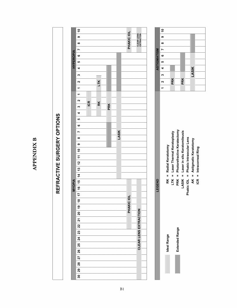
B1
AP
PE
ND
IX A
REF
RA
CTI
VE S
UR
GER
Y O
PTIO
NS
MYO
PIA
H
YPER
OPI
A
30
29
28
27
26
25
24
23
22
21
20
1918
1716
1514
1312
1110
9 8
7 6
5 4
3 2
1 1
2 3
4 5
6 7
8 9
10
ICR
RK
LTK
PR
K
LA
SIK
PH
AK
IC IO
L
PH
AK
IC IO
L
C
LEA
R L
ENS
EXTR
AC
TIO
N
CLE
AR
LEN
S EX
TRA
CTO
N
LEG
END
AST
IGM
ATI
SM
1 2
3 4
5 6
7 8
9 10
Idea
l Ran
ge
RK
=
Rad
ial K
erat
otom
y
LTK
= La
ser T
herm
al K
erat
opla
sty
PR
K
Exte
nded
Ran
ge
PRK
= Ph
otor
efra
ctiv
e K
erat
ecto
my
LA
SIK
= La
ser i
n si
tu K
erat
omile
usis
PRK
Ph
akic
IOL
= Ph
akic
Intr
aocu
lar L
ens
AK
= A
stig
mat
ic K
erat
otom
y
LASI
K
ICR
= In
trac
orne
al R
ing
AP
PE
ND
IX B
