neuroscience: the biological bases of behavior 8-10% of ap exam
TRANSCRIPT
Neuroscience: The Biological Bases of Behavior
8-10% of AP Exam
Communication in the Nervous System
Neurons The body’s electrochemical system
(nervous system) contains the brain, spinal cord and billions of nerves all formed by neurons.
Neurons have 3 tasks: 1. receive information 2. carry information 3. pass information
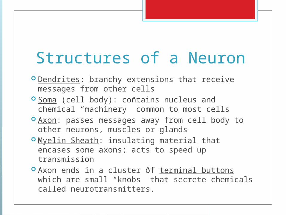
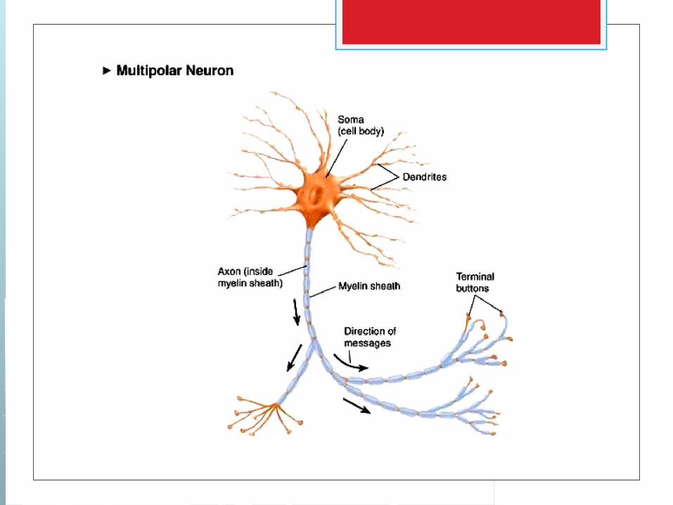
Structures of a Neuron Dendrites: branchy extensions that receive
messages from other cells Soma (cell body): contains nucleus and chemical
“machinery” common to most cells Axon: passes messages away from cell body to
other neurons, muscles or glands Myelin Sheath: insulating material that encases
some axons; acts to speed up transmission Axon ends in a cluster of terminal buttons which
are small “knobs” that secrete chemicals called neurotransmitters.
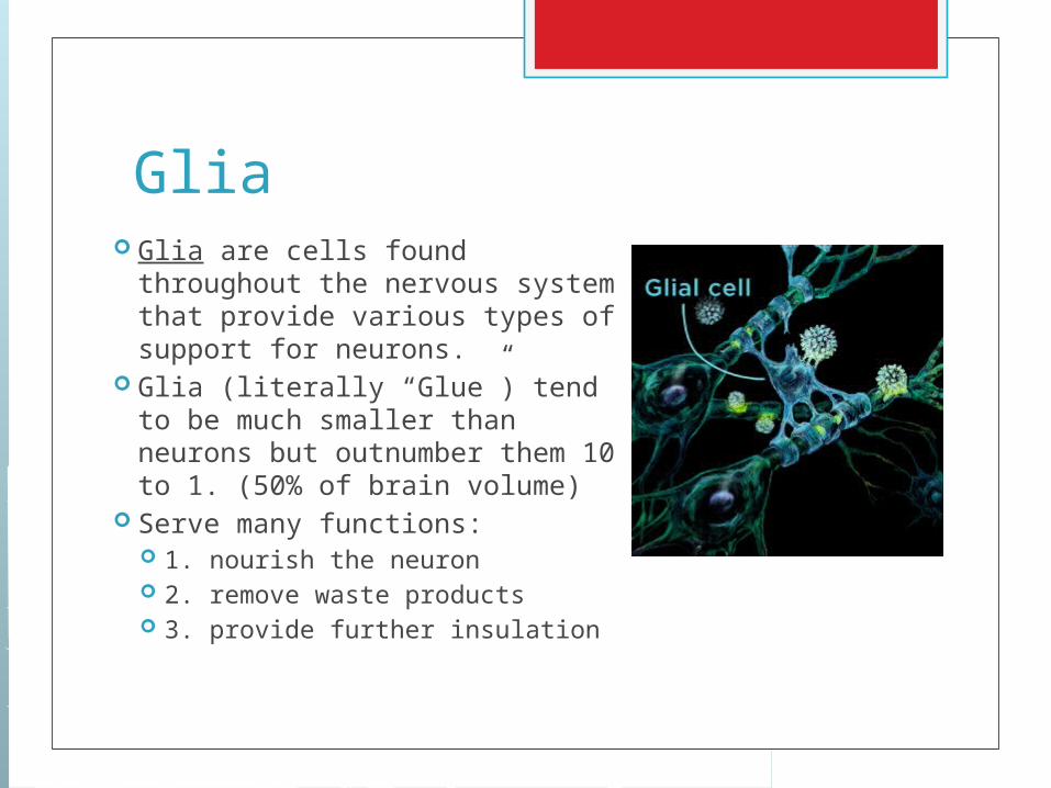
Glia Glia are cells found
throughout the nervous system that provide various types of support for neurons.
Glia (literally “Glue”) tend to be much smaller than neurons but outnumber them 10 to 1. (50% of brain volume)
Serve many functions: 1. nourish the neuron 2. remove waste products 3. provide further insulation

The Neural Impulse What happens when a neuron is stimulated? Both inside and outside the neuron are fluids
containing electrically charged atoms and molecules called ions.
Positively charged sodium (Na) and potassium (K) ions and negatively charged chloride ions flow back forth across the cell membrane.
The difference in flow rates leads to a slightly higher concentration of negatively charged ions inside the cell.
The resting potential of a neuron is its stable, negative charge when the cell is inactive.

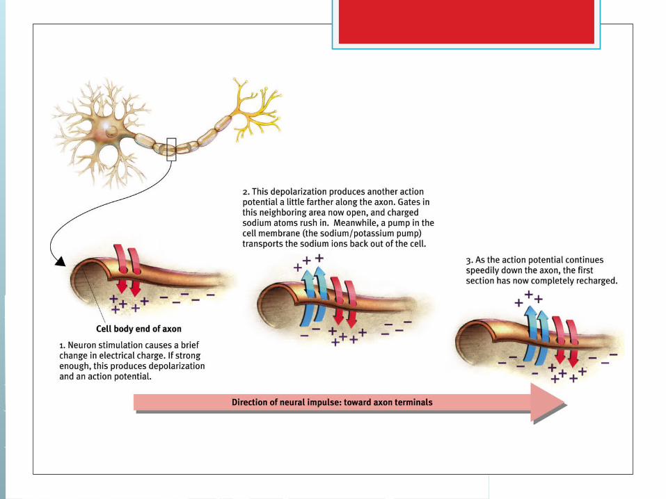
The Action Potential When the neuron is stimulated, channels in
its cell membrane open. For a brief moment positively charged
sodium (Na) ions rush in. For an instant, the neuron’s charge is less
negative or even positive, creating what is called an action potential.
Like a spark traveling along a trail of gunpowder, the voltage change races down the axon.

Refractory Period After the firing of an action potential, the
channels in the cell membrane that opened to let in (Na) ions close up.
Some time is needed before they are ready to open again and the neuron cannot fire until then.
This period of time after an action potential during another action potential cannot begin is called the refractory period.
How is neural impulse like flushing a toilet?
1. What is the action potential?
2. What is the refractory period?
3. What is the resting period?
JUST LIKE A NEURON!!!!
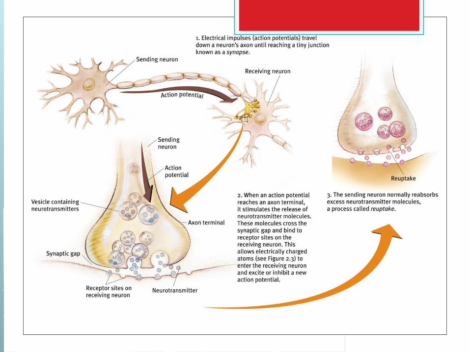
The Synapse: Where Neurons Meet Neuronal communication takes place
without the neurons actually touching one another.
These neural transmissions take place in a synapse: a microscopic gap between the terminal buttons of one neuron and the dendrite of another.
The neuron that sends a signal across the gap is called the presynaptic neuron.
The neuron that receives the signal is called the postsynaptic neuron.
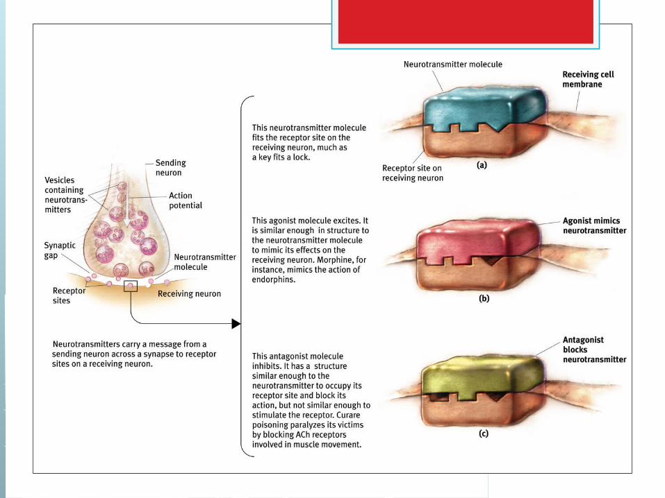
Neurotransmitters How do messages travel across the gaps between
neurons? The arrival of an action potential at an axon’s
terminal buttons triggers the release of neurotransmitters.
Neurotransmitters are chemicals that transmit information from one neuron to another.
These neurotransmitters are then released into the synaptic cleft where they may bind with special molecules in the postsynaptic cell at various receptor sites.
These receptor sites are specifically “tuned” to recognize and respond to some neurotransmitters but not to others.
Postsynaptic Potentials When a neurotransmitter and a receptor molecule
combine, reactions in the cell membrane cause a postsynaptic potential (PSP); a voltage charge at a receptor site.
Two types of messages can be sent from cell to cell: excitatory and inhibitory.
An excitatory PSP is a positive voltage shift that increases the likelihood that the postsynaptic neuron will fire an action potential.
An inhibitory PSP is a negative voltage shift that decreases the likelihood that the postsynaptic neuron will fire an action potential.
Reuptake Neurotransmitters that are not absorbed
at postsynaptic receptor sites are typically “reabsorbed” by the presynaptic neuron.
This process of reabsorbing non-binding neurotransmitters in the synapse is called reuptake.
Neurotransmitters Specific neurotransmitters work at specific
kinds of synapses, binding to receptor sites. However a neurotransmitter cannot bind to
just any site. The binding process operates much like a
“lock and key” scenario. Resultantly specific transmitters can deliver
signals at only certain locations on cell membranes.
How Drugs Affect Neurotransmission Although synaptic receptor sites are
sensitive to specific neurotransmitters, sometimes they can be “fooled” by other chemical substances.
An agonist is a chemical that mimics the action of a neurotransmitter, binding to receptor sites and causing a PSP.
An antagonist is a chemical that opposes the action of a neurotransmitter, occupying its receptor sites, preventing a PSP.
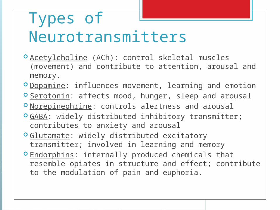
Types of Neurotransmitters Acetylcholine (ACh): control skeletal muscles
(movement) and contribute to attention, arousal and memory.
Dopamine: influences movement, learning and emotion Serotonin: affects mood, hunger, sleep and arousal Norepinephrine: controls alertness and arousal GABA: widely distributed inhibitory transmitter;
contributes to anxiety and arousal Glutamate: widely distributed excitatory transmitter;
involved in learning and memory Endorphins: internally produced chemicals that
resemble opiates in structure and effect; contribute to the modulation of pain and euphoria.
Organization of the Nervous System
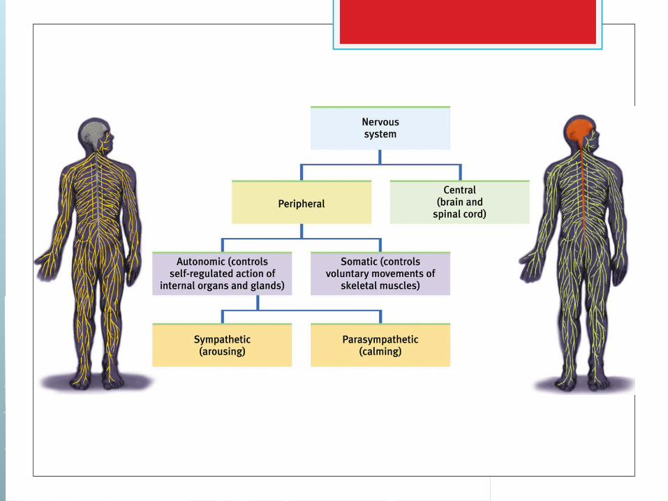
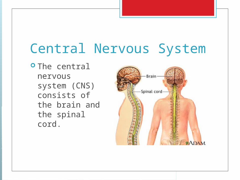
Central Nervous System The central
nervous system (CNS) consists of the brain and the spinal cord.
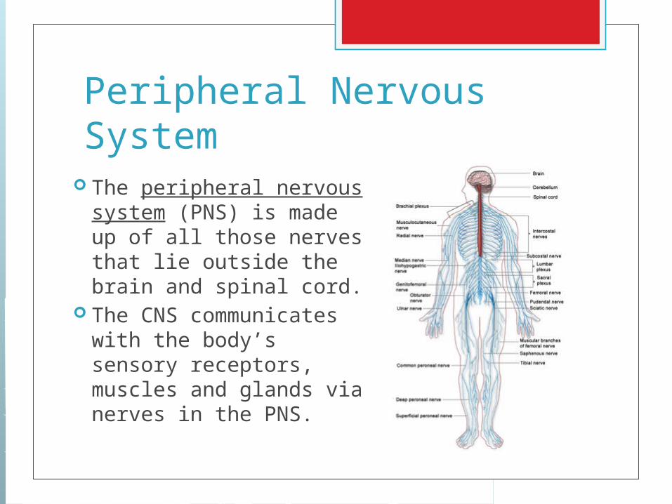
Peripheral Nervous System
The peripheral nervous system (PNS) is made up of all those nerves that lie outside the brain and spinal cord.
The CNS communicates with the body’s sensory receptors, muscles and glands via nerves in the PNS.
The Somatic Nervous System One of the two divisions of the PNS is known as
the somatic nervous system. The somatic nervous system (SNS)is made up
of nerves that connect to voluntary skeletal muscles and to sensory receptors.
The nerves of the SNS carry information from receptors in the skin, muscles and joints to the CNS and that carry information from the CNS to the muscles.
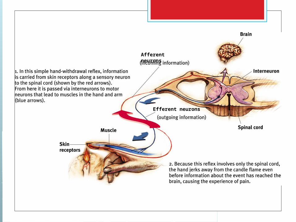
Types of Nerves in the SNS The functions of the SNS require three
specific types of nerves: Afferent (sensory) neurons: carry info
inward to the CNS from sensory receptor sights.
Interneurons: carry info throughout the brain and spinal cord
Efferent (motor) neurons: carry info outward from the CNS to muscles and glands.
Afferent neurons
Efferent neurons
The Autonomic Nervous System The autonomic nervous system (ANS):
controls automatic, involuntary, visceral functions that people don’t normally think about.
These functions include heart rate, digestion and perspiration.
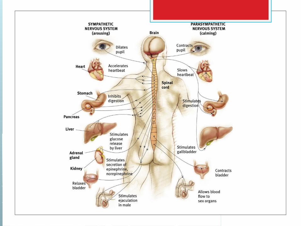
The Autonomic Nervous System The ANS can be subdivided into two
branches: sympathetic and parasympathetic.
Sympathetic nervous system: branch of the ANS that mobilizes and arouses the body’s resource’s for emergencies.
Parasympathetic nervous system: branch of the ANS that calms down and conserves bodily resources.

Looking Inside the BrainHistorical and Contemporary Research Strategies and Technology
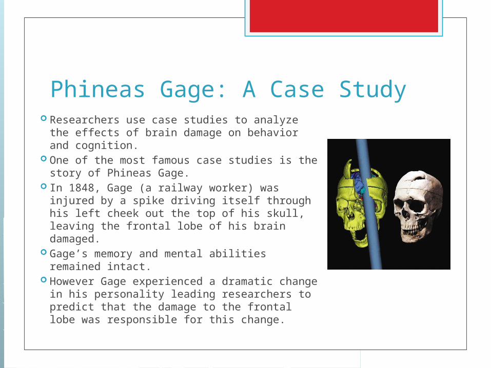
Phineas Gage: A Case Study Researchers use case studies to analyze the
effects of brain damage on behavior and cognition.
One of the most famous case studies is the story of Phineas Gage.
In 1848, Gage (a railway worker) was injured by a spike driving itself through his left cheek out the top of his skull, leaving the frontal lobe of his brain damaged.
Gage’s memory and mental abilities remained intact.
However Gage experienced a dramatic change in his personality leading researchers to predict that the damage to the frontal lobe was responsible for this change.
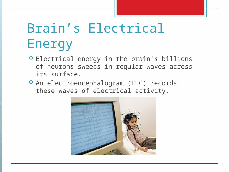
Brain’s Electrical Energy Electrical energy in the brain’s billions of
neurons sweeps in regular waves across its surface.
An electroencephalogram (EEG) records these waves of electrical activity.
Scanning Brain Structure Two types of brain scanners allows researchers
a glimpse into the structure of the brain itself. Computerized tomography (CT Scan):
computer-enhanced x-ray of brain structure; creates a vivid image of a horizontal slice of the brain
Magnetic resonance imaging (MRI): technique that uses magnets and radio waves to produce images that distinguish between different types of soft tissue in the brain.
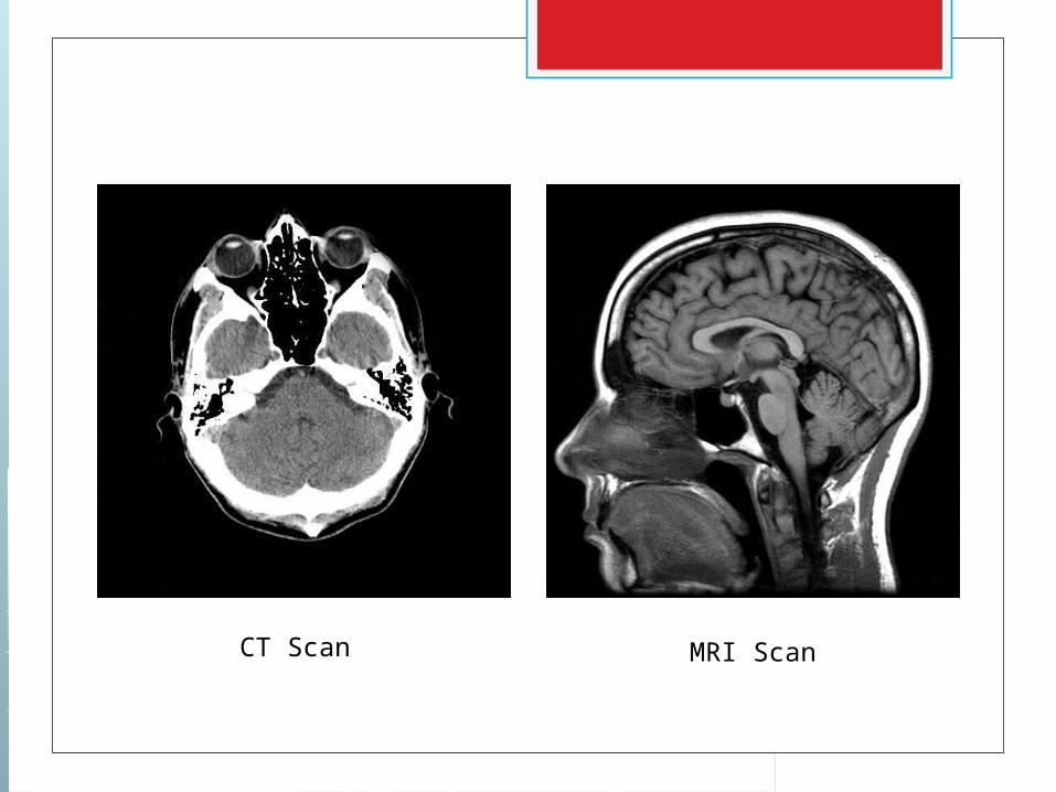
CT Scan MRI Scan
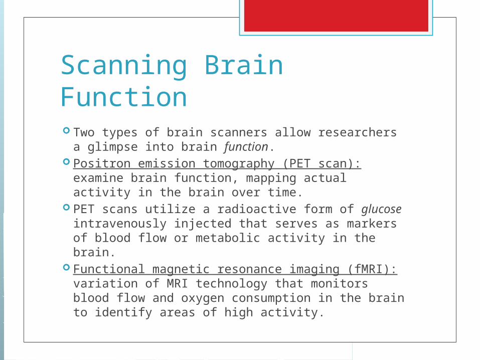
Scanning Brain Function Two types of brain scanners allow researchers a
glimpse into brain function. Positron emission tomography (PET scan):
examine brain function, mapping actual activity in the brain over time.
PET scans utilize a radioactive form of glucose intravenously injected that serves as markers of blood flow or metabolic activity in the brain.
Functional magnetic resonance imaging (fMRI): variation of MRI technology that monitors blood flow and oxygen consumption in the brain to identify areas of high activity.
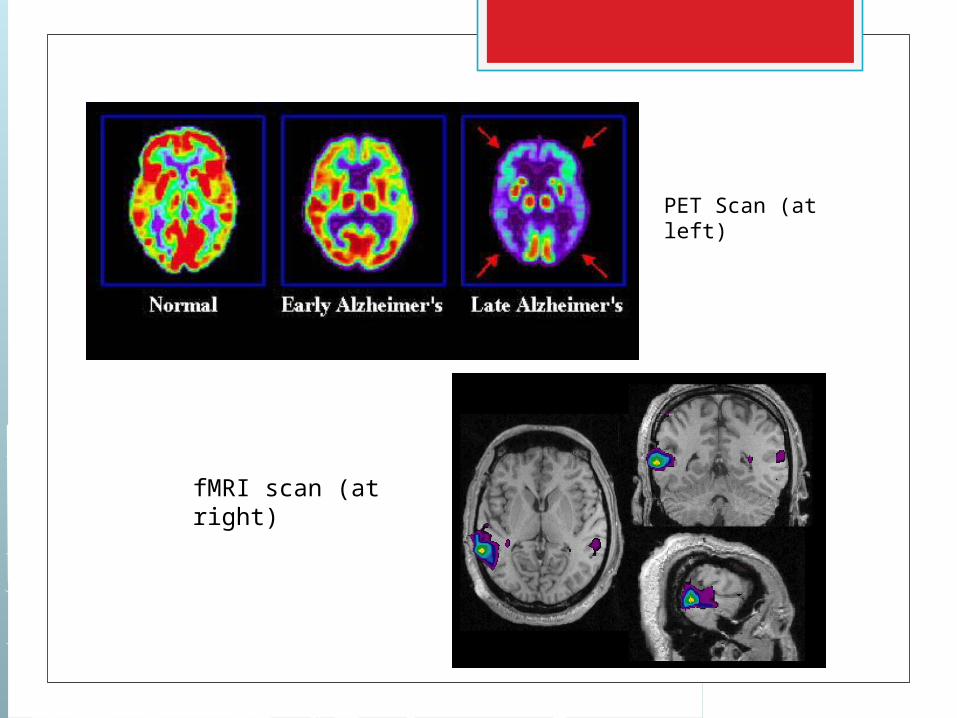
PET Scan (at left)
fMRI scan (at right)
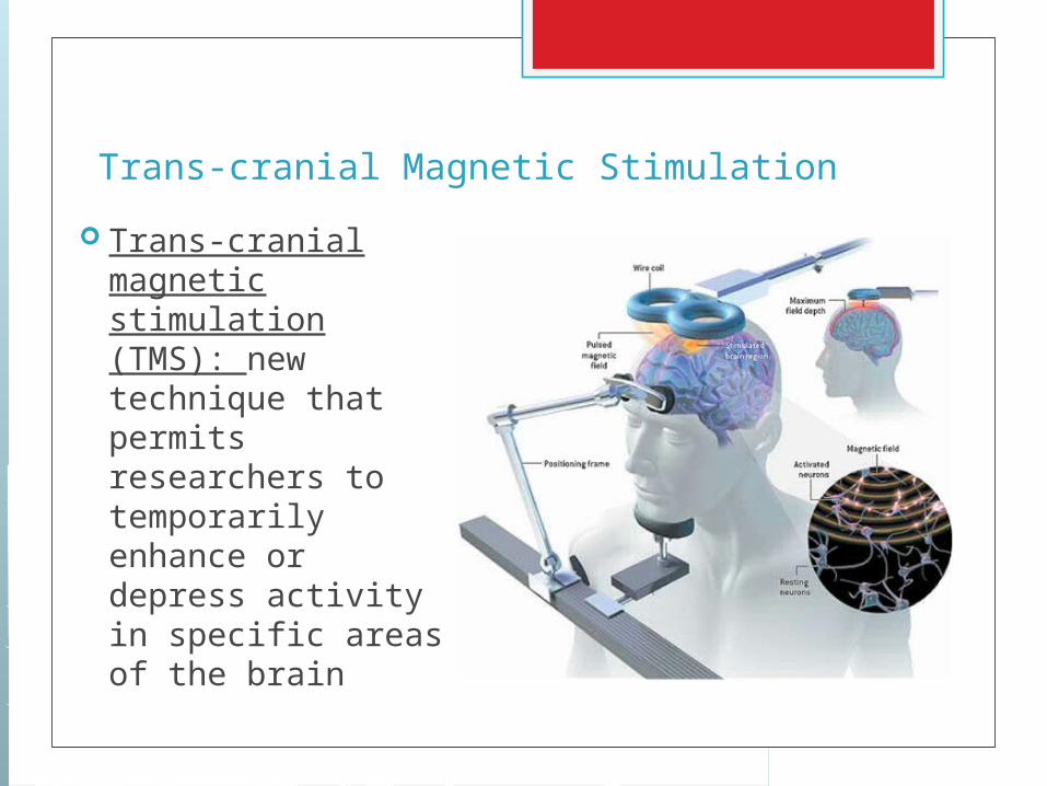
Trans-cranial Magnetic Stimulation
Trans-cranial magnetic stimulation (TMS): new technique that permits researchers to temporarily enhance or depress activity in specific areas of the brain
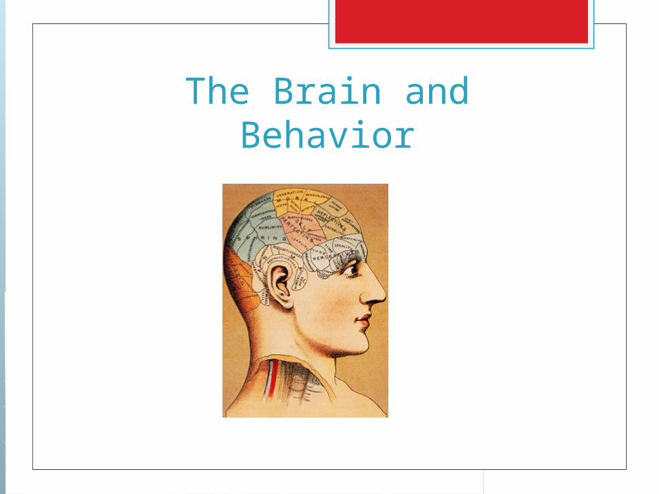
The Brain and Behavior
Structures and Functions in the Brain
Brainstem: transmits information between the brain and the rest of the body
Medulla: regulation of unconscious function such as heartbeat and breathing
Pons: involved in sleep and arousal Reticular formation: group of fibers that
carry stimulation related to sleep and arousal; modulation of reflexes, breathing and pain perception
Structures and Functions in the Brain
Thalamus: relay center for the cerebral cortex; handles incoming and outgoing sensory signals
Cerebellum: coordinates fine muscle movement and balance

Structures and Functions in the Brain
Limbic system: doughnut shaped system of neural structures at the border of the brainstem and cerebral hemispheres.
The limbic system includes: 1. amygdala: involved in emotion and
aggression 2. hippocampus: involved in the process
of new memories and learning 3. hypothalamus: regulation of basic
biological needs including hunger, thirst and homeostasis
The Cerebral Cortex Cerebrum: largest and most complex part
of the human brain; area responsible for most complex mental activities
Cerebral Cortex: intricately folded outer layer of the cerebrum
Cerebrum is divided into two halves called hemispheres (right and left)
The two hemispheres are connected by a thick band of fibers called the corpus callosum.
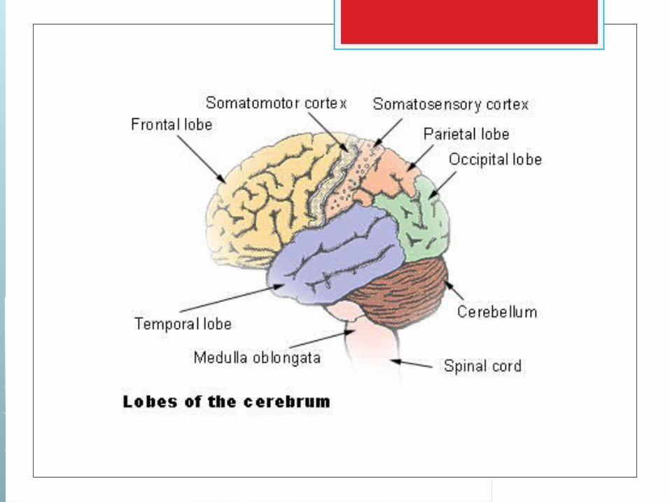
Lobes of the Cerebral Cortex Occipital Lobe: cortical areas where visual
signals are sent and visual processing begins (visual cortex)
Parietal Lobe: receives sensory input for touch and body positions; includes the somatosensory cortex
Temporal Lobe: responsible for the processing of language and audition (auditory cortex)
Frontal Lobe: involved in speaking and muscle movement (includes the motor cortex) and in making plans and judgment (prefrontal cortex)

Brain Plasticity
Brain Plasticity It was once believed that significant
changes in the anatomy and organization of the brain were limited to early periods of development.
However, new research has shown that anatomical structure and functional organization is more malleable than widely assumed.
This capacity for modification of the brain is known as plasticity.
Brain Plasticity Recent studies into brain plasticity have
shown: 1. Aspects of experience can actually shape
features of brain structure. 2. Damage to incoming sensory pathways or
destruction of brain tissue can lead to neural “reorganization”. Healthy neurons attempt to compensate for
the loss of nearby neurons. 3. The adult brain is now known to create
new neurons, a process called neurogenesis. Seen in the hippocampus and olfactory bulb.
Right Brain/Left BrainCerebral Laterality
Hemispheric Specialization Hemispheric specialization: the notion
that each hemisphere of the brain (left and right) serve separate and distinct functions
Language and the Left Hemisphere
Hints of hemispheric specialization date back to the 1860s and the work of Paul Broca.
Broca was treating a patient who had been unable to speak for 30 years.
After the patient died, lesions on the left side of the frontal lobe identified the probable cause of the speech deficiency.
Since then, many cases have shown this area of the motor cortex (now called Broca’s area) plays an important role in the production of speech.

Language and the Left Hemisphere
In 1874, German researcher Carl Wernicke discovered that after damage to a specific area of the left temporal lobe people could speak only meaningless words.
Today, this region of the temporal lobe (known as Wernicke’s area) is thought to be responsible comprehension and expression of language.
Split-Brain Research In split-brain surgery the corpus callosum
(the band of fibers that connects the cerebral hemispheres) is severed.
This is done to reduce severe seizures in a limited number of patients.
Moreover, the surgery provides researchers with the ability to study people who have had their brain literally split in two.
Split-Brain Research To appreciate the logic behind split-brain
research, you must appreciate the logic behind it.
Each hemisphere’s primary connections are to the opposite side of the body.
To oversimplify, vision and hearing processed on the right side is routed and interpreted on the left side of the brain and vice versa.
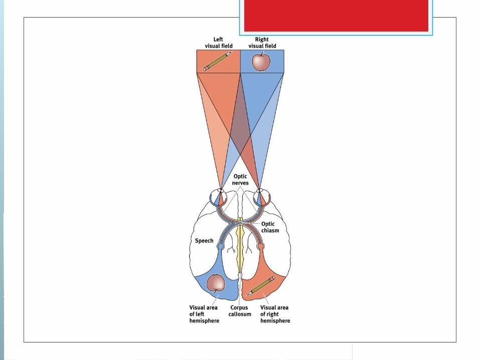

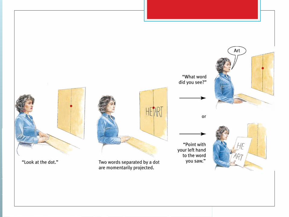
Split-Brain Research In a classic study of split-brain patients, Roger
Sperry and Michael Gazziniga presented visual stimuli in a single visual field (right or left) so that the stimuli would be sent to only one hemisphere.
When pictures were flashed to the right visual field (and thus sent to the left hemisphere), the subjects were able to name and describe (i.e. speak) the object depicted.
However the subjects were not able to describe the same objects when they were flashed in the left visual field (thus sent to the right hemisphere).
Split-Brain Research Sperry and Gazzaniga’s research supported the theory
that each hemisphere is responsible for different tasks. Further studies on non-split-brain patients have
concluded that: 1. The left hemisphere is usually better on tasks
involving verbal processing, such as language, speech and writing, analytical thought and objectivity (math).
2. The right hemisphere is usually better on non-verbal tasks, including visual-spatial perception, creativity, intuition and interpretation of body language.

The Endocrine System
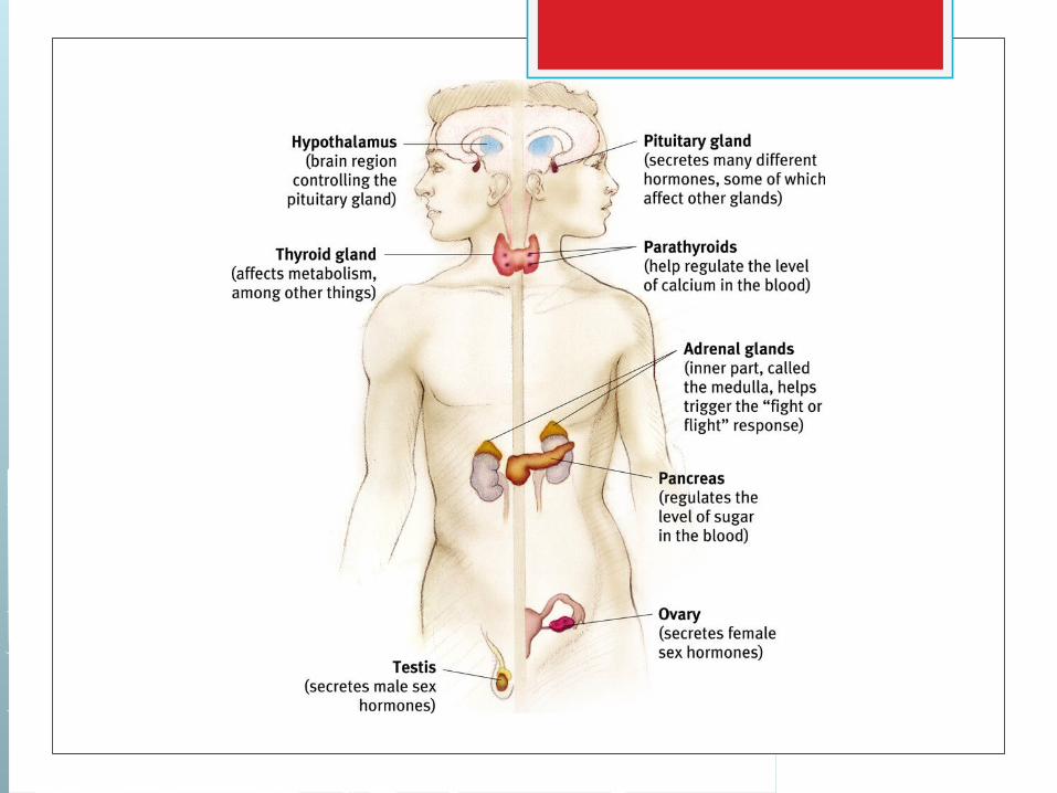
The Endocrine System Endocrine system: consists of glands that
secrete chemicals into the bloodstream that help control bodily function
The messengers within the endocrine system are called hormones.
Once released, hormones diffuse through the bloodstream and bind to special receptors on target cells.
In comparison to neurotransmitters, hormones are transmitted over a longer path at a slower place and usually in brief bursts throughout the day.
Types and Function of Glands Much of the endocrine system is
controlled by the nervous system via the hypothalamus which connects to the pituitary gland.
Pituitary gland: master gland; responsible for regulating the actions of other glands
Thyroid gland: regulates energy levels Adrenal gland: help to arouse the body in
times of stress