neurones by the uncharged form of amitriptyline and related tricyclic
TRANSCRIPT
B 1995 Stockton Press All rights reserved 0007-1188/95 $12.00 $
Potent block of potassium currents in rat isolated sympatheticneurones by the uncharged form of amitriptyline and relatedtricyclic compoundsJulian R.A. Wooltorton & 'Alistair Mathie
Department of Pharmacology, Royal Free Hospital School of Medicine, Rowland Hill Street, London NW3 2PF
1 The block of K+ currents by amitriptyline and the related tricyclic compounds cyproheptadine anddizocilpine was studied in dissociated rat sympathetic neurones by whole-cell voltage-clamp recording.2 Cyproheptadine (30 uM) inhibited the delayed-rectifier current (Kv) by 92% and the transient current(KA) by 43%. For inhibition of KV, cyproheptadine had a KD of 2.2 lM. Dizocilpine (30 pM) inhibitedKV by 26% and KA by 22%. The stereoisomers of dizocilpine were equally potent at blocking KV andKA-3 Amitriptyline, a weak base, was significantly more effective in blocking KV at pH 9.4 (KD= 0.46 pM)where the ratio of charged to uncharged drug was 50:50 compared with pH 7.4 (KD=11.9 pM) where theratio was 99:1.4 N-methylamitriptyline (10 gM), the permanently charged analogue of amitriptyline, inhibited Kv byonly 2% whereas in the same cells amitriptyline (10 pM) inhibited Kv by 36%.5 Neither amitriptyline nor N-methylamitriptyline had a detectable effect on Kv when added to theintracellular solution.6 It is concluded that the uncharged form of amitriptyline is approximately one hundred times morepotent in blocking Kv than the charged form. However, this does not seem to be due to unchargedamitriptyline having better access to an intracellular binding site.
Keywords: Amitriptyline; N-methylamitriptyline; cyproheptadine; dizocilpine; potassium currents; rat sympathetic neurones;whole-cell voltage-clamp; pH
Introduction
Previously, we have shown that two voltage-gated potassium(K+) currents in rat superior cervical ganglion (SCG) neu-rones, the delayed-rectifier current Kv and the transient cur-rent KA, can be inhibited by certain tricyclic antidepressants(e.g. amitriptyline and imipramine) and other structurally re-lated compounds such as chlorpromazine (Wooltorton &Mathie, 1993). Potassium channels play an important role inthe regulation of neuronal membrane potential and neuronalexcitability (Cook & Quast, 1990; Halliwell, 1990), thereforecompounds like these which inhibit K+ currents will have anumber of actions on neurones which may be either ther-apeutic or toxic.
This study was divided into two parts. In the first series ofexperiments, we have extended our observations to two furthertricyclic compounds; cyproheptadine and dizocilpine. Cypro-heptadine is an antagonist at 5-hydroxytryptamine and hista-mine (HI) receptors used clinically in certain allergies such ashay-fever and occasionally in migraine (Laurence & Bennett,1992); while dizocilpine (MK-801) is a non-competitiveNMDA receptor antagonist with anticonvulsant properties(Wong et al., 1986).
In the second series of experiments, we have studied in moredetail the mechanism of action of these tricyclic compounds ininhibiting Kv. In particular, since all of the active compoundsare weak bases, we have determined whether the protonated(charged) or unprotonated (uncharged) form of these mole-cules is more important for their inhibitory action by varyingthe pH of the extracellular recording solution and by using apermanently charged analogue of amitriptyline, N-methyla-mitriptyline.
'Author for correspondence.
Some of the results in this study have been published inabstract form (Wooltorton & Mathie, 1994; 1995).
Methods
Cell dissociation
Neurones were dissociated from rat superior cervical ganglia(SCGs) by a method modified from one described previously(Beech et al., 1991; Bernheim et al., 1991). Sprague-Dawleyrats of either sex (age 7-8 days) were killed by decapitation.The SCGs were removed and placed at 370C in a modifiedHank's solution containing 20 iu ml- papain for 20 min. Theganglia were then incubated in a mixture of 400 iu ml-' col-lagenase (Type I) and 16 mg ml-' dispase (Grade II) for45 min and triturated every 15 min. Cells were then cen-trifuged and resuspended in Leibovitz L-15 medium. Isolatedneurones were kept at 4°C and used for recording within 10 h.
Solutions
The external solution (in mM) was NaCl 150, KCl 2.5, CaCl22.5, MgCl2 1, HEPES 10, glucose 8, TTX 0.0005 and the pHwas adjusted with NaOH as required (physiologicalpH = 7.4). The internal solution (in mM) was: KCl 145,MgCl2 5, HEPES 5, BAPTA 10 and the pH was adjusted to7.4 with KOH.
Stock solutions of all compounds were made up in dis-tilled water except for cyproheptadine, which was made upin 50% ethanol, and were kept at -20°C until required.They were then added to the external solutions at suitableconcentrations shortly before the experiment. Solutions wereapplied to the cells by continuous perfusion of the chamber
Bridsh Joumal of Phamacology (1995) 116, 2191 - 2200
J.R.A. Wooltorton & A. Mathie Block of Kv by amitriptyline
during recording. The perfusion rate was 4-5 ml min-' andcomplete exchange of the bath solution occurred within 20-40 s.
Papain, collagenase, cyproheptadine, tacrine and ami-triptyline were obtained from Sigma, (+ )- and (-)-dizocilpinefrom Semat Technical (RBI), dispase from Boehringer Man-nheim and Leibovitz L-15 from Life Technologies (Gibco). N-methylamitriptyline was a generous gift from Dr F. J. Ehlert.
Current recording and analysis
Currents were recorded in the whole-cell configuration of thepatch-clamp technique (Hamill et al., 1981) at 20-230C withan Axopatch ID amplifier. Patch pipettes had resistances ofbetween 2 and 5 MO. During whole-cell recording, series re-sistance was 14.3 ± 0.8 MC and the whole-cell capacitance was14.1 + 0.8 pF (n = 60). Currents were not leak-subtracted.
Voltage protocols Protocol (1): To measure Kv and KA, cellswere held at -30 mV and a conditioning pulse to -120 mVwas applied for 500 ms, once every 5 s, before a test pulse of400 ms to 0 mV. The peak current, measured within 10 ms ofthe step to 0 mV, has been shown to be predominantly KA,while the sustained current measured as an average over100 ms, 297 ms following the step was predominantly Kv (seeWooltorton & Mathie, 1993).
Protocol (2): To measure Kv in isolation, cells were held at-70 mV and stepped once every 6 s to -50 mV for 30 ms toinactivate any residual KA, then depolarized to a test potentialof + 10 mV for 150 ms before stepping back first to -50 mVfor 30 ms then to the holding potential. KV was measured asthe average current over 25 ms, 116.5 ms following the step to+ 10 mV.
Protocol (3): To study the steady state inactivation ofKA andKV, cells were held at -70 mV and stepped once every 5 s topotentials between - 120 mV and + 30 mV in 10 mV incre-ments for 2 s, and depolarized to + 50 mV for 90 ms. KA wasmeasured at the time where peak current after the depolarizingstep from - 120 mV occurred. Kv was measured as the mean
current over 25 ms, 62.5 ms after the depolarizing step.Currents were low-pass filtered at 5 kHz, digitized at 0.5-
5 kHz and recorded and analysed using an IBM-compatiblePC, pClamp version 5.5 or version 5.7 with a TL-l Labmasteror a Digidata 1200 interface (Axon Instruments) and Excelversion 4.0 (Microsoft). Statistical tests were carried out usinga Student's t test. P values of less than 0.05 were consideredsignificant. All data are expressed as mean ± s.e.mean and n isthe number of cells.
The concentration of amitriptyline and N-methylami-triptyline entering a cell from the recording pipette was cal-culated as described in Stansfeld & Mathie (1993) (see alsoBeech et al., 1991; Pusch & Neher, 1988). Briefly, previousstudies have shown that BAPTA (mol. wt. 476.4) diffused intosympathetic neurones (Rs = 3.5 MCI; Cs = 36 pF) with a timeconstant, TB of 362 s (Beech et al., 1991; Stansfeld & Mathie,1993). Thus BAPTA diffusion into any similar sized neurone ofinterest can be calculated from equation 1:
TBu= (RuCu32/RsCs312)rB [1]
where TB= 362s, Rs = 3.5 MCI, Cs= 36 pF, Ru and Cu are theseries resistance and whole-cell capacitance of the neurone ofinterest and TBU is the time constant of BAPTA entry into thisneurone. Once this is known, the time constant for entry of anycompound (of reasonably similar molecular weight) can becalculated from equation 2. So, for example, for N-methyla-mitriptyline:
TaiM = TBu(MamiM/MBAPTA) [2]
where TamM is the time constant for N-methylamitriptyline andManM and MBAPTA are the two compounds respective mole-cular weights (476.4 and 419.3). The concentration of N-me-thylamitriptyline inside the cell is then known for any timefollowing breakthrough and from equation 3 and the valuesobtained from the amitriptyline dose-response curve (see Re-sults), the degree of block of Kv at any time followingbreakthrough can be calculated if it is assumed that thecharged form of amitriptyline blocks from inside the cell. Thisis shown for amitriptyline in Figure 10a and N-methylami-triptyline in Figure l0b.
Chlorpromazine Cyproheptadine
/N\
N-methylamitriptyline TacrineNH2
Dizocilpine (MK-801)
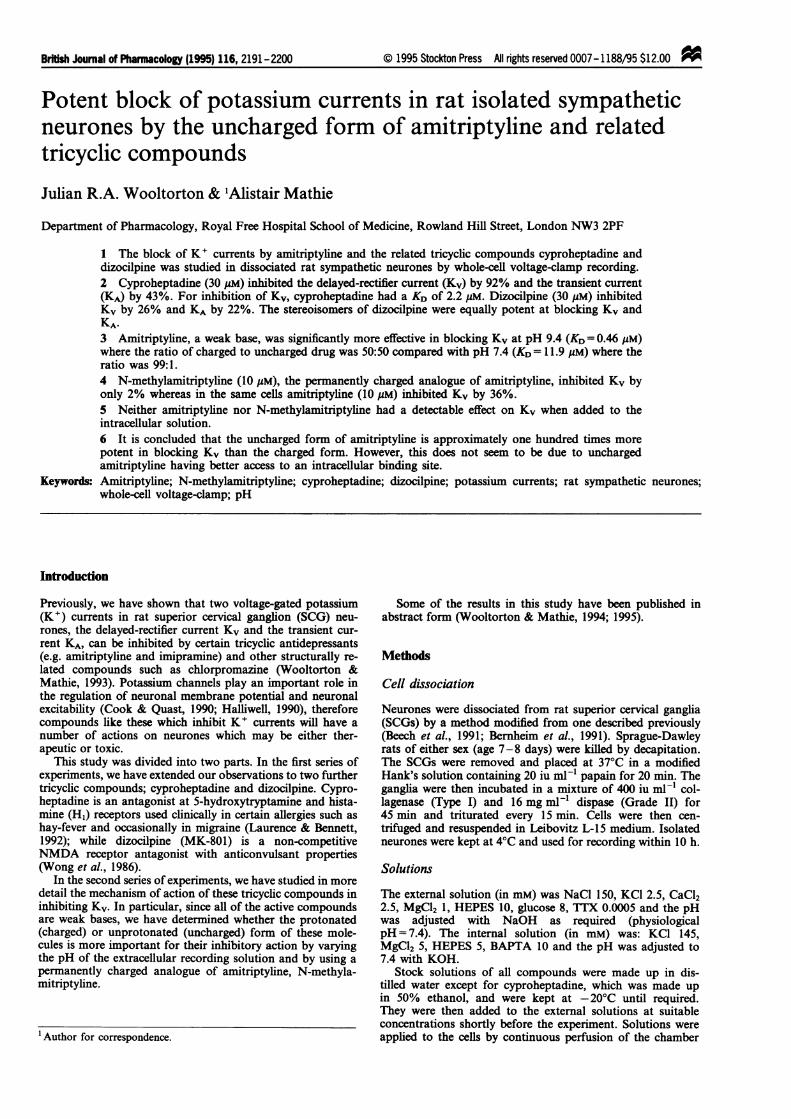
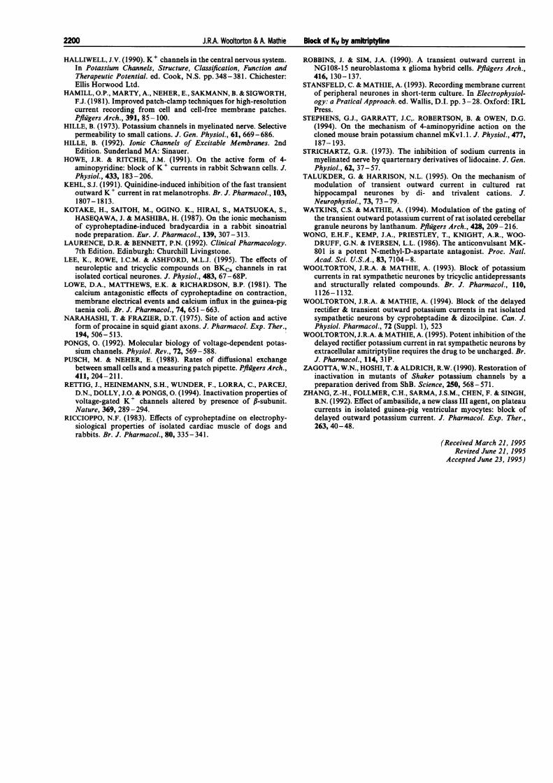
Figure 1 Chemical structures of the tricyclic compounds investigated.
Amitriptyline Imipramine
2192
J.R.A. Wooltorton & A. Mathie Block of Kv by amitriptyline
Structures of tricyclic compounds
The structures of the compounds studied are shown in Figure1. They all possess a tricyclic region. Imipramine, amitripty-line, chlorpromazine and cyproheptadine each has a tertiaryamine group on the central side chain substituent, positionedthree carbon atoms from the cyclic structure, which differsslightly for each drug. In contrast, tacrine and dizocilpine haveno such side chain. N-methylamitriptyline is a permanentlycharged analogue of amitriptyline, having the same structureexcept for a quaternary amine group in place of the tertiaryamine group.
Results
Block of KA and Kv by cyproheptadine and dizodilpine
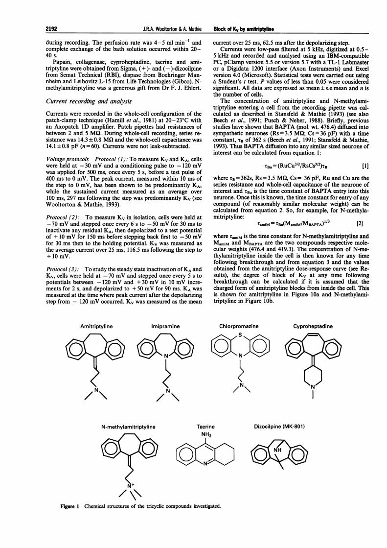
Both the transient and sustained potassium currents (KA andKv) can be evoked by voltage protocol 1 (see Methods) and are
illustrated in Figure 2a. The control peak current (at 0 mV) forthe cells used in this study was 2417 ± 191 pA (n = 60) while thecontrol sustained current was 621 ± 42 pA.
Figure 2 also illustrates the effect of various concentrationsof cyproheptadine on KA and Kv. The inhibition of bothcurrents was concentration-dependent and there appeared tobe selectivity for Kv over KA (Figure 2b). For example, 30 gMcyproheptadine produced an inhibition of Kv of 92.2 ± 1.6%(n = 6) but inhibited KA by only 43.3 ± 2.7% (data not shown).Figure 2a also shows the hyperpolarization-activated chloridecurrent of these neurones during the prepulse to -120 mV(Clark & Mathie, 1995). It is of interest that cyproheptadinecauses a slight enhancement of this current.
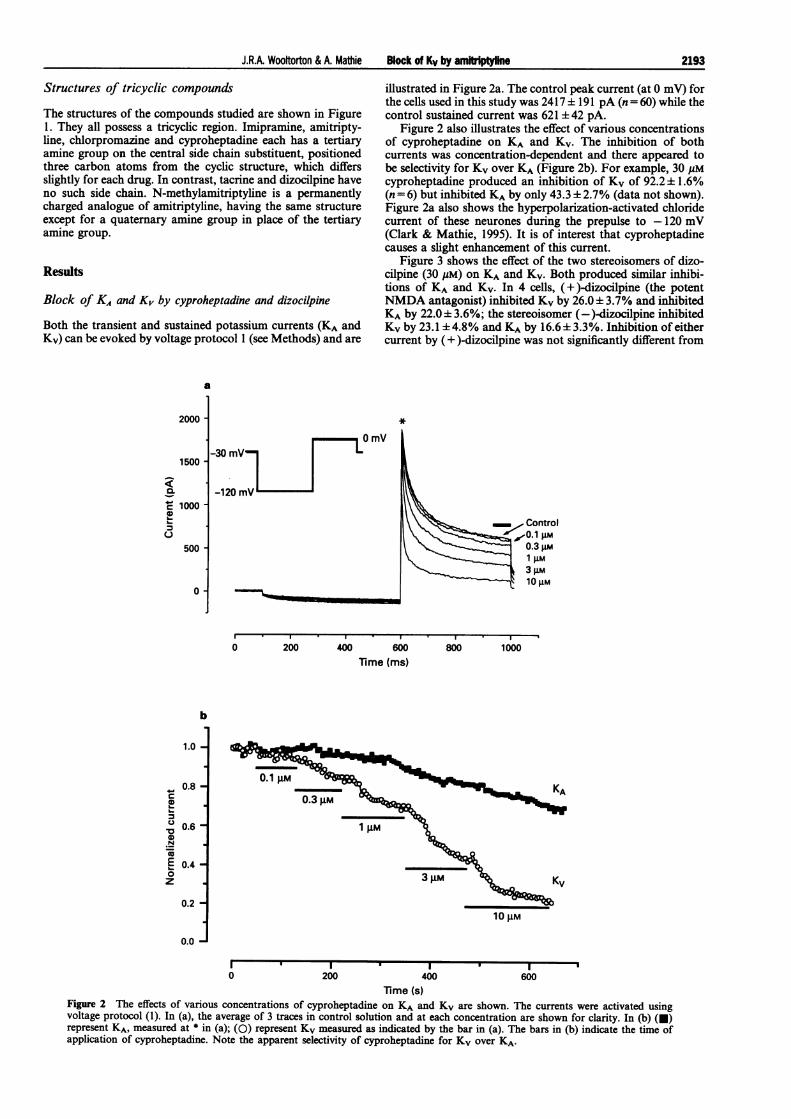
Figure 3 shows the effect of the two stereoisomers of dizo-cilpine (30 gM) on KA and KV. Both produced similar inhibi-tions of KA and KV. In 4 cells, (+)-dizocilpine (the potentNMDA antagonist) inhibited KV by 26.0± 3.7% and inhibitedKA by 22.0 3.6%; the stereoisomer (-)-dizocilpine inhibitedKV by 23.1 ±4.8% and KA by 16.6±3.3%. Inhibition of eithercurrent by (+ )-dizocilpine was not significantly different from
a
2000 -
1500
a
z 1000-0
500 -
0 -
*
0 200 400 600lime (ms)
1
800 1000
n 0.60-0E 0.6 -
z
0.2-
0.0 -
0.1 IM
0.3 gMKA
I I I, I I
0 200 400 600Time (s)
Figure 2 The effects of various concentrations of cyproheptadine on KA and KV are shown. The currents were activated usingvoltage protocol (1). In (a), the average of 3 traces in control solution and at each concentration are shown for clarity. In (b) (U)represent KA, measured at * in (a); (0) represent KV measured as indicated by the bar in (a). The bars in (b) indicate the time ofapplication of cyproheptadine. Note the apparent selectivity of cyproheptadine for Kv over KA.
2193
b
1.0
0.8 -I
2- -bf
a
75C0C.
(+)-Dizocilpine (-)-Dizocilpine
.
0 50 100 150 200 250 300Time (s)
Fgure 3 The effect of 30pM (+)- and (-)-dizocilpine on KA (0)and Kv (U) is shown. In this cell, (+)-dizocilpine inhibited Kv by28% and KA by 19%. (-)-Dizocilpine inhibited Kv by 19% and KAby 17%. Currents were activated using voltage protocol (1).
inhibition by (-)-dizocilpine (P>0.05). The non-selectivity ofdizocilpine for either current and its lower potency comparedwith cyproheptadine is consistent with our previous hypothesisthat, for block of potassium channels by a family of tricycliccompounds, a central chain, as is present in amitriptyline andcyproheptadine for example, is important for the selectivity ofblock for Kv over KA (Wooltorton & Mathie, 1993).
Concentration-dependent inhibition of Kv bycyproheptadine and amitriptyline
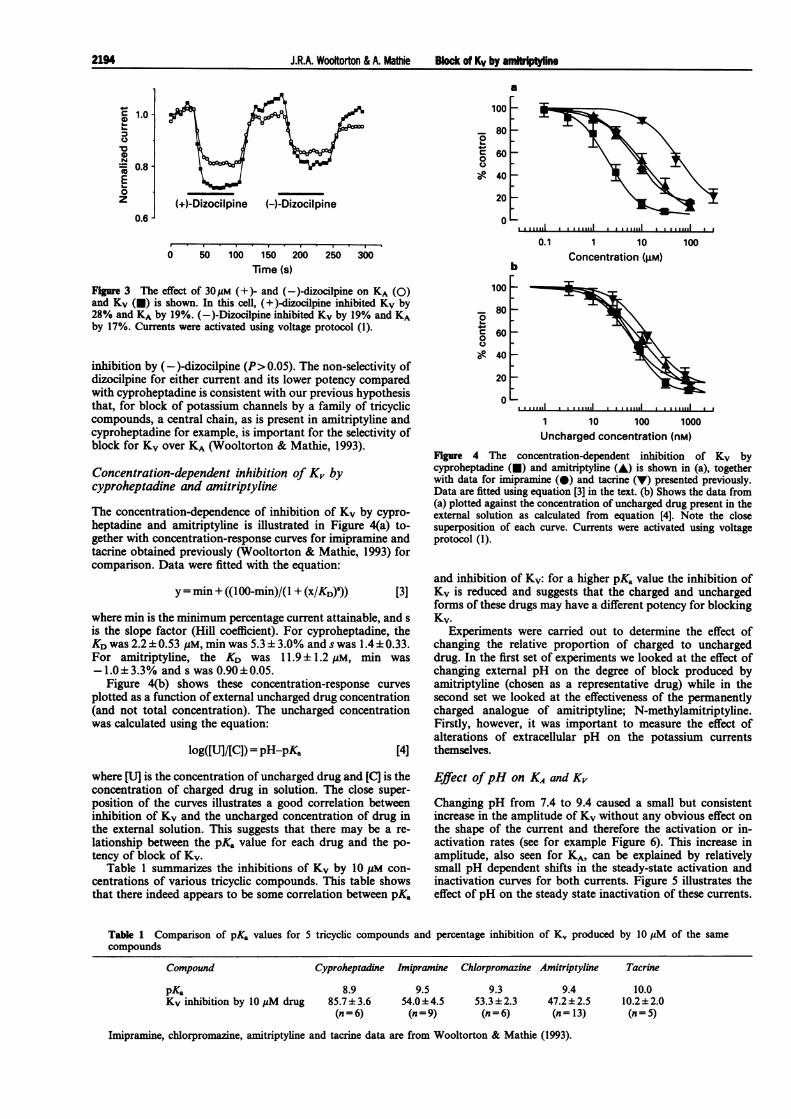
The concentration-dependence of inhibition of Kv by cypro-heptadine and amitriptyline is illustrated in Figure 4(a) to-gether with concentration-response curves for imipramine andtacrine obtained previously (Wooltorton & Mathie, 1993) forcomparison. Data were fitted with the equation:
y = min + ((100-min)/(1 + (x/KD)5)) [3]
where min is the minimum percentage current attainable, and sis the slope factor (Hill coefficient). For cyproheptadine, theKD was 2.2 ± 0.53 yM, min was 5.3 ± 3.0% and s was 1.4 ± 0.33.For amitriptyline, the KD was 11.9 ± 1.2 yM, min was-1.0±3.3% and s was 0.90±0.05.
Figure 4(b) shows these concentration-response curvesplotted as a function of external uncharged drug concentration(and not total concentration). The uncharged concentrationwas calculated using the equation:
log([U]/[C]) = pH-pKa [4]
soL-
0
1-0
100
80
60
40
20
0
0.1 1 10Concentration (gM)b
100
..
1 10 100 1000Uncharged concentration (nM)
Figure 4 The concentration-dependent inhibition of Kv bycyproheptadine (U) and amitriptyline (A) is shown in (a), togetherwith data for imipramine (0) and tacrine (V) presented previously.Data are fitted using equation [3] in the text. (b) Shows the data from(a) plotted against the concentration of uncharged drug present in theexternal solution as calculated from equation [4]. Note the closesuperposition of each curve. Currents were activated using voltageprotocol (1).
and inhibition of Kv: for a higher pKa value the inhibition ofKv is reduced and suggests that the charged and unchargedforms of these drugs may have a different potency for blockingKv.
Experiments were carried out to determine the effect ofchanging the relative proportion of charged to unchargeddrug. In the first set of experiments we looked at the effect ofchanging external pH on the degree of block produced byamitriptyline (chosen as a representative drug) while in thesecond set we looked at the effectiveness of the permanentlycharged analogue of amitriptyline; N-methylamitriptyline.Firstly, however, it was important to measure the effect ofalterations of extracellular pH on the potassium currentsthemselves.
where [U] is the concentration of uncharged drug and [C] is theconcentration of charged drug in solution. The close super-position of the curves illustrates a good correlation betweeninhibition of Kv and the uncharged concentration of drug inthe external solution. This suggests that there may be a re-lationship between the pKa value for each drug and the po-tency of block of Kv.
Table 1 summarizes the inhibitions of Kv by 10 yM con-centrations of various tricyclic compounds. This table showsthat there indeed appears to be some correlation between pKa
Effect ofpH on KA and K,
Changing pH from 7.4 to 9.4 caused a small but consistentincrease in the amplitude of Kv without any obvious effect onthe shape of the current and therefore the activation or in-activation rates (see for example Figure 6). This increase inamplitude, also seen for KA, can be explained by relativelysmall pH dependent shifts in the steady-state activation andinactivation curves for both currents. Figure 5 illustrates theeffect of pH on the steady state inactivation of these currents.
Table 1 Comparison of pKa values for 5 tricyclic compounds and percentage inhibition of Kv produced by 10 jiM of the same
compounds
Compound Cyproheptadine Imipramine Chlorpromazine Amitriptyline
PKa 8.9
Kv inhibition by 10 uM drug 85.7± 3.6(n = 6)
9.554.0±4.5(n = 9)
9.353.3 ± 2.3(n = 6)
9.447.2±2.5(n= 13)
Tacrine
10.010.2±2.0(n = 5)
Imipramine, chlorpromazine, amitriptyline and tacrine data are from Wooltorton & Mathie (1993).
C 1.0U)
0
U)N= 0.8
0z
0.6
100
80
60
40
20
0I 1LJI aI,L ,,,,1 ,
2194
I I I I MI I I 1 I. ll
J.R.A. Wooftorton & A. Mathie Block of Kv by amkrW!ne
J.R.A. Wooltorton & A. Mathie Block of Kv by amitkiptyline
50 mV
a-70 mV-
A_ -120 ~mV
I0 _
1000 pAL10 Ms
e
1.0-
0.8-a,
a, 0.6--ON
'aE 0.4-0z
0.2-
0.0-
* Z.= ^is ,8^s
. % % zAn, A* .,t: w
. .
... ...., +, .,. .....
.
. .
. ..
. .
. ..
: * :.
.. .. .. .
t : ^., ., t %, %
: * :* t v' ' :ants'
1.0-
c 0.8
, 0.6-a,.N
E 0.40z
0.2-
'A
'U
'p. ,.
4,,
U
-120 -100 -80 -60 -40 -20Conditioning potential (mV)
-60 -40 -20 0 20 40Conditioning potential (mV)
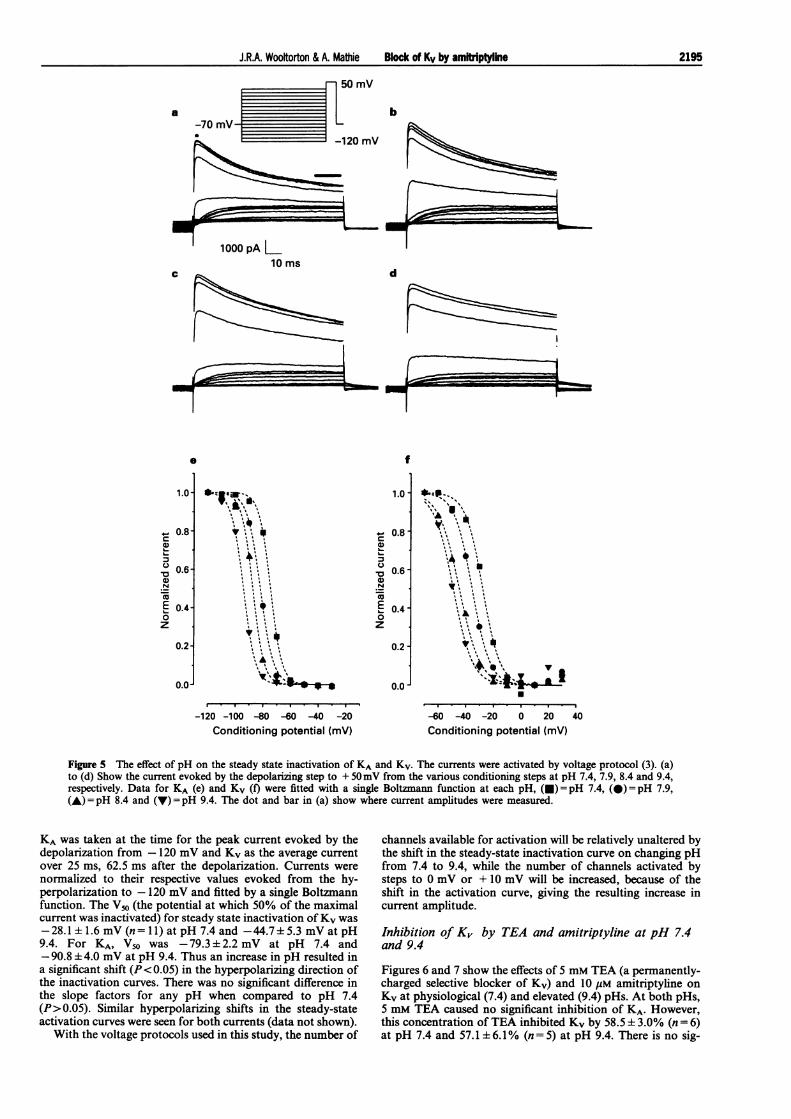
Figure 5 The effect of pH on the steady state inactivation of KA and Kv. The currents were activated by voltage protocol (3). (a)to (d) Show the current evoked by the depolarizing step to + 50mV from the various conditioning steps at pH 7.4, 7.9, 8.4 and 9.4,respectively. Data for KA (e) and Kv (f) were fitted with a single Boltzmann function at each pH, (0) = pH 7.4, (@)= pH 7.9,(A) = pH 8.4 and (V) = pH 9.4. The dot and bar in (a) show where current amplitudes were measured.
KA was taken at the time for the peak current evoked by thedepolarization from - 120 mV and Kv as the average currentover 25 ms, 62.5 ms after the depolarization. Currents were
normalized to their respective values evoked from the hy-perpolarization to -120 mV and fitted by a single Boltzmannfunction. The V50 (the potential at which 50% of the maximalcurrent was inactivated) for steady state inactivation ofKv was
-28.1 ± 1.6 mV (n=11) at pH 7.4 and -44.7+5.3 mV at pH9.4. For KA, V50 was -79.3+2.2 mV at pH 7.4 and-90.8 ± 4.0 mV at pH 9.4. Thus an increase in pH resulted ina significant shift (P< 0.05) in the hyperpolarizing direction ofthe inactivation curves. There was no significant difference inthe slope factors for any pH when compared to pH 7.4(P>0.05). Similar hyperpolarizing shifts in the steady-stateactivation curves were seen for both currents (data not shown).
With the voltage protocols used in this study, the number of
channels available for activation will be relatively unaltered bythe shift in the steady-state inactivation curve on changing pHfrom 7.4 to 9.4, while the number of channels activated bysteps to 0 mV or + 10 mV will be increased, because of theshift in the activation curve, giving the resulting increase incurrent amplitude.
Inhibition of K, by TEA and amitriptyline at pH 7.4and 9.4
Figures 6 and 7 show the effects of 5 mM TEA (a permanently-charged selective blocker of Kv) and 10 uM amitriptyline on
Kv at physiological (7.4) and elevated (9.4) pHs. At both pHs,5 mM TEA caused no significant inhibition of KA. However,this concentration ofTEA inhibited Kv by 58.5 t 3.0% (n = 6)at pH 7.4 and 57.1 + 6.1% (n = 5) at pH 9.4. There is no sig-
b
dc
f
0.0 -
m
I
2195
1.
I

J.RA. Wotoon & A. MaiNe Block of Kv by am-trpt
nificant difference between these values (P> 0.05). Note thatthe wash off was very similar at both pHs for TEA (Figure 6b).This suggests that the effect of TEA on potassium channels isnot pH-dependent, i.e. the binding of the drug molecule to thechannel protein is unaffected by pH.
At pH 7.4, 10 uM amitriptyline blocked Kv to a similardegree to that seen before (Wooltorton & Mathie, 1993):47.7 ± 5.0% (n = 10). In contrast to TEA, however, inhibitionof this current, in the same 10 cells, was significantly larger atthe elevated pH (79.7 ± 5.6%) illustrating a distinct pH-de-pendence for amitriptyline. It can also be seen that the timetaken for wash-off was much longer for amitriptyline at theelevated pH (Figure 6c).
Concentration-dependent inhibition of K, byamitriptyline at pH 7.4 and 9.4
Figure 8 shows a concentration-response curve for amitripty-line blocking Kv at both pH 7.4 and 9.4. Figure 8a illustratesthe percentage inhibition of Kv plotted against total drugconcentration. The KD for amitriptyline at pH 7.4 was11.9 ± 1.2 pM with minimum attainable current of- 1.0 ± 3.3% and slope of 0.90+ 0.05 as shown in Figure 4. AtpH 9.4, amitriptyline was about 25 times more potent with aKD of 0.46±0.08 pM, a minimum attainable current of16.8±4.2% and a slope of 0.98±0.13 (n=5-10 cells at eachconcentration). To confirm that this result was not unique toamitriptyline, similar experiments were carried out with cy-proheptadine and tacrine. At pH 9.4, 120 nM cyproheptadineinhibited Kv by 67% (n = 2) while 623 nM tacrine inhibited Kvby 25 ±2% (n = 3). These drug concentrations would produceno detectable inhibition of Kv at pH 7.4 (see Figure 4); how-
ever, they were calculated to contain the same concentration ofuncharged drug as 3 uM cyproheptadine (uncharged con-centration, 90 nM) and 50 gM tacrine (uncharged concentra-tion, 125 nM) at pH 7.4, concentrations which significantlyinhibit Kv at this pH (76±7%, n=3 and 40±4%, n=3, re-spectively, P< 0.05). These results illustrate the correlation
100
90
80
70
C60- TW.2 n=10250T
C
*40
TEA 5 mM Amitriptyline 10 gIM
a Control pH 9.4Control/wash pH 7.4
10CoMoAmit pH 7.4
10 gM Amit pH 9.4
a200 pA30 ms lVmV-70 mV -0 mV
Figue 7 Percentage inhibition of Kv by 5mM TEA and 10Mamitriptyline. The inhibition of Kv of amitriptyline is significantlygreater (P<0.05) at pH 9.4 compared to pH 7.4. Currents wereactivated using voltage protocol (2).
100
8040
:30r*
pH 7.4 pH 9.4
60
40
20
TEA TEA
pH 7.4 pH 9.4
Amit
60 s Amit
0
U-
a
=~ i %......1.... .....1.... .....1......I"....""I.... A\ i
.I.~~~~~~~~I.II
10 100 1000 10000 100 000
b Total concentration (nM)
10 100 1000
Uncharged concentration (nM)10 000
Figure 6 The effects of 10 M amitriptyline and 5mM TEA on Kv atpH 7.4 and 9.4 are shown. Currents in (a) were activated by voltageprotocol (2). In (b) and (c) (U) represent data at pH 7.4 and (0)data at pH 9.4. In this cell 10pM amitriptyline (Amit) inhibited Kvby 59% at pH 7.4 and 95% at pH 9.4. TEA (5mM) inhibited Kv by66% at both pHs. Note the slow wash of amitriptyline at pH 9.4.
FIgure 8 Concentration-dependent inhibition of Kv by amitriptylineat pH 7.4 (U) and pH 9.4 (0) are shown in (a). Data are fitted withequation [3]. (b) Shows the data from (a) plotted against unchargeddrug concentration calculated from equation [4]. Currents wereactivated by voltage protocol (1).
b1.2-
2 1.0-4-a0 0.8-a 0.6-*2 0.4-,, 0.2-U.
0.0-c
1.22 1.0-
0 0.8-0c 0.60
I 0.4&L 0.2U-
0.0-
2196
J.R.A. Wootorton & A. Mathie Block of Kv by amdrlptyne
between external uncharged drug concentration and potencyof block.
Figure 8b shows the percentage inhibition of amitriptylineplotted as a function of uncharged concentration. If the ex-ternal uncharged form of the drug was the only form whichcaused blockade ofKv then the curves for data obtained at thetwo pHs would be superimposed in Figure 8b. In fact, there issome discrepancy between the two curves (a discrepancy alsoseen when comparing the effectiveness of the same unchargedconcentration of tacrine at the two pHs, above). By taking thepercentage inhibition as a function of the concentration ofboth the uncharged molecules and the charged molecules, theapproximate contribution of each form can be calculated.Assuming that both species act at the same site but have dif-ferent affinities, then at 50% inhibition the equation:
{[u]/KU} + {[C]/KC} = 1 [5]holds, where KU and KC are the respective equilibrium dis-sociation constants of the uncharged and charged forms (seeHowe & Ritchie, 1991). By assuming that these are constant atthe different pHs, simultaneous equations can be solved toreveal an approximate potency ratio between the two forms:
a
At pH 7.4At pH 9.4Thus
119/KU+ 11781/KC= 1230/KU + 230/KC =1
KC/KU = 11551/111 = 103.4
This would suggest that the uncharged form of amitriptyline isapproximately 100 times more potent than the charged form.
Inhibition of Kv by N-methylainitriptyline
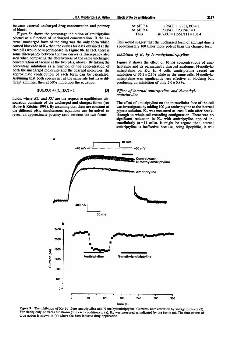
Figure 9 shows the effect of 10 gM concentrations of ami-triptyline and its permanently charged analogue, N-methyla-mitriptyline on Kv. In 4 cells, amitriptyline caused aninhibition of 36.2± 2.1% while in the same cells, N-methyla-mitriptyline was significantly less effective at blocking Kv,producing an inhibition of only 2.0± 0.8%.
Effect of internal amitriptyline and N-methyl-amitriptyline
The effect of amitriptyline on the intracellular face of the cellwas investigated by adding 100 pM amitriptyline to the internalpipette solution. Kv was measured at least 3 min after break-through to whole-cell recording configuration. There was nosignificant reduction in Kv with amitriptyline applied in-tracellularly (n= 11 cells). It might be argued that internalamitriptyline is ineffective because, being lipophilic, it will
10mV
-70 mV E1 - - -50 mV
% Control/washw N-methylamitriptyline
i Amitriptyline
400 pA
30 ms
0I
Amitriptyline N-methylamitriptyline
0 60 120 180 240 300 360
Time (s)Figure 9 The inhibition of Kv by 1OpM amitriptyline and N-methylamitriptyline. Currents were activated by voltage protocol (2).For clarity only 12 traces are shown (3 in each condition) in (a). Kv was measured as indicated by the bar in (a). The time course ofdrug action is shown in (b) where the bars indicate drug application.
b2400 -
2000 -
1600 -
1200 -
800 -
400-
0
ca,6-)
2197
J.R.A. Wooltorton & A. Mathie Block of Kv by amitriptyline
a
4001
aI-
100
0
b
5 10 15
800i
a
600
400
200
0 6 12 18 24
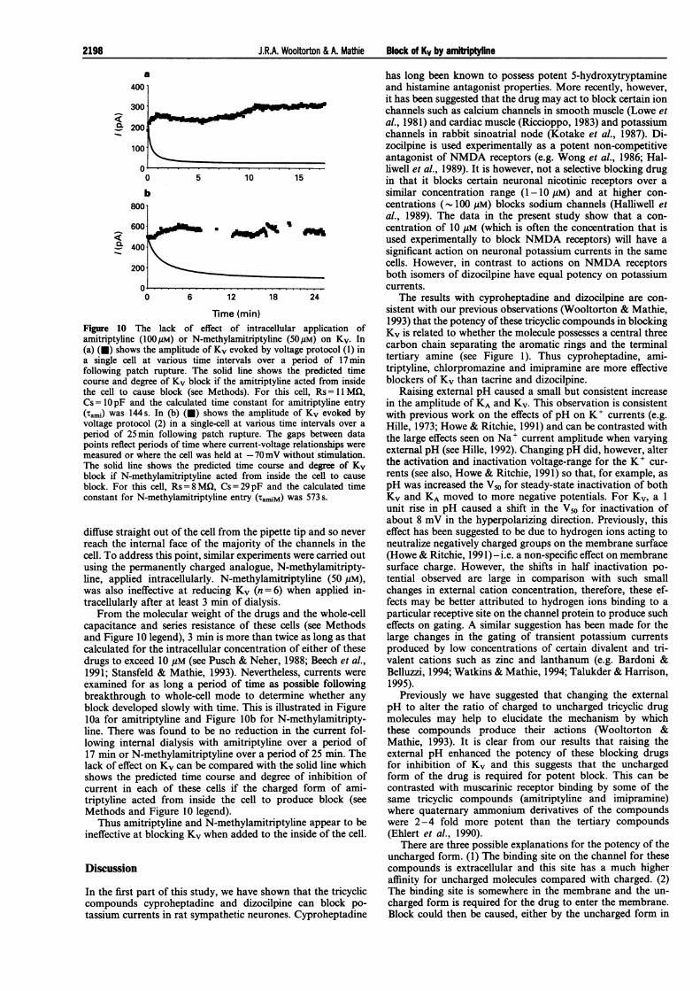
Time (min)Figure 10 The lack of effect of intracellular application ofamitriptyline (100pM) or N-methylamitriptyline (50pM) on Kv. In(a) (v) shows the amplitude of Kv evoked by voltage protocol (1) ina single cell at various time intervals over a period of 17minfollowing patch rupture. The solid line shows the predicted timecourse and degree of Kv block if the amitriptyline acted from insidethe cell to cause block (see Methods). For this cell, Rs = MCI,Cs=1OpF and the calculated time constant for amitriptyline entry(rami) was 144 s. In (b) (l) shows the amplitude of Kv evoked byvoltage protocol (2) in a single-cell at various time intervals over a
period of 25min following patch rupture. The gaps between datapoints reflect periods of time where current-voltage relationships weremeasured or where the cell was held at -70mV without stimulation.The solid line shows the predicted time course and degree of Kvblock if N-methylamitriptyline acted from inside the cell to cause
block. For this cell, Rs = 8 MQ, Cs = 29pF and the calculated timeconstant for N-methylamitriptyline entry (TAM) was 573 s.
diffuse straight out of the cell from the pipette tip and so neverreach the internal face of the majority of the channels in thecell. To address this point, similar experiments were carried outusing the permanently charged analogue, N-methylamitripty-line, applied intracellularly. N-methylamitriptyline (50 gM),was also ineffective at reducing Kv (n =6) when applied in-tracellularly after at least 3 min of dialysis.
From the molecular weight of the drugs and the whole-cellcapacitance and series resistance of these cells (see Methodsand Figure 10 legend), 3 min is more than twice as long as thatcalculated for the intracellular concentration of either of thesedrugs to exceed 10 gM (see Pusch & Neher, 1988; Beech et al.,1991; Stansfeld & Mathie, 1993). Nevertheless, currents wereexamined for as long a period of time as possible followingbreakthrough to whole-cell mode to determine whether anyblock developed slowly with time. This is illustrated in FigurelOa for amitriptyline and Figure lOb for N-methylamitripty-line. There was found to be no reduction in the current fol-lowing internal dialysis with amitriptyline over a period of17 min or N-methylamitriptyline over a period of 25 min. Thelack of effect on Kv can be compared with the solid line whichshows the predicted time course and degree of inhibition ofcurrent in each of these cells if the charged form of ami-triptyline acted from inside the cell to produce block (seeMethods and Figure 10 legend).
Thus amitriptyline and N-methylamitriptyline appear to beineffective at blocking Kv when added to the inside of the cell.
Discussion
In the first part of this study, we have shown that the tricycliccompounds cyproheptadine and dizocilpine can block po-tassium currents in rat sympathetic neurones. Cyproheptadine
has long been known to possess potent 5-hydroxytryptamineand histamine antagonist properties. More recently, however,it has been suggested that the drug may act to block certain ionchannels such as calcium channels in smooth muscle (Lowe etal., 1981) and cardiac muscle (Riccioppo, 1983) and potassiumchannels in rabbit sinoatrial node (Kotake et al., 1987). Di-zocilpine is used experimentally as a potent non-competitiveantagonist of NMDA receptors (e.g. Wong et al., 1986; Hal-liwell et al., 1989). It is however, not a selective blocking drugin that it blocks certain neuronal nicotinic receptors over asimilar concentration range (1-10 M) and at higher con-centrations (- 100 uM) blocks sodium channels (Halliwell etal., 1989). The data in the present study show that a con-centration of 10 yM (which is often the concentration that isused experimentally to block NMDA receptors) will have asignificant action on neuronal potassium currents in the samecells. However, in contrast to actions on NMDA receptorsboth isomers of dizocilpine have equal potency on potassiumcurrents.
The results with cyproheptadine and dizocilpine are con-sistent with our previous observations (Wooltorton & Mathie,1993) that the potency of these tricyclic compounds in blockingKv is related to whether the molecule possesses a central threecarbon chain separating the aromatic rings and the terminaltertiary amine (see Figure 1). Thus cyproheptadine, ami-triptyline, chlorpromazine and imipramine are more effectiveblockers of Kv than tacrine and dizocilpine.
Raising external pH caused a small but consistent increasein the amplitude of KA and Kv. This observation is consistentwith previous work on the effects of pH on K+ currents (e.g.Hille, 1973; Howe & Ritchie, 1991) and can be contrasted withthe large effects seen on Na+ current amplitude when varyingexternal pH (see Hille, 1992). Changing pH did, however, alterthe activation and inactivation voltage-range for the K+ cur-rents (see also, Howe & Ritchie, 1991) so that, for example, aspH was increased the V50 for steady-state inactivation of bothKv and KA moved to more negative potentials. For Kv, a 1unit rise in pH caused a shift in the V50 for inactivation ofabout 8 mV in the hyperpolarizing direction. Previously, thiseffect has been suggested to be due to hydrogen ions acting toneutralize negatively charged groups on the membrane surface(Howe & Ritchie, 1991) -i.e. a non-specific effect on membranesurface charge. However, the shifts in half inactivation po-tential observed are large in comparison with such smallchanges in external cation concentration, therefore, these ef-fects may be better attributed to hydrogen ions binding to aparticular receptive site on the channel protein to produce sucheffects on gating. A similar suggestion has been made for thelarge changes in the gating of transient potassium currentsproduced by low concentrations of certain divalent and tri-valent cations such as zinc and lanthanum (e.g. Bardoni &Belluzzi, 1994; Watkins & Mathie, 1994; Talukder & Harrison,1995).
Previously we have suggested that changing the externalpH to alter the ratio of charged to uncharged tricyclic drugmolecules may help to elucidate the mechanism by whichthese compounds produce their actions (Wooltorton &Mathie, 1993). It is clear from our results that raising theexternal pH enhanced the potency of these blocking drugsfor inhibition of Kv and this suggests that the unchargedform of the drug is required for potent block. This can becontrasted with muscarinic receptor binding by some of thesame tricyclic compounds (amitriptyline and imipramine)where quaternary ammonium derivatives of the compoundswere 2-4 fold more potent than the tertiary compounds(Ehlert et al., 1990).
There are three possible explanations for the potency of theuncharged form. (1) The binding site on the channel for thesecompounds is extracellular and this site has a much higheraffinity for uncharged molecules compared with charged. (2)The binding site is somewhere in the membrane and the un-charged form is required for the drug to enter the membrane.Block could then be caused, either by the uncharged form in
2198
I0 ffta
J.R.A. Wooftorton & A. Mathie Block of Kv by amirptylhe 2199
the membrane or the charged form gaining access to the insideof the channel. (3) The binding site (or access to it) is in-tracellular (for either charged or uncharged form) and theuncharged form is required for the drug to cross the mem-brane.
Our experiments where amitriptyline and N-methylami-triptyline were added to the internal solution and were in-effective would seem to provide strong evidence againstproposal (3), namely, that the binding site for these com-pounds is intracellular. This can be contrasted with the me-chanism of action of 4-aminopyridine, which is thought toblock K+ currents by binding to an intracellular binding site(e.g. Howe & Ritchie, 1991; Choquet & Korn, 1992; Stephenset al., 1994). Experiments where 4-aminopyridine or its per-manently charged analogue, 4-aminopyridine methiodide,were applied to the intracellular solution have shown clearlythat these drugs act on an intracellular site of the potassiumchannel mKy 1.1 to cause block (Stephens et al., 1994). Thissite is suggested to be at or near to the 'binding site' used byeither the N-terminal ball peptide or the fi subunit of in-activating K channels to cause N-type inactivation (see e.g.Zagotta et al., 1990; Pongs, 1992; Rettig et al.,1994). Fur-thermore, a similar mechanism and site of action has beenproposed for a number of other drugs that block potassiumchannels, in particular, quaternary ammonium ions actingfrom inside the cell (e.g Armstrong, 1971; French & Shouki-mas, 1981).
The major mechanism of action of local anaesthetic agentson sodium channels is also thought to involve the unchargedform of the molecule crossing the membrane, becomingcharged and then reaching its binding site on the channelprotein from the inside of the cell (e.g. Strichartz, 1973;Courtney, 1975; Narahashi & Frazier, 1975), although a dif-ferent mechanism must apply to the neutral local anaestheticagent, benzocaine (see Hille, 1992). From the results in thisstudy, we must propose a different mechanism of action forblock of Kv by the tricyclic compounds tested. The char-acteristics of block show a number of similarities with block ofIK(f) in rat melanotrophs by quinidine (Kehl, 1991) althoughquinidine increased the rate ofdecay of IK(f); an effect increasedby membrane depolarization. Block by quinidine did not seemto be due to the drug acting from inside the cell. Furthermore,block by quinidine was neither voltage- nor use-dependentwhich is similar to what we have previously reported for tri-cyclic block of Kv (Wooltorton & Mathie, 1993).
The simplest conclusion drawn from the results with qui-
nidine was that the quinidine binding site was on the externalface of the membrane (Kehl, 1991) similar to proposal (1)above. Our observation that the charged form of amitriptylinehas a finite blocking action; although calculated as beingaround 100 fold less potent than uncharged amitriptyline;provides some support for proposal (1), namely an externalbinding site with a high affinity for the uncharged form andsuggests proposal (2) is less likely to be correct.
Because the uncharged form of these compounds conferspotency and this form exists at low concentrations aroundneutral pH, the effectiveness of these compounds in inhibitingKv will be increased if extracellular pH becomes alkaline. Itcan be calculated that the uncharged concentration will beapproximately doubled for 0.3 of a pH unit rise in extracellularpH. It is noteworthy that activation of GABAA, AMPA/kai-nate and NMDA receptors can give rise to extracellular al-kaline shifts (Chesler & Kaila, 1992). These can be measuredwith pH microelectrodes as being 0.1 to 0.2 of a pH unit in sizeand this recorded response is thought to be only a fraction ofthe actual synaptic pH changes that occur due to the limitedtemporal and spatial resolution of such measurements (Chesler& Kaila, 1992).
In addition to the effects described here, it has been shownrecently that a number of the same tricyclic compounds arepotent blockers of calcium-activated K+ currents in rat cor-tical neurones (Lee et al.,1995). However, none of the com-pounds used is a selective blocker of potassium currents (seeabove and Wooltorton & Mathie, 1993), indeed all of them areused primarily in clinical or experimental situations for otherpharmacological actions which they possess. Despite this,block of K+ currents will be evident at therapeutic con-centrations ofmany of these tricyclic compounds (see Benet &Williams, 1990) and will be a major consideration at higherconcentrations. Block of K+ currents will prolong action po-tentials in both neuronal and cardiac tissue (Robbins & Sim,1990; Zhang et al., 1992) and this could easily account for theconvulsive and dysrythmic adverse actions of tricyclic com-pounds when they are taken in overdose.
The equipment used for these experiments was obtained on theMRC project grant and a BBSRC research grant. We are extremelygrateful to Dr F.J. Ehlert (U.C. Irvine) for the gift of N-methylamitriptyline.
References
ARMSTRONG, C.M. (1971). Interaction of tetraethylammonium ionderivatives with the potassium channels of giant axons. J. Gen.Physiol., 58, 413-437.
BARDONI, R. & BELLUZZI, 0. (1994). Modifications of A-currentkinetics in mammalian central neurones induced by extracellularzinc. J. Physiol., 479, 389-400.
BEECH, D.J., BERNHEIM, L., MATHIE, A. & HILLE, B. (1991).Intracellular Ca2+ buffers disrupt muscarinic suppression ofCa2+ current and M current in rat sympathetic neurons. Proc.Natl. Acad. Sci. U.S.A., 88, 652-656.
BENET, L.Z. & WILLIAMS, R.L. (1990). Design and optimisation ofdosage regimens; pharmacokinetic data. In Goodman & Gilman'sThe Pharmacological Basis of Therapeutics. 8th edition, ed.Gilman, A.G., Rall, T.W., Nies, A.S. & Taylor, P. pp. 1650-1735. Oxford: Pergamon Press.
BERNHEIM, L., BEECH, D.J. & HILLE, B. (1991). A diffusiblemessenger mediates one of the pathways coupling receptors toCa2+ channels in rat sympathetic neurons. Neuron, 6, 859-867.
CHESLER, M. & KAILA, K. (1992). Modulation of pH by neuronalactivity. Trends Neurosci., 15, 396- 402.
CHOQUET, D. & KORN, H. (1992). Mechanism of 4-aminopyridineaction on voltage-gated potassium channels in lymphocytes. J.Gen. Physiol., 99, 217-240.
CLARK, S. & MATHIE, A. (1995). A hyperpolarisation-activatedchloride current in acutely isolated rat superior cervical ganglion(SCG) neurons. J. Physiol., 485, 48-49P.
COOK, N.S. & QUAST, U. (1990). Potassium channel pharmacology.In Potassium Channels, Structure, Classification, Function andTherapeutic Potential. ed. Cook, N.S. pp. 181-255. Chichester:Ellis Horwood Ltd.
COURTNEY, K.R. (1975). Mechanism of frequency-dependentinhibition of sodium currents in frog myelinated nerve by thelidocaine derivative GEA 968. J. Pharmacol. Exp. Ther., 195,225-236.
EHLERT, F.J., DELEN, F.M., YUN, S.H. & LIEM, H.A. (1990). Theinteraction of amitriptyline, doxepin, imipramine and their N-methyl quarternary ammonium derivatives with subtypes ofmuscarinic receptors in brain and heart. J. Pharmacol. Exp.Ther., 253, 13- 19.
FRENCH, R.J. & SHOUKIMAS, J.J. (1981). Blockage of squid axonpotassium conductance by internal tetra-N-alkylammonium ionsof various sizes. Biophys. J., 34, 271-291.
HALLIWELL, R.F., PETERS, J.A. & LAMBERT, J.J. (1989). Themechanism of action and pharmacological specificity of theanticonvulsant NMDA antagonist MK-801: a voltage clampstudy on neuronal cells in culture. Br. J. Pharmacol., 96, 480-494.
2200 J.R.A. Wooltorton & A. Mathie Block of Kv by amftiptyline
HALLIWELL, J.V. (1990). K+ channels in the central nervous system.In Potassium Channels, Structure, Classification, Function andTherapeutic Potential. ed. Cook, N.S. pp. 348-381. Chichester:Ellis Horwood Ltd.
HAMILL, O.P., MARTY, A., NEHER, E., SAKMANN, B. & SIGWORTH,F.J. (1981). Improved patch-clamp techniques for high-resolutioncurrent recording from cell and cell-free membrane patches.Pflugers Arch., 391, 85-100.
HILLE, B. (1973). Potassium channels in myelinated nerve. Selectivepermeability to small cations. J. Gen. Physiol., 61, 669-686.
HILLE, B. (1992). Ionic Channels of Excitable Membranes. 2ndEdition. Sunderland MA: Sinauer.
HOWE, J.R. & RITCHIE, J.M. (1991). On the active form of 4-aminopyridine: block of K+ currents in rabbit Schwann cells. J.Physiol., 433, 183-206.
KEHL, S.J. (1991). Quinidine-induced inhibition of the fast transientoutward K+ current in rat melanotrophs. Br. J. Pharmacol., 103,1807- 1813.
KOTAKE, H., SAITOH, M., OGINO. K., HIRAI, S., MATSUOKA, S.,HASEQAWA, J. & MASHIBA, H. (1987). On the ionic mechanismof cyproheptadine-induced bradycardia in a rabbit sinoatrialnode preparation. Eur. J. Pharmacol., 139, 307- 313.
LAURENCE, D.R. & BENNETT, P.N. (1992). Clinical Pharmacology.7th Edition. Edinburgh: Churchill Livingstone.
LEE, K., ROWE, I.C.M. & ASHFORD, M.L.J. (1995). The effects ofneuroleptic and tricyclic compounds on BKca channels in ratisolated cortical neurones. J. Physiol., 483, 67 - 68P.
LOWE, D.A., MATTHEWS, E.K. & RICHARDSON, B.P. (1981). Thecalcium antagonistic effects of cyproheptadine on contraction,membrane electrical events and calcium influx in the guinea-pigtaenia coli. Br. J. Pharmacol., 74, 651-663.
NARAHASHI, T. & FRAZIER, D.T. (1975). Site of action and activeform of procaine in squid giant axons. J. Pharmacol. Exp. Ther.,194, 506-513.
PONGS, 0. (1992). Molecular biology of voltage-dependent potas-sium channels. Physiol. Rev., 72, 569- 588.
PUSCH, M. & NEHER, E. (1988). Rates of diffusional exchangebetween small cells and a measuring patch pipette. Pflzigers Arch.,411, 204-211.
RETTIG, J., HEINEMANN, S.H., WUNDER, F., LORRA, C., PARCEJ,D.N., DOLLY, J.O. & PONGS, 0. (1994). Inactivation properties ofvoltage-gated K+ channels altered by presence of fl-subunit.Nature, 369, 289-294.
RICCIOPPO, N.F. (1983). Effects of cyproheptadine on electrophy-siological properties of isolated cardiac muscle of dogs andrabbits. Br. J. Pharmacol., 80, 335-341.
ROBBINS, J. & SIM, J.A. (1990). A transient outward current inNG108-15 neuroblastoma x glioma hybrid cells. Pflugers Arch.,416, 130-137.
STANSFELD, C. & MATHIE, A. (1993). Recording membrane currentof peripheral neurones in short-term culture. In Electrophysiol-ogy: a Pratical Approach. ed. Wallis, D.I. pp.3-28. Oxford: IRLPress.
STEPHENS, G.J., GARRATT, J.C,. ROBERTSON, B. & OWEN, D.G.(1994). On the mechanism of 4-aminopyridine action on thecloned mouse brain potassium channel mKvl .1. J. Physiol., 477,187- 193.
STRICHARTZ, G.R. (1973). The inhibition of sodium currents inmyelinated nerve by quarternary derivatives of lidocaine. J. Gen.Physiol., 62, 37- 57.
TALUKDER, G. & HARRISON, N.L. (1995). On the mechanism ofmodulation of transient outward current in cultured rathippocampal neurones by di- and trivalent cations. J.Neurophysiol., 73, 73-79.
WATKINS, C.S. & MATHIE, A. (1994). Modulation of the gating ofthe transient outward potassium current of rat isolated cerebellargranule neurons by lanthanum. Pflugers Arch., 428, 209-216.
WONG, E.H.F., KEMP, J.A., PRIESTLEY, T., KNIGHT, A.R., WOO-DRUFF, G.N. & IVERSEN, L.L. (1986). The anticonvulsant MK-801 is a potent N-methyl-D-aspartate antagonist. Proc. Nati.Acad. Sci. U.S.A., 83, 7104-8.
WOOLTORTON, J.R.A. & MATHIE, A. (1993). Block of potassiumcurrents in rat sympathetic neurones by tricyclic antidepressantsand structurally related compounds. Br. J. Pharmacol., 110,1126-1132.
WOOLTORTON, J.R.A. & MATHIE, A. (1994). Block of the delayedrectifier & transient outward potassium currents in rat isolatedsympathetic neurons by cyproheptadine & dizocilpine. Can. J.Physiol. Pharmacol., 72 (Suppl. 1), 523
WOOLTORTON, J.R.A. & MATHIE, A. (1995). Potent inhibition of thedelayed rectifier potassium current in rat sympathetic neurons byextracellular amitriptyline requires the drug to be uncharged. Br.J. Pharmacol., 114, 31P.
ZAGOTTA, W.N., HOSHI, T. & ALDRICH, R.W. (1990). Restoration ofinactivation in mutants of Shaker potassium channels by apreparation derived from ShB. Science, 250, 568-571.
ZHANG, Z.-H., FOLLMER, C.H., SARMA, J.S.M., CHEN, F. & SINGH,B.N. (1992). Effect of ambasilide, a new class III agent, on plateaucurrents in isolated guinea-pig ventricular myocytes: block ofdelayed outward potassium current. J. Pharmacol. Exp. Ther.,263, 40-48.
(Received March 21, 1995Revised June 21, 1995
Accepted June 23, 1995)