neurohumoral behavior in cardiac pacemaker patients controlled · tance control, as well as cardiac...
TRANSCRIPT

284 June 2000
Progress in Biomedical Research
Introduction
Under normal circumstances, physiologic chronotropyis influenced by the sympathetic and parasympatheticnervous system, permitting cardiovascular adaptationto various situations such as rest, postural changes,physical effort, defecation, and others. Reflex brady-cardia and tachycardia, which are essential in these cir-cumstances and mainly triggered by arterial barorecep-tors and the cardiopulmonary reflex, are dependent onpressure changes within the cardiac chambers and thegreat vessels (aorta and pulmonary artery).The autonomic nervous system is crucial for the regu-lation of heart rate, contractility, and vascular resis-tance control, as well as cardiac output, blood flow dis-tribution, and blood pressure regulation.Under physiologic conditions, the most important fac-tor regulating myocardial contractility is the norepi-nephrine concentration in cardiac sympathetic nerveendings. The most rapid modifications in cardiac con-
tractility have been observed to occur after changes inthe intensity of adrenergic nerve impulses [1].When stimulated, the adrenal medulla releases epi-nephrine, which is carried to the heart where it stimu-lates beta-adrenergic receptors, increasing cardiac con-tractility. This mechanism is not as fast as norepineph-rine release from cardiac nerve endings, but it can bevital to preserve cardiac output in some particular con-ditions.If we consider stroke volume to be constant and car-diac output to be linearly correlated with heart rate, theability to modify the heart rate will be an importantmechanism in the control of cardiac output [2].Therefore, the importance of the heart rate in main-taining cardiac output in patients with chronotropicincompetence is reflected in their inability to increasecardiac output, even when myocardial contractility isentirely normal. In those patients, a rate-adaptive pace-
Neurohumoral Behavior in Cardiac Pacemaker Patients Controlled bythe Autonomic Nervous System with Closed Loop Stimulation
S. A. D. NISHIÓKA, M. MARTINELLI Fº, H. LOPES, F. CONTIM, R. COSTAUniversity of Sao Paulo, Medical School, Heart Institute (InCor), Sao Paulo, Brazil
Summary
The current goal for sensor-driven pacing is to find a physiological system to correct chronotropic incompetence.The objectives of this study were to evaluate changes in the heart rate effected by sympathetic sensor-driven pac-ing and to compare these with normal sinus function. Fifteen patients with AV block and a pacemaker controlledby Closed Loop Stimulation were studied. Six were female, 9 male, with ages ranging from 37 to 80 years. Theywere divided into Group I (8 patients with chronotropic incompetence) and Group II (7 patients with normal sinusfunction). All patients performed the Valsalva maneuver and tilt table test under catecholamine and renine dosages.For the 1st stage, the programmed pacing mode was: DDD, LR = 60 ppm, URL = 0.85 * (220 - age); the 2nd stagewas in DDDR for Group I and VVIR for Group II. Tilt table test: In group I, heart rate variations occurred only inDDDR (after inclining). In Group II, heart rate changes were similar for both modes. For Group I, cathecholaminedosages were higher in DDD (p < 0.05) than in DDDR. The phase II and IV (Valsalva maneuver) did not changein Group I, DDD, but had near physiological behavior with sensor activated. The sympathetic SDP provided neu-rohumoral adjustment for patients with AV block and chronotropic incompetence.
Key words
Heart rate response, artificial cardiac stimulation, chronotropic incompetence, Closed Loop Stimulation, circulat-ing catecholamines

June 2000 285
Progress in Biomedical Research
tricular block, with ages ranging from 37 to 80 years(mean 54.7 years) were prospectively studied. Theyunderwent a pacemaker implantation or pacemakerreplacement [7] between 09/97 to 03/99. Thesepatients were divided into 2 groups. Group I consistedof 8 patients with a third degree atrioventricular blockplus sick sinus syndrome. Group II consisted of 7patients with normal sinus function and a third degreeatrioventricular block. Patients' NYHA functionalclasses for heart failure and heart disease are describedon Table 1.Clinical follow-up for Group I patients ranged from 14to 31 months (mean 22.3), while Group II patientswere followed for 2 months.All the patients had normal left-ventricular function asobserved by echocardiography (left-ventricular ejec-tion fraction (LVEF) > 0.60), except for 2 patients fromGroup II: one patient had ischaemic cardiomyopathy(LVEF = 0.52) and another had aortic valve disease(LVEF = 0.43).
Study DesignThe first stage of the study consisted of performingexaminations thirty days after implanting the pace-maker and programming it to a DDD mode, with abasic rate of 60 ppm and an upper tracking rate of0.85 * (220 - age). All patients underwent tilt table test-ing (TTT) with plasma epinephrine (EP), norepineph-
maker can guarantee that an adequate heart rate ismaintained [3,4].Among the many rate-adaptive artificial heart stimula-tion systems currently in use, we will evaluate the per-formance of a physiologic sensor model, called a sym-pathetic biosensor, that is able to capture the intracavi-tary myocardial signals, and which uses myocardialcontractility as an indicator of the sympathetic tone [5].
Objective
The aims of this study were: • to compare this pacemaker's biosensor heart-rate
response with sinus node performance in patientswith normal chronotropic response, and
• to evaluate the heart rate changes due to sensorresponse to modifications in myocardial contractili-ty in patients with chronotropic incompetence.
These systems will be evaluated using tests that pro-duce changes in heart dynamics, thus inducing reflexresponses by the parasympathetic and sympathetic ner-vous system. In order to do that, catecholamine andplasma renin activity measurements will be conductedbefore and after tilt table testing. A Valsalva maneuverwill also be performed for all patients [6].
Methodes
The artificial heart stimulation system implanted wasthe INOS2 CLS (Biotronik, Germany), a biosensorgoverned by the ventricular contractions that providethe heart rate autoregulation [5] (Figure 1).
PatientsFifteen patients with an advanced/complete atrioven-
Figure 1. Closed loop system.
Table 1. Studied patients, cardiomyopathy and NYHA class.

286 June 2000
Progress in Biomedical Research
rine (NE), and plasma renin activity (PRA) measure-ments undertaken during baseline (after one hour of bedrest) and after a 60º inclination (Figure 2). After the endof the TTT, a Valsalva maneuver was performed.Immediately after this, Group I patients had their pace-maker programmed to DDDR whereas Group IIpatients were reprogrammed to VVIR. Other thanreprogramming the mode, the rest was unchanged.In a 15-day follow-up after sensor activation, theexaminations were repeated with the new pacing modeused in the second stage of the study.The pacemaker programming mode was maintained inDDDR for Group I patients during follow-up through04/00. Group II had their pacemaker sensor deactivat-ed while atrioventricular synchronization took place(DDD mode).
Tilt Table TestingIn present practice, tilt table testing is used to assesscardiovascular changes related to postural modifica-tions, as well as to evaluate syncope of unknown etiol-ogy [8].The degree of postural hypotension, the heart rateresponse, and the circulating catecholamine levelswere evaluated after a bed-rest period which was fol-lowed by tilting to an upright position in order to eval-uate sensor rate response in both groups. This wasdone in both the first and second stages of the study. TTT was undertaken after the patient had rested on hisor her back for 1 hour. After this, the patient was thentilted upright at 60º.
Blood Sample CollectionAfter the venous puncture, the patient was kept in bed
in a calm setting with the lights turned low. Plasma EPand NE were taken in cooled flasks. PRA was alsomeasured during baseline. New blood samples weretaken 10 minutes after tilting upright in order to mea-sure EP, NE and PRA.
Plasma Catecholamine MeasurementsEpinephrine and norepinephrine were measured byhigh performance liquid chromatography [9].
Measure of Plasma Renin Activity PRA was measured by radioimmunoassay for angiotensine I in the presence of angiotensinase inhibitors.
Valsalva ManeuverThe Valsalva maneuver is normally performed in dailyactivities such as lifting weights, coughing, defecation,vomiting, pushing heavy objects and the expulsiveperiod during childbirth. It consists of a forced exhala-tion against a closed glottis, with a subsequent rise inintrathoracic pressure (ITP). The increase in ITP pro-motes sudden changes in preload and afterload. As thepressure increases, it is transferred to the heart cham-bers and great vessels, leading to an increased pressurein the right atrium and intra-thoracic veins with conse-quent decline in venous return. According to Frank-Starling's law, the VM produces a decrease in myocar-dial fiber distension and a drop in stroke volume [10]. The VM was accomplished with the patient in a supineposition. After a regular inspiration, the patients blowsinto a manometer to maintain a level of pressure rang-ing from 20 to 40 mmHg for 10 to 30 seconds. Thispressure is enough to cause a drop in pulse pressureand a reflex peripheral vasoconstriction. Healthy indi-viduals can attain up 40 to 60 mmHg of intrathoracicpressure during the maneuver.
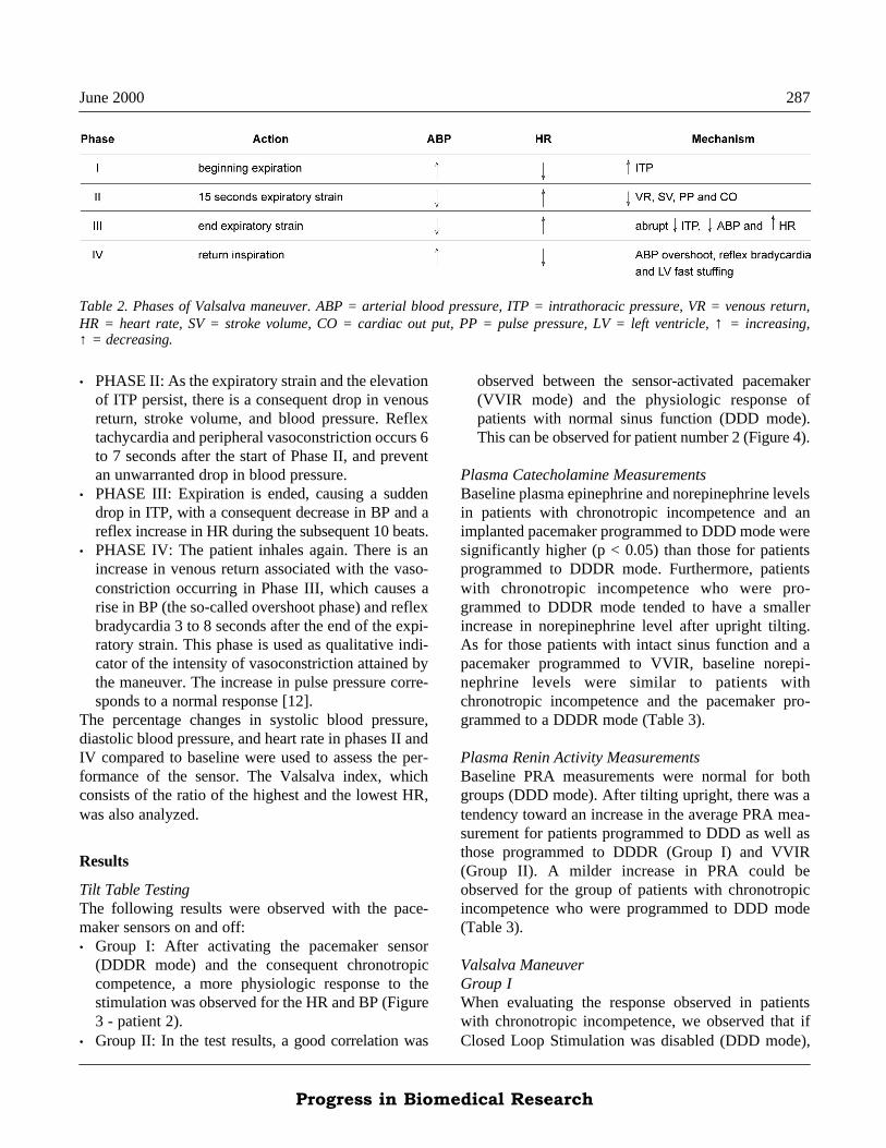
Phases of the Valsalva Maneuver The normal response to the VM has been divided in 4phases [11] (Table 2): • PHASE I: With the initial expiration, the increase in
intrathoracic and intraabdominal pressure is trans-mitted to the left ventricle (LV) and aorta, producingan increase in stroke volume (SV) and transport ofblood into the peripheral arteries. The sum of theseeffects produces a rise in blood pressure (BP). Pulsepressure is unchanged, since there is a rise in bothdiastolic and systolic pressures. The transientincrease in BP leads to reflex bradycardia.
Figure 2. Study designer.
June 2000 287
Progress in Biomedical Research
Table 2. Phases of Valsalva maneuver. ABP = arterial blood pressure, ITP = intrathoracic pressure, VR = venous return,HR = heart rate, SV = stroke volume, CO = cardiac out put, PP = pulse pressure, LV = left ventricle, ↑ = increasing,↑ = decreasing.
• PHASE II: As the expiratory strain and the elevationof ITP persist, there is a consequent drop in venousreturn, stroke volume, and blood pressure. Reflextachycardia and peripheral vasoconstriction occurs 6to 7 seconds after the start of Phase II, and preventan unwarranted drop in blood pressure.
• PHASE III: Expiration is ended, causing a suddendrop in ITP, with a consequent decrease in BP and areflex increase in HR during the subsequent 10 beats.
• PHASE IV: The patient inhales again. There is anincrease in venous return associated with the vaso-constriction occurring in Phase III, which causes arise in BP (the so-called overshoot phase) and reflexbradycardia 3 to 8 seconds after the end of the expi-ratory strain. This phase is used as qualitative indi-cator of the intensity of vasoconstriction attained bythe maneuver. The increase in pulse pressure corre-sponds to a normal response [12].
The percentage changes in systolic blood pressure,diastolic blood pressure, and heart rate in phases II andIV compared to baseline were used to assess the per-formance of the sensor. The Valsalva index, whichconsists of the ratio of the highest and the lowest HR,was also analyzed.
Results
Tilt Table TestingThe following results were observed with the pace-maker sensors on and off:• Group I: After activating the pacemaker sensor
(DDDR mode) and the consequent chronotropiccompetence, a more physiologic response to thestimulation was observed for the HR and BP (Figure3 - patient 2).
• Group II: In the test results, a good correlation was
observed between the sensor-activated pacemaker(VVIR mode) and the physiologic response ofpatients with normal sinus function (DDD mode).This can be observed for patient number 2 (Figure 4).
Plasma Catecholamine MeasurementsBaseline plasma epinephrine and norepinephrine levelsin patients with chronotropic incompetence and animplanted pacemaker programmed to DDD mode weresignificantly higher (p < 0.05) than those for patientsprogrammed to DDDR mode. Furthermore, patientswith chronotropic incompetence who were pro-grammed to DDDR mode tended to have a smallerincrease in norepinephrine level after upright tilting.As for those patients with intact sinus function and apacemaker programmed to VVIR, baseline norepi-nephrine levels were similar to patients withchronotropic incompetence and the pacemaker pro-grammed to a DDDR mode (Table 3).
Plasma Renin Activity MeasurementsBaseline PRA measurements were normal for bothgroups (DDD mode). After tilting upright, there was atendency toward an increase in the average PRA mea-surement for patients programmed to DDD as well asthose programmed to DDDR (Group I) and VVIR(Group II). A milder increase in PRA could beobserved for the group of patients with chronotropicincompetence who were programmed to DDD mode(Table 3).
Valsalva Maneuver Group IWhen evaluating the response observed in patientswith chronotropic incompetence, we observed that ifClosed Loop Stimulation was disabled (DDD mode),

288 June 2000
Progress in Biomedical Research
phases II and IV of the Valsalva maneuver could not bewell defined. However, after activation of Closed LoopStimulation (DDDR mode), it was possible not only todefine all Valsalva maneuver phases but also to main-tain a physiological response in phases I, II and III. Inone patient a slight paradoxical HR response could benoted at the start of phase IV (Figure 5).
Group II This group of patients presented an almost physiologi-cal response to the Valsalva maneuver during each of itsphases when programmed to the VVIR mode comparedto the DDD mode. Although the curve behavior wassimilar under both conditions, there was a slight para-doxical HR response during phase IV of the ValsalvaManeuver. It is interesting to note that, in this group, the2 patients who had left-ventricular dysfunction showedan abnormal response to the Valsalva maneuver (a"square wave" response), which remained unchanged inboth situations (patients 9 and 14, Figure 6).
Valsalva Index For Group I, the mean value was near 1.0 in the DDDmode, indicating a flat heart rate trend without anyresponse to the Valsalva maneuver. In the DDDR
mode, the Valsalva index increased to 1.4. For GroupII, the mean value of the Valsalva index was 1.3 inDDD as well as in VVIR (Table 4).
Discussion
The present study was based on the principle that aclinical indication of a new stimulation system, a heartrate responsive pacing driven by the sympathetic tone,demands the maximum advantage of knowledge, alongwith the favorable general behavior shown by manyauthors [13]. More consistent information concerningthe intrinsic mechanisms of the pacing system, mainlyinvolving the neurohumoral changes, are still neces-sary to improve its application [14].First of all, our initial expectation was justified by thegeneral findings of this study: The VVIR pacing mode(Group II) provided similar behavior to the physiolog-ical stimulation (DDD mode with normal sinus func-tion) during resting conditions, upright tilting, and theValsalva maneuver. Two patients with chronotropicincompetence had a more physiologic response duringTTT and the Valsalva maneuver when the pacemakersensor was activated (DDDR mode). The inclusion of Valsalva maneuver in our methodolo-
Figure 3. Tilt table testing (Patient 6 - Group I). A: inactivesensor (DDD, C mode). In this situation, the heart rate didnot increase after inclination at 60°. B: active sensor(DDD,R mode). We observed an increase in heart rate afterinclination at 60° and also arterial blood pressureincreased.
Figure 4. Tilt table testing (Patient 13 - Group II). A: inac-tive sensor (DDD, C mode). B: active sensor (VVI, R mode).In both conditions A (DDD,C mode) and B (VVI,R mode),there was an increase in heart rate and arterial blood pres-sure after inclination at 60°.

June 2000 289
Progress in Biomedical Research
gy in order to try to reproduce daily activities allowedadditional clinical outcomes to be obtained. The para-doxical HR response observed during phase IV of theValsalva maneuver can be related to an increase inmyocardial contractility recognized by the sensor. Theincrease in contractility is produced by a rise in venousreturn together with an increase in peripheral resis-tance [15]. In spite of the paradoxical rise in HR dur-ing phase IV of the Valsalva maneuver, patientsremained asymptomatic during the entire examination. The Valsalva Index values observed in Group I (DDDmode) were a consequence of an unchanged heart rateduring phases II and IV of Valsalva maneuver, butunder DDDR-mode sensor activation there was a ten-dency to the index normalization. In Group II, we
observed similar variations in phases II and IV for bothconditions (DDD and VVIR modes), providing a nearnormal index value. Concerning the catecholamine behavior, we observedinteresting responses in both groups. In Group II(NSF), the NE and EP plasma levels, showed normaland similar values during resting and tilting conditions(DDD and VVIR modes). Group I patients had anabnormal plasma catecholamine level that increasedduring resting conditions (DDD mode), which indi-cates an important sympathetic influence. Under tilt-ing, the plasma level rose less than expected. Yet, dur-ing activated sensor (DDDR mode), NE and EP restinglevels normalized. This is similar to the observed NSFpatient's behavior.
Table 3. Epinephrine (EP), norepinephrine (NE) and plasma renin activity (PRA) plasmatic values obtained in patients ofGroups I and Groups II.

290 June 2000
Progress in Biomedical Research
Figure 5. Phases of Valsalva maneuver (patient 3) with AVblock and chronotropic incompetence (Group I). A: inactivesensor (DDD,C mode). B: active sensor (DDD,R mode).
Figure 6a. Phases of Valsalva maneuver (patient 9) with AVblock and NSF (Group II). A: inactive sensor (DDD,Cmode). B: active sensor (VVI,R mode).
Figure 6b. Phases of Valsalva maneuver (patient 14) withAV block and NSF (Group II). A: inactive sensor (DDD,Cmode). B: active sensor (VVI,R mode).Table 4. Mean value of the Valsalva index in Group I and II.

June 2000 291
Progress in Biomedical Research
[5] Schaldach M. What is closed loop stimulation? Prog BiomedRes 1998; 2(3): 49-55.
[6] Chirst T, Brattström A, Kühn H, et al. Effect of circulatingcathecholamines on the pacing rate of the closed loop stimu-lation pacemaker. Prog Biomed Res. 1998; 3(3): 143-6.
[7] Lukl J, Doupal V, Sovova E, et al. Incidence and significanceof chronotropic incompetence in patients with indications forprimary pacemaker implantation or pacemaker replacement.PACE. 1999; 22: 1284-91.
[8] Kenny RA, Ingram A, Bayliss J. Head up tilt: a useful toll forinvestigating syncope. Lancet. 1986; 2: 1352-54.
[9] Goldstein DS, Feuerstein G, Izzo Jr. JL, et al. Validity andreliability of liquid chromatography with eletrochemicaldetection for measuring plasma levels of norepinephrine inman. Life Sciences. 1981. 28: 467-75.
[10] Porth CJM, Bamrah VS. The Valsalva maneuver: mecha-nisms and clinical implications. Heart Lung. 1987; 13: 507.
[11] Sharpey-Shafer EP. Effect of respiratory acts on circulation.In: Handbook of Physiology. Section 2: Circulation III.American Physiological Society, Washington DC, USA,1965: 1975.
[12] Sharpey-Shafer EP. Effects of Valsalva maneuver on the nor-mal and failing circulation. Br Med J. 1955; 1: 693.
[13] Andrade JCS. Cardiac contractility sensor evaluation in aDDDR system - a multicenter study. Prog Biomed Res. 1998;3(3): 137-42.
[14] Graux P, Guyomar Y, Heuls S, et al. Closed loop stimulationand neurocardiogenic syncope. Prog Biomed Res. 1999; 4(4):449-51.
[15] Katz AM. Regulation of myocardial contractility 1958-1983.J Am Coll Cardiol. 1983; 1: 126.
These findings clearly showed that patients withchronotropic incompetence were not able to increasethe heart rate according to the humoral release whenthe sensor was deactivated (DDD mode). In addition,the significant decrease in plasma catecholamine levelsduring sensor activation (DDDR mode) reinforced ouroutcomes.In conclusion, the present study, which was proposedin order to evaluate new findings concerning a sympa-thetically-mediated pacing system, seems to have beenan important contribution to the knowledge of theintrinsic mechanism involved in its documented phys-iological performance.
References
[1] Beierholm EA, Grantham RN, O'Keefe DD, et al. Effects ofacid-base changes, hypoxia, and catecholamines on ventricu-lar performance. Am J Physiol. 1975; 228: 1555.
[2] Mitchell JH, Wallace AG, Skinner Jr. NS. Intrinsic effects ofheart rate on left ventricular performance. Am J Physiol.1963; 205: 42.
[3] Narahara KA, Blettel ML. Effect of rate on left ventricularvolumes and ejection fraction during chronic ventricular pac-ing. Circulation. 1983; 67: 323.
[4] Ströbel JS, Kay GN. Programming of sensor driven pace-makers. Cardiol Clin. 2000; 18(1): 157-76.