nematode panagrellus redivivus
TRANSCRIPT

Nucleic Acids Research, Vol. 20, No. 7 1623-1628
Unusual features of the retroid element PAT from thenematode Panagrellus redivivus
Yves de Chastonay+, Heinz Felder, Christopher Link1, Pierre Aeby2, Heinz Tobler andFritz MulIer*Institute of Zoology, University of Fribourg, Perolles, CH-1700 Fribourg, Switzerland, 1Department ofBiology, University of Denver, Colorado, USA and 2Baxter Diagnostic, BonnstraBe 9, CH-3186Dudingen, Switzerland
Received December 30, 1991; Revised and Accepted February 25, 1992
ABSTRACTThe PAT retroid transposable elements differ fromother retroids in that they have a 'split direct repeat'structure, i.e., an internal 300bp sequence is foundrepeated, about one half at each element extremity. Avery abundant transcript of about 900nt, the start ofwhich maps to the preferentially deleted portion of PATelements, is detected on total Panagrellus redivivusRNA bearing Northern blots. A potentiallycorresponding ORF encodes a protein of 265 residueshaving a carboxy terminal Cystein motif, believed to beexclusively characteristic of the GAG protein in retroidelements. A much fainter, 1 800nt long transcript, is alsodetected on Northern blots and maps slightlydownstream of the first ORF. The predicted proteinsequence of this region bears motifs typical of reversetranscriptase and RNaseH, as found in the Pol genesof retroid elements. Peptide motif similarities aregreatest with the DIRS-1 element derived fromDictyostelium discoideum. The possibility of using PATelements as transposon tagging system forCaenorhabditis elegans is discussed.
INTRODUCTIONMobile genetic elements have been found in a wide variety oforganisms, ranging from prokaryotes to higher eukaryotes andhumans (for review, see 1). Two main classes of transposableelements can be distiguished, for each of which a different modeof transposition, as well as several structural features, arecharacteristic. The first class, retroid elements, are mobilizedvia full length RNA molecules and require reverse transcriptionprior to possible integration in the genome (2, 3). These can besubdivided into long terminal repeat (LTR) containing and non-LTR containing elements , both of which, however, contain groupspecific antigen (GAG) and RNA dependent DNA polymerase(RT) (4, 5). First strand cDNA is usually primed by a tRNAmolecule and synthesis proceeds outwards through the 5' LTR
EMBL accession no. X60774
(for review, see 6). The LTRs then play a key role by allowingordered inter- or intrastrand exchanges during reversetranscription (7, 8). In the case of non-LTR containing elements,several priming possibilities that avoid the need for strandexchanges have been reported, including priming by host DNAat the insertion site, a mechanism involving terminal uridinetransferase enzyme (for review, see 5) and protein priming (9).For the non-retroid counterparts, all transposition intermediatesappear to be composed of DNA. A distinctive structural featureof these elements is the presence of short inverted repeats (IRs)at the element termini and their encoding a so-called transposasefunction (4). One element, DIRS-1 of Dictyostelium discoideum,shares properties of both classes (i.e., IRs and RT), having ledto the proposition of an intramolecular priming mechanism forretroid replication (10).
Representatives of either class have been found in the phylumNematoda, among which the non-retroid Tc family ofCaenorhabditis elegans (11) and the retrovirus-like element TASof Ascaris lumbricoides (12). A further nematode transposon,the PAT element of Panagrellus redivivus, was discovered upontransposition in a C. elegans unc-22 gene homologue, the locusof mutation to the twitcher phenotype (13). Like most transposingentities, the PAT element belongs to the repetitive DNA fraction.Distribution is random and elements are represented between 10and 50 times per haploid genome, depending on strain. In thispaper we present the overall structure, relevant open readingframes (ORFs) and transcription of PAT, in an attempt to classifythe element among other transposing entities and to evaluateperspectives.
MATERIALS AND METHODSNematode cultures, DNA isolation, restriction digestion, TBEelectrophoresis and Southern blot hybridizationP. redivivus strain Sc was cultivated as described for C. elegansby Sulston and Brenner (14). E. coli OP5OSR were given as foodsource and microbial contamination was avoided by the addition
* To whom correspondence should be addressed+ Present address: ESSEX Chemie AG, Tribschenstr. 11, CH-6005 Luzern, Switzerland
k.) 1992 Oxford University Press

1624 Nucleic Acids Research, Vol. 20, No. 7
of 10 Ag nystatin and 200 tzg streptomycin per ml culture medium.Chromosomal DNA was isolated as outlined by Sulston andHodgkin (15) and restriction digestions were carried out accordingto the manufacturers specifications. Agarose gels forelectrophoresis were cast in TBE buffer and transferred ontonitrocellulose membranes with 20 x SSC (16). Hybridization withnick-translated probes was performed in 5 x Denhardt (16)/ 0. 1 %SDS/ 4 x SSC/ 10% Na pyrophosphate/ 10 mM EDTA at 42°Cfor 16 h. Filters were washed 2x45 min in 0.1 xSSC/ 0.1I%SDS at 62°C, dried and exposed to X-ray films at -70'C witha tungstate screen. Dehybridization of filters was carried out at70°C in two changes of 0.1 % SDS/ 50% formamide, followedby one change of 0. 1 %SDS/ 0. 1 x SSC, for 30 min each. Filterswere checked for absence of residual signal by film expositionprior to re-hybridization.
DNA sequencing and computer analysisNucleotide sequences were established following the chaintermination method (17), using SequenaseTm enzyme, accordingto the manufacturors specifications. Single stranded DNA wasobtained as described in the Amersham sequencing handbook forM13 clones, and according to the procedure recommended byStratagene with VCS-M 13 helper phage for pBS-M 13 clones.Sequence analysis was performed with the PC-Gene 6.26program and homologies were searched through the SwissProt 13and EMBL release 21 data banks. Protein alignments wereobtained with the Clustal program of PC-Gene, having allcomputation parameters set to maximum values, except that thewindow size was set to 10 and transitions were weighted twiceas likely as transversions.
RNA isolation, electrophoresis and Northern blothybridizationTotal RNA was extracted from P. redivivus cultures as describedfor C. elegans (D. Pilgrim, personal communications),glyoxalated, electrophoresed and transferred onto Biotransmembrane (ICN-Schwarz-Mann) following the manufacturersrecommendations. RNA markers, i.e., total RNA and RNAladder (BRL), were visualized by staining the corresponding laneswith ethidium bromide. Hybridization was carried out in 50%formamide/ 2 x SSC/ 10 x Denhardt/ 1 % SDS/ 50mM sodiumphosphate buffer pH 6.5/ 100 mg each, yeast tRNA and denaturedsalmon sperm DNA, by adjunction of the labelled probe after3 hrs prehybridization at 50°C. Hybridization was persued24-65 hrs and followed by 4 changes of 2 x SSC/ 0.1 % SDS,2 x 5 min at room temperature and 2 x45 min at 60'C. The filterwas then washed for 30 min in 0. 1 x SSC/ 0.1 % SDS at roomtemperature prior to exposition. Dehybridization technique wasthat used for Southern blots.
RESULTSPAT elements have unusual split DR structures
For the analysis of PAT element structure, we have isolatedseveral clones from a P. redivivus strain C 15 genomic library.Each of the screened entities contains single SstI and SphIrestriction sites near their left and right ends, respectively (cf.restriction map Fig. iB). In order to define the extremities ofPAT elements, we have sequenced the borders of two
representatives, i.e., the sequences upstream of SstI anddownstream of SphI. Homology is nearly complete up to a
specific position from where on the sequences diverge. Fig. 1A
Figure 1. (A) PAT42 and PATI border sequences aligned. Nucleotides belongingto the entities are in capitals, insertion site bases are in lower case letters. Furthercomparison shows the PATI insertion site with the corresponding empty site ofthe unc-22 homologue of P. redivi'us. (B) DR arrangement in PAT42. Thedifferent DR halves are represented as empty and full triangles. EcoRI patternis shown and restriction sites referred to in the text are indicated above the elementdiagram in bold letters. (C) Alignment of the DR halves of PAT42. Differencesare indicated by shading and missing bases by a hyphen. (D) P. redivivus strainC 15 and Sc genomic blot of EcoRI digested DNA, hybridized with the internal1.35kb EcoRI fragment shown in Figure IB. Length markers are indicated besidethe genomic lanes and the 1.35kb band, implying conservation of DR arrangement,is shown by an arrow.
*11
I.
s.4...,
C4 _
Nucleic Acids Research, Vol. 20, No. 7 1625
shows the alignment of the PAT42 border sequences with thoseof PATl. Furthermore, the unc-22 sequence of P. redivivus isaligned with the PATI insertion site in the mentioned gene,confirming the positions previously defined as element termini.A feature found for many transposable elements is the presence
of direct or inverted repeats at both ends. Single stranded M13clone hybridizations had shown that PAT elements contain directrepeats (13). Sequencing data of the PAT42 element, however,revealed that these are not LTRs as expected from retroidelements. Instead, an internal 300bp region is found, split, aboutone half at each end of the element (Fig. iB). We refer to thePAT element direct repeat (DR) organization as 'split DR'structure. Fig. IC presents the nucleotide sequence of the internal300bp DR of PAT42, aligned with the two 148bp border halfDR sequences. To which DR half the central AAC trinucleotidebelongs, cannot be deduced since AAC is also found at both endsof PATI and PAT42 (see also Fig. IA). The presence of AAC
(A)
PAT 42 D
r9- cc =,r Ft EC tl: -
X 3 8 8 aI I i
PAT 1
PAT 31
PAT 32
inside the internal DR, however, suggests that one border AACrepeat belongs to the transposable element rather than that theborder copies of AAC represent one host trinucleotide and aduplication of the sequence resulting from insertional gap repair.
In order to see whether the split DR structure is representativefor PAT elements, we hybridized a Southern blot of EcoRIdigested genomic P. redivivus DNA with a labelled PAT42 EcoRI1.35kb DNA fragment, containing the internal DR (proberepresented by a black bar in Fig. iB). This probe lights up asa very strong 1.35 kb band on the blot (Fig. ID), indicating thatPAT elements do indeed have the split DR structure in common.As expected, many weaker bands are also detected, most if notall of which are due to crosshybridization with border DRsequences. The conclusion that the lengths of these bands arevariable was foregone, since they are generated by one EcoRIsite inside the element and another one in its environment.
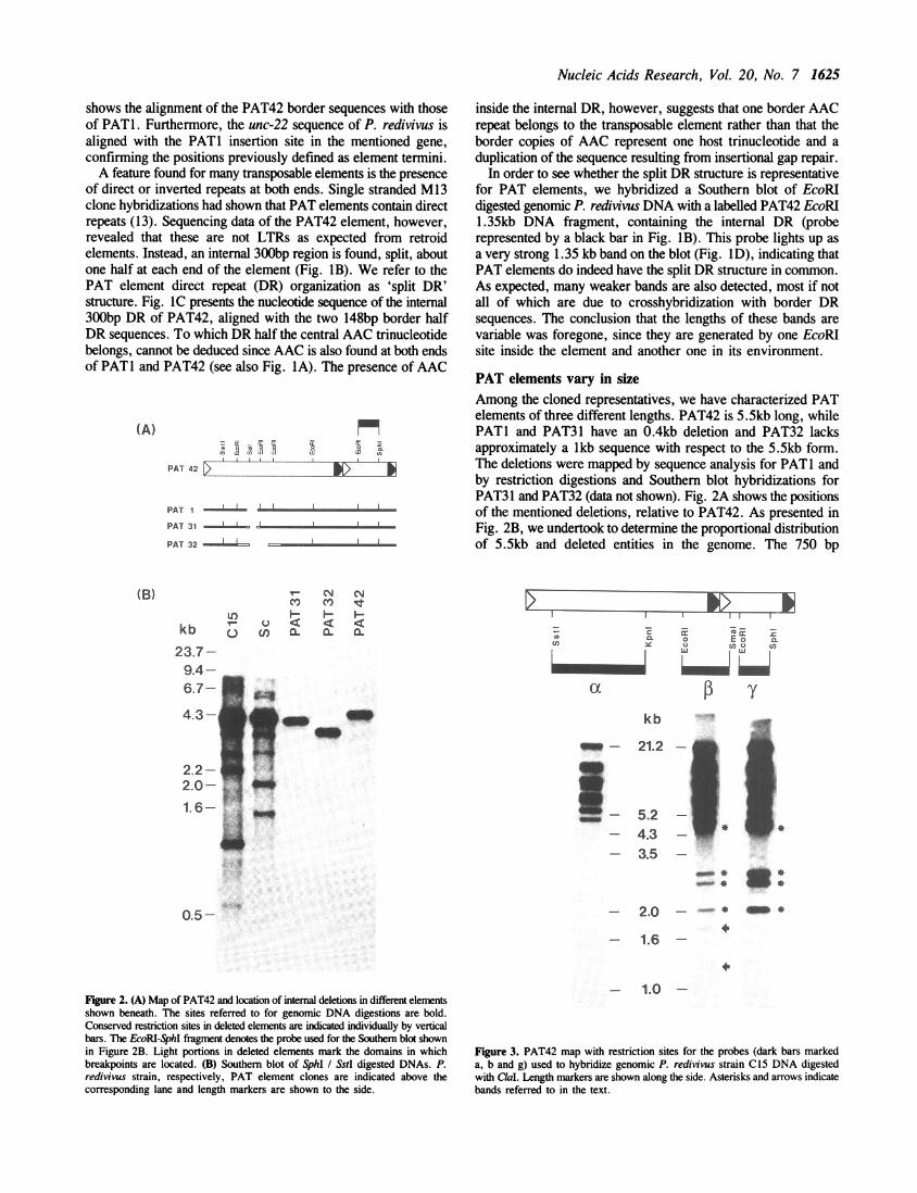
PAT elements vary in sizeAmong the cloned representatives, we have characterized PATelements of three different lengths. PAT42 is 5.5kb long, whilePATI and PAT31 have an 0.4kb deletion and PAT32 lacksapproximately a lkb sequence with respect to the 5.5kb form.The deletions were mapped by sequence analysis for PATI andby restriction digestions and Southern blot hybridizations forPAT31 and PAT32 (data not shown). Fig. 2A shows the positionsof the mentioned deletions, relative to PAT42. As presented inFig. 2B, we undertook to determine the proportional distributionof 5.5kb and deleted entities in the genome. The 750 bp
_C, CIMcn cs eSU' I I-) I-45
u < < <u en aX a. a
9.4-6.7-
I I ~~~~I II Ii_: - i-
W
CC
-
U) a 0 ~~Ec o.
kb
am-- 21.2 -5 a
- 5.2 -U- 4.3 - _- 3.5 -
- 2.0 * -*
- 1.6 -
Figure 2. (A) Map of PAT42 and location of internal deletions in different elementsshown beneath. The sites referred to for genomic DNA digestions are bold.Conserved restriction sites in deleted elements are indicated individually by verticalbars. The EcoRI-SphI fragment denotes the probe used for the Southern blot shownin Figure 2B. Light portions in deleted elements mark the domains in whichbreakpoints are located. (B) Southern blot of SphI / SstI digested DNAs. P.redivivus strain, respectively, PAT element clones are indicated above thecorresponding lane and length markers are shown to the side.
- 1.0 -
Figure 3. PAT42 map with restriction sites for the probes (dark bars markeda, b and g) used to hybridize genomic P. redivivus strain C15 DNA digestedwith ClaI. Length markers are shown along the side. Asterisks and arrows indicatebands referred to in the text.
(B)
kb
23.7-
0.5-
1626 Nucleic Acids Research, Vol. 20, No. 7
EcoRIISphI fragment (black bar in Fig. 2A) was used ashybridization probe on a genomic blot of SphIlSstI digested DNA.The bands lighting up are generated by the two restriction siteslocated inside the different elements. A strong band of about 4.6kb lights up in both genomic tracks, corresponding in length tothe cloned copy PAT42. Several shorter bands are visible in bothgenomic tracks. In strain C 15, at least two of these representinternally deleted elements as deduced from length homologiesto the controls PAT31 and PAT32. At this point, we could notdiscern whether the shorter bands in the genomic tracks are dueto restriction site heterogeneities in different elements or todeletion events of further extent. The elucidation of PAT elementstructure was therefore also attacked by a further series ofhybridizations.
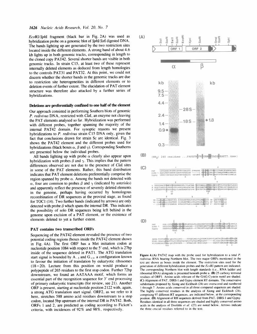
Deletions are preferentially confined to one half of the element
Our approach consisted in performing Southern blots of genomicP. redivivus DNA, restricted with ClaI, an enzyme not cleavingthe PAT elements analysed so far. Hybridization was performedwith different probes, together spanning the majority of theinternal PAT42 domain. For synoptic reasons we presenthybridizations to P. redivivus strain Cl 5 DNA only, given thefact that conclusions drawn for strain Sc are identical. Fig. 3shows the PAT42 element and the different probes used forhybridizations (black boxes a, and -y). Corresponding Southernsare presented below the individual probes.
All bands lighting up with probe a clearly also appear upon
hybridization with probes and y. This implies that the patterndifferences observed are not due to the presence of ClaI sitesin some of the PAT elements. Rather, this band distributionindicates that PAT element deletions preferentially comprise theregion spanned by probe oz. Among the bands not detected witha, four are common to probes and -y (indicated by asterisks)and apparently reflect the presence of severely deleted elementsin the genome, perhaps having occurred by homologousrecombination of DR sequences at the proviral stage, as foundfor TOC 1 (14). Two further bands (indicated by arrows) are onlydetected with probe which spans the internal DR. This indicatesthe possibility of solo DR sequences being left behind in thegenome upon excision of a PAT element, or the existence ofelements deleted to yet a further extent.
PAT contains two transcribed ORFs
Sequencing of the PAT42 element revealed the presence of twopotential coding regions (boxes inside the PAT42 element shownin Fig. 4A). The first ORF has a Met initiation codon at
nucleotide position 1084 with respect to the 5' end, which is 27bpinside of the sequence deleted in PAT1. The ATG translationstart signal is bounded by A-3 and G +1, a configuration knownto favour the initiation of translation by eukaryotic ribosomes(18-20). Lecture from this position on would produce a
polypeptide of 265 residues to the first stop codon. Further 72bpdownstream, we found an AATAAA motif, which forms an
essential part of the recognition sequence for 3' end processingof primary eukaryotic transcripts (for review, see 21). AnotherORF is present, starting at nucleotide position 2122 with, again,a strong ATG translation start signal. ORF2, as we refer to ithere, stretches 588 amino acid residues downstream to a stopcodon, located Sbp upstream of the internal DR in PAT42. Both,ORFs 1 and 2, are predicted as coding according to Fickett'scriteria, with incidences of 92% and 98%, respectively.
?DAS '!
Figure 4.(A) PAT42 map with the probe used for hybridization to a total P.redivivus RNA bearing Northern blot. The two major ORFs mentioned in thetext are shown as boxes inside the element. The restriction sites used for thegeneration of different hybridization probes and the EcoRI pattern are indicated.The corresponding Northern blot with length standards (i.e., RNA ladder andribosomal RNA) alongside is presented beneath probe a. (B) 25 carboxy terminalresidues of ORFI. Amino acids relevant of the GAG Cystein motif are shaded.(C) Alignment of PAT, DIRS-1 and Gypsy element RT domains. The conservativesubdomains proposed by Xiong and Eickbush (24) are overscored and numberedI through 7. Amino acids conserved in all three compared sequences are shaded.The highly conserved residues in the analysis of Xiong and Eickbush (24),comprising 97 different RT sequences, are indicated below, at the correspondingposition. (D) Alignment of RH sequences derived from PAT, DIRS-1 and Gypsy.Residues identical in all three sequences are shaded and highly conserved aminoacids in the analysis of Doolittle et al. (23) are noted below. Arrows indicatethe three crucial residues referred to in the text.
i*I i~

Nucleic Acids Research, Vol. 20, No. 7 1627
Protein sequence analysis based on prediction from nucleic acidstudies revealed some striking features for ORFs1 and 2. Fig. 4Bshows the 25 putative carboxy terminal residues of ORFI, bearinga typical Cys motif, which is the correct location for a motif foundexclusively in GAG proteins of retroid elements to date (22, 23).Typical for ORF2 is the presence of RT and RNaseH (RH)motifs, separated by a 103 residue tether, again, characteristicfeatures of retroid elements. Fig. 4C shows the alignment of RTsequences derived from PAT42, DIRS-1 and Gypsy. DIRS-1 isthe closest match to PAT42 among the sequences analysed byXiong and Eickbush (24). 21 out of 178 residues are identicalbetween the compared sequences, not taking account ofconservative changes. Individually, homologies are 49/178between PAT and DIRS-1, 41/178 between DIRS-1 and Gypsyand 38/178 between Gypsy and PAT. These figures make of PATthe most distantly related retroid element in the Gypsy family,but this assignment is stronger than to other retroid elementclasses (analysis not presented). Fig. 4D shows alignment of theRH domains derived from the same elements. Once again,DIRS-1 is the element to which PAT has most similarity whenthe RHs analysed by Doolittle et al. (23) and PAT are compared.Three residues, reported to be absolutely crucial for activity ofMMLV and E. coli RH (25, 26), are also found in the threecompared RHs, namely Asp'o, Glu48 and Asp70. These residuesare indicated by arrows in Fig. 4D.
In order to confirm transcription of the different regions inPAT elements, we performed a series of Northern blotexperiments. Hybridization to total P. redivivus bearing Northernblot with probe a is shown in Fig. 4A. This probe lights up asa very strong band of 900nt after 2.5 hr filter exposition. Theorientation of this transcript was confirmed left to right byhybridization with T3 and T7 probes obtained from a PAT42SphIISstI pBS-M13 clone. Hybridization with subclonesdemonstrated that the transcript starts slightly upstream of thefirst Sall site (far left) and ends just downstream of the third EcoRIsite (data not shown). This indicates that the region spanned byORFI is transcribed in the foreseen orientation. Furthermore,the lack of shorter bands implies that elements deleted in thisregion do not transcribe ORFI sequences. Another transcript,about 1800nt long, is detected after 15 day film exposition uponhybridization with a probe adjacent to a, namely the KpnI/SmaIfragment (see Fig. 4A).
Fig. 5 presents a diagram of ORFs 1 and 2 with putativefunctional domains boxed. No protease-like domain was identifiedon account of known motifs, a situation only found in severalmembers of the LINES family and in the DIRS-1 element thusfar (23). Also similar to CaMV, CERV, DIRS-l and someelements of the LINES family, is the lack of an accurate fingermotif in what is believed to be an Integrase (Int) domain. Onthe other hand, a tether separating RT and RH was not foundin the Gypsy (including DIRS-1) and copia family elements. Amore consistent approach to retroid element classification, though,resides in amino acid homology scans through well defined RTregions of different retroids (23, 24). Using the Clustal program
265M
I 204 307 434 5881~~~ ~~~~~~~1RT tether RH tnt?1
Figure 5. General repartition diagram of putative functional domains in ORFs1 and 2. Numbers indicate amino acid position with respect to the methioninecodon thought to act as initiator.
of PC gene, the PAT element RT scores best with DIRS-1(Fig. 4C) and very similar results are obtained comparing RHsequences (Fig. 4D). The distant relationship of PAT and DIRS-lto the Gypsy family, however, seems to imply the necessity ofcreating a novel class of retroid elements.
DISCUSSIONA rather unusual feature of the retroid PAT elements is their splitDR structure (Fig. lB and C). The only precedent of internalDR sequences was reported for TOC1 of Chlamydomonasreinhardtii (27). The fact that the half DRs are alternate(A...BA..B), implies that PAT elements are not composed oftwo different transposons having inserted into or next to oneanother. If DR sequences were to serve for strand exchangesduring reverse transcription, the maintenance of overall sequencearrangement would probably require several jumps for first strandcDNA synthesis as proposed for TOC1 (28 <:fs >), a peculiarityin terms of retroid elements (8). PAT lacks homology to the 3'end of a tRNA molecule near any one DR sequence, but thepossibility of reverse transcription initiating at, and proceedingoutwards through, a DR sequence cannot be excluded. Anoteworthy example in this context is the exotic nucleic acidpriming mode, reported for copia elements (29), where initiationstarts at a sequence homologous to an internal stretch of a tRNAmolecule. An alternative to outwards cDNA synthesis initiationis terminal priming, either by protein as found for HBV (9), by3' addition of uridine residues and poly-A priming, or by reversetranscription upon ligating of the element template to the genomicinsertion site (for review, see 5).
Several aspects concerning replication of PAT elements remainunanswered, among others, proof is lacking with respect to theexistence of virus-like particles (VLPs) as predicted by thepresence of a putative GAG protein. If, as shown for Ty (30,31), VLPs were to be the site of reverse transcription, it shouldbe interesting to analyse the nature and number of encapsidatedPAT molecules to gain more knowledge on the replication cycle.It seems clear, however, that PAT encodes functions necessaryfor replication via RNA intermediates.
In most retroid elements, the stoichiometry of GAG and Polprotein expression is regulated by translation from the samemessenger molecule and occasional frameshifting to produceslighter levels of GAG-Pol fusion protein than GAG protein alone.This mechanism was well studied for Ty elements (32) and HIV(33). An exception to this retroid feature is found in the case ofHBV, which uses separate initiation codons for GAG and Pol,albeit, from the same messenger molecule (34). PAT elementsare outsider retroids in this respect since GAG and Pol generatediscrete transcripts, as deduced from the lack of precursor andthe difference of signal intensity on Northern blots. Thus thestoichiometric proportion of GAG to Pol protein seems to beregulated at the transcriptional level. Several small potential codingregions were detected further 3'wards, none , however, containinga putative transmembrane domain, as one would expect of Envin infectious retroviruses. Hence, PAT presents the features forthe classification as nonviral retroelement (35).The majority of elements are 5.5kb long but several internally
deleted forms are also found in both analysed nematode strains.The extent of deletion in different elements is variable and someevents could be the consequence of recombination betweenhomologous DR segments. It is not clear when, during thereplication cycle or at the proviral stage, these deletions take
2_ _ .

1628 Nucleic Acids Research, Vol. 20, No. 7
place. Retroviral recombination has been shown to occur duringreverse transcription, probably by template switching), and forTy, such deletion events take place during or immediately afterreverse transcription, but before genome integration (37). In PATelements, deletions are confined preferentially to one half of theentities. The lack of shorter bands on the Northern blot of Fig. 4implies that in PAT1, an element of the 0.4kb deletion form,the start of the major 900nt transcript along with the transcriptionpromotor are lost. We therefore refer to PAT 1 as non-autonomous and presume that the missing function was providedin trans. This element did, however, retain the sequences requiredin cis for transposition (13), and might therefore be used asmutator element for a transposon tagging system.The nematode C. elegans is favored by a growing population
of molecular biologists because of its easiness to handle, shortgeneration time, small genome size, detailed knowledge of itsgenome, development and cell lineage, and because many findingscan be extrapolated to other species including man (38).Transposons have been identified (i.e., Tc elements), but therequirement of relatively high copy numbers for efficienttransposition (39) make their use as tagging system quite tedious.Moreover, Tc elements are characterized by their target sitespecificity (11), which could account for a limited number ofaffectable genes. Not only do P and PAT elements behave similarlywith respect to the non-reciprocal effect on fertility in crossesbetween high and low copy number strains (40, 13), but the notionthat PAT elements may provide a useful tagging system for C.elegans is indicated by the following observations. First, P.redivivus and C. elegans are closely related nematodes, andpossible transposition of PAT elements in the latter gains supportwhen considering P element mobility in non-Drosophilids (41).Second, the elements actively transpose and cause gene disruptionat their insertion site. Third, partially deleted, non-autonomouselements can be mobilized and integrated if the missing functionis provided in trans. Fourth, the germline is concerned bytransposition of PAT elements, as deduced from the stably inheritedunc-22 mutant (13). Fifth, PAT DNA probes do not crosshybridizewith C. elegans DNA, thus facilitating screening pro cedures. Onthe other hand, mobility of PAT elements could be impaired inC. elegans by the presence of repressors, inappropriatetranscription signals or the lack of positively regulating host factors(for review, see 5). DNA injection experiments have beenestablished (42, 43) and expression of genes in transgenic C.elegans was found to be correct (44). Thus, inspirations canhenceforth be gathered from transposon tagging and enhancertrapping P element systems in Drosophila (45). Moreover,perspectives are opened with recently found marker genes suchas roller (46) and genes known to suppress recognizable phenotypes(47), as well as the recently described use of inducible promotersin such systems (48). Libraries of strains with single PAT elementinsertions would be likely to greatly facilitate gene recognition andcharacterization in C. elegans.
ACKNOWLEDGEMENTS
We thank Mr. H. Gachoud for art work and photography andMrs. C. Dedual as well as Mrs. C. Stempfel for excellenttechnical assistance. This work was supported by grants No.3.113-0.85 and 3.141-0.88 from the Swiss National ScienceFoundation.
REFERENCES1. Berg, D. and Howe, M. (1989) Mobile DNA, American Society for
Microbiology, Washington, DC.2. Boeke, J.D., Garfinkel, D.J., Styles, C.A. and Fink, G.R. (1985) Cell, 40,
491 -500.3. Varmus, H. (1988) Science, 240, 1427 - 1434.4. Finnegan, D.J. (1989) TIG, 5, 103-107.5. Boeke, J.D. and Corces, V.G. (1989) Annu. Rev. Microbiol.,43, 403-434.6. Varmus, H. (1989) in Berg, D.E. and Howe, M.M. (eds.) Mobile DNA,
American Society for Microbiology, Washington, DC, 53- 108.7. Arkhipova, I.R., Mazo, A.M., Cherkasova, V.A., Gorelova, T.V., Schuppe,
N.G. and Ilyin, Y.V. (1986) Cell, 44, 555-563.8. Panganiban, A.T. and Fiore, D. (1988) Science, 241, 1064-1069.9. Bartenschlager, R. and Schaller, H. (1988) EMBO J., 7, 4185-4192.
10. Cappello, J., Handelsman, K. and Lodish, H.F. (1985) Cell, 43, 105 - 115.11. Emmons, S.W. (1988) in Wood, W.B. (ed.) 7he Nematode Caenorhabditis
elegans, Cold Spring Harbor Laboratory Press, NY, 47-79.12. Aeby, P., Spicher, A., de Chastonay, Y., Muller, F. and Tobler, H. (1986)
EMBO J., 5, 3353-3360.13. Link, C.D., Graf-Whitsel, J. and Wood, W.B. (1987) Proc. Natl. Acad.
Sci. USA, 84, 5325-5329.14. Suiston, J.E. and Brenner, S. (1974) Genetics, 77, 95-104.15. Sulston, J.E. and Hodgkin, J. (1988) in Wood, W.B. (ed.) The Nematode
Caenorhabditis elegans, Cold Spring Harbor Laboratory Press, NY,587 -606.
16. Maniatis,T., Fritsch, E.F. and Sambrook, J. (1982) Molecular Cloning, ALaboratory Manual. Cold Spring Harbor Laboratory Press, NY.
17. Sanger, F., Nicklen, S. and Coulson, A.R. (1977) Proc. Natl. Acad. Sci.,74, 5463-5467.
18. Kozak, M. (1984) Nature, 308, 241 -246.19. Kozak, M. (1986) Cell, 44, 283-292.20. Morle, F., Lopez, B., Henni, T. and Godet, J. (1985) EMBO J., 4,
1245- 1250.21. Wickens, M. (1990) TIBS, 15, 277-281.22. Covey, S.N. (1986) Nucl. Acids Res., 14, 623-633.23. Doolittle, R.F., Feng, D.-F., Johnson, M.S. and McClure, M.A. (1989)
Quart. Rex'. Biol., 64, 1-30.24. Xiong, Y. and Eickbush, T.H. (1990) EMBO J., 9, 3353-3362.25. Repaske, R., Harley, J.W., Kavilick, M.F., O'Neil, R.R. and Austin, J.B.
(1989) J. Virol., 63, 1460- 1464.26. Kanaya, S. et al. (1990) J. Biol. Chem., 265, 4615.27. Day, A., Schirmer-Rahire, M., Kuchka, M.R., Mayfield, S.P. and Rochaix,
J.-D. (1988) EMBO J., 7, 1917-1927.28. Day, A. and Rochaix, J. -D. (1991) Nucl. Acids Res., 19, 1259-1266.29. Kikuchi, Y., Ando, Y. and Shiba,T. (1986) Nature, 323, 824-826.30. Garfinkel, D.J., Boeke, J.D. and Fink, G.R. (1985) Cell, 42, 507-517.31. Mellor, J., Malim, M.H., Gull, K., Tuite, M.F. and McReady, Dibbayawan,
T., Kingsman, S.M. and Kingsman, A.J.(1985) Nature, 318, 583-586.32. Kingsman, A.J. and Kingsman, S.M. (1988) Cell, 53, 333-335.33. Wilson, W., Braddock, M., Adams, S.E., Rathjen, P.D., Kingsman, S.M.
and Kingsman, A.J. (1988) Cell, 55, 1159-1169.34. Schlicht, H.J., Radziwill, G. and Schaller, H. (1989) Cell, 56, 85-92.35. Hull, R. and Will, H. (1989) TIG, 5, 357-359.36. Goodrich, D.W. and Duesberg, P.H. (1990) Proc. Natl. Acad. Sci. USA,
87, 2052-2056.37. Xu, H. and Boeke, J.D. (1987) Proc. Natl. Acad. Sci. USA, 84, 8553-8557.38. Roberts, L. (1990) Science, 248, 1310-1313.39. Babity, J.M., Starr, T.V.B. and Rose, A.M. (1990) Mol. Gen. Genet., 222,
65 -70.40. Rubin, G.M., Kidwell, M.G. and Bingham, P.M. (1982) Cell, 29, 987-994.41. O'Brochta, D.A. and Handler, A.M. (1988) Proc. Natl. Acad. Sci. USA,
85, 6052-6056.42. Stinchcomb, D.T., Shaw, J.E., Carr, S.H. and Hirsch, D. (1985) Mol. Cell.
Biol., 5, 3484-3496.43. Fire, A. (1986) EMBO J., 5, 2673-2680.44. Fire, A. and Waterston, R.H. (1989) EMBO J., 11, 3419-3428.45. Cooley, L., Kelley, R. and Spradling, A. (1988) Science, 239, 1121 - 1128.46. Kramer, J.M., French, R.P., Park, E.-C. and Johnson, J.J. (1990) Mol.
Cell. Biol., 10, 2081 - 2089.47. Kimble, J., Hodgkin, J., Smith, T. and Smith, J. (1982) Nature, 229,
456-458.48. Sass, H. (1990) Gene, 89, 179-186.