necrotic lymphoma in a patient with post-transplantation...
TRANSCRIPT
148 Ultrasonography 34(2), April 2015 e-ultrasonography.org
Necrotic lymphoma in a patient with post-transplantation lymphoproliferative disorder: ultrasonography and CT findings with pathologic correlation
Minsu Lee1,2, Sang Kyum Kim3, Yong Eun Chung1,2, Jin-Young Choi1,2, Mi-Suk Park1,2, Joon Seok Lim1,2, Myeong-Jin Kim1,2, Honsoul Kim1,2
1Department of Radiology, 2Research Institute of Radiological Science, Severance Hospital,
Yonsei University College of Medicine, Seoul; 3Department of Pathology, Yonsei University
College of Medicine, Seoul, Korea
http://dx.doi.org/10.14366/usg.14050pISSN: 2288-5919 • eISSN: 2288-5943
Ultrasonography 2015;34:148-152
Seventeen months after kidney transplantation for the treatment of nephrotic syndrome, a retroperitoneal mass was incidentally detected in a 30-year-old man during routine follow-up. Ultrasonography revealed a mass measuring 5.5 cm×4.3 cm located between the liver and the atrophic right kidney, which showed markedly heterogeneous internal echogenicity. Contrast-enhanced computed tomography displayed a mild degree of enhancement only at the periphery of the mass, while the center lacked perceivable intensification. The patient underwent surgical resection. The final pathological diagnosis was non-Hodgkin lymphoma (diffuse large B-cell lymphoma), and extensive necrosis was observed on microscopic examination. We found that the prominent heterogeneous echogenicity of the mass (an unusual finding of lymphoma) demonstrated on ultrasonography is a result of extensive necrosis, which may sometimes occur in patients with post-transplantation lymphoproliferative disorder.
Keywords: Lymphoma; Ultrasonography; Immunocompromised host
Received: November 1, 2014Revised: December 1, 2014Accepted: December 1, 2014
Correspondence to:Honsoul Kim, MD, Department of Radiology, Yonsei University College of Medicine, 50-1 Yonsei-ro, Seodaemun-gu, Seoul 120-752, Korea
Tel. +82-2-2228-7400Fax. +82-2-393-3035E-mail: [email protected]
CASE REPORT
This is an Open Access article distributed under the terms of the Creative Commons Attribution Non-Commercial License (http://creativecommons.org/licenses/by-nc/3.0/) which permits unrestricted non-commercial use, distribution, and reproduction in any medium, provided the original work is properly cited.
Copyright © 2015 Korean Society of Ultrasound in Medicine (KSUM)
How to cite this article: Lee M, Kim SK, Chung YE, Choi JY, Park MS, Lim JS, et al. Necrotic lymphoma in a patient with post-transplantation lymphoproliferative disorder: ultrasonography and CT findings with pathologic correlation. Ultrasonography. 2015 Apr;34(2):148-152.
Introduction
Lymphoma involving the abdomen usually demonstrates on ultrasonography as a homogeneous mass with marked hypoechogenicity. Non-homogeneous echogenicity is considered an unlikely finding for lymphoma, essentially because lymphoma lesions are mostly homogeneous and rarely demonstrate necrosis [1,2]. Post-transplantation lymphoproliferative disorder (PTLD) represents a range of lymphoid proliferation, from abnormal lymphoid hyperplasia to malignant lymphoma, that occurs after organ transplantation and administration of immunosuppressive agents. Immunosuppression and Epstein-Barr virus (EBV) infection are the two major risk factors associated with the development of PTLD. The imaging findings of lymphoma associated with PTLD can be nonspecific and atypical. Here, we describe a lymphoma case associated with PTLD that presented on ultrasonography as a mass with striking heterogeneous echogenicity. This study was approved by the Institutional Review Board
Necrotic lymphoma in a PTLD patient
e-ultrasonography.org Ultrasonography 34(2), April 2015 149
of our institution, and a waiver of informed consent was obtained.
Case Report
A 30-year-old man with nephrot ic syndrome underwent successful renal transplantation from a cadaveric donor. Prior to transplantation, he had been diagnosed as a hepatitis B virus carrier with liver cirrhosis and had been treated with a stent graft for aortic dissection. After transplantation, he received a triple
immunosuppressive regimen that consisted of tacrolimus (4 mg/day), mycophenolatemofetil (MMF; 2,000 mg/day), and prednisolone (5 mg/day). Seventeen months after transplantation, he denied any specific symptoms, but his laboratory tests showed elevated aspartate transaminase and alanine transaminase levels during regular follow-up.
Abdominal ultrasonography for further evaluation of hepatitis was performed using Philips iU22 Ultrasound System (Philips Healthcare, Best, The Netherlands) with a 1.0-5.0-MHz (C5-1)
A B
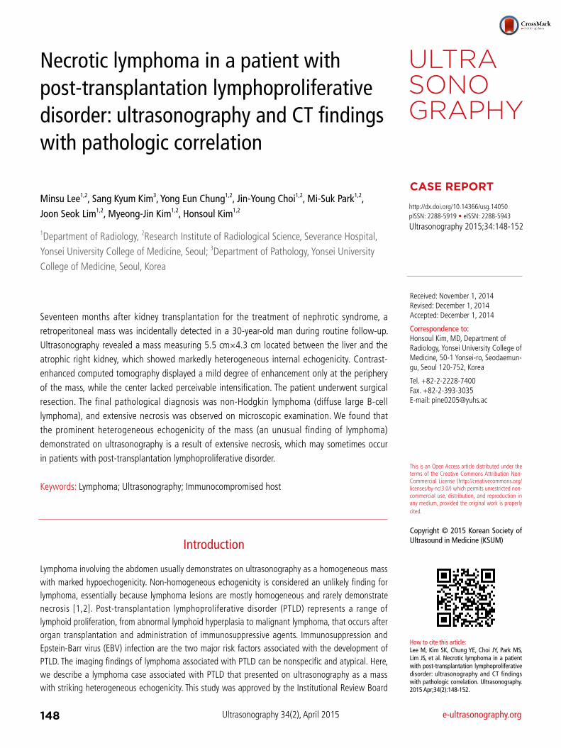
Fig. 1. A 31-year-old man who underwent kidney transplantation 17 months prior. A. Routine follow-up ultrasonography shows an incidental mass (arrows) located between the liver and the atrophic right kidney with markedly heterogeneous echogenicity on the B-mode. B. On color Doppler ultrasonography, no flow signal can be detected within the mass (arrows). C, D. Pre-contrast computed tomography (CT) (C) and CT performed during the hepatic venous phase (D) demonstrate a mass (arrows) located at the right retroperitoneal space, abutting the liver, atrophic right kidney, and duodenum. A mild degree of enhancement is observed at the mass periphery but not in the central portion, suggesting the necrosis of the inner portion.
C D
Minsu Lee, et al.
150 Ultrasonography 34(2), April 2015 e-ultrasonography.org
wideband convex transducer. Underlying liver cirrhosis with multiple nodular lesions and atrophic native kidneys were observed. In addition, an incidental mass measuring 5.5 cm×4.3 cm with marked heterogeneous internal echogenicity was found between the liver and the atrophic right kidney (Fig. 1A, B). The patient underwent subsequent triple-phase contrast-enhanced abdominal computed tomography (CT) for further evaluation of the mass. The CT revealed a 5.5-cm-sized right-sided retroperitoneal mass that showed mild peripheral enhancement, but the center of the mass seemed to lack perceivable intensification, suggesting that extensive necrosis had occurred. The mass was abutting the right kidney, liver segment 6, and the duodenum (Fig. 1C, D). The retroperitoneal location of the mass and the assumed extensive necrotic change led to the impression of retroperitoneal sarcoma. Desmoid tumor, inflammatory pseudo-tumor, and tuberculous abscess were other disease entities
considered for the differential diagnosis. We excluded lymphoma because extensive necrosis is a very unusual finding.
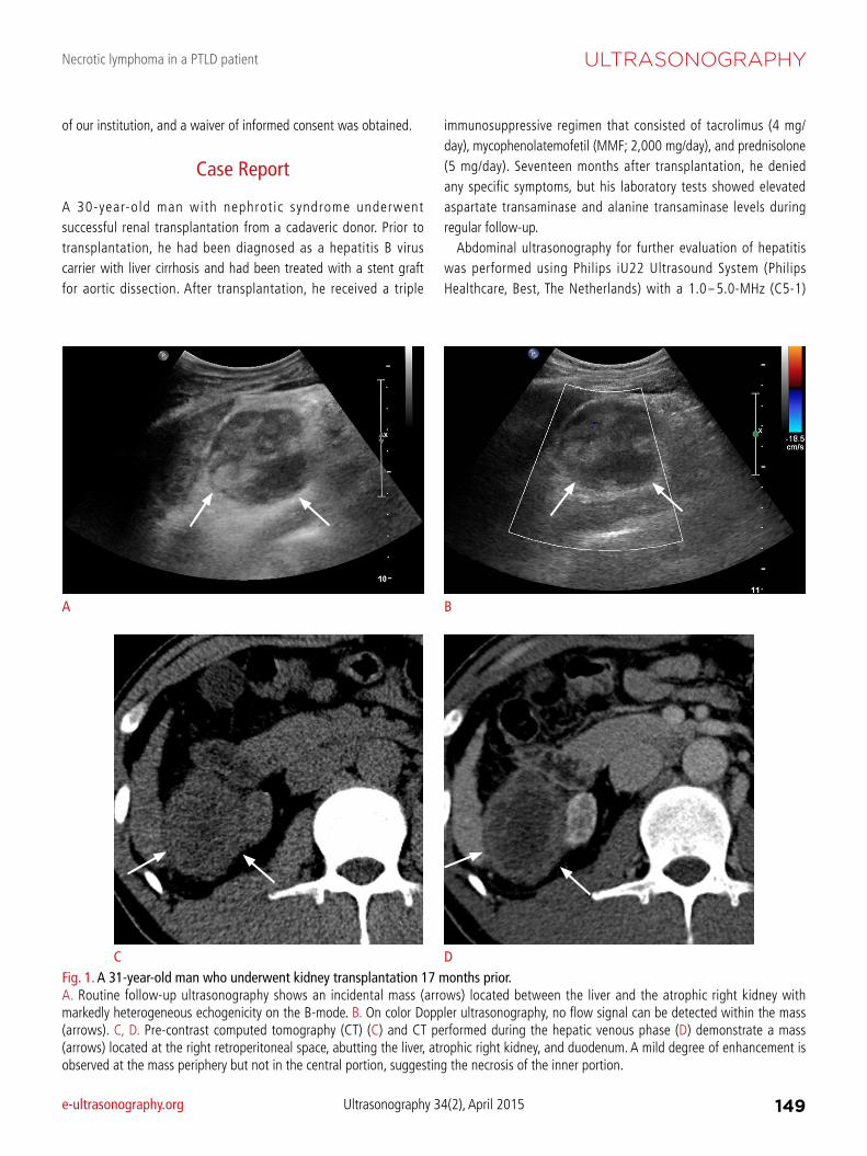
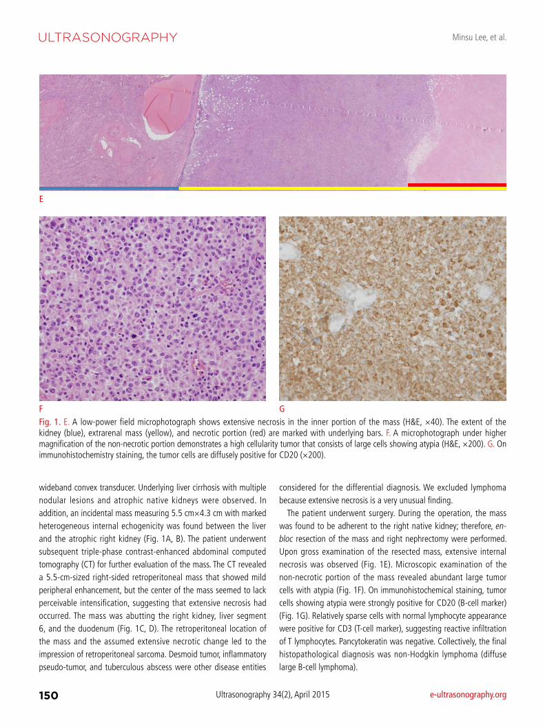
The patient underwent surgery. During the operation, the mass was found to be adherent to the right native kidney; therefore, en-bloc resection of the mass and right nephrectomy were performed. Upon gross examination of the resected mass, extensive internal necrosis was observed (Fig. 1E). Microscopic examination of the non-necrotic portion of the mass revealed abundant large tumor cells with atypia (Fig. 1F). On immunohistochemical staining, tumor cells showing atypia were strongly positive for CD20 (B-cell marker) (Fig. 1G). Relatively sparse cells with normal lymphocyte appearance were positive for CD3 (T-cell marker), suggesting reactive infiltration of T lymphocytes. Pancytokeratin was negative. Collectively, the final histopathological diagnosis was non-Hodgkin lymphoma (diffuse large B-cell lymphoma).
Fig. 1. E. A low-power field microphotograph shows extensive necrosis in the inner portion of the mass (H&E, ×40). The extent of the kidney (blue), extrarenal mass (yellow), and necrotic portion (red) are marked with underlying bars. F. A microphotograph under higher magnification of the non-necrotic portion demonstrates a high cellularity tumor that consists of large cells showing atypia (H&E, ×200). G. On immunohistochemistry staining, the tumor cells are diffusely positive for CD20 (×200).
F G
E
Necrotic lymphoma in a PTLD patient
e-ultrasonography.org Ultrasonography 34(2), April 2015 151
After surgery, the doses of tacrolimus and MMF were reduced to 1 mg/day and 500 mg/day, respectively. The patient managed well and did not show evidence of rejection of the transplanted renal allograft. The patient received three cycles of chemotherapy with a regimen of cyclophosphamide, doxorubicin, vincristine, prednisolone, and rituximab (R-CHOP). No evidence of lymphoma recurrence was observed during the postoperative surveillance period of 20 months.
Discussion
On ultrasonography, lymphomas presenting as a mass usually demonstrate homogenous, markedly-low echogenicity irrespective of the nodal or extra-nodal status [3-5]. This is because lymphoma is fundamentally a homogeneous tumor that generates very few internal reflections. Under rare circumstances, however, if the lymphoma mass becomes heterogeneous, atypical imaging findings may be encountered. For example, a reported lymphoma case involving the liver and a hepatoduodenal ligament that carried intermingled adipose cells within the mass presented as a hyperechoic mass [6].
PTLD consists of a wide spectrum of conditions associated with lymphoid proliferation that occurs after organ transplantation and the administration of immunosuppressive agents. This malady ranges from reactive, polyclonal hyperplasia to aggressive non-Hodgkin lymphoma. In the literature, PTLD is described as occurring most frequently during the first year after transplantation, with the highest incidence after small bowel transplantation (~20%), followed by lung, heart, liver, and kidney transplantation (1%-10%) [7]. Immunosuppression and EBV infection status are two major factors associated with the development of PTLD [8,9].
In the current article, we report a post-transplantation lymphoma case that presented as a retroperitoneal mass showing striking heterogeneous echogenicity on ultrasonography. The patient underwent surgical resection, and we performed a radiological-pathological correlation to account for such atypical imaging findings. As such marked inhomogeneity revealed on ultrasonography is very unusual for lymphoma [1,2], we mistakenly excluded lymphoma from our differential diagnosis. Based on the gross and microscopic features of the surgical specimen, the heterogeneous echogenicity was attributed to extensive necrosis within the mass. Unlike untreated lymphomas, which rarely contain necrotic portions, lymphomas in post-transplantation conditions have been reported to occasionally show central areas of necrosis [10,11], which was probably the case in our patient as well. Knowledge of such atypical ultrasonography features of lymphoma associated with PTLD seems to be important because ultrasonography is frequently used as the initial surveillance modality after transplantation; if the radiologist
is familiar with these atypical imaging features, confusion can be avoided.
In conclusion, lymphoma can occasionally present on ultrasono-graphy as a mass with markedly heterogeneous echogenicity, which represents internal necrosis when associated with the conditions of PTLD. If the patient has a clinical history of organ transplantation, is receiving immunosuppressive therapies, and has atypical findings, the clinician is cautioned not to exclude lymphoma from the differential diagnosis, and early tissue confirmation is indicated.
ORCID: Minsu Lee: http://orcid.org/0000-0003-1943-3677; Sang Kyum Kim:
http://orcid.org/0000-0003-0768-9923; Yong Eun Chung: http://orcid.org/0000-
0003-0811-9578; Jin-Young Choi: http://orcid.org/0000-0002-9025-6274; Mi-Suk
Park: http://orcid.org/0000-0001-5817-2444; Joon Seok Lim: http://orcid.org/0000-
0002-0334-5042; Myeong-Jin Kim: http://orcid.org/0000-0001-7949-5402; Honsoul
Kim: http://orcid.org/0000-0002-2367-234X
Conflict of InterestNo potential conflict of interest relevant to this article was reported.
References
1. Rettenbacher T. Sonography of peripheral lymph nodes part 1: normal findings and B-image criteria. Ultraschall Med 2010;31:344-362.
2. Cui XW, Hocke M, Jenssen C, Ignee A, Klein S, Schreiber-Dietrich D, et al. Conventional ultrasound for lymph node evaluation, update 2013. Z Gastroenterol 2014;52:212-221.
3. Carroll BA, Ta HN. The ultrasonic appearance of extranodal abdominal lymphoma. Radiology 1980;136:419-425.
4. Soyer P, Van Beers B, Teillet-Thiebaud F, Grandin C, Kazerouni F, Barge J, et al. Hodgkin's and non-Hodgkin's hepatic lymphoma: sonographic findings. Abdom Imaging 1993;18:339-343.
5. Gazelle GS, Lee MJ, Hahn PF, Goldberg MA, Rafaat N, Mueller PR. US, CT, and MRI of primary and secondary liver lymphoma. J Comput Assist Tomogr 1994;18:412-415.
6. Kim H, Kim KW, Park MS, Kim H. Lymphoma presenting as an echogenic periportal mass: sonographic findings. J Clin Ultrasound 2008;36:437-439.
7. Gottschalk S, Rooney CM, Heslop HE. Post-transplant lymphopro-liferative disorders. Annu Rev Med 2005;56:29-44.
8. Paya CV, Fung JJ, Nalesnik MA, Kieff E, Green M, Gores G, et al. Epstein-Barr virus-induced posttransplant lymphoproliferative disorders. ASTS/ASTP EBV-PTLD Task Force and The Mayo Clinic Organized International Consensus Development Meeting. Transplantation 1999;68:1517-1525.
9. Penn I, Hammond W, Brettschneider L, Starzl TE. Malignant lympho-mas in transplantation patients. Transplant Proc 1969;1:106-112.
10. Borhani AA, Hosseinzadeh K, Almusa O, Furlan A, Nalesnik M.
Minsu Lee, et al.
152 Ultrasonography 34(2), April 2015 e-ultrasonography.org
Imaging of posttransplantation lymphoproliferative disorder after solid organ transplantation. Radiographics 2009;29:981-1000.
11. Sauter A, Faul C, Bitzer M, Bares R, Kraus S, Fenchel M, et al.
Imaging findings in immunosuppressed patients with Epstein Barr virus-related B cell malignant lymphoma. AJR Am J Roentgenol 2010;194:W141-W149.