neck formation and growth. main topographic regions in ...anat.lf1.cuni.cz/souhrny/ofa_a6.pdf ·...
TRANSCRIPT
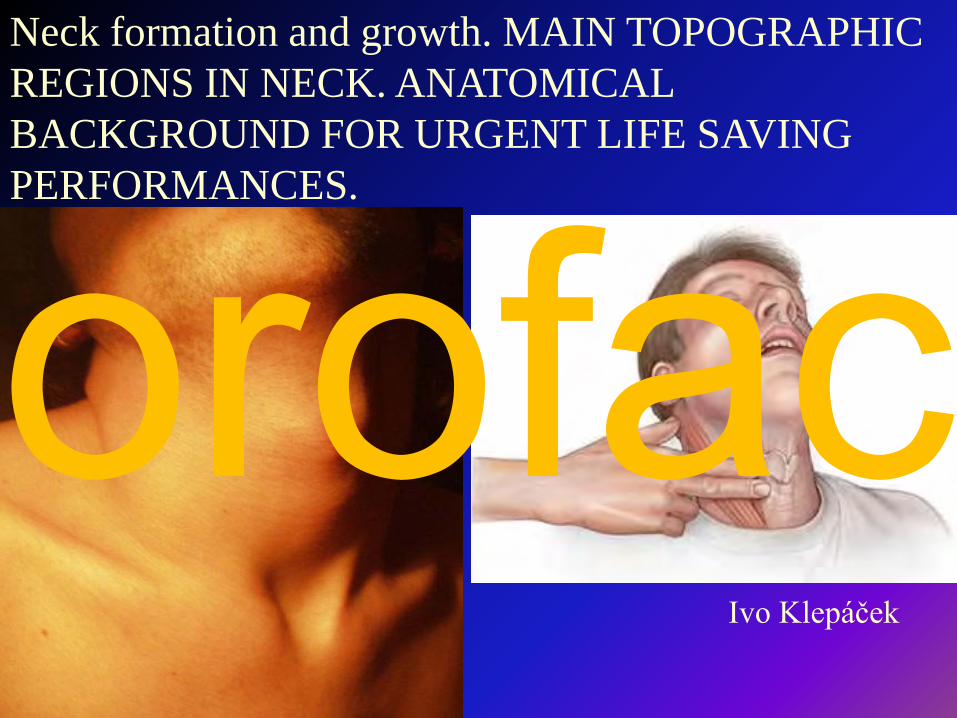
Neck formation and growth. MAIN TOPOGRAPHIC REGIONS IN NECK. ANATOMICAL BACKGROUND FOR URGENT LIFE SAVING PERFORMANCES.
Ivo Klepáček
orofac
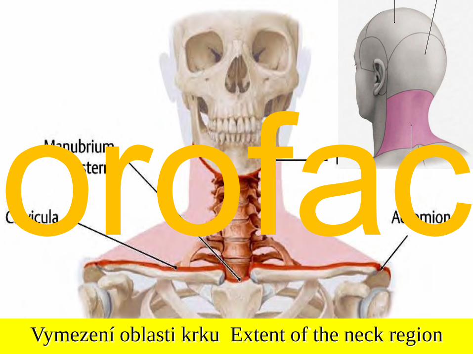
Vymezení oblasti krku Extent of the neck region
orofac
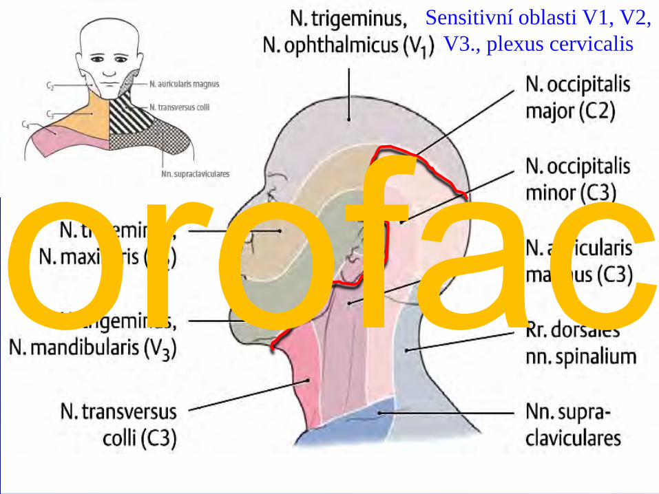
Sensitivní oblasti V1, V2, V3., plexus cervicalis
orofac
*
*
*
*
**
*
*
orofac
orofac
orofac
cranial caudalmiddleorofac
orofac
orofac
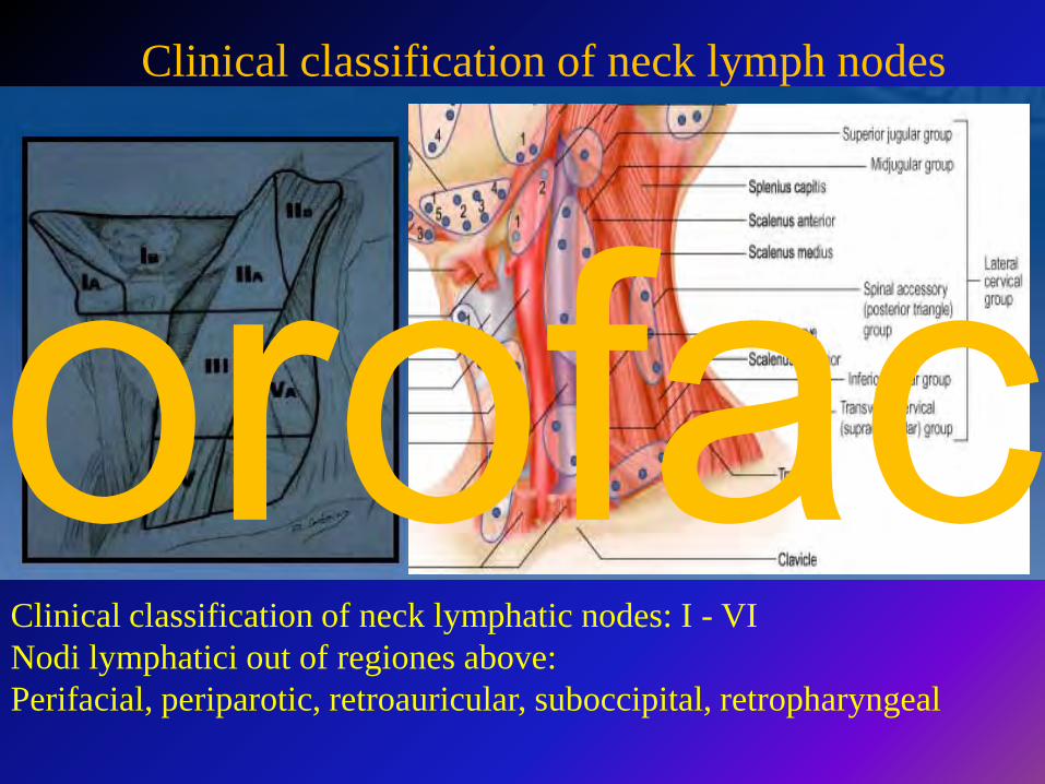
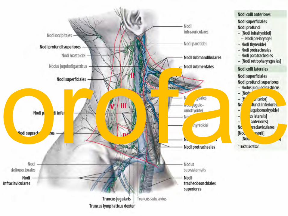
Clinical classification of neck lymphatic nodes: I - VINodi lymphatici out of regiones above:Perifacial, periparotic, retroauricular, suboccipital, retropharyngeal
Clinical classification of neck lymph nodes
orofac
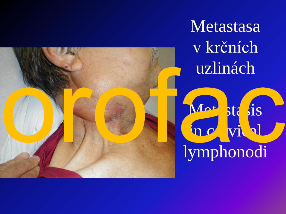
Metastasa v krčních uzlinách
Metastasis in cervical
lymphonodiorofac

TOPOGRAPHIC REGIONS
and SPACESorofac
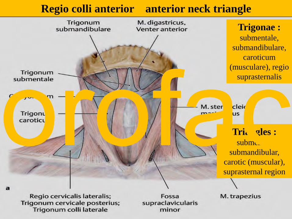
Trigonae : submentale,
submandibulare, caroticum
(musculare), regio suprasternalis
Regio colli anterior anterior neck triangle
Triangles : submental,
submandibular, carotic (muscular), suprasternal region
orofac
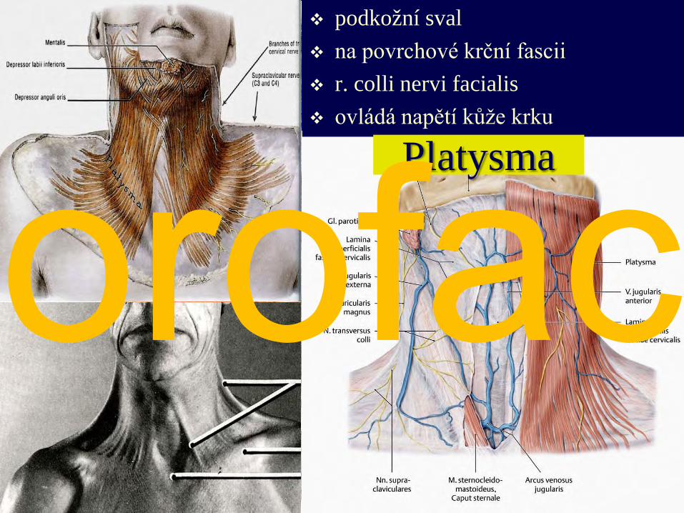
podkožní sval na povrchové krční fascii r. colli nervi facialis ovládá napětí kůže krku
Platysma
orofac

proc. mastoideusmanubrium sterni, claviculan.accessorius (XI) + branches from plexus cervicalis
Sternocleidomastoid m. sternocleidomastoideus
orofac
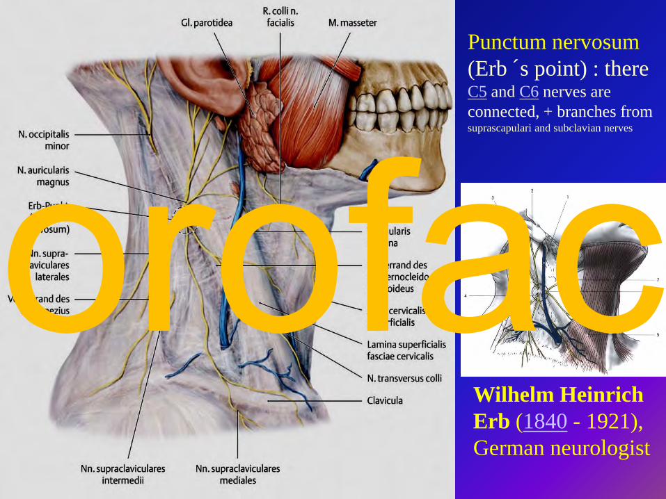
Wilhelm Heinrich Erb (1840 - 1921), German neurologist
Punctum nervosum (Erb ´s point) : there C5 and C6 nerves are connected, + branches from suprascapulari and subclavian nerves
orofac
orofac
orofac
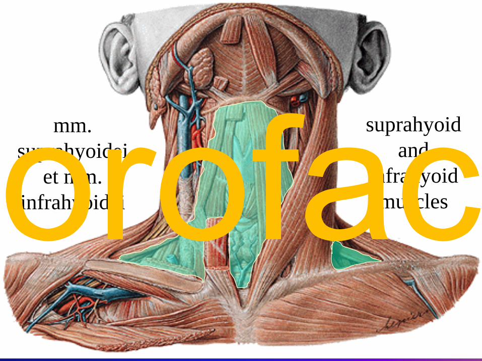
mm. suprahyoidei
et mm. infrahyoidei
suprahyoid and
infrahyoid musclesorofac
orofac
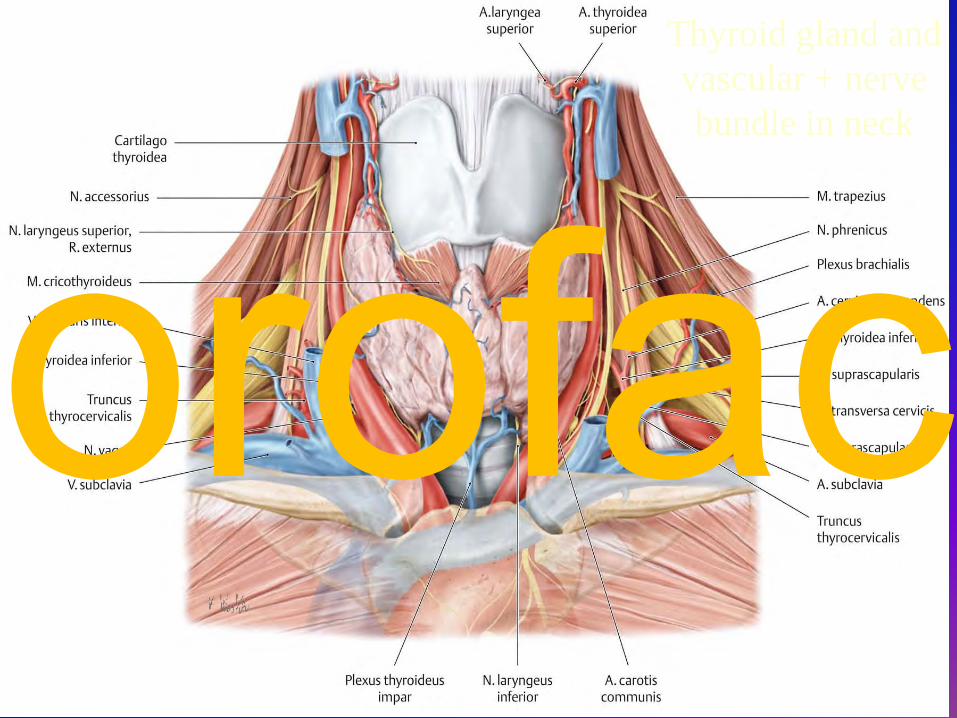
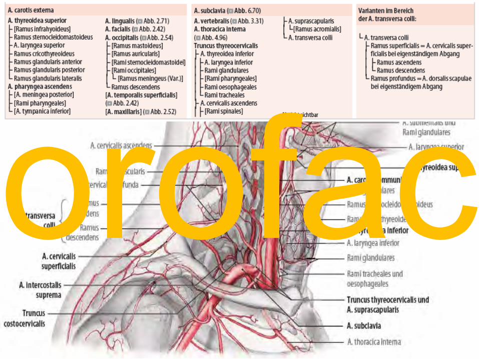
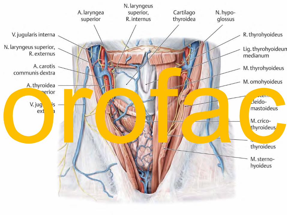
Thyroid gland andvascular + nerve bundle in neck
orofac
orofac
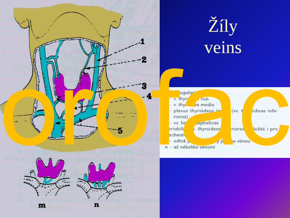
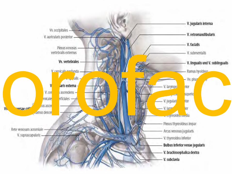
Žílyveins
orofac
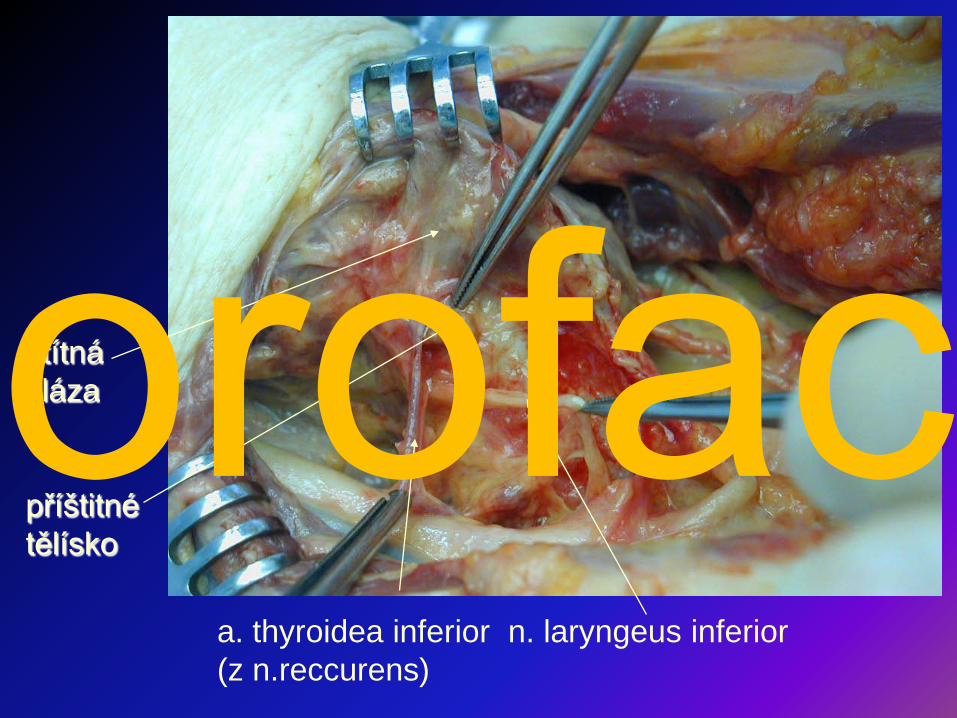
štítná žláza
příštitné tělísko
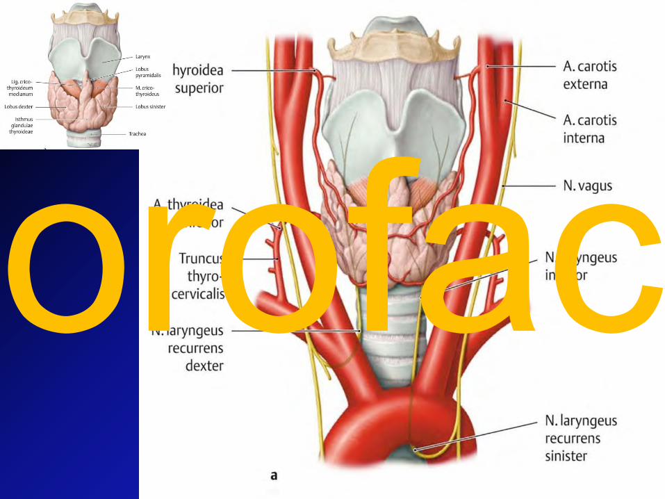
a. thyroidea inferior n. laryngeus inferior (z n.reccurens)
orofac
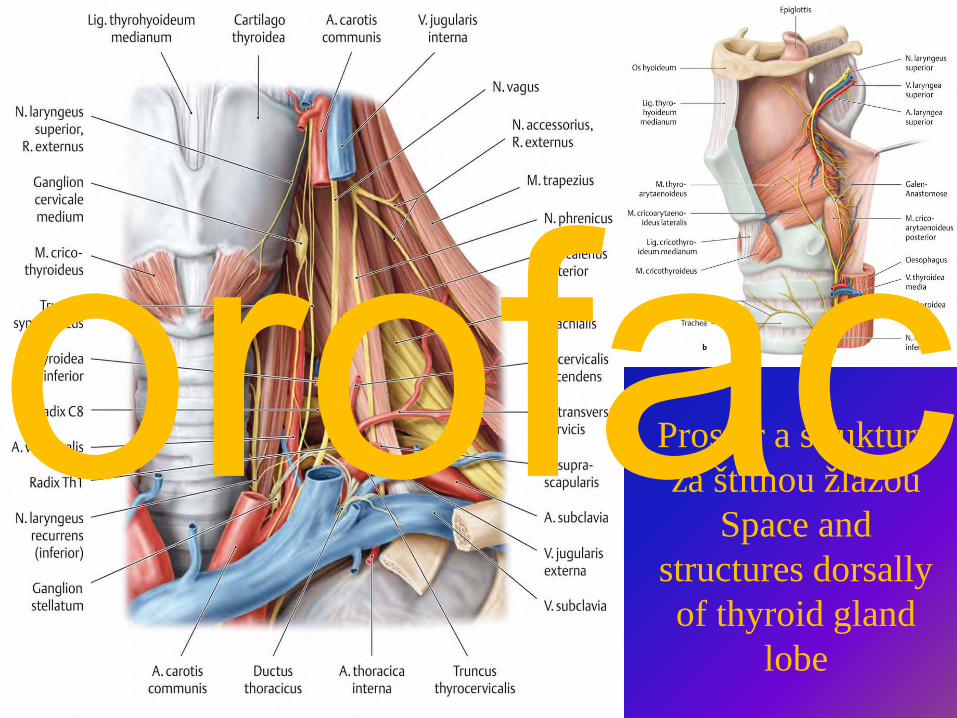
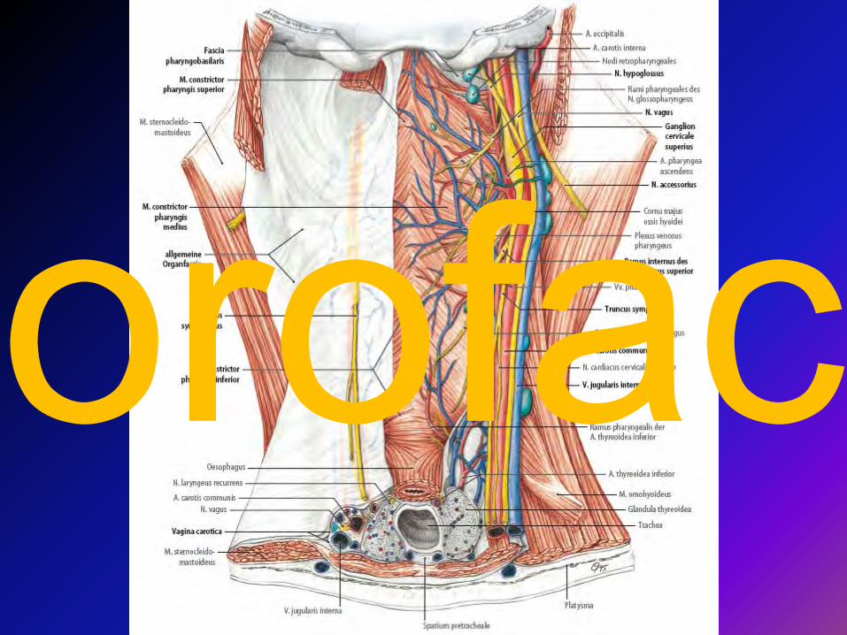
Prostor a struktury za štítnou žlázou
Space andstructures dorsallyof thyroid gland
lobe
orofac
orofac
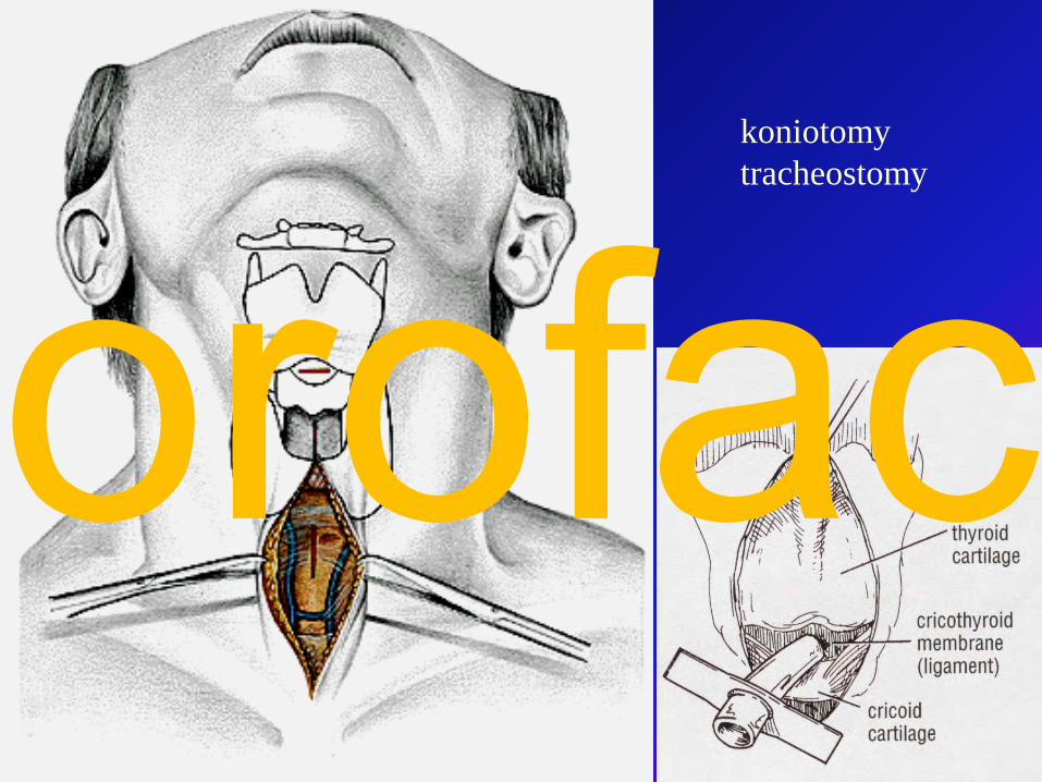
koniotomytracheostomy
orofac
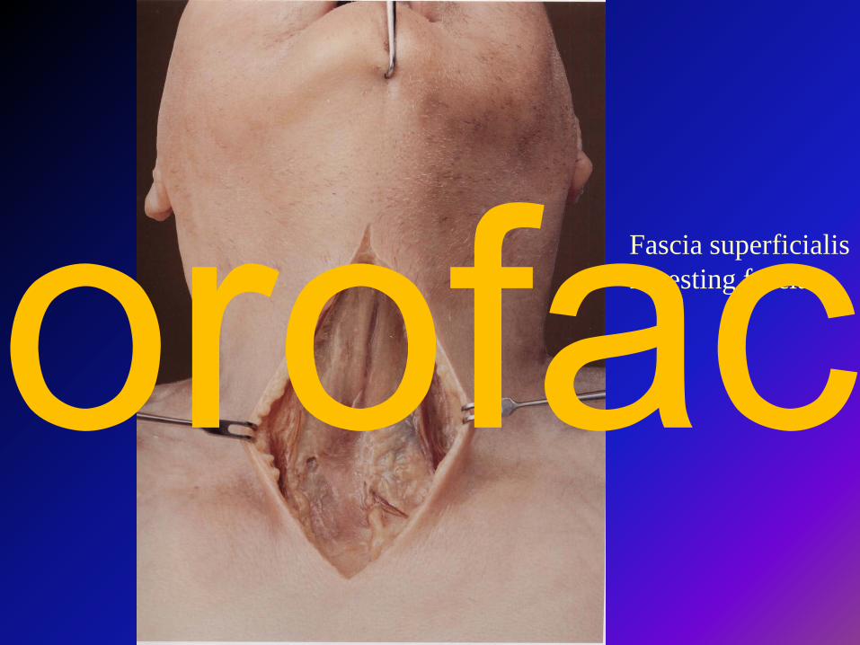
Fascia superficialisInvesting fasciaorofac
Fascia superficialisInvesting fascia
Proťatacutorofac
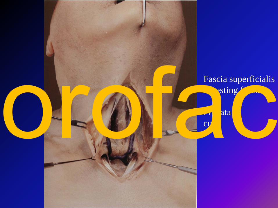
Fascia pretrachealisPretracheal fascia
S otvoremwindowedorofac
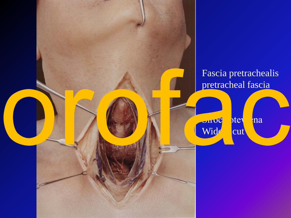
Fascia pretrachealis pretracheal fascia
Široce otevřenaWidely cutorofac
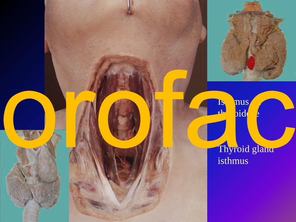
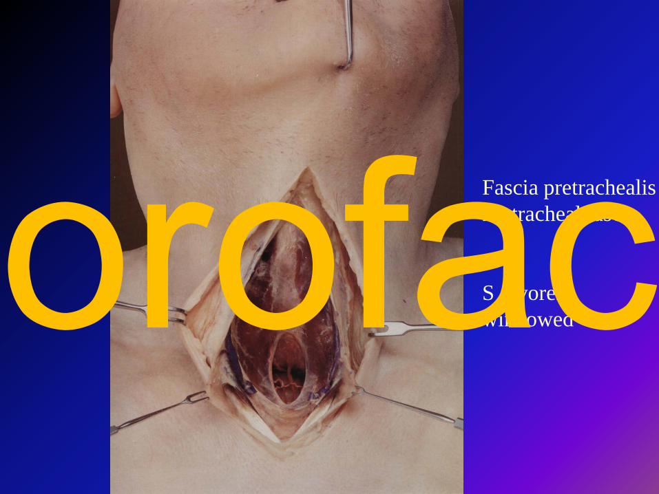
Isthmus gl. thyroideae
Thyroid glandisthmus
orofac
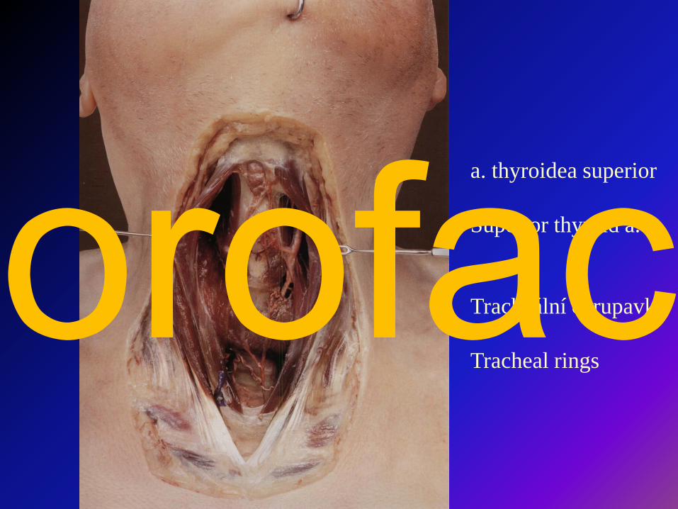
a. thyroidea superior
Superior thyroid a.
Tracheální chrupavky
Tracheal ringsorofac
orofac
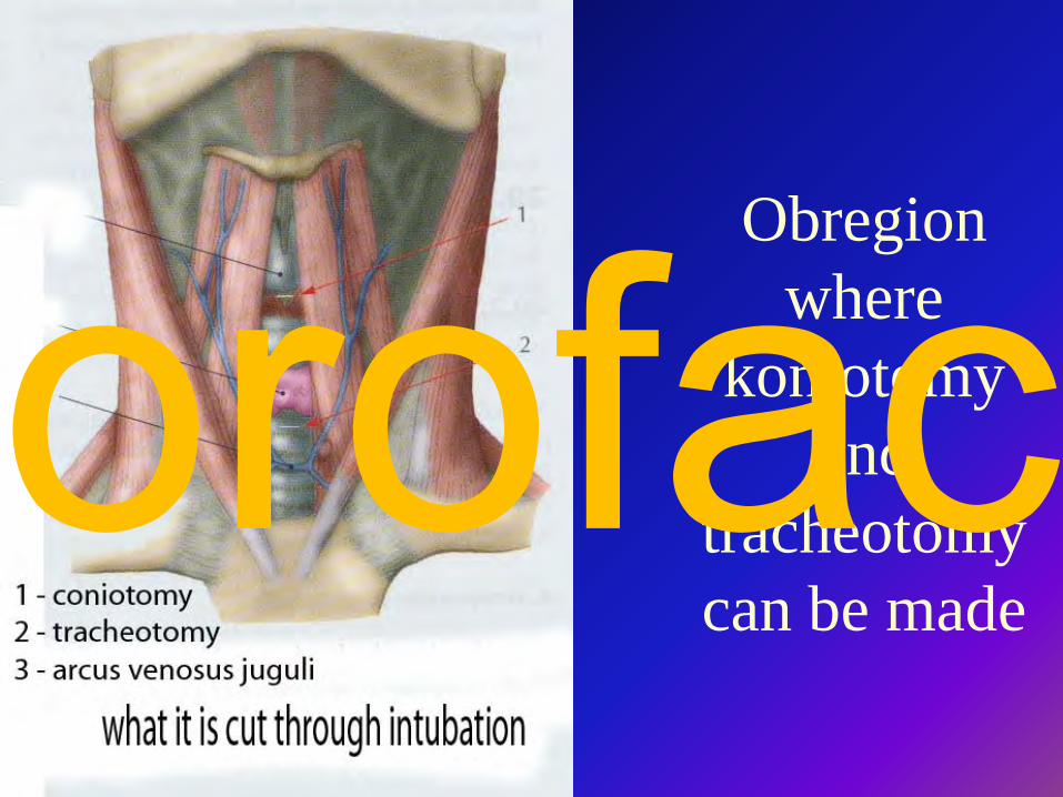
Obregionwhere
koniotomyand
tracheotomycan be made
orofac
orofac
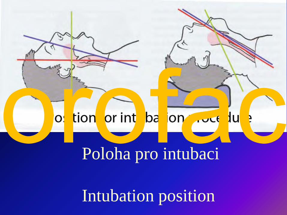
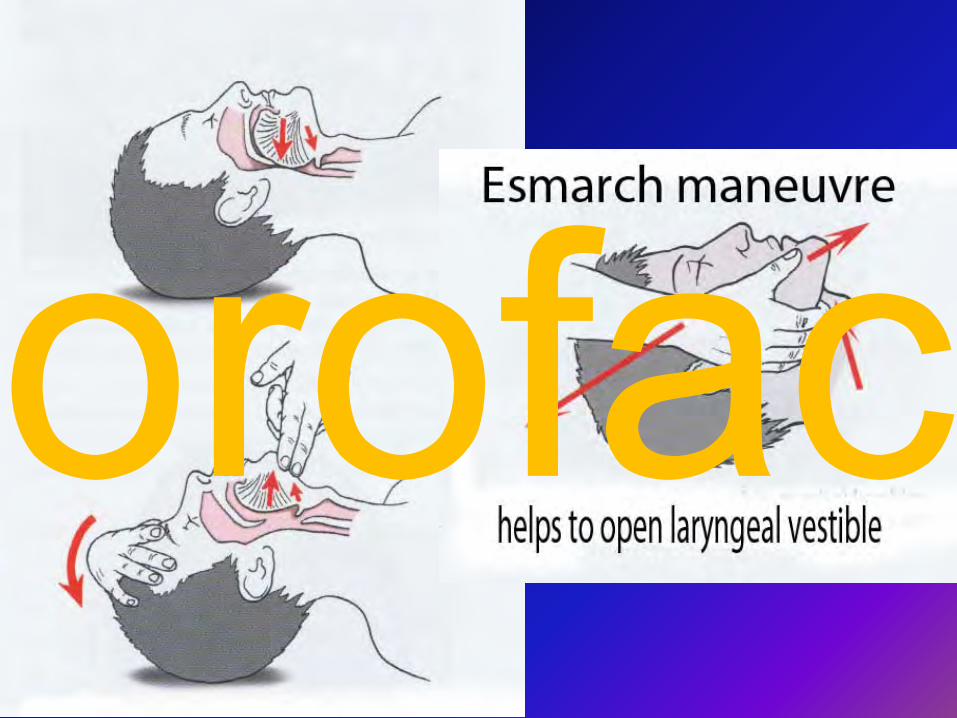
Poloha pro intubaci
Intubation position
orofac
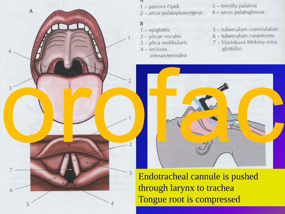
Endotracheal cannule is pushedthrough larynx to tracheaTongue root is compressed
orofac
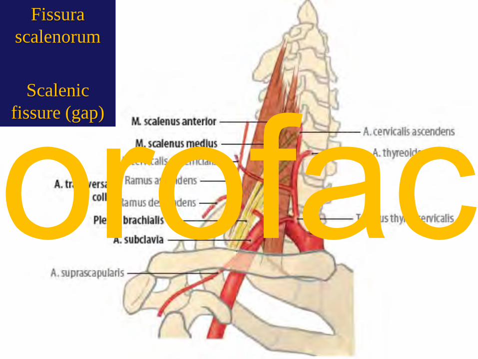
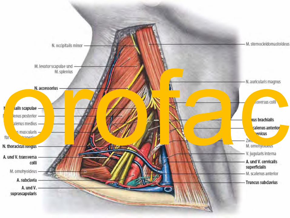
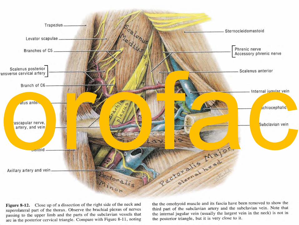
Fissurascalenorum
Scalenicfissure (gap)
orofac
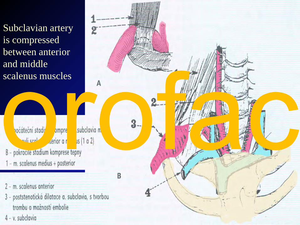
Subclavian artery is compressed between anterior and middle scalenus musclesorofac
orofac
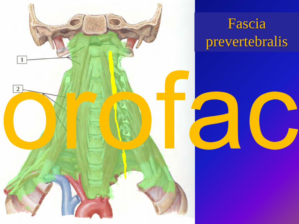
Tooth development and eruptionFascia
prevertebralis
orofac
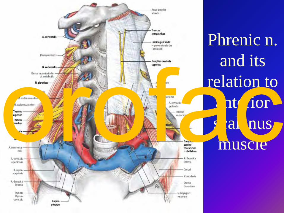
Phrenic n. and its
relation to anterior scalenus muscleorofac
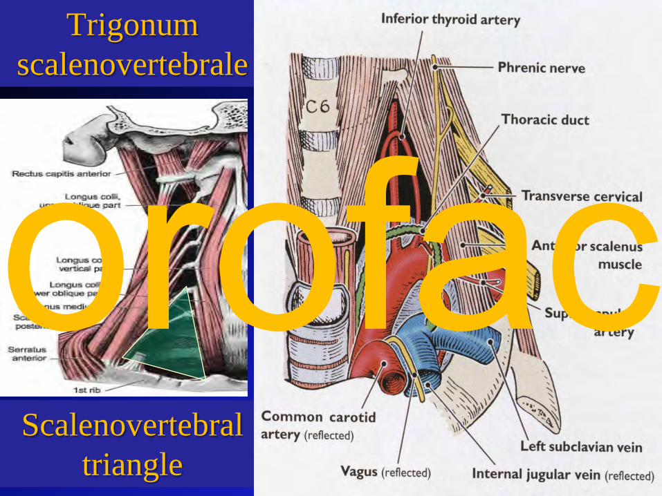
Trigonumscalenovertebrale
Scalenovertebraltriangle
orofac
orofac
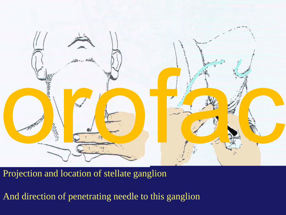
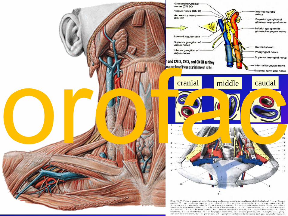
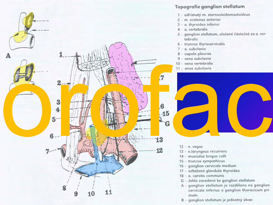
Projection and location of stellate ganglion
And direction of penetrating needle to this ganglion
orofac
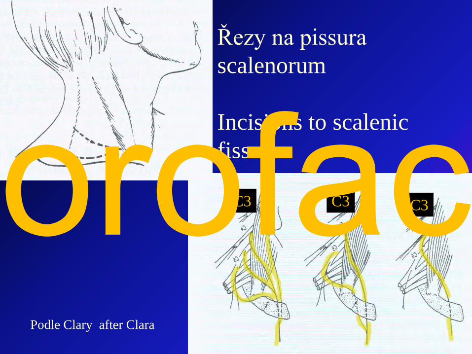
Podle Clary after Clara
Řezy na pissura scalenorum
Incisions to scalenic fissure
C3 C3 C3orofac
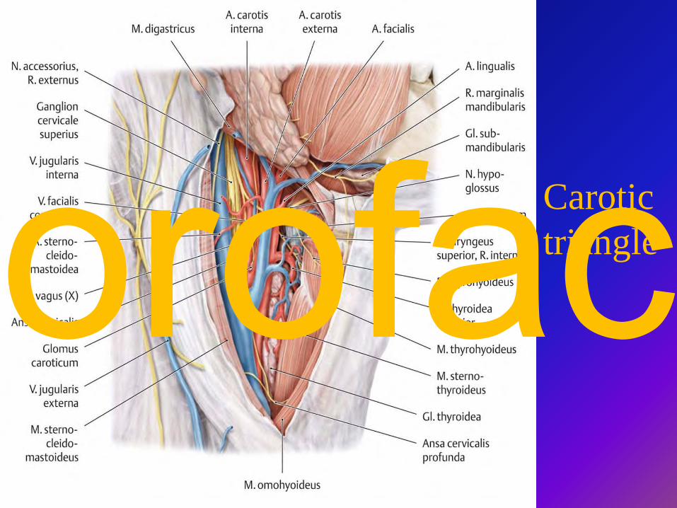
Carotic triangleorofac
orofac
orofac
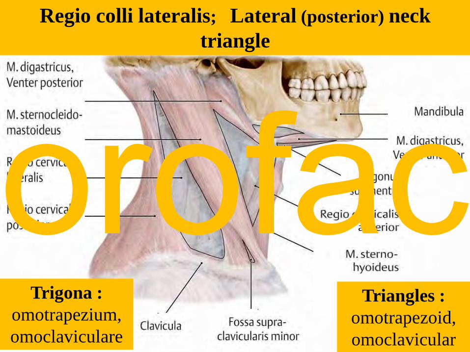
Trigona : omotrapezium, omoclaviculare
Triangles : omotrapezoid, omoclavicular
Regio colli lateralis; Lateral (posterior) neck triangle
orofac
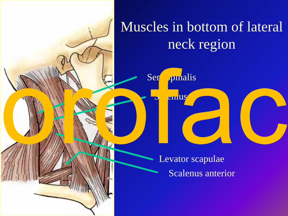
Muscles in bottom of lateral neck region
Semispinalis
Levator scapulae
Splenius
Scalenus anterior
orofac
orofac
orofac
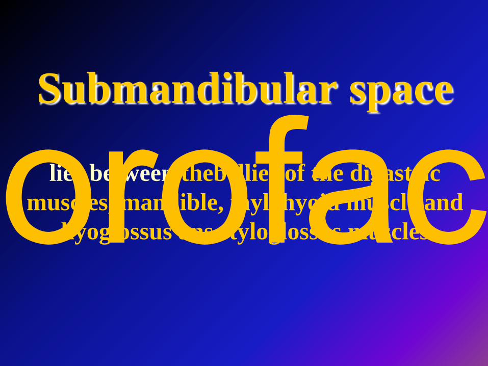
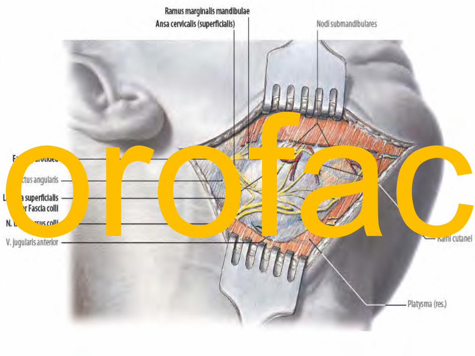
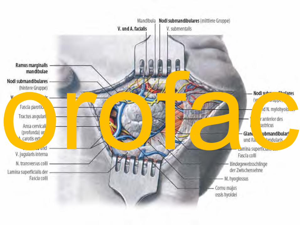
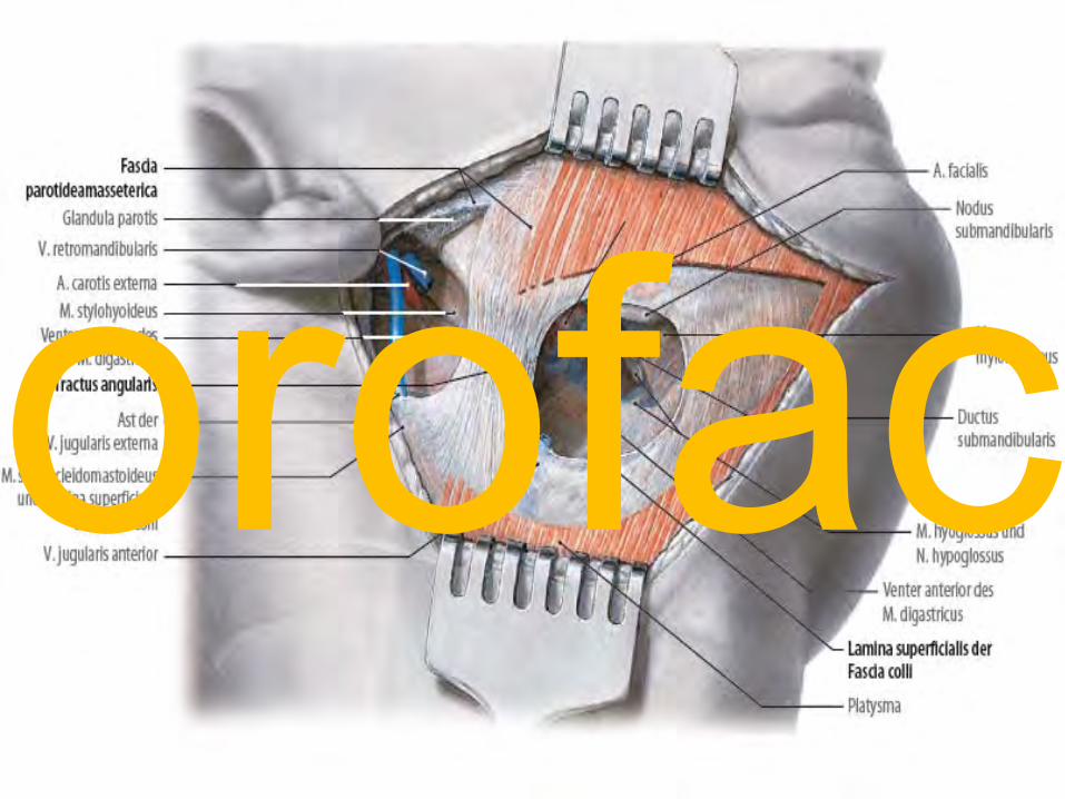
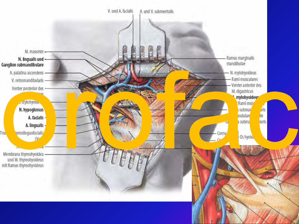
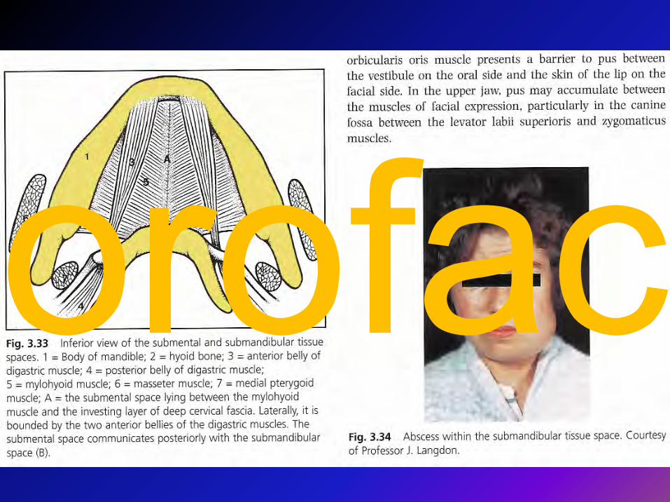
Submandibular space
lies between thebellies of the digastric muscles, mandible, mylohyoid muscle and
hyoglossus ans styloglossus musclesorofac
orofac
orofac
orofac
orofac
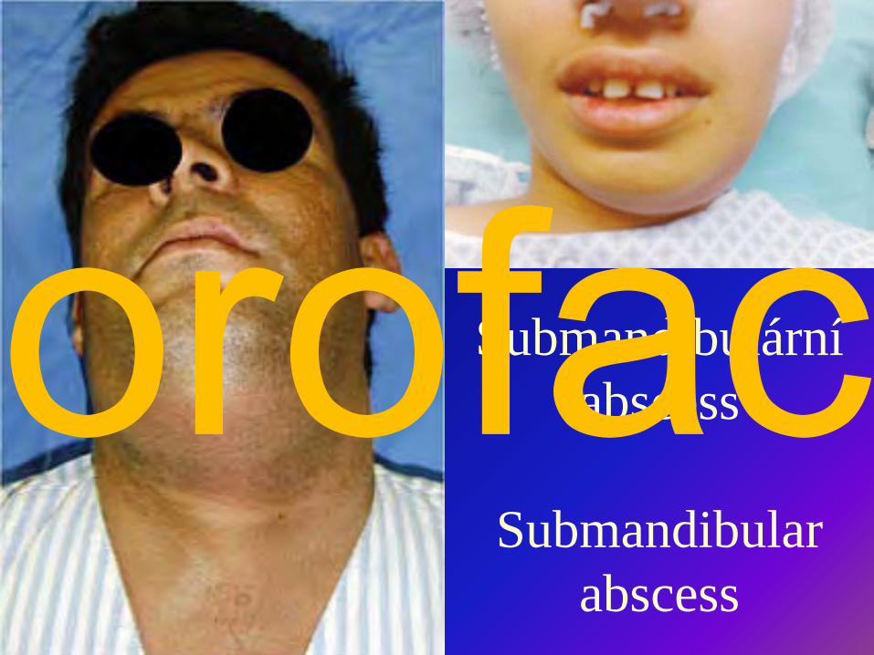
Submandibulární abscess
Submandibular abscess
orofac
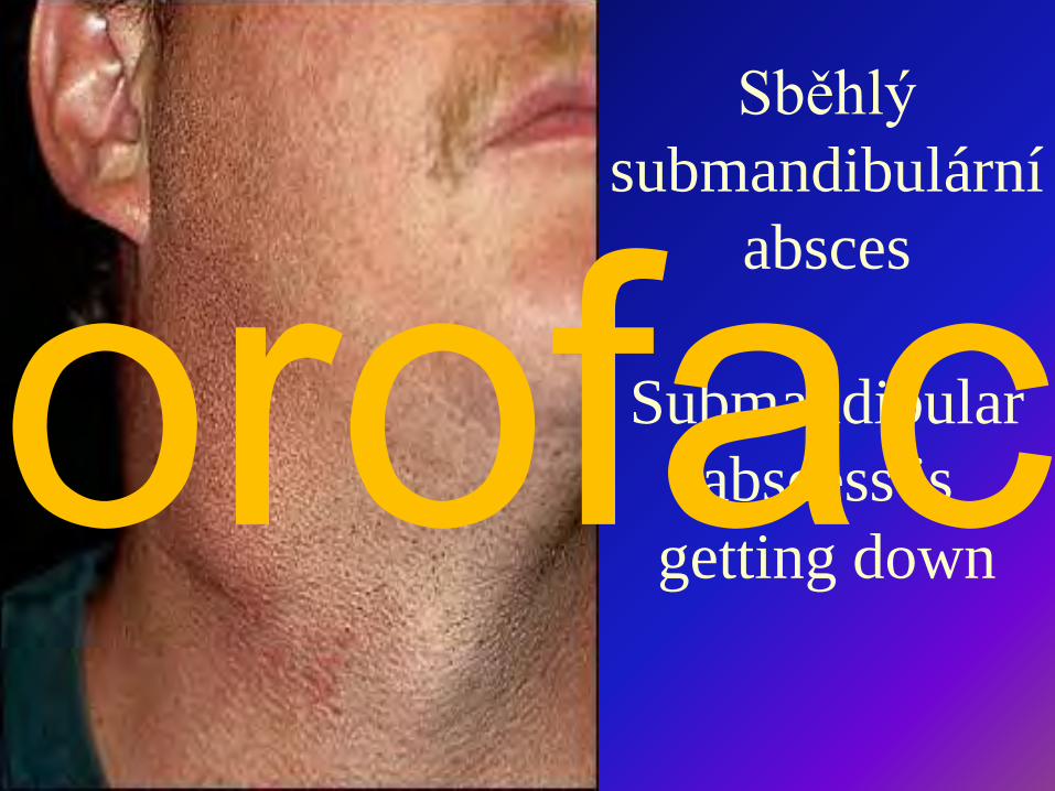
Sběhlý submandibulární
absces
Submandibular abscess is
getting downorofac

Submental space
lies between the mylohyoid muscles and the investing layer of deep cervical fascia
superficiallyorofac
orofac
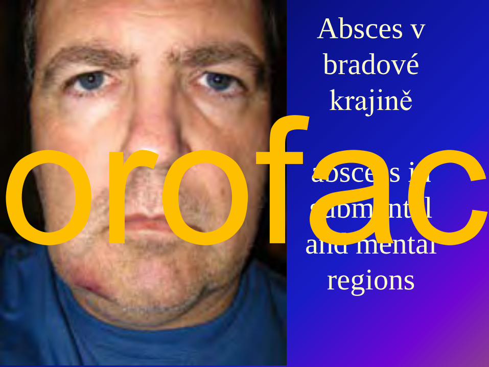
Absces v bradové krajině
abscess in submental and mental
regionsorofac
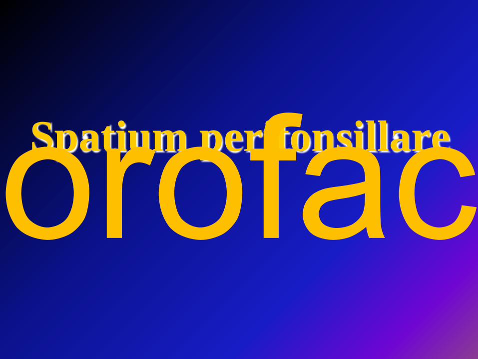
Spatium peritonsillareorofac
orofac
orofac
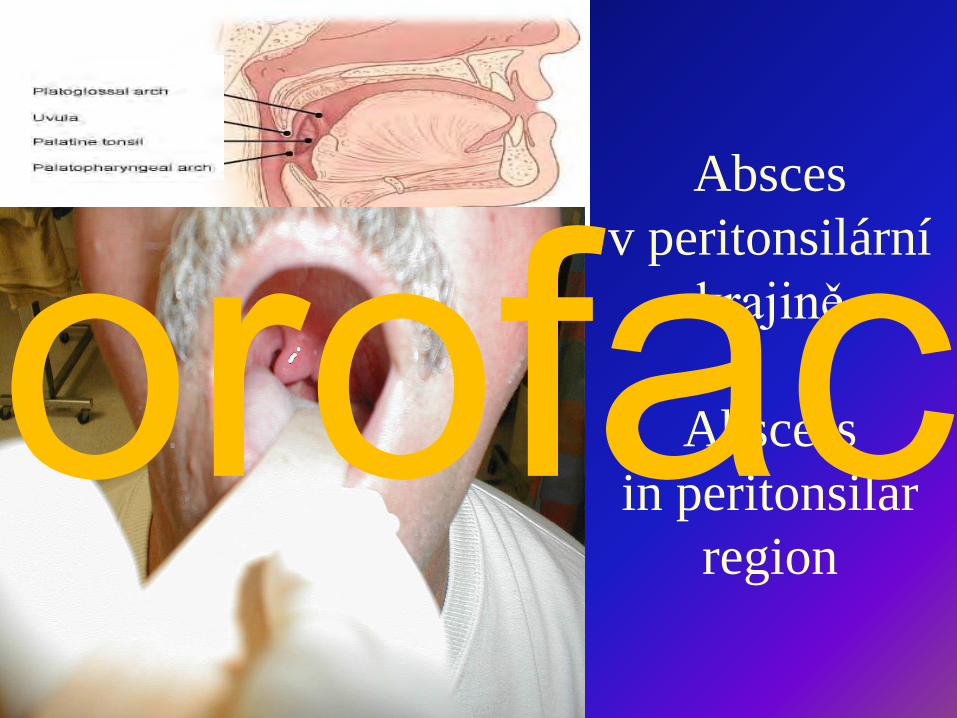
Absces v peritonsilární
krajině
Abscess in peritonsilar
region
orofac
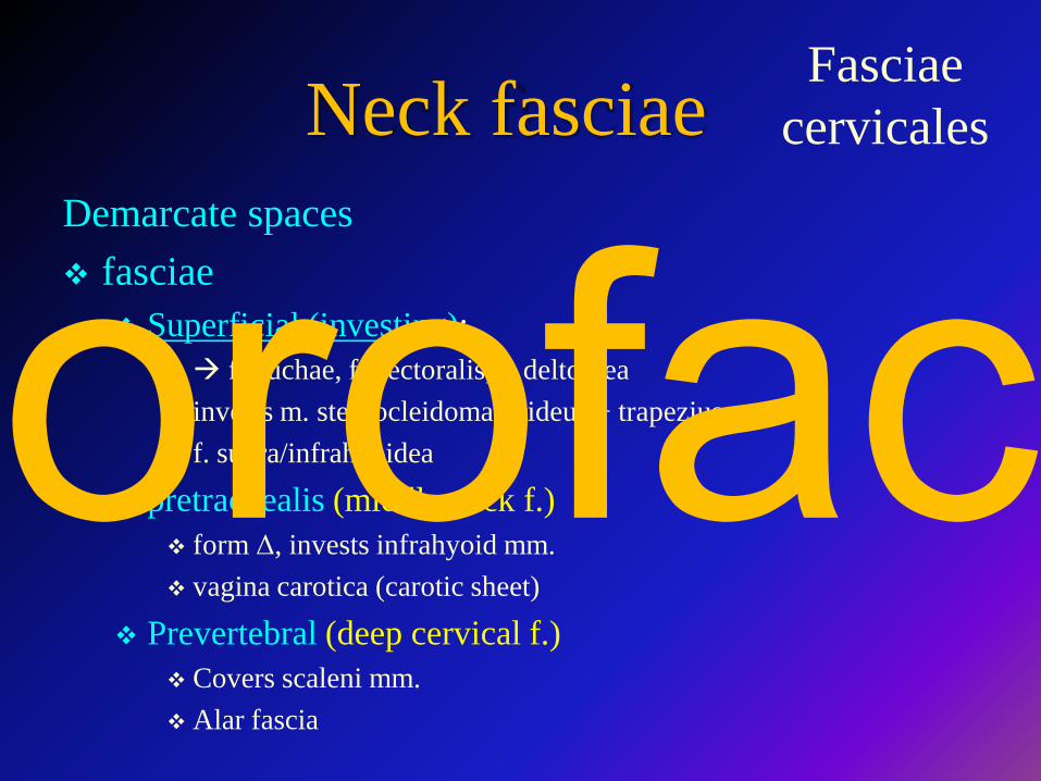
Neck fasciaeDemarcate spaces fasciae
Superficial (investing): f. nuchae, f. pectoralis, f. deltoidea invests m. sternocleidomastoideus + trapezius f. supra/infrahyoidea
pretrachealis (middle neck f.) form Δ, invests infrahyoid mm. vagina carotica (carotic sheet)
Prevertebral (deep cervical f.) Covers scaleni mm. Alar fascia
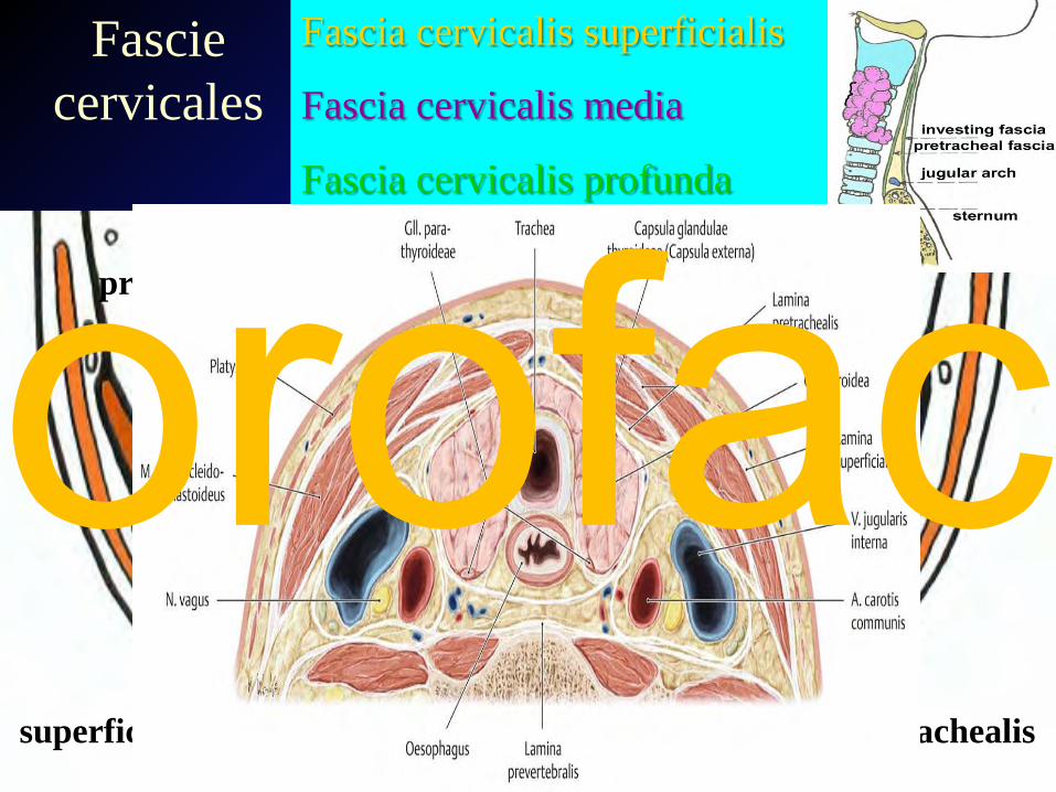
Fasciae cervicales
orofac
pretrachealis
prevertebralis
superficialis
Fascie cervicales
Fascia cervicalis superficialis
Fascia cervicalis media
Fascia cervicalis profunda
orofac
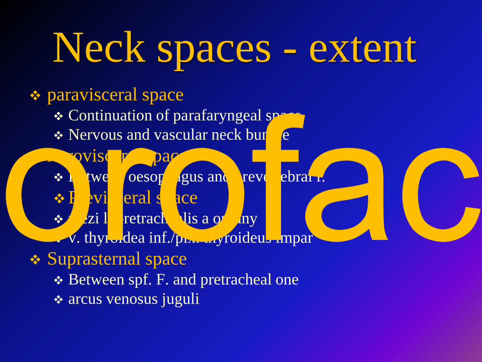
Neck spaces - extent paravisceral space
Continuation of parafaryngeal space Nervous and vascular neck bundle
retrovisceral space Between oesophagus and prevertebral f.Previsceral space mezi l. pretrachealis a orgány v. thyroidea inf./plx. thyroideus impar
Suprasternal space Between spf. F. and pretracheal one arcus venosus juguli
orofac
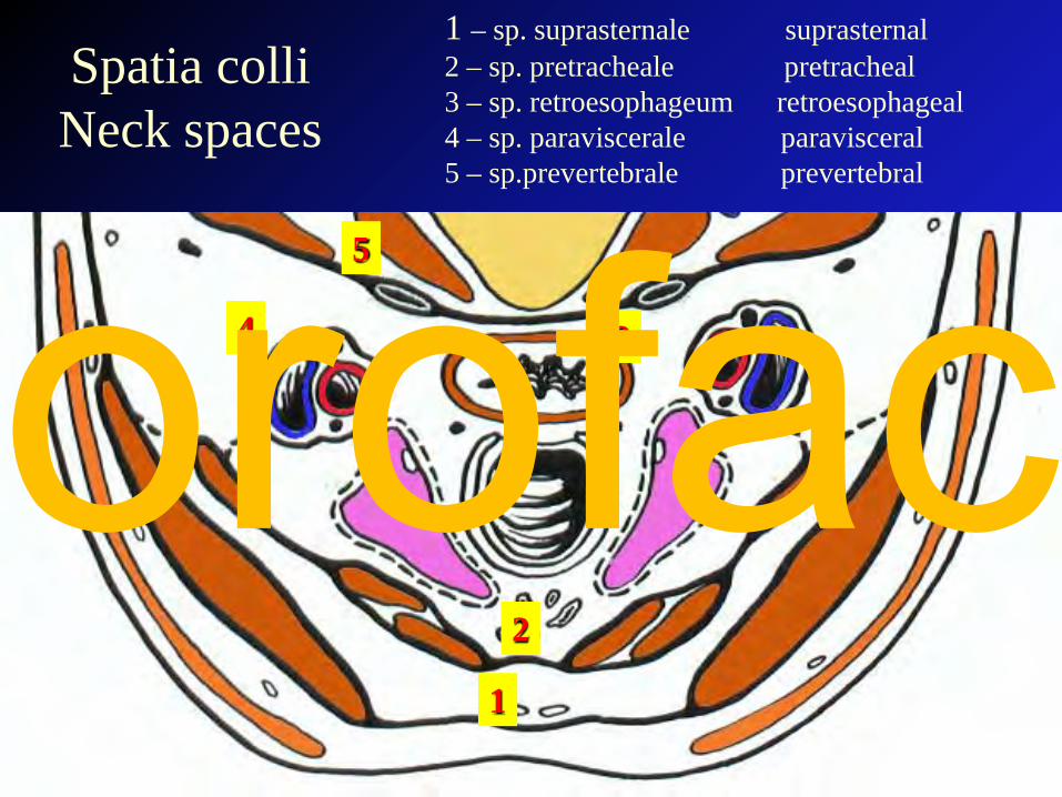
Spatia colli Neck spaces
1
2
34
5
1 – sp. suprasternale suprasternal2 – sp. pretracheale pretracheal3 – sp. retroesophageum retroesophageal4 – sp. paraviscerale paravisceral5 – sp.prevertebrale prevertebral
orofac
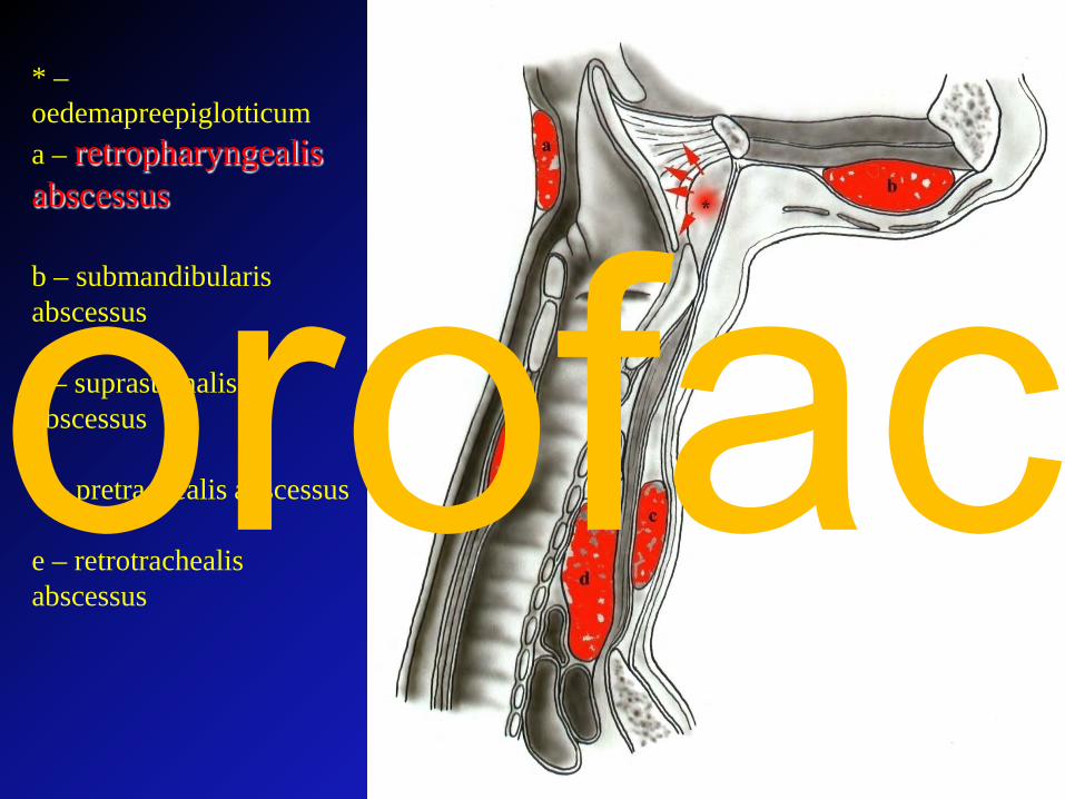
* –oedemapreepiglotticuma – retropharyngealis abscessus
b – submandibularis abscessus
c – suprasternalis abscessus
d – pretrachealis abscessus
e – retrotrachealis abscessus
orofac
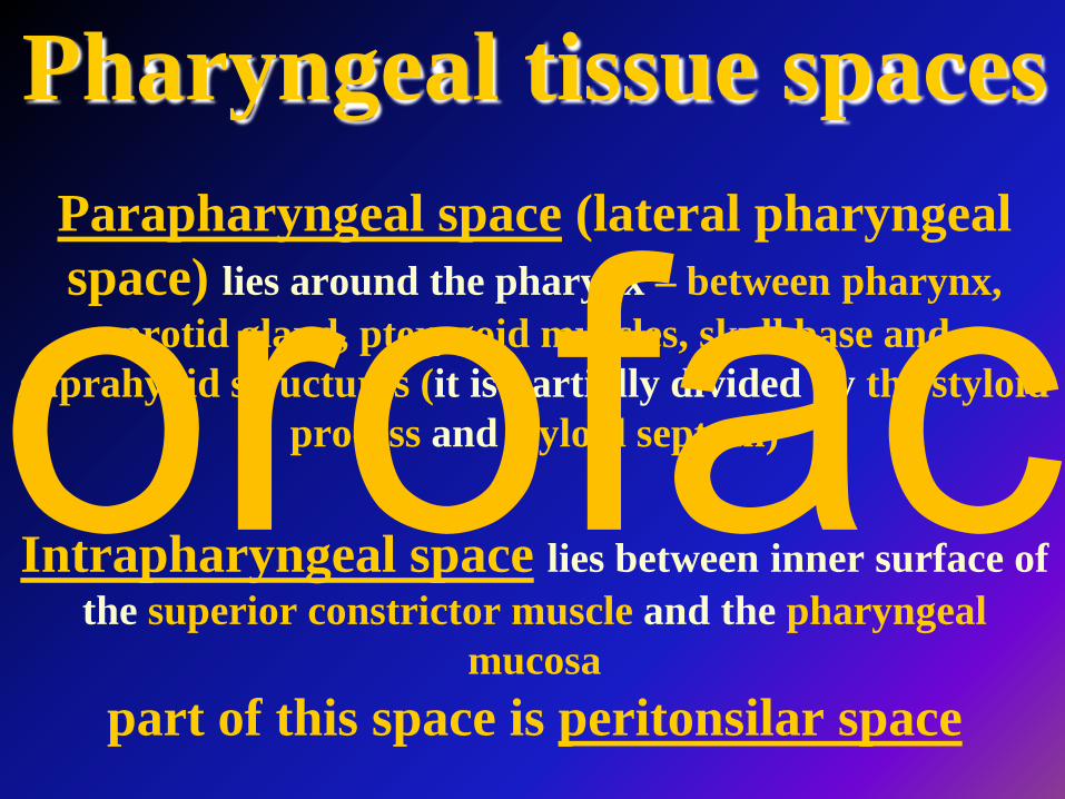
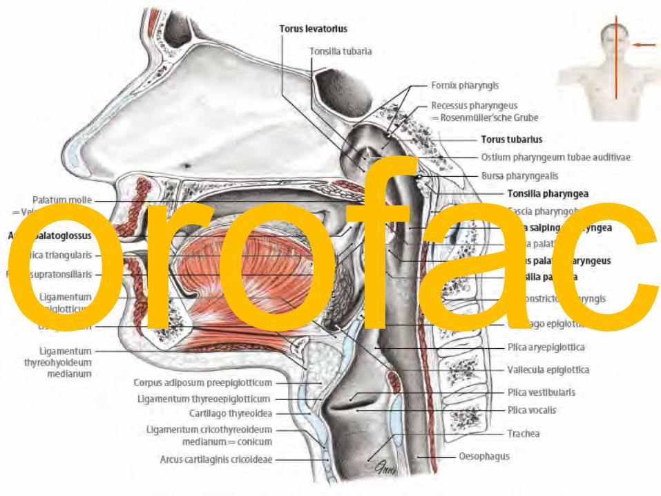
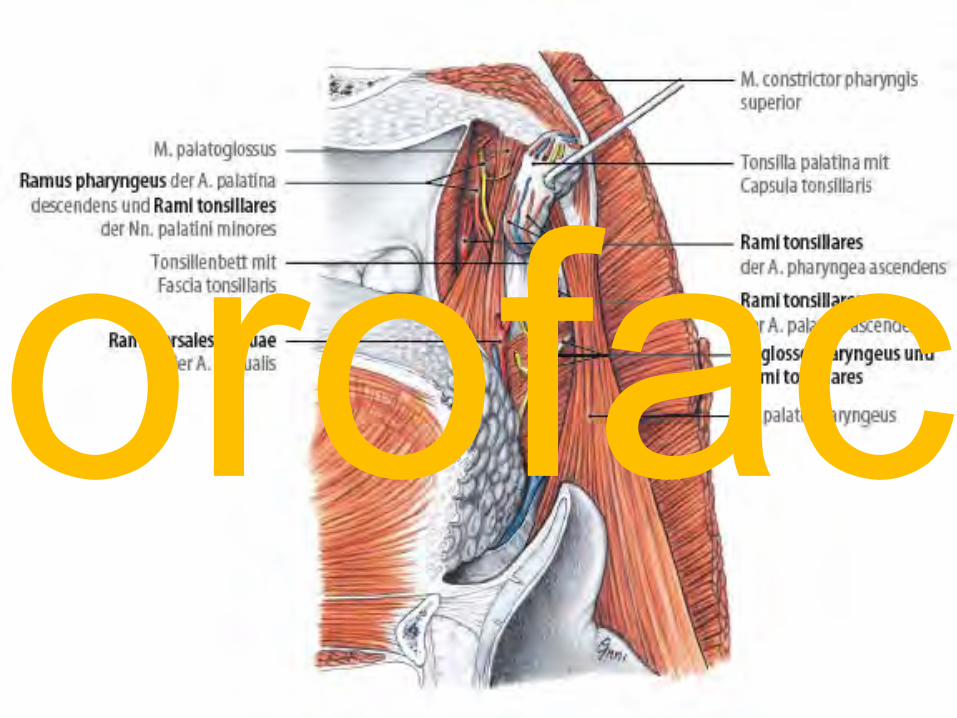
Pharyngeal tissue spacesParapharyngeal space (lateral pharyngeal space) lies around the pharynx – between pharynx,
protid gland, pterygoid muscles, skull base and suprahyoid structures (it is partially divided by the styloid
process and styloid septum)
Intrapharyngeal space lies between inner surface of the superior constrictor muscle and the pharyngeal
mucosapart of this space is peritonsilar space
orofac
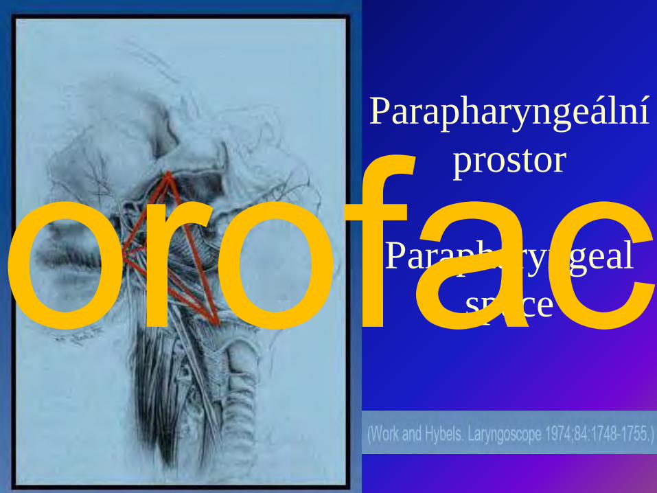
Parapharyngeální prostor
Parapharyngeal spaceorofac
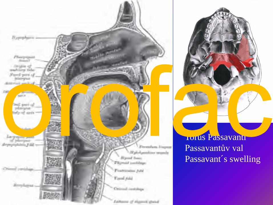
Torus PassavantiPassavantův valPassavant´s swelling
orofac
orofac

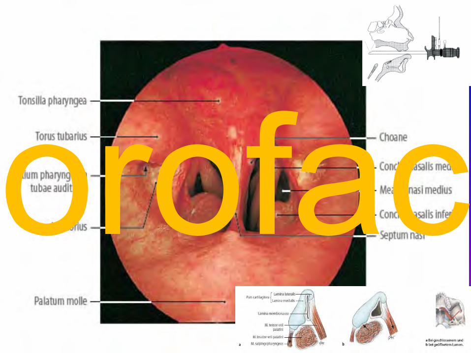
Tensor rozšiřuje ústí levator zahajuje rozšíření
tensor dilates tubalevator starts opening process of
tuba
orofac
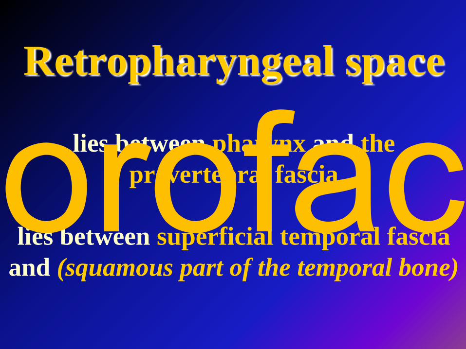
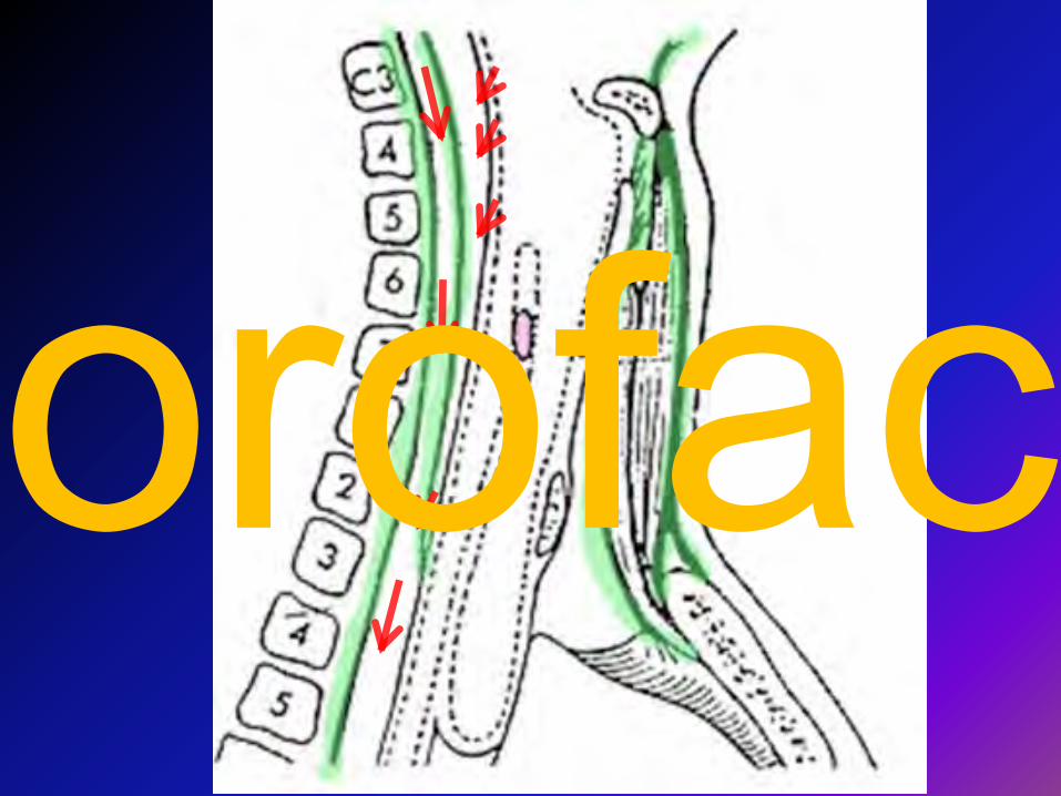
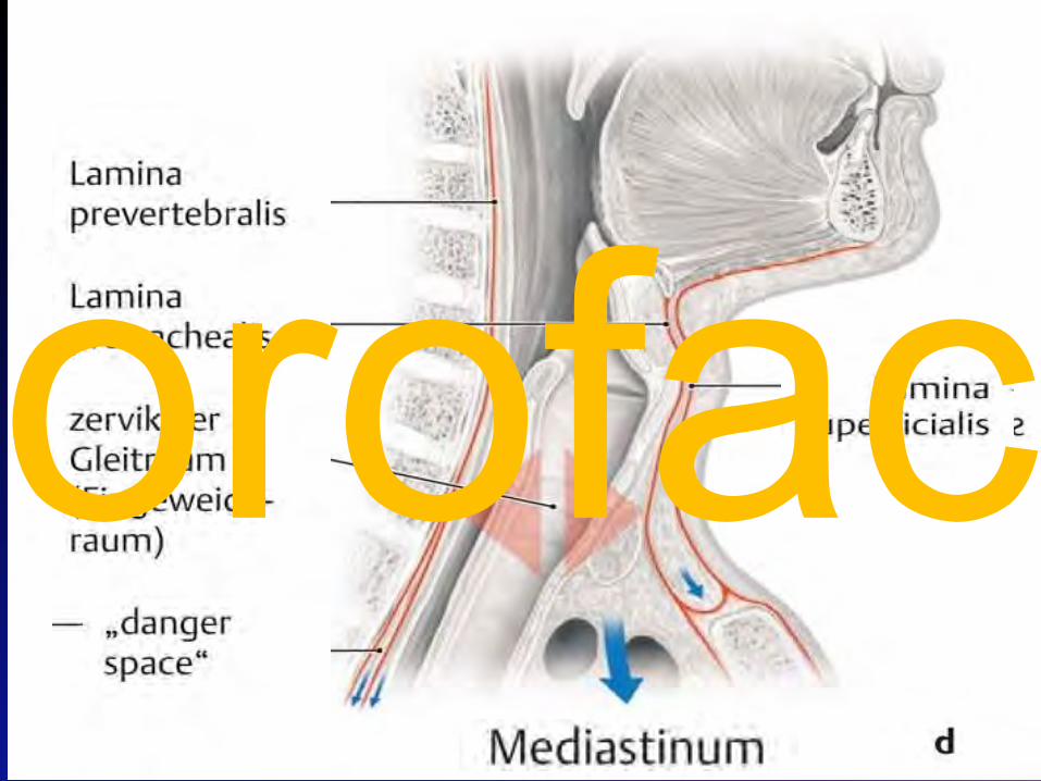
Retropharyngeal space
lies between pharynx and the prevertebral fascia
lies between superficial temporal fascia and (squamous part of the temporal bone)
orofac
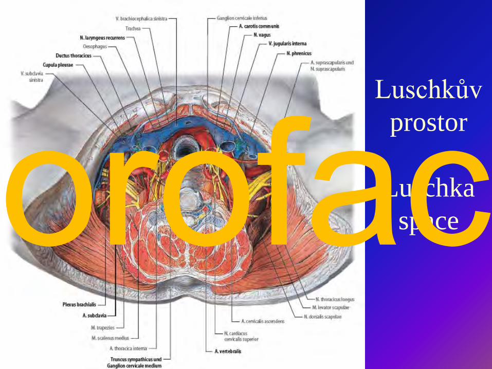
Luschkův prostor
Luschka spaceorofac
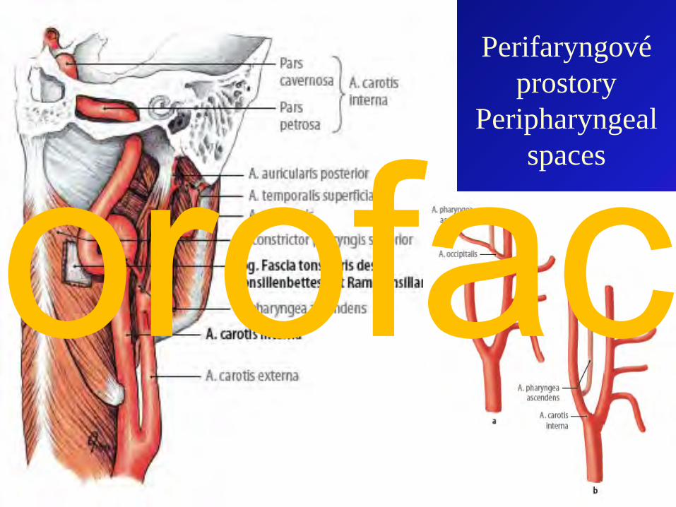
Perifaryngové prostory
Peripharyngeal spaces
orofac
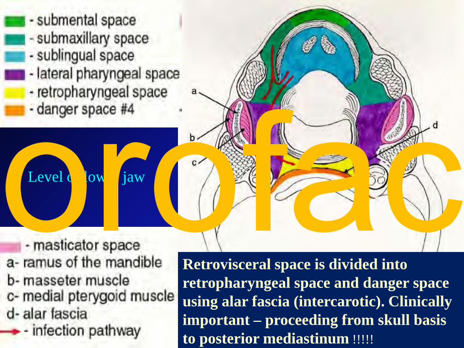
Retrovisceral space is divided into retropharyngeal space and danger space using alar fascia (intercarotic). Clinically important – proceeding from skull basis to posterior mediastinum !!!!!
Level of lower jaworofac
orofac
orofac
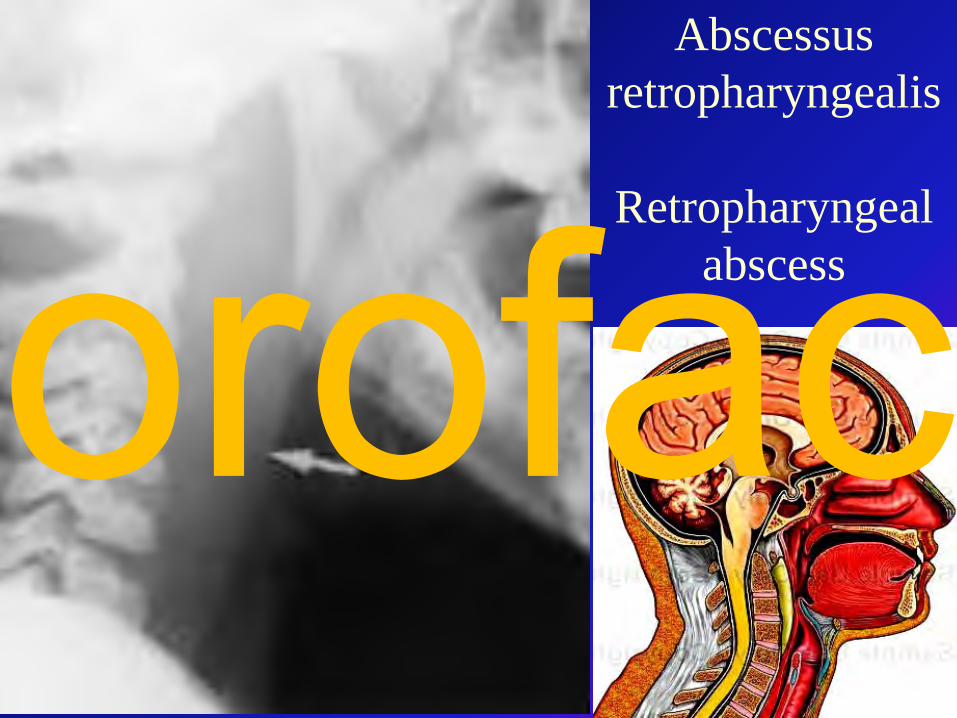
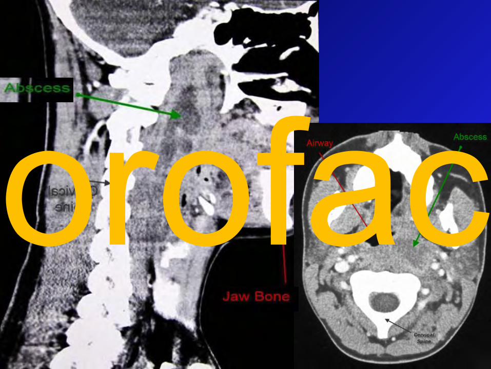
Abscessus retropharyngealis
Retropharyngeal abscessorofac
orofac
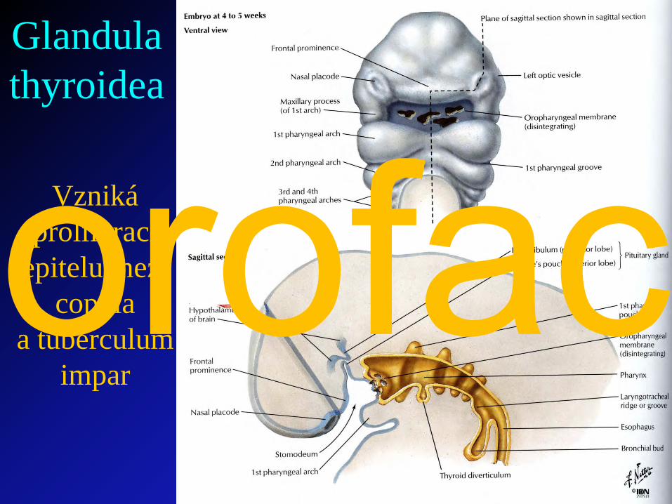
Vzniká proliferací
epitelu mezi copula
a tuberculumimpar
Glandula thyroidea
orofac
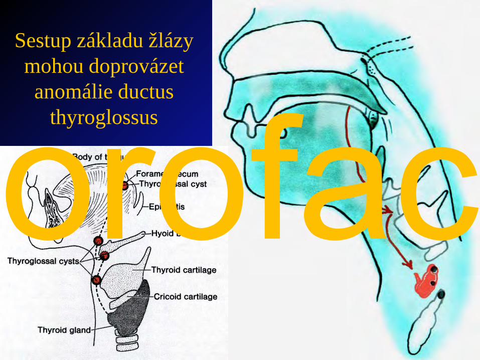
Sestup základu žlázy mohou doprovázet anomálie ductus
thyroglossusorofac
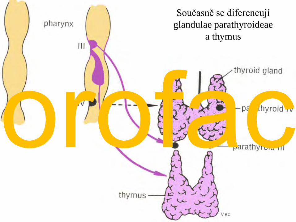
Současně se diferencují glandulae parathyroideae
a thymus
orofac
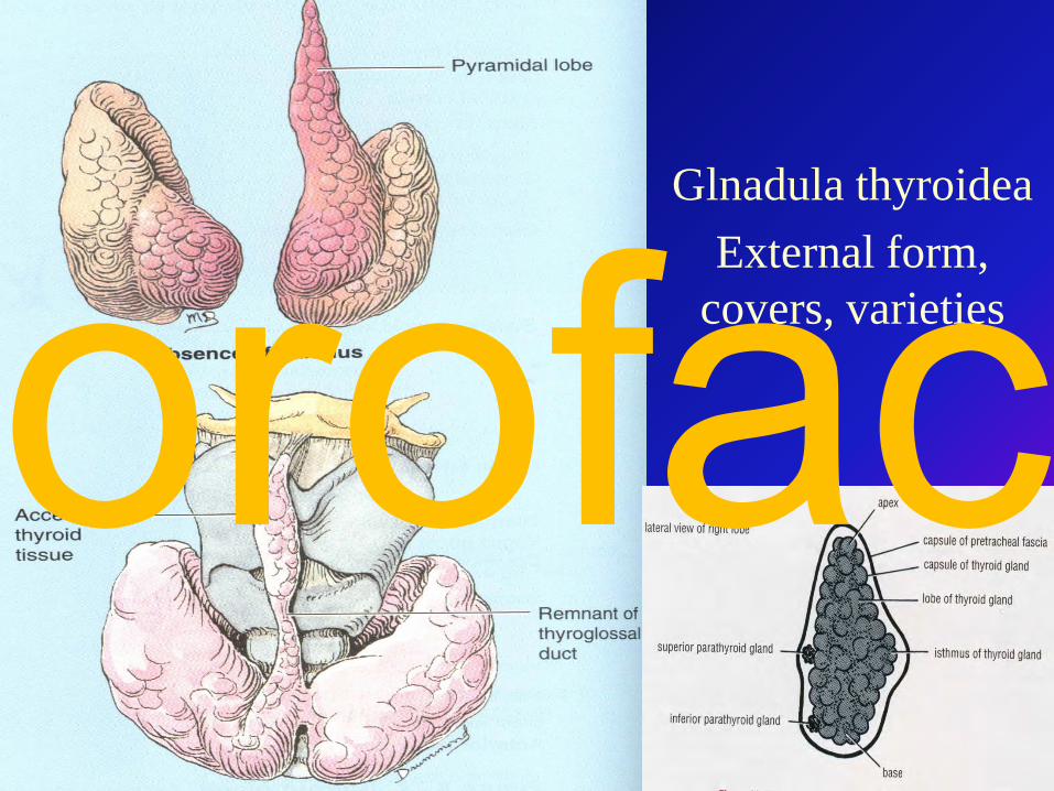
Glnadula thyroideaExternal form,
covers, varietiesorofac
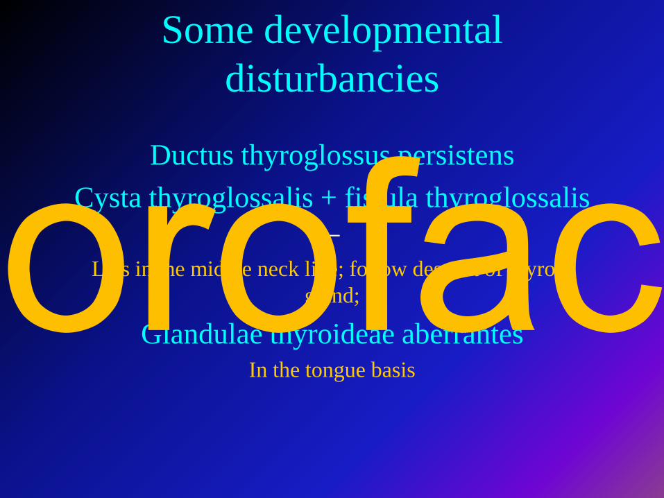
Some developmental disturbancies
Ductus thyroglossus persistensCysta thyroglossalis + fistula thyroglossalis
–Lies in the middle neck line; follow descent of thyroid
gland;
Glandulae thyroideae aberrantesIn the tongue basis
orofac
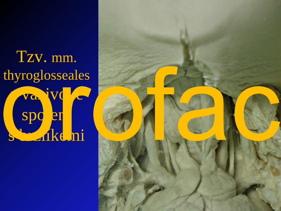
Tzv. mm. thyroglossealesa vazivové
spojení s brzlíkemiorofac
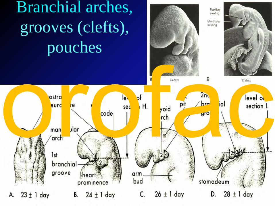
Branchial arches,grooves (clefts),
pouches
orofac
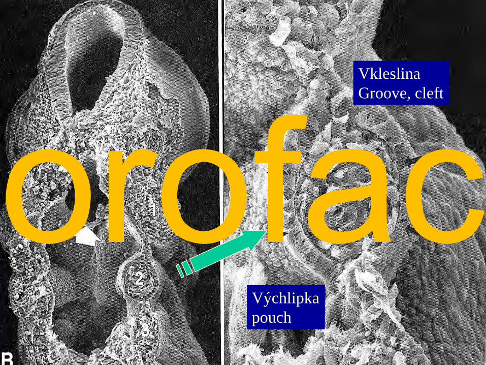
Výchlipkapouch
VkleslinaGroove, cleft
orofac
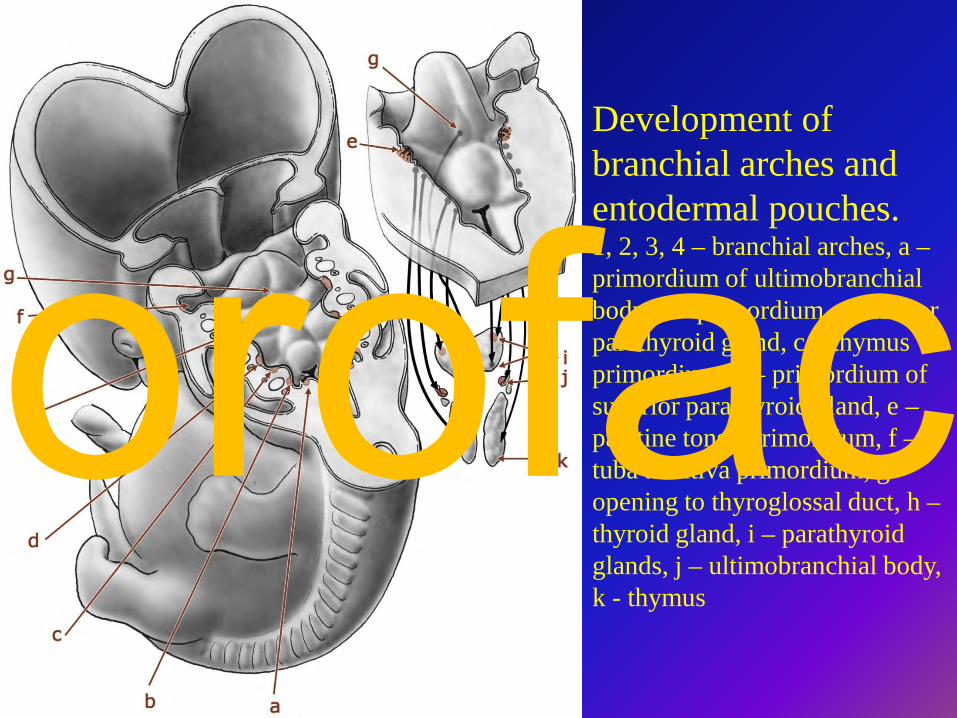
Development of branchial arches and entodermal pouches. 1, 2, 3, 4 – branchial arches, a –primordium of ultimobranchial body, b – primordium of inferior parathyroid gland, c – thymus primordium, d – primordium of superior parathyroid gland, e –palatine tonsil primordium, f –tuba auditiva primordium, g –opening to thyroglossal duct, h –thyroid gland, i – parathyroid glands, j – ultimobranchial body, k - thymus
orofac
27 / 4A
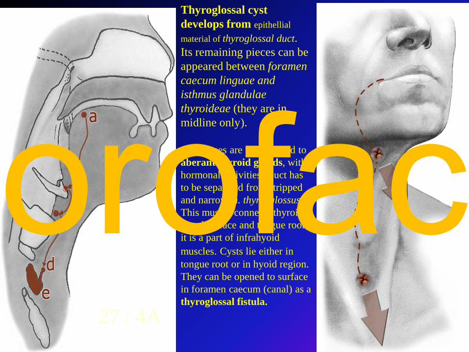
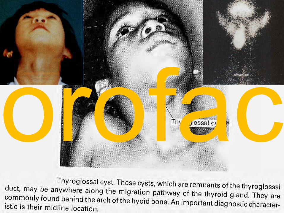
Thyroglossal cyst develops from epithellial material of thyroglossal duct. Its remaining pieces can be appeared between foramen caecum linguae and isthmus glandulae thyroideae (they are in midline only).
Sometimes are transformed to aberant thyroid glands, with hormonal activities. Duct has to be separated from stripped and narrow m. thyreoglossus. This muscle connects thyroid gland surface and tongue root; it is a part of infrahyoid muscles. Cysts lie either in tongue root or in hyoid region. They can be opened to surface in foramen caecum (canal) as a thyroglossal fistula.
orofac
orofac
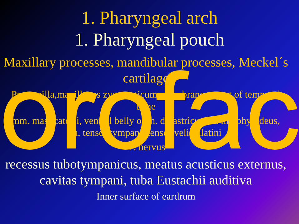
1. Pharyngeal arch1. Pharyngeal pouch
Maxillary processes, mandibular processes, Meckel´s cartilage
Premaxilla,maxilla, os zygomaticum, membranous part of temporal bone
mm. masticatorii, ventral belly of m. digastricus, m. mylohyoideus, m. tensor tympani, tensor veli palatini
V. nervus
recessus tubotympanicus, meatus acusticus externus, cavitas tympani, tuba Eustachii auditiva
Inner surface of eardrum
orofac
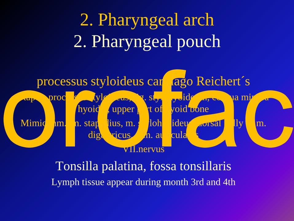
2. Pharyngeal arch2. Pharyngeal pouch
processus styloideus cartilago Reichert´sStapes, processus styloideus, lig. stylohyoideum, cornua minora
hyoidei, upper part of hyoid boneMimic mm., m. stapedius, m. stylohyoideus, dorsal belly of m.
digastricus, mm. auricularesVII.nervus
Tonsilla palatina, fossa tonsillarisLymph tissue appear during month 3rd and 4th
orofac
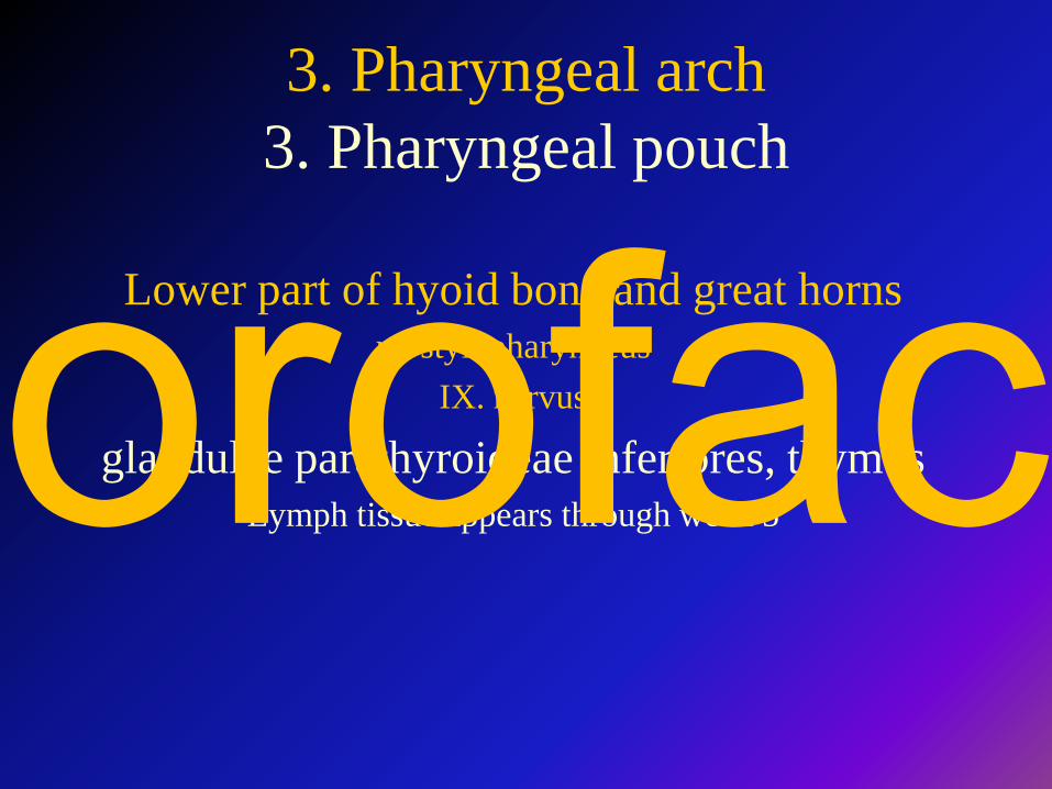
3. Pharyngeal arch3. Pharyngeal pouch
Lower part of hyoid bone and great horns m. stylopharyngeus
IX. nervus
glandulae parathyroideae inferiores, thymus Lymph tissue appears through week 5orofac
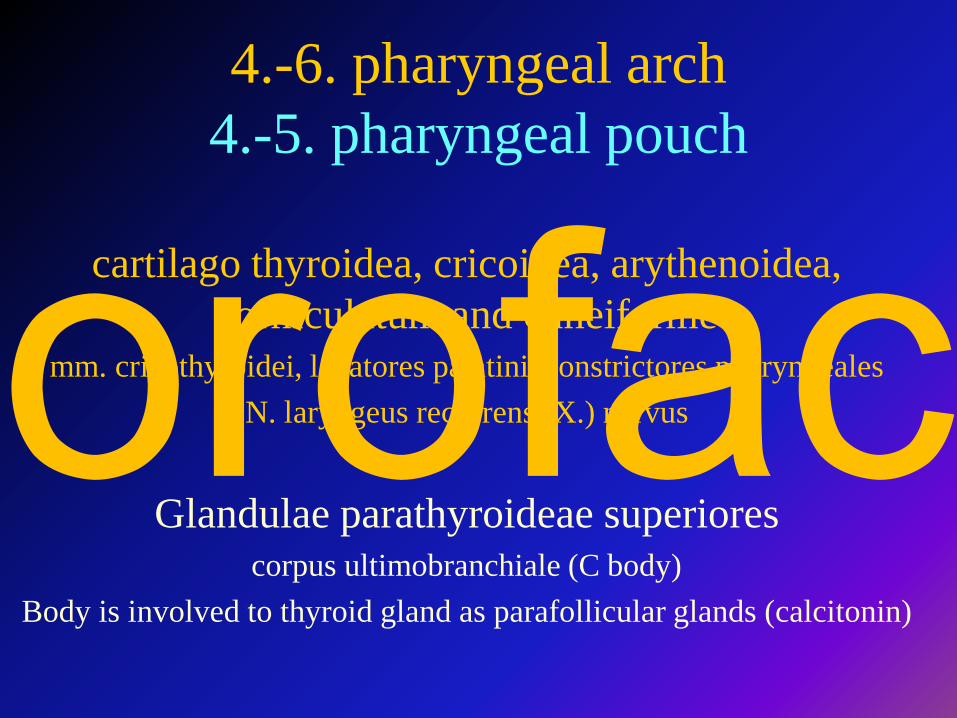
4.-6. pharyngeal arch4.-5. pharyngeal pouch
cartilago thyroidea, cricoidea, arythenoidea, corniculatum and cuneiforme
mm. cricothyroidei, levatores palatini, constrictores pharyngealesN. laryngeus recurrens (X.) nervus
Glandulae parathyroideae superiorescorpus ultimobranchiale (C body)
Body is involved to thyroid gland as parafollicular glands (calcitonin)
orofac
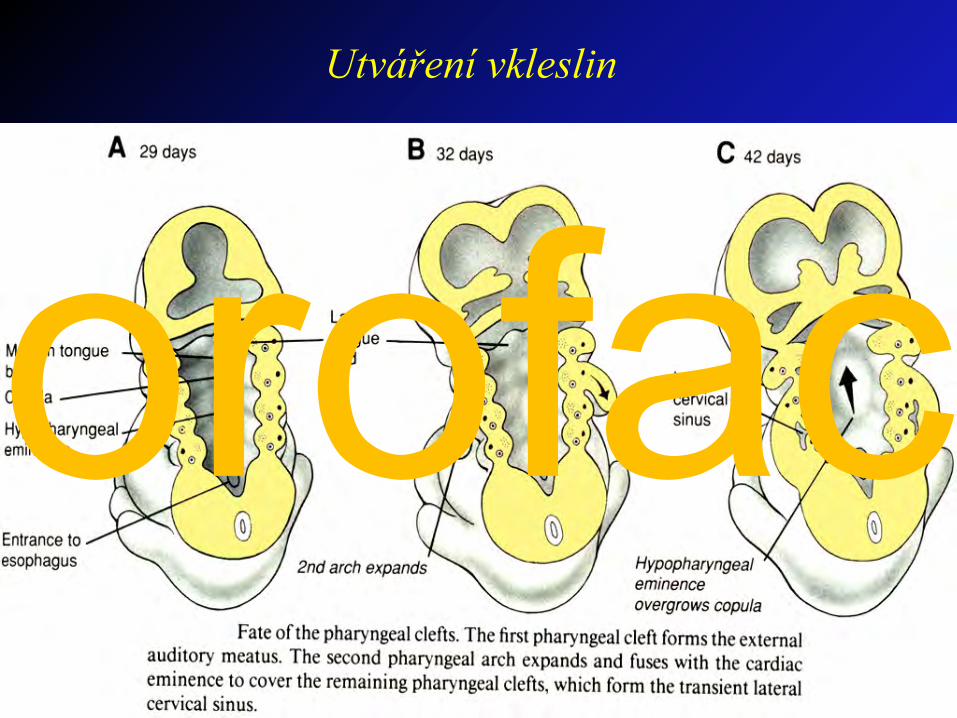
Utváření vkleslin
orofac
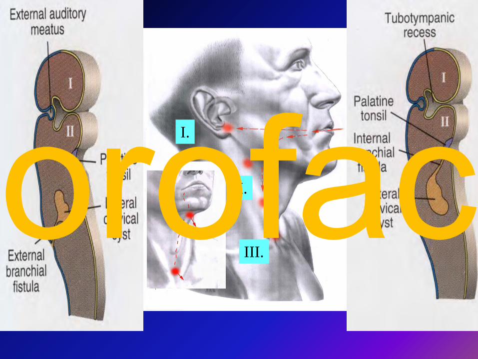
I.
II.
III.orofac
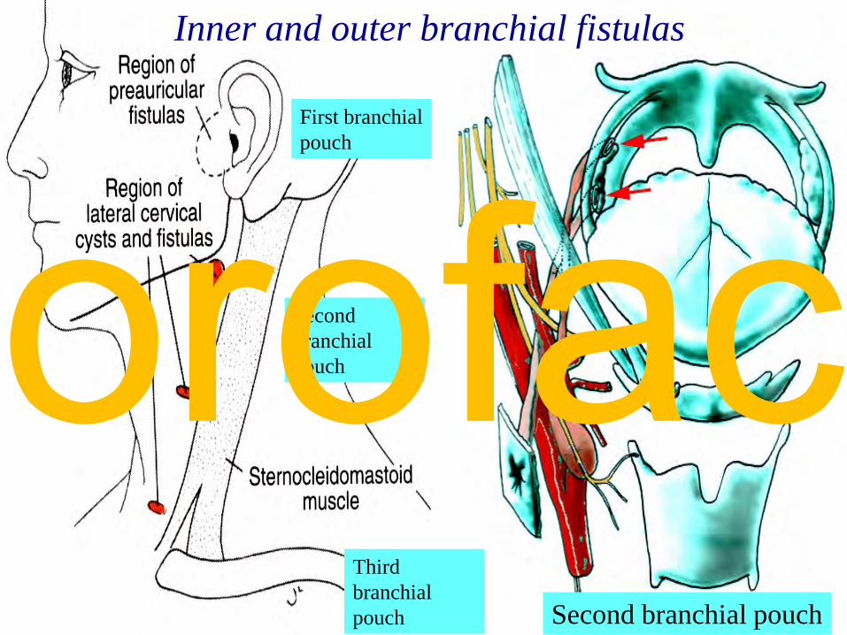
Inner and outer branchial fistulas
Second branchial pouch
Second branchial pouch
Third branchial pouch
First branchial pouch
orofac
LiteratureM. Dykes : Anatomy
2th edition, Mosby 2002
R. Čihák: Anatomie 1, 2, 3Grada Publishing 2003
or s.snell: Clinical anatomy for Medical Students6th edition, Lippincott, Williams & Wilkins
G.J.ToRToRa : Principles of Human Anatomy4th edition, Williams & Wilkins
k.l.MooRe, a.F.Dalley: Clinically Oriented Anatomy4th edition, Williams & Wilkins
F.h.neTTeR: anatomický atlas člověkaGrada Avicenum 2003
orofac