nccn overview testicular cancer a carcinoma, choriocarcinoma, yolk sac tumor, and teratoma....
TRANSCRIPT

© JNCCN–Journal of the National Comprehensive Cancer Network | Volume 10 Number 4 | April 2012
502
Overview An estimated 8590 new cases of testicular cancer will be diagnosed in the United States in 2012.1 Germ cell tumors (GCTs) constitute 95% of malig-nant tumors arising in the testes. These tumors also occur occasionally in extragonadal primary sites, but they are still managed the same as testicular GCTs. Although GCTs are uncommon tumors that consti-tute only 2% of all human malignancies, they are the most common solid tumor in men between 15 and 34 years of age. In addition, the worldwide incidence of these tumors has more than doubled in the past 40 years.
Several risk factors for GCT development have been identified, including history of a GCT, positive family history, cryptorchidism, testicular dysgenesis,
NCCN
Testicular CancerClinical Practice Guidelines in Oncology
Robert J. Motzer, MD; Neeraj Agarwal, MD; Clair Beard, MD; Sam Bhayani, MD; Graeme B. Bolger, MD; Mark K. Buyyounouski, MD, MS; Michael A. Carducci, MD; Sam S. Chang, MD; Toni K. Choueiri, MD; Shilpa Gupta, MD; Steven L. Hancock, MD; Gary R. Hudes, MD; Eric Jonasch, MD; Timothy M. Kuzel, MD; Clayton Lau, MD; Ellis G. Levine, MD; Daniel W. Lin, MD; Kim A. Margolin, MD; M. Dror Michaelson, MD, PhD; Thomas Olencki, DO; Roberto Pili, MD; Thomas W. Ratliff, MD; Bruce G. Redman, DO; Cary N. Robertson, MD; Charles J. Ryan, MD; Joel Sheinfeld, MD; Jue Wang, MD; and Richard B. Wilder, MD
NCCN Clinical Practice Guidelines in Oncology for Testicular Cancer
Key WordsNCCN Clinical Practice Guidelines, NCCN Guidelines, testicular cancer, germ cell tumors, alpha-fetoprotein, lactate dehydro-genase, human chorionic gonadotropin, cisplatin, seminoma, nonseminoma (JNCCN 2012;10:502–535)
NCCN Categories of Evidence and ConsensusCategory 1: Based upon high-level evidence, there is uniform NCCN consensus that the intervention is appropriate.Category 2A: Based upon lower-level evidence, there is uniform NCCN consensus that the intervention is appropriate.Category 2B: Based upon lower-level evidence, there is NCCN consensus that the intervention is appropriate.Category 3: Based upon any level of evidence, there is major NCCN disagreement that the intervention is appropriate.
All recommendations are category 2A unless otherwise noted.
Clinical trials: NCCN believes that the best management for any cancer patient is in a clinical trial. Participation in clinical trials is especially encouraged.
Please NoteThe NCCN Clinical Practice Guidelines in Oncology (NCCN Guidelines®) are a statement of consensus of the authors regarding their views of currently accepted ap-proaches to treatment. Any clinician seeking to apply or consult the NCCN Guidelines® is expected to use indepen-dent medical judgment in the context of individual clinical circumstances to determine any patient’s care or treatment. The National Comprehensive Cancer Network® (NCCN®) makes no representation or warranties of any kind regarding their content, use, or application and disclaims any respon-sibility for their applications or use in any way.
© National Comprehensive Cancer Network, Inc. 2012, All rights reserved. The NCCN Guidelines and the illustrations herein may not be reproduced in any form without the express written permission of NCCN.
Disclosures for the NCCN Testicular Cancer Panel
At the beginning of each NCCN Guidelines panel meeting, panel members disclosed any financial support they have received from industry. Through 2008, this information was published in an aggregate statement in JNCCN and online. Furthering NCCN’s commitment to public transparency, this disclosure process has now been expanded by listing all potential conflicts of interest respective to each individual expert panel member.
Individual disclosures for the NCCN Testicular Cancer Panel members can be found on page 535. (The most recent version of these guidelines and accompanying disclosures, including levels of compensation, are available on the NCCN Web site at www.NCCN.org.)
These guidelines are also available on the Internet. For the latest update, visit www.NCCN.org.

Testicular Cancer
NCCNGuidelines®
© JNCCN–Journal of the National Comprehensive Cancer Network | Volume 10 Number 4 | April 2012
503
Journal of the National Comprehensive Cancer Network
Text continues on p. 521
and Klinefelter syndrome. GCTs are classified as semi-noma or nonseminoma. Nonseminomatous tumors of-ten include multiple cell types, including embryonal cell carcinoma, choriocarcinoma, yolk sac tumor, and teratoma. Teratomas are considered to be either ma-ture or immature, depending on whether adult-type differential cell types or partial somatic differentiation, similar to that present in the fetus, is found. Rarely, a teratoma histologically resembles a somatic cancer, such as sarcoma or adenocarcinoma, and is then re-ferred to as a teratoma with malignant transformation.
The serum tumor markers alpha-fetoprotein (AFP), lactate dehydrogenase (LDH), and β-human chorionic gonadotropin (β-HCG) are critical in di-agnosing GCTs, determining prognosis, and assess-ing treatment outcome. These should be determined
before, during, and after treatment and throughout the follow-up period. Serum tumor markers are use-ful for monitoring all stages of nonseminomas. They are also useful in monitoring metastatic seminomas, because elevated marker levels is the early sign of relapse.
LDH is a less-specific marker than AFP and β-HCG. AFP is a serum tumor marker produced by nonseminomatous cells (embryonal carcinoma, yolk-sac tumor) and may be seen at any stage. The approximate half-life of AFP is 5 to 7 days. A non-seminoma, therefore, is associated with elevated se-rum concentrations of AFP. When patients with a histologically “pure” testicular seminoma have an elevated level of AFP, it is generally assumed that an undetected focus of nonseminoma is present.2,3
NCCN Testicular Cancer Panel Members*Robert J. Motzer, MD/Chair†Þ
Memorial Sloan-Kettering Cancer CenterNeeraj Agarwal, MD‡
Huntsman Cancer Institute at the University of UtahSClair Beard, MD§
Dana-Farber/Brigham and Women’s Cancer CenterSam Bhayani, MDω
Siteman Cancer Center at Barnes-Jewish Hospital and Washington University School of Medicine
Graeme B. Bolger, MD†University of Alabama at BirminghamComprehensive Cancer Center
SMark K. Buyyounouski, MD, MS§Fox Chase Cancer Center
Michael A. Carducci, MD†ÞThe Sidney Kimmel Comprehensive Cancer Center at Johns Hopkins
Sam S. Chang, MDωVanderbilt-Ingram Cancer Center
Toni K. Choueiri, MD†ÞDana-Farber/Brigham and Women’s Cancer Center
Shilpa Gupta, MD†H. Lee Moffitt Cancer Center & Research Institute
Steven L. Hancock, MD§ÞStanford Cancer Institute
Gary R. Hudes, MD†‡Fox Chase Cancer Center
Eric Jonasch, MD†The University of Texas MD Anderson Cancer Center
Timothy M. Kuzel, MD‡Robert H. Lurie Comprehensive Cancer Center ofNorthwestern University
Clayton Lau, MDωCity of Hope Comprehensive Cancer Center
Ellis G. Levine, MD†Roswell Park Cancer Institute
Daniel W. Lin, MDωUniversity of Washington/Seattle Cancer Care Alliance
Kim A. Margolin, MD†‡Fred Hutchinson Cancer Research Center/Seattle Cancer Care Alliance
M. Dror Michaelson, MD, PhD†Massachusetts General Hospital Cancer Center
Thomas Olencki, DO†The Ohio State University Comprehensive Cancer Center – James Cancer Hospital and Solove Research Institute
Roberto Pili, MD†Roswell Park Cancer Institute
Thomas W. Ratliff, MD†St. Jude Children’s Research Hospital/University of Tennessee Cancer Institute
Bruce G. Redman, DO†University of Michigan Comprehensive Cancer Center
Cary N. Robertson, MDωDuke Cancer Institute
Charles J. Ryan, MD†ωUCSF Helen Diller Family Comprehensive Cancer Center
Joel Sheinfeld, MDωMemorial Sloan-Kettering Cancer Center
Jue Wang, MD†UNMC Eppeley Cancer Center atThe Nebraska Medical Center
*SRichard B. Wilder, MD§H. Lee Moffitt Cancer Center & Research Institute
NCCN Staff: Mary Dwyer, MS, and Rashmi Kumar, PhD
KEY:
*Writing Committee Member
Subcomittee: sPrinciples of Radiotherapy for Pure Testicular Seminoma
Specialties: †Medical Oncology; ÞInternal Medicine; ‡Hematology/Hematology Oncology; §Radiotherapy/Radiation Oncology; and ωUrology

© JNCCN–Journal of the National Comprehensive Cancer Network | Volume 10 Number 4 | April 2012
504
Testicular Cancer Version 1:2012
Clinical trials: NCCN believes that the best management of any cancer patient is in a clinical trial. Participation in clinical trials is especially encouraged. All recommendations are category 2A unless otherwise indicated.
WORKUP PRIMARYTREATMENTb
PATHOLOGICDIAGNOSIS
ab
Quantitative analysis of beta subunit.Though rare, when a patient presents with rapidly increasing beta-HCG, symptoms related to disseminated disease and a testicular mass, chemotherapy can be initiated immediately without waiting for a biopsy diagnosis.
c
d
Mediastinal primary site seminoma should be treated by risk status used for gonadal seminomas with etoposide/cisplatin for 4 cycles orbleomycin/etoposide/cisplatin for 3 cycles.
If AFP-positive, treat as nonseminoma.
Suspicioustesticularmass
Nonseminomatous germ cell tumor(includes mixed seminoma tumors andseminoma histology with elevated AFP)
•••
Discuss sperm bankingRadical inguinal orchiectomyConsider inguinal biopsy ofcontralateral testis if:
Suspicious ultrasound forintratesticularabnormalitiesCryptorchid testisMarked atrophy
➤
➤
➤
••••••
H&PAlpha-fetoprotein (AFP)beta-hCG
Chemistry profileChest x-ray
a
LDH
• Testicular ultrasound
CLINICAL STAGE
••
••••
Abdominal/pelvic CTChest CT if:
Positive abdominal CT or abnormalchest x-ray
Repeat beta-HCG, LDH,AFPBrain MRI, if clinically indicatedBone scan, if clinically indicatedDiscuss sperm banking
➤
f
StageIA, IB
StageIIA, IIB
StageIIC, III
See Primary Treatment and Follow-Up(page 506)
See Primary Treatment and Follow-Up(page 506)
See Primary Treatment and Follow-Up(page 506)
ef
PET scan is not clinically indicated for nonseminoma.Elevated values should be followed after orchiectomy with repeated determination to allow precise staging.
StageIS
See Primary Treatment and Follow-Up(page 506)
POSTDIAGNOSTIC WORKUPe
•••••
Abdominal/pelvic CTRepeat beta-HCG, LDH,AFPBrain MRI, if clinically indicatedBone scan, if clinically indicatedDiscuss sperm banking
± chest imagingf
Stage IA, IB, IS:See Primary Treatment (page 508)
Stage IIA, IIB:See Treatment (page 508)Primary
Stage IIC, IIIA, IIIB, IIIC,and brain metastasis:See Treatment (page 510)Primary
Pure seminoma germ cell tumor(pure seminoma histology and AFPnegative may have elevated beta-HCG)
c
d;

NCCN Clinical Practice Guidelines in Oncology
© JNCCN–Journal of the National Comprehensive Cancer Network | Volume 10 Number 4 | April 2012
505
Testicular Cancer Version 1:2012
Version1.2012, 01-17-12 ©2012 National Comprehensive Cancer Network, Inc. All rights reserved. The NCCN Guidelines® and this illustration may not be reproduced in any form without the express written permission of NCCN®.
WORKUP PRIMARYTREATMENTb
PATHOLOGICDIAGNOSIS
ab
Quantitative analysis of beta subunit.Though rare, when a patient presents with rapidly increasing beta-HCG, symptoms related to disseminated disease and a testicular mass, chemotherapy can be initiated immediately without waiting for a biopsy diagnosis.
c
d
Mediastinal primary site seminoma should be treated by risk status used for gonadal seminomas with etoposide/cisplatin for 4 cycles orbleomycin/etoposide/cisplatin for 3 cycles.
If AFP-positive, treat as nonseminoma.
Suspicioustesticularmass
Nonseminomatous germ cell tumor(includes mixed seminoma tumors andseminoma histology with elevated AFP)
•••
Discuss sperm bankingRadical inguinal orchiectomyConsider inguinal biopsy ofcontralateral testis if:
Suspicious ultrasound forintratesticularabnormalitiesCryptorchid testisMarked atrophy
➤
➤
➤
••••••
H&PAlpha-fetoprotein (AFP)beta-hCG
Chemistry profileChest x-ray
a
LDH
• Testicular ultrasound
CLINICAL STAGE
••
••••
Abdominal/pelvic CTChest CT if:
Positive abdominal CT or abnormalchest x-ray
Repeat beta-HCG, LDH,AFPBrain MRI, if clinically indicatedBone scan, if clinically indicatedDiscuss sperm banking
➤
f
StageIA, IB
StageIIA, IIB
StageIIC, III
See Primary Treatment and Follow-Up(page 506)
See Primary Treatment and Follow-Up(page 506)
See Primary Treatment and Follow-Up(page 506)
ef
PET scan is not clinically indicated for nonseminoma.Elevated values should be followed after orchiectomy with repeated determination to allow precise staging.
StageIS
See Primary Treatment and Follow-Up(page 506)
POSTDIAGNOSTIC WORKUPe
•••••
Abdominal/pelvic CTRepeat beta-HCG, LDH,AFPBrain MRI, if clinically indicatedBone scan, if clinically indicatedDiscuss sperm banking
± chest imagingf
Stage IA, IB, IS:See Primary Treatment (page 508)
Stage IIA, IIB:See Treatment (page 508)Primary
Stage IIC, IIIA, IIIB, IIIC,and brain metastasis:See Treatment (page 510)Primary
Pure seminoma germ cell tumor(pure seminoma histology and AFPnegative may have elevated beta-HCG)
c
d;
PURE SEMINOMA
© JNCCN–Journal of the National Comprehensive Cancer Network | Volume 10 Number 4 | April 2012
506
Testicular Cancer Version 1:2012
Clinical trials: NCCN believes that the best management of any cancer patient is in a clinical trial. Participation in clinical trials is especially encouraged. All recommendations are category 2A unless otherwise indicated.
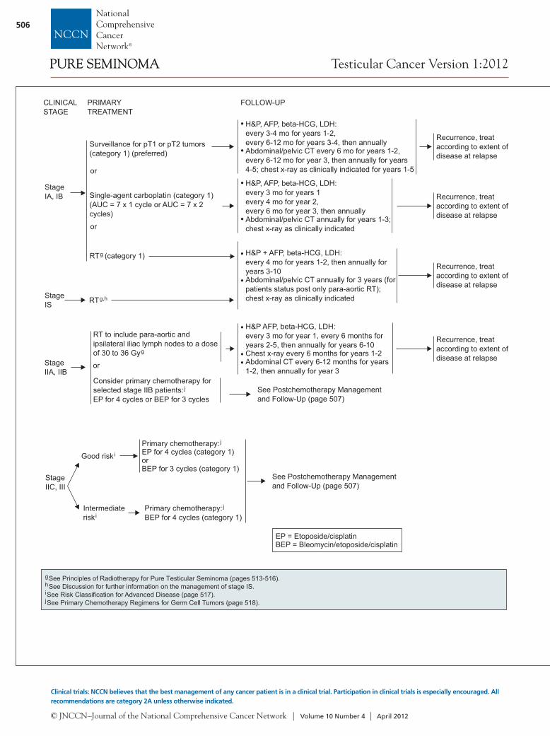
CLINICALSTAGE
StageIA, IB
FOLLOW-UPPRIMARYTREATMENT
Surveillance for pT1 or pT2 tumors
Single-agent carboplatin(AUC = 7 x 1 cycle or AUC = 7 x 2cycles)
(category 1) (preferred)
(category 1)
RT (category 1)g
•
•
H&P, AFP, beta-HCG, LDH:every mo for years ,every 6-12 mo for years , then annuallyAbdominal/pelvic CT every 6 mo for years 1-2,every 6-12 mo for year 3, then annually for years4-5; chest x-ray as clinically indicated for years 1-5
3-4 1-23-4
Recurrence, treataccording to extent ofdisease at relapse
Recurrence, treataccording to extent ofdisease at relapse
gSee Principles of Radiotherapy for Pure Testicular Seminoma (pages 513-516).See Discussion for further information on the management of stage IS.
See Risk Classification for Advanced Disease (page 517).See Primary Chemotherapy Regimens for Germ Cell Tumors (page 518).
hij
StageIS RTg,h
or
or•
•
H&P, AFP, beta-HCG, LDH:every mo for yearsevery 4 mo for year 2,
hen annuallyAbdominal/pelvic CT
3 1
every 6 mo for year 3, tannually for years 1-3;
chest x-ray as clinically indicated
H&P + AFP, beta-HCG, LDH:every 4 mo for years 1-2, then annually foryears 3-10Abdominal/pelvic CT annually for 3 years (forpatients status post only para-aortic RT);chest x-ray as clinically indicated
Recurrence, treataccording to extent ofdisease at relapse
StageIIA, IIB
RT to include para-aortic andipsilateral iliac lymph nodes to a doseof 30 to 36 Gy
Consider p
EP for 4 cycles or
g
jrimary chemotherapy for
selected stage IIB patients:BEP for 3 cycles
H&P
Chest x-ray every 6 months for years 1-2Abdominal CT every 6-12 months for years1-2, then annually for year 3
AFP, beta-HCG, LDH:every 3 mo for year 1, every 6 months foryears 2-5, then annually for years 6-10
Recurrence, treataccording to extent ofdisease at relapse
See Postchemotherapy Managementand Follow-Up (page 507)
StageIIC, III
Primary chemotherapy:EP for 4 cycles (category 1)orBEP for 3 cycles (category 1)
j
Good riski
Intermediateriski
Primary chemotherapy:j
BEP for 4 cycles (category 1)
See Postchemotherapy Managementand Follow-Up (page 507)
or
FOLLOW-UP
H&P + chest x-ray
every 2 mo forfor
then annuallyAbdominal/pelvic CT
Post RPLND:3-6 mo, then asclinically indicatedAfter all otherprimarymanagement asclinically indicated
PET scan as clinicallyindicated
,AFP, beta-HCG, LDH:
year 1,every 3 mo year 2,every 6 mo for year 3and 4,
➤
➤
Recurrence,SeeSecond-LineTherapy(page 512)
Progressive disease(growing mass orrising markers)
See Second-Line Therapy forNonseminoma (page 512)
Chest,abdominal, pelvicCT scanSerum tumormarkers
No residual massor residual mass
3 cm andnormal markers
Residualmass(> 3 cm)andnormalmarkers
Surveillance
Consider RPLND, iftechnically feasibleorSecond-linechemotherapyorRT (category 2B)
k
l
g
PET scan(approximately6 wk post-chemotherapy)
Positive
Negative Surveillance
STAGE IIB, IIC, III AFTER PRIMARYTREATMENT WITH CHEMOTHERAPY
POST-CHEMOTHERAPYMANAGEMENT
gSee Principles of Radiotherapy for Pure Testicular Seminoma (pages 513-516).If viable seminoma found by retroperitoneal lymph node dissection (RPLND), see page 510 (residual embryonal, yolk sac, choriocarcinoma, or seminomaelements).
See Second-Line or Subsequent Chemotherapy Regimens for Metastatic Germ Cell Tumors (page 519).
k
l
EP = Etoposide/cisplatinBEP = Bleomycin/etoposide/cisplatin
PURE SEMINOMAPURE SEMINOMA

NCCN Clinical Practice Guidelines in Oncology
© JNCCN–Journal of the National Comprehensive Cancer Network | Volume 10 Number 4 | April 2012
507
Testicular Cancer Version 1:2012
Version1.2012, 01-17-12 ©2012 National Comprehensive Cancer Network, Inc. All rights reserved. The NCCN Guidelines® and this illustration may not be reproduced in any form without the express written permission of NCCN®.
CLINICALSTAGE
StageIA, IB
FOLLOW-UPPRIMARYTREATMENT
Surveillance for pT1 or pT2 tumors
Single-agent carboplatin(AUC = 7 x 1 cycle or AUC = 7 x 2cycles)
(category 1) (preferred)
(category 1)
RT (category 1)g
•
•
H&P, AFP, beta-HCG, LDH:every mo for years ,every 6-12 mo for years , then annuallyAbdominal/pelvic CT every 6 mo for years 1-2,every 6-12 mo for year 3, then annually for years4-5; chest x-ray as clinically indicated for years 1-5
3-4 1-23-4
Recurrence, treataccording to extent ofdisease at relapse
Recurrence, treataccording to extent ofdisease at relapse
gSee Principles of Radiotherapy for Pure Testicular Seminoma (pages 513-516).See Discussion for further information on the management of stage IS.
See Risk Classification for Advanced Disease (page 517).See Primary Chemotherapy Regimens for Germ Cell Tumors (page 518).
hij
StageIS RTg,h
or
or•
•
H&P, AFP, beta-HCG, LDH:every mo for yearsevery 4 mo for year 2,
hen annuallyAbdominal/pelvic CT
3 1
every 6 mo for year 3, tannually for years 1-3;
chest x-ray as clinically indicated
H&P + AFP, beta-HCG, LDH:every 4 mo for years 1-2, then annually foryears 3-10Abdominal/pelvic CT annually for 3 years (forpatients status post only para-aortic RT);chest x-ray as clinically indicated
Recurrence, treataccording to extent ofdisease at relapse
StageIIA, IIB
RT to include para-aortic andipsilateral iliac lymph nodes to a doseof 30 to 36 Gy
Consider p
EP for 4 cycles or
g
jrimary chemotherapy for
selected stage IIB patients:BEP for 3 cycles
H&P
Chest x-ray every 6 months for years 1-2Abdominal CT every 6-12 months for years1-2, then annually for year 3
AFP, beta-HCG, LDH:every 3 mo for year 1, every 6 months foryears 2-5, then annually for years 6-10
Recurrence, treataccording to extent ofdisease at relapse
See Postchemotherapy Managementand Follow-Up (page 507)
StageIIC, III
Primary chemotherapy:EP for 4 cycles (category 1)orBEP for 3 cycles (category 1)
j
Good riski
Intermediateriski
Primary chemotherapy:j
BEP for 4 cycles (category 1)
See Postchemotherapy Managementand Follow-Up (page 507)
or
FOLLOW-UP
H&P + chest x-ray
every 2 mo forfor
then annuallyAbdominal/pelvic CT
Post RPLND:3-6 mo, then asclinically indicatedAfter all otherprimarymanagement asclinically indicated
PET scan as clinicallyindicated
,AFP, beta-HCG, LDH:
year 1,every 3 mo year 2,every 6 mo for year 3and 4,
➤
➤
Recurrence,SeeSecond-LineTherapy(page 512)
Progressive disease(growing mass orrising markers)
See Second-Line Therapy forNonseminoma (page 512)
Chest,abdominal, pelvicCT scanSerum tumormarkers
No residual massor residual mass
3 cm andnormal markers
Residualmass(> 3 cm)andnormalmarkers
Surveillance
Consider RPLND, iftechnically feasibleorSecond-linechemotherapyorRT (category 2B)
k
l
g
PET scan(approximately6 wk post-chemotherapy)
Positive
Negative Surveillance
STAGE IIB, IIC, III AFTER PRIMARYTREATMENT WITH CHEMOTHERAPY
POST-CHEMOTHERAPYMANAGEMENT
gSee Principles of Radiotherapy for Pure Testicular Seminoma (pages 513-516).If viable seminoma found by retroperitoneal lymph node dissection (RPLND), see page 510 (residual embryonal, yolk sac, choriocarcinoma, or seminomaelements).
See Second-Line or Subsequent Chemotherapy Regimens for Metastatic Germ Cell Tumors (page 519).
k
l
EP = Etoposide/cisplatinBEP = Bleomycin/etoposide/cisplatin
PURE SEMINOMAPURE SEMINOMA
© JNCCN–Journal of the National Comprehensive Cancer Network | Volume 10 Number 4 | April 2012
508
Testicular Cancer Version 1:2012
Clinical trials: NCCN believes that the best management of any cancer patient is in a clinical trial. Participation in clinical trials is especially encouraged. All recommendations are category 2A unless otherwise indicated.
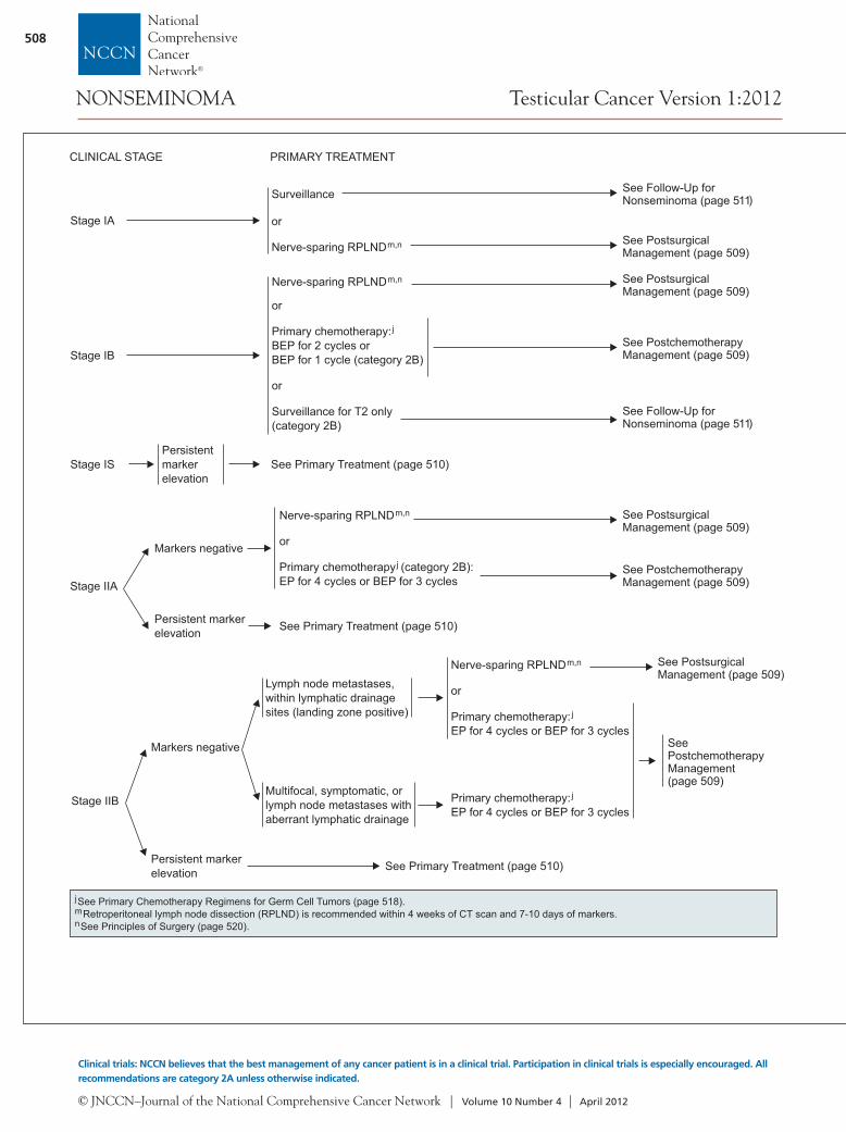
CLINICAL STAGE
Stage IA
Surveillance
or
Nerve-sparing RPLNDm,n
Stage IB
Nerve-sparing RPLND
or
Primary chemotherapy:BEP for 2 cycles orBEP for 1 cycle (category 2B)
or
Surveillance for T2(category 2B)
m,n
j
only See Follow-Up forNonseminoma (page 511)
See Follow-Up forNonseminoma (page 511)
Stage ISPersistentmarkerelevation
See PostchemotherapyManagement (page 509)
See PostsurgicalManagement (page 509)
See PostsurgicalManagement (page 509)
jmn
See Primary Chemotherapy Regimens for Germ Cell Tumors (page 518).Retroperitoneal lymph node dissection (RPLND) is recommended within 4 weeks of CT scan and 7-10 days of markers.See Principles of Surgery (page 520).
PRIMARY TREATMENT
See Primary Treatment (page 510)
Stage IIA
Markers negative
Persistent markerelevation
Nerve-sparing RPLND
or
Primary chemotherapy (category 2B):EP for 4 cycles or BEP for 3 cycles
m,n
j
See Primary Treatment (page 510)
See PostchemotherapyManagement (page 509)
See PostsurgicalManagement (page 509)
Stage IIB
Markers negative
Persistent markerelevation
Lymph node metastases,within lymphatic drainagesites (landing zone positive)
Multifocal, symptomatic, orlymph node metastases withaberrant lymphatic drainage
Primary chemotherapy:EP for 4 cycles or BEP for 3 cycles
j
Nerve-sparing RPLND
or
Primary chemotherapy:EP for 4 cycles or BEP for 3 cycles
m,n
j
SeePostchemotherapyManagement(page 509)
See PostsurgicalManagement (page 509)
See Primary Treatment (page 510)
Negative markers,residual mass ( 1 cm)on CT scan
≥
Nerve-sparing bilateral RPLNDorSurveillance(category 2B)
m,n
Negative markers, nomass or residual mass(< 1 cm) on CT scan
Nerve-sparing bilateral RPLND(category 2B)orSurveillance(category 2B)
m,n
POSTCHEMOTHERAPY MANAGEMENT
Stage IB, IIA, IIBtreated with primarychemotherapy
See Follow-Up forNonseminoma (page 511)
jSee Primary Chemotherapy Regimens for Germ Cell Tumors (page 518).mn
Retroperitoneal lymph node dissection (RPLND) is recommended within 4 weeks of CT scan and 7-10 days of markers.See Principles of Surgery (page 520).
POSTSURGICAL MANAGEMENT
pN0
pN1
Surveillance (preferred)orChemotherapy:EP for 2 cyclesor BEP for 2 cycles
j
Surveillance
pN2
Chemotherapy (preferred):EP for 2 cycles or BEP for 2 cyclesor
j
Surveillance
pN3C (preferred)hemotherapy :EP for 4 cyclesor BEP for 3 cycles
j
See Follow-Up forNonseminoma (page 511)
Stage IA, IB, IIA, IIBtreated with nerve-sparing RPLND
EP = Etoposide/cisplatinBEP = Bleomycin/etoposide/cisplatin
NONSEMINOMA

NCCN Clinical Practice Guidelines in Oncology
© JNCCN–Journal of the National Comprehensive Cancer Network | Volume 10 Number 4 | April 2012
509
Testicular Cancer Version 1:2012
Version1.2012, 01-17-12 ©2012 National Comprehensive Cancer Network, Inc. All rights reserved. The NCCN Guidelines® and this illustration may not be reproduced in any form without the express written permission of NCCN®.
CLINICAL STAGE
Stage IA
Surveillance
or
Nerve-sparing RPLNDm,n
Stage IB
Nerve-sparing RPLND
or
Primary chemotherapy:BEP for 2 cycles orBEP for 1 cycle (category 2B)
or
Surveillance for T2(category 2B)
m,n
j
only See Follow-Up forNonseminoma (page 511)
See Follow-Up forNonseminoma (page 511)
Stage ISPersistentmarkerelevation
See PostchemotherapyManagement (page 509)
See PostsurgicalManagement (page 509)
See PostsurgicalManagement (page 509)
jmn
See Primary Chemotherapy Regimens for Germ Cell Tumors (page 518).Retroperitoneal lymph node dissection (RPLND) is recommended within 4 weeks of CT scan and 7-10 days of markers.See Principles of Surgery (page 520).
PRIMARY TREATMENT
See Primary Treatment (page 510)
Stage IIA
Markers negative
Persistent markerelevation
Nerve-sparing RPLND
or
Primary chemotherapy (category 2B):EP for 4 cycles or BEP for 3 cycles
m,n
j
See Primary Treatment (page 510)
See PostchemotherapyManagement (page 509)
See PostsurgicalManagement (page 509)
Stage IIB
Markers negative
Persistent markerelevation
Lymph node metastases,within lymphatic drainagesites (landing zone positive)
Multifocal, symptomatic, orlymph node metastases withaberrant lymphatic drainage
Primary chemotherapy:EP for 4 cycles or BEP for 3 cycles
j
Nerve-sparing RPLND
or
Primary chemotherapy:EP for 4 cycles or BEP for 3 cycles
m,n
j
SeePostchemotherapyManagement(page 509)
See PostsurgicalManagement (page 509)
See Primary Treatment (page 510)
Negative markers,residual mass ( 1 cm)on CT scan
≥
Nerve-sparing bilateral RPLNDorSurveillance(category 2B)
m,n
Negative markers, nomass or residual mass(< 1 cm) on CT scan
Nerve-sparing bilateral RPLND(category 2B)orSurveillance(category 2B)
m,n
POSTCHEMOTHERAPY MANAGEMENT
Stage IB, IIA, IIBtreated with primarychemotherapy
See Follow-Up forNonseminoma (page 511)
jSee Primary Chemotherapy Regimens for Germ Cell Tumors (page 518).mn
Retroperitoneal lymph node dissection (RPLND) is recommended within 4 weeks of CT scan and 7-10 days of markers.See Principles of Surgery (page 520).
POSTSURGICAL MANAGEMENT
pN0
pN1
Surveillance (preferred)orChemotherapy:EP for 2 cyclesor BEP for 2 cycles
j
Surveillance
pN2
Chemotherapy (preferred):EP for 2 cycles or BEP for 2 cyclesor
j
Surveillance
pN3C (preferred)hemotherapy :EP for 4 cyclesor BEP for 3 cycles
j
See Follow-Up forNonseminoma (page 511)
Stage IA, IB, IIA, IIBtreated with nerve-sparing RPLND
EP = Etoposide/cisplatinBEP = Bleomycin/etoposide/cisplatin
NONSEMINOMA

© JNCCN–Journal of the National Comprehensive Cancer Network | Volume 10 Number 4 | April 2012
510
Testicular Cancer Version 1:2012
Clinical trials: NCCN believes that the best management of any cancer patient is in a clinical trial. Participation in clinical trials is especially encouraged. All recommendations are category 2A unless otherwise indicated.
CLINICALSTAGE
IntermediateriskStage IIIB
i
Poor riskStage IIIC
i
Good riskStage ISStage IIA,Stage IIB, S1Stage IICStage IIIA
i
S1
Primarychemotherapy:j
EP for 4 cyclesorBEP for 3 cycles
Primarychemotherapy:j
BEP for 4 cycles
Clinical trial(preferred)or
BEP for 4 cyclesorVIP for 4 cycles inselected patients
Primarychemotherapy:j
p
Completeresponse,negativemarkers
Partial response,residual masseswith normal AFPand beta-HCGlevels
q
Incompleteresponseq
Surgicalresection ofall residualmasses
Surveillance (category 2B)orBilateral RPLND category 2B)m,n ± nerve-sparing (
Teratoma ornecrosis
Residualembryonal, yolksac,choriocarcinoma, orseminoma elements
Chemotherapyfor 2 cycles(EP or TIP orVIP /VeIP )
jj
ll
See Second-LineTherapy (page 512)
See Follow-Up forNonseminoma(page 511)
Surveillance
Primary chemotherapy + RT± surgery, if clinically indicated
j
ijlmno
See Risk Classification for Advanced Disease (page 517).See Primary Chemotherapy Regimens for Germ Cell Tumors (page 518).See Second-Line or Subsequent Chemotherapy Regimens for Metastatic Germ Cell Tumors (page 519).Retroperitoneal lymph node dissection (RPLND) is recommended within 4 weeks of CT scan and 7-10 days of markers (category 2B).See Principles of Surgery (page 520).Patients should receive adequate treatment for brain metastases, in addition to cisplatin-based chemotherapy.Patients who may not tolerate bleomycin.There is limited predictive value for PET scan for residual masses.
pq
PRIMARY TREATMENT POSTCHEMOTHERAPYMANAGEMENT
FOLLOW-UP FOR NONSEMINOMA
Follow-Up for Stage IA, IB on Surveillance Only
Year
1
2
3
4
5
6+
Months between H&P,markers, chest x-ray
1-2
2
3
4
6
12
Months betweenabdominal CT
3-4
4-6
6-12
6-12
12
12-24
Recurrence, See Second-LineTherapy (page 512)
Table 1 Follow-Up After Complete Response to Chemotherapy andRPLND
Year
1
2
3
4
5
6+
Months between H&P,markers, chest x-ray
(category 2B forchest x-ray frequency)
2-3
2-3
3-6
6
6-12
12
Months betweenabdominal/pelvic CT
6
6-12
12
12
12
As clinicallyindicated
Table 2
Follow-Up After RPLND Only
Months between H&P,markers, chest x-ray
(category 2B forchest x-ray frequency)
2-3
2-3
3-6
6
6-12
12
Months betweenabdominal/pelvic CT
Baseline
As indicated
As indicated
As indicated
As indicated
As indicated
Table 3
Year
1
2
3
4
5
6+
Brainmetastaseso
EP = Etoposide/cisplatinBEP = Bleomycin/etoposide/cisplatinTIP = Paclitaxel/ifosfamide/cisplatinVeIP = Vinblastine/ifosfamide/cisplatinVIP = Etoposide/ifosfamide/cisplatin
NONSEMINOMA

NCCN Clinical Practice Guidelines in Oncology
© JNCCN–Journal of the National Comprehensive Cancer Network | Volume 10 Number 4 | April 2012
511
Testicular Cancer Version 1:2012
Version1.2012, 01-17-12 ©2012 National Comprehensive Cancer Network, Inc. All rights reserved. The NCCN Guidelines® and this illustration may not be reproduced in any form without the express written permission of NCCN®.
CLINICALSTAGE
IntermediateriskStage IIIB
i
Poor riskStage IIIC
i
Good riskStage ISStage IIA,Stage IIB, S1Stage IICStage IIIA
i
S1
Primarychemotherapy:j
EP for 4 cyclesorBEP for 3 cycles
Primarychemotherapy:j
BEP for 4 cycles
Clinical trial(preferred)or
BEP for 4 cyclesorVIP for 4 cycles inselected patients
Primarychemotherapy:j
p
Completeresponse,negativemarkers
Partial response,residual masseswith normal AFPand beta-HCGlevels
q
Incompleteresponseq
Surgicalresection ofall residualmasses
Surveillance (category 2B)orBilateral RPLND category 2B)m,n ± nerve-sparing (
Teratoma ornecrosis
Residualembryonal, yolksac,choriocarcinoma, orseminoma elements
Chemotherapyfor 2 cycles(EP or TIP orVIP /VeIP )
jj
ll
See Second-LineTherapy (page 512)
See Follow-Up forNonseminoma(page 511)
Surveillance
Primary chemotherapy + RT± surgery, if clinically indicated
j
ijlmno
See Risk Classification for Advanced Disease (page 517).See Primary Chemotherapy Regimens for Germ Cell Tumors (page 518).See Second-Line or Subsequent Chemotherapy Regimens for Metastatic Germ Cell Tumors (page 519).Retroperitoneal lymph node dissection (RPLND) is recommended within 4 weeks of CT scan and 7-10 days of markers (category 2B).See Principles of Surgery (page 520).Patients should receive adequate treatment for brain metastases, in addition to cisplatin-based chemotherapy.Patients who may not tolerate bleomycin.There is limited predictive value for PET scan for residual masses.
pq
PRIMARY TREATMENT POSTCHEMOTHERAPYMANAGEMENT
FOLLOW-UP FOR NONSEMINOMA
Follow-Up for Stage IA, IB on Surveillance Only
Year
1
2
3
4
5
6+
Months between H&P,markers, chest x-ray
1-2
2
3
4
6
12
Months betweenabdominal CT
3-4
4-6
6-12
6-12
12
12-24
Recurrence, See Second-LineTherapy (page 512)
Table 1 Follow-Up After Complete Response to Chemotherapy andRPLND
Year
1
2
3
4
5
6+
Months between H&P,markers, chest x-ray
(category 2B forchest x-ray frequency)
2-3
2-3
3-6
6
6-12
12
Months betweenabdominal/pelvic CT
6
6-12
12
12
12
As clinicallyindicated
Table 2
Follow-Up After RPLND Only
Months between H&P,markers, chest x-ray
(category 2B forchest x-ray frequency)
2-3
2-3
3-6
6
6-12
12
Months betweenabdominal/pelvic CT
Baseline
As indicated
As indicated
As indicated
As indicated
As indicated
Table 3
Year
1
2
3
4
5
6+
Brainmetastaseso
EP = Etoposide/cisplatinBEP = Bleomycin/etoposide/cisplatinTIP = Paclitaxel/ifosfamide/cisplatinVeIP = Vinblastine/ifosfamide/cisplatinVIP = Etoposide/ifosfamide/cisplatin
NONSEMINOMA

© JNCCN–Journal of the National Comprehensive Cancer Network | Volume 10 Number 4 | April 2012
512
Testicular Cancer Version 1:2012
Clinical trials: NCCN believes that the best management of any cancer patient is in a clinical trial. Participation in clinical trials is especially encouraged. All recommendations are category 2A unless otherwise indicated.
• ChemotherapyConventionaldose therapy(VeIP or TIP)orHigh-dosechemotherapy
l
➤
➤
Favorableprognosis:
Low markersLow volumeComplete responseon first-line therapyTestis primary
s
•••
•
Unfavorable prognosis:Incomplete responseHigh markersHigh volumeExtratesticular primaryLate relapse
s
•••••
Incompleteresponse orrelapse
Completeresponse Follow-up
Relapse
lSee Second-Line or Subsequent Chemotherapy Regimens for Germ Cell Tumors (page 519).rIt is preferred that patients with recurrent nonseminoma be treated at centers with expertise in the management of this disease.
No priorchemotherapy
Priorchemotherapy
Treat as per risk status on page 510
•
•
•
Chemotherapy
Best supportive care
l
➤
➤
High-dosechemotherapy(preferred if notpreviously given) orClinical trial
Surgical salvage should beconsidered if solitary site
•
•
•
Chemotherapy
High-dose chemotherapy (category 2B)Surgical salvage should be considered ifsolitary siteBest supportive care
l
➤
➤
➤
Clinical trial (preferred)orConventional dose therapy (VeIP or TIP)or
PalliativechemotherapyorRT
l
RECURRENCEr SECOND-LINE THERAPY
sExamples of systems used to estimate prognosis are:1) Lorch A, Beyer J, Bascoul-Mollevi C, et al. Prognostic factors in patients with metastatic germ cell tumors who experienced treatment failure with cisplatin-based first-line chemotherapy. International Prognostic Factors Study Group. J Clin Oncol 2010;28:4906-4911.2) Einhorn LH, Williams SD, Chamness A, et al. High-dose chemotherapy and stem-cell rescue for metastatic germ-cell tumors. N Engl J Med2007;357:340-348.3) Motzer RJ, Geller NL, Tan CC, et al. Salvage chemotherapy for patients with germ cell tumors. The Memorial Sloan-Kettering Cancer Centerexperience (1979-1989). Cancer 1991;67:1305-1310.
PersistentdiseaseorRelapse
Persistent diseaseor Relapse
PRINCIPLES OF RADIOTHERAPY FOR PURE TESTICULAR SEMINOMA
General Principles•
•
••
•
•
Modern radiotherapy involves smaller fields and lower doses than were used in the past. References are provided to support currentrecommended management.Risk-adapted management using tumor size > 4 cm and rete testis invasion for stage I seminoma is discouraged. This is based on avalidation study in 2010, which revealed that tumor size > 4 cm and rete testis invasion were not predictors of relapse.Linear accelerators with > 6 MV photons should be used when possible.The mean dose (Dmean) and dose delivered to 50% of the volume (D50%) of the kidneys, liver, and bowel are lower with CT-basedanteroposterior-posteroanterior (AP-PA) 3-dimensional conformal radiation therapy (3D-CRT) than intensity-modulated radiationtherapy (IMRT). As a result, the risk of second cancers arising in the kidneys, liver, or bowel may be lower with 3D-CRT than IMRT,and IMRT is not recommended.Timing of radiotherapy:
Radiotherapy should start within 7 weeks after orchiectomy.Patients should be treated 5 days per week.Patients who miss a fraction should be treated to the same total dose and with the same fraction size, extending the overalltreatment time slightly.
Antiemetic medication significantly improves nausea.
1,2
34
➤
➤
➤
See NCCN Clinical Practice Guidelines (NCCN Guidelines) in Oncology forAntiemesis (available in this issue and online at www.NCCN.org). Antiemetic prophylaxis is encouraged at least 2 hours beforeeach treatment, and some cases may require more frequent dosing.
A discussion of semen analysis and sperm banking before orchiectomy is recommended in patients who wish to preserve fertility. Ifsperm banking is desired, it should be performed before imaging and the delivery of adjuvant therapy.
Preparation for Radiotherapy• 5,6
Treatment Planning Principles• A noncontrast CT simulation should be performed with the patient supine, arms at his sides, in the treatment position.
Immobilization with a cast may be used to improve the reproducibility of patient setup.All patients, with the exception of those who have undergone bilateral orchiectomy, should be treated with a scrotal shield.The legs should be separated by a rolled towel of approximately the same diameter as the scrotal shield and its stand.
➤
➤
VeIP = Vinblastine/ifosfamide/cisplatinTIP = Paclitaxel/ifosfamide/cisplatin
NONSEMINOMA

NCCN Clinical Practice Guidelines in Oncology
© JNCCN–Journal of the National Comprehensive Cancer Network | Volume 10 Number 4 | April 2012
513
Testicular Cancer Version 1:2012
Version1.2012, 01-17-12 ©2012 National Comprehensive Cancer Network, Inc. All rights reserved. The NCCN Guidelines® and this illustration may not be reproduced in any form without the express written permission of NCCN®.
• ChemotherapyConventionaldose therapy(VeIP or TIP)orHigh-dosechemotherapy
l
➤
➤
Favorableprognosis:
Low markersLow volumeComplete responseon first-line therapyTestis primary
s
•••
•
Unfavorable prognosis:Incomplete responseHigh markersHigh volumeExtratesticular primaryLate relapse
s
•••••
Incompleteresponse orrelapse
Completeresponse Follow-up
Relapse
lSee Second-Line or Subsequent Chemotherapy Regimens for Germ Cell Tumors (page 519).rIt is preferred that patients with recurrent nonseminoma be treated at centers with expertise in the management of this disease.
No priorchemotherapy
Priorchemotherapy
Treat as per risk status on page 510
•
•
•
Chemotherapy
Best supportive care
l
➤
➤
High-dosechemotherapy(preferred if notpreviously given) orClinical trial
Surgical salvage should beconsidered if solitary site
•
•
•
Chemotherapy
High-dose chemotherapy (category 2B)Surgical salvage should be considered ifsolitary siteBest supportive care
l
➤
➤
➤
Clinical trial (preferred)orConventional dose therapy (VeIP or TIP)or
PalliativechemotherapyorRT
l
RECURRENCEr SECOND-LINE THERAPY
sExamples of systems used to estimate prognosis are:1) Lorch A, Beyer J, Bascoul-Mollevi C, et al. Prognostic factors in patients with metastatic germ cell tumors who experienced treatment failure with cisplatin-based first-line chemotherapy. International Prognostic Factors Study Group. J Clin Oncol 2010;28:4906-4911.2) Einhorn LH, Williams SD, Chamness A, et al. High-dose chemotherapy and stem-cell rescue for metastatic germ-cell tumors. N Engl J Med2007;357:340-348.3) Motzer RJ, Geller NL, Tan CC, et al. Salvage chemotherapy for patients with germ cell tumors. The Memorial Sloan-Kettering Cancer Centerexperience (1979-1989). Cancer 1991;67:1305-1310.
PersistentdiseaseorRelapse
Persistent diseaseor Relapse
PRINCIPLES OF RADIOTHERAPY FOR PURE TESTICULAR SEMINOMA
General Principles•
•
••
•
•
Modern radiotherapy involves smaller fields and lower doses than were used in the past. References are provided to support currentrecommended management.Risk-adapted management using tumor size > 4 cm and rete testis invasion for stage I seminoma is discouraged. This is based on avalidation study in 2010, which revealed that tumor size > 4 cm and rete testis invasion were not predictors of relapse.Linear accelerators with > 6 MV photons should be used when possible.The mean dose (Dmean) and dose delivered to 50% of the volume (D50%) of the kidneys, liver, and bowel are lower with CT-basedanteroposterior-posteroanterior (AP-PA) 3-dimensional conformal radiation therapy (3D-CRT) than intensity-modulated radiationtherapy (IMRT). As a result, the risk of second cancers arising in the kidneys, liver, or bowel may be lower with 3D-CRT than IMRT,and IMRT is not recommended.Timing of radiotherapy:
Radiotherapy should start within 7 weeks after orchiectomy.Patients should be treated 5 days per week.Patients who miss a fraction should be treated to the same total dose and with the same fraction size, extending the overalltreatment time slightly.
Antiemetic medication significantly improves nausea.
1,2
34
➤
➤
➤
See NCCN Clinical Practice Guidelines (NCCN Guidelines) in Oncology forAntiemesis (available in this issue and online at www.NCCN.org). Antiemetic prophylaxis is encouraged at least 2 hours beforeeach treatment, and some cases may require more frequent dosing.
A discussion of semen analysis and sperm banking before orchiectomy is recommended in patients who wish to preserve fertility. Ifsperm banking is desired, it should be performed before imaging and the delivery of adjuvant therapy.
Preparation for Radiotherapy• 5,6
Treatment Planning Principles• A noncontrast CT simulation should be performed with the patient supine, arms at his sides, in the treatment position.
Immobilization with a cast may be used to improve the reproducibility of patient setup.All patients, with the exception of those who have undergone bilateral orchiectomy, should be treated with a scrotal shield.The legs should be separated by a rolled towel of approximately the same diameter as the scrotal shield and its stand.
➤
➤
VeIP = Vinblastine/ifosfamide/cisplatinTIP = Paclitaxel/ifosfamide/cisplatin

© JNCCN–Journal of the National Comprehensive Cancer Network | Volume 10 Number 4 | April 2012
514
Testicular Cancer Version 1:2012
Clinical trials: NCCN believes that the best management of any cancer patient is in a clinical trial. Participation in clinical trials is especially encouraged. All recommendations are category 2A unless otherwise indicated.
Stage I
Special Considerations
Dose: For stages IA, IB, and IS, a total dose of 20.0 Gy (midplane) in 10 daily 2.0-Gy fractions is recommended for the minority ofpatients who prefer adjuvant treatment realizing that there is a high likelihood of salvage should a relapse occur during surveillance.Para-aortic (PA) strip fields - field arrangement:
In patients with no history of pelvic or scrotal surgery, para-aortic strip irradiation may be delivered with opposed AP-PA fields. Theweights of the fields may be equal.
Recent nodal mapping studies suggest that fields should target the retroperitoneal lymph nodes but not necessarily the ipsilateralrenal hilar nodes (see lateral borders).Superior and inferior borders: borders may be determined by bony anatomy.
- T11.- The inferior border should be placed at the inferior border of vertebral body L5.
Lateral borders:- Conventionally, PA strip fields are approximately 10 cm wide, encompassing the tips of the transverse processes of
the para-aortic vertebrae.- The location of the kidneys within the PA strip fields varies from patient to patient.
For patients whose kidneys are relatively medial, small renal blocks may be added at the level of T12. Theright and left kidney D50% should be 8 Gy (i.e., no more than 50% of each kidney can receive 8 Gy orhigher). If only one kidney is present, the kidney D15% should be 20 Gy (i.e., no more than 15% of thevolume of the kidney can receive 20 Gy or higher).An alternative 3D-CRT planning technique is to base the lateral borders on vascular structures on atreatment planning CT scan without contrast. The aorta and inferior vena cava may be contoured on the CTscan; one should allow a 1.2- to 1.9-cm margin on the aorta and inferior vena cava to include the para-aortic,paracaval, interaortocaval, and preaortic nodes in the clinical target volume. The planning target volumeis then established by uniformly expanding the clinical target volume by 0.5 cm in all directions to account fortreatment setup errors. A uniform 0.7-cm margin should be provided on the planning target volume to theblock edge to take beam penumbra into account (Figure 1, ).
Ipsilateral pelvic surgery (e.g., inguinal herniorrhaphy or orchiopexy) may alter the lymphatic drainage of the testis. As a result,irradiation of the ipsilateral iliac and inguinal lymph nodes, including the surgical scar from prior surgery, has been advocated even instage I patients. Given the large volume of tissue that would be irradiated and the resulting increased risks of late effects, othermanagement approaches are recommended for these patients.
910
11,12
1310,14
3
3
11,15
3
12,17
The superior border should be placed at the bottom of vertebral body
16
see page 516
Stage IIA-B(modified dog-leg fields and cone down).
There is no break between the 2 phases.For clinical stage IIA-B patients, treatment is delivered in 2 consecutive AP-PA phases
Modified dog-leg fields:Dose: the initial phase consists of treatment of modified dog-leg fields to 20.0 Gy (midplane) in 10 daily 2.0-Gy fractions or25.5 Gy in 15 daily 1.7-Gy fractions.Target: the fields should include the retroperitoneal and proximal ipsilateral iliac lymph nodes.
Modified dog-leg fields as described by Classen et al. are preferred.- Care should be taken to ensure coverage of the ipsilateral common, external, and proximal internal iliac lymph nodes
down to the top of the acetabulum.- The fields can be set up using bony landmarks or by contouring the vascular structures, as for stage I.
The superior border should be placed at the bottom of vertebral body T11.The inferior border should be placed at the top of the acetabulum.The medial border for the lower aspect of the modified dog-leg fields extends from the tip of thecontralateral transverse process of the fifth lumbar vertebra toward the medial border of the ipsilateralobturator foramen.The lateral border for the lower aspect of the modified dog-leg fields is defined by a line from the tip ofthe ipsilateral transverse process of the fifth lumbar vertebra to the superolateral border of the ipsilateralacetabulum.Preferably, one should contour the aorta and inferior vena cava from the bottom of the T11 vertebrainferiorly and ipsilateral iliac arteries and veins down to the top of the acetabulum. One should provide a1.2- to 1.9-cm margin on these vascular structures for the clinical target volume. The planning targetvolume is then established by uniformly expanding the clinical target volume by 0.5 cm in all directions toaccount for treatment setup errors. A uniform 0.7-cm margin should be provided on the planning targetvolume to the block edge to take beam penumbra into account (Figure 2, ).It is not necessary to include the ipsilateral inguinal nodes or the inguinal scar in the AP-PA fields unlessthe patient has a history of ipsilateral pelvic surgery (e.g., inguinal herniorrhaphy or orchiopexy).
Cone Down:Dose: the second phase (cone down) of the radiotherapy consists of daily 2-Gy fractions to a cumulative total dose ofapproximately 30 Gy for stage IIA and 36 Gy for stage IIB.Target: the nodal mass (gross tumor volume) must be contoured. A uniform, 2-cm margin from the gross tumor volume to blockedge should be provided for the AP-PA cone down fields (Figure 3, ).
1718
19
2019
11,15
163
12
19
see page 516
see page 516
PRINCIPLES OF RADIOTHERAPY FOR PURE TESTICULAR SEMINOMAPRINCIPLES OF RADIOTHERAPY FOR PURE TESTICULAR SEMINOMA

NCCN Clinical Practice Guidelines in Oncology
© JNCCN–Journal of the National Comprehensive Cancer Network | Volume 10 Number 4 | April 2012
515
Testicular Cancer Version 1:2012
Version1.2012, 01-17-12 ©2012 National Comprehensive Cancer Network, Inc. All rights reserved. The NCCN Guidelines® and this illustration may not be reproduced in any form without the express written permission of NCCN®.
Stage I
Special Considerations
Dose: For stages IA, IB, and IS, a total dose of 20.0 Gy (midplane) in 10 daily 2.0-Gy fractions is recommended for the minority ofpatients who prefer adjuvant treatment realizing that there is a high likelihood of salvage should a relapse occur during surveillance.Para-aortic (PA) strip fields - field arrangement:
In patients with no history of pelvic or scrotal surgery, para-aortic strip irradiation may be delivered with opposed AP-PA fields. Theweights of the fields may be equal.
Recent nodal mapping studies suggest that fields should target the retroperitoneal lymph nodes but not necessarily the ipsilateralrenal hilar nodes (see lateral borders).Superior and inferior borders: borders may be determined by bony anatomy.
- T11.- The inferior border should be placed at the inferior border of vertebral body L5.
Lateral borders:- Conventionally, PA strip fields are approximately 10 cm wide, encompassing the tips of the transverse processes of
the para-aortic vertebrae.- The location of the kidneys within the PA strip fields varies from patient to patient.
For patients whose kidneys are relatively medial, small renal blocks may be added at the level of T12. Theright and left kidney D50% should be 8 Gy (i.e., no more than 50% of each kidney can receive 8 Gy orhigher). If only one kidney is present, the kidney D15% should be 20 Gy (i.e., no more than 15% of thevolume of the kidney can receive 20 Gy or higher).An alternative 3D-CRT planning technique is to base the lateral borders on vascular structures on atreatment planning CT scan without contrast. The aorta and inferior vena cava may be contoured on the CTscan; one should allow a 1.2- to 1.9-cm margin on the aorta and inferior vena cava to include the para-aortic,paracaval, interaortocaval, and preaortic nodes in the clinical target volume. The planning target volumeis then established by uniformly expanding the clinical target volume by 0.5 cm in all directions to account fortreatment setup errors. A uniform 0.7-cm margin should be provided on the planning target volume to theblock edge to take beam penumbra into account (Figure 1, ).
Ipsilateral pelvic surgery (e.g., inguinal herniorrhaphy or orchiopexy) may alter the lymphatic drainage of the testis. As a result,irradiation of the ipsilateral iliac and inguinal lymph nodes, including the surgical scar from prior surgery, has been advocated even instage I patients. Given the large volume of tissue that would be irradiated and the resulting increased risks of late effects, othermanagement approaches are recommended for these patients.
910
11,12
1310,14
3
3
11,15
3
12,17
The superior border should be placed at the bottom of vertebral body
16
see page 516
Stage IIA-B(modified dog-leg fields and cone down).
There is no break between the 2 phases.For clinical stage IIA-B patients, treatment is delivered in 2 consecutive AP-PA phases
Modified dog-leg fields:Dose: the initial phase consists of treatment of modified dog-leg fields to 20.0 Gy (midplane) in 10 daily 2.0-Gy fractions or25.5 Gy in 15 daily 1.7-Gy fractions.Target: the fields should include the retroperitoneal and proximal ipsilateral iliac lymph nodes.
Modified dog-leg fields as described by Classen et al. are preferred.- Care should be taken to ensure coverage of the ipsilateral common, external, and proximal internal iliac lymph nodes
down to the top of the acetabulum.- The fields can be set up using bony landmarks or by contouring the vascular structures, as for stage I.
The superior border should be placed at the bottom of vertebral body T11.The inferior border should be placed at the top of the acetabulum.The medial border for the lower aspect of the modified dog-leg fields extends from the tip of thecontralateral transverse process of the fifth lumbar vertebra toward the medial border of the ipsilateralobturator foramen.The lateral border for the lower aspect of the modified dog-leg fields is defined by a line from the tip ofthe ipsilateral transverse process of the fifth lumbar vertebra to the superolateral border of the ipsilateralacetabulum.Preferably, one should contour the aorta and inferior vena cava from the bottom of the T11 vertebrainferiorly and ipsilateral iliac arteries and veins down to the top of the acetabulum. One should provide a1.2- to 1.9-cm margin on these vascular structures for the clinical target volume. The planning targetvolume is then established by uniformly expanding the clinical target volume by 0.5 cm in all directions toaccount for treatment setup errors. A uniform 0.7-cm margin should be provided on the planning targetvolume to the block edge to take beam penumbra into account (Figure 2, ).It is not necessary to include the ipsilateral inguinal nodes or the inguinal scar in the AP-PA fields unlessthe patient has a history of ipsilateral pelvic surgery (e.g., inguinal herniorrhaphy or orchiopexy).
Cone Down:Dose: the second phase (cone down) of the radiotherapy consists of daily 2-Gy fractions to a cumulative total dose ofapproximately 30 Gy for stage IIA and 36 Gy for stage IIB.Target: the nodal mass (gross tumor volume) must be contoured. A uniform, 2-cm margin from the gross tumor volume to blockedge should be provided for the AP-PA cone down fields (Figure 3, ).
1718
19
2019
11,15
163
12
19
see page 516
see page 516
PRINCIPLES OF RADIOTHERAPY FOR PURE TESTICULAR SEMINOMAPRINCIPLES OF RADIOTHERAPY FOR PURE TESTICULAR SEMINOMA

© JNCCN–Journal of the National Comprehensive Cancer Network | Volume 10 Number 4 | April 2012
516
Testicular Cancer Version 1:2012
Clinical trials: NCCN believes that the best management of any cancer patient is in a clinical trial. Participation in clinical trials is especially encouraged. All recommendations are category 2A unless otherwise indicated.
Figure 2Figure 1 Figure 3
PRINCIPLES OF RADIOTHERAPY FOR PURE TESTICULAR SEMINOMA
References1. Chung P, Warde P. Stage I seminoma: adjuvant treatment is effective but is it necessary? J Natl Cancer Inst 2011;103:194-196.2. Chung P. Prognostic factors for relapse in stage I seminoma: a validation study [abstract]. J Clin Oncol 2010;28:Abstract 4535.3. Zilli T, Boudreau C, Doucet R, et al. Bone marrow-sparing intensity-modulated radiation therapy for stage I seminoma. Acta Oncol 2011;50:555-562.4. Hall EJ, Wuu CS. Radiation-induced second cancers: the impact of 3D-CRT and IMRT. Int J Radiat Oncol Biol Phys 2003;56:83-88.5. Ragni G, Somigliana E, Restelli L, et al. Sperm banking and rate of assisted reproduction treatment: insights from a 15-year cryopreservation program for
male cancer patients. Cancer 2003;97:1624-1629.6. Saito K, Suzuki K, Iwasaki A, et al. Sperm cryopreservation before cancer chemotherapy helps in the emotional battle against cancer. Cancer
2005;104:521-524.7. Oliver RT, Mead GM, Rustin GJ, et al. Randomized trial of carboplatin versus radiotherapy for stage I seminoma: mature results on relapse and
contralateral testis cancer rates in MRC TE19/EORTC 30982 study (ISRCTN27163214). J Clin Oncol 2011;29:957-962.8. Mead GM, Fossa SD, Oliver RT, et al. Randomized trials in 2466 patients with stage I seminoma: patterns of relapse and follow-up. J Natl Cancer Inst
2011;103:241-249.9. Garmezy B, Pagliaro LC. Choosing treatment for stage I seminoma: who should get what? Oncology (Williston Park) 2009;23:753, 759.10. Fossa SD, Horwich A, Russell JM, et al. Optimal planning target volume for stage I testicular seminoma: a Medical Research Council randomized trial.
Medical Research Council Testicular Tumor Working Group. J Clin Oncol 1999;17:1146-1154.11. Dinniwell R, Chan P, Czarnota G, et al. Pelvic lymph node topography for radiotherapy treatment planning from ferumoxtran-10 contrast-enhanced
magnetic resonance imaging. Int J Radiat Oncol Biol Phys 2009;74:844-851.12. McMahon CJ, Rofsky NM, Pedrosa I. Lymphatic metastases from pelvic tumors: anatomic classification, characterization, and staging. Radiology
2010;254:31-46.13. Bruns F, Bremer M, Meyer A, et al. Adjuvant radiotherapy in stage I seminoma: is there a role for further reduction of treatment volume? Acta Oncol
2005;44:142-148.14. Classen J, Schmidberger H, Meisner C, et al. Para-aortic irradiation for stage I testicular seminoma: results of a prospective study in 675 patients. A trial
of the German testicular cancer study group (GTCSG). Br J Cancer 2004;90:2305-2311.15. Shih HA, Harisinghani M, Zietman AL, et al. Mapping of nodal disease in locally advanced prostate cancer: rethinking the clinical target volume for pelvic
nodal irradiation based on vascular rather than bony anatomy. Int J Radiat Oncol Biol Phys 2005;63:1262-1269.16. Boujelbene N, Cosinschi A, Khanfir K, et al. Pure seminoma: a review and update. Radiat Oncol 2011;6:90.17. Jones WG, Fossa SD, Mead GM, et al. Randomized trial of 30 versus 20 Gy in the adjuvant treatment of stage I testicular seminoma: a report on Medical
Research Council Trial TE18, European Organisation for the Research and Treatment of Cancer Trial 30942 (ISRCTN18525328). J Clin Oncol2005;23:1200-1208.
18. Choo R, Sandler H, Warde P, et al. Survey of radiation oncologists: practice patterns of the management of stage I seminoma of testis in Canada and aselected group in the United States. Can J Urol 2002;9:1479-1485.
19. Classen J, Schmidberger H, Meisner C, et al. Radiotherapy for stages IIA/B testicular seminoma: final report of a prospective multicenter clinical trial. JClin Oncol 2003;21:1101-1106.
20. Paly J, Efstathiou J, Hedgire S, et al. Mapping patterns of nodal metastases in seminoma: rethinking the para-aortic field [abstract]. Int J Radiat Oncol BiolPhys 2011;81:Abstract 88.
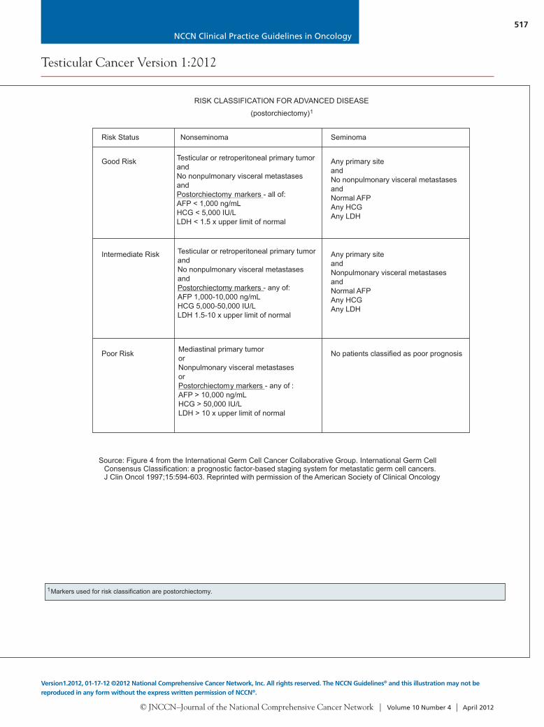
Risk Status Nonseminoma Seminoma
Good Risk Any primary siteandNo nonpulmonary visceral metastasesandNormal AFPAny HCGAny LDH
Intermediate Risk Any primary siteandNonpulmonary visceral metastasesandNormal AFPAny HCGAny LDH
Poor Risk No patients classified as poor prognosis
Source: Figure 4 from the International Germ Cell Cancer Collaborative Group. International Germ CellConsensus Classification: a prognostic factor-based staging system for metastatic germ cell cancers. J Clin Oncol 1997;15:594-603. Reprinted with permission of the American Society of Clinical Oncology.
1Markers used for risk classification are postorchiectomy.
Testicular or retroperitoneal primary tumorandNo nonpulmonary visceral metastasesand
- all of:AFP < 1,000 ng/mLHCG < 5,000 IU/LLDH < 1.5 x upper limit of normal
Postorchiectomy markers
Testicular or retroperitoneal primary tumorandNo nonpulmonary visceral metastasesand
- any of:AFP 1,000-10,000 ng/mLHCG 5,000-50,000 IU/LLDH 1.5-10 x upper limit of normal
Postorchiectomy markers
Mediastinal primary tumororNonpulmonary visceral metastasesor
- any of :AFP > 10,000 ng/mLHCG > 50,000 IU/LLDH > 10 x upper limit of normal
Postorchiectomy markers
(postorchiectomy)1
RISK CLASSIFICATION FOR ADVANCED DISEASE
NCCN Clinical Practice Guidelines in Oncology
© JNCCN–Journal of the National Comprehensive Cancer Network | Volume 10 Number 4 | April 2012
517
Testicular Cancer Version 1:2012
Version1.2012, 01-17-12 ©2012 National Comprehensive Cancer Network, Inc. All rights reserved. The NCCN Guidelines® and this illustration may not be reproduced in any form without the express written permission of NCCN®.
Figure 2Figure 1 Figure 3
PRINCIPLES OF RADIOTHERAPY FOR PURE TESTICULAR SEMINOMA
References1. Chung P, Warde P. Stage I seminoma: adjuvant treatment is effective but is it necessary? J Natl Cancer Inst 2011;103:194-196.2. Chung P. Prognostic factors for relapse in stage I seminoma: a validation study [abstract]. J Clin Oncol 2010;28:Abstract 4535.3. Zilli T, Boudreau C, Doucet R, et al. Bone marrow-sparing intensity-modulated radiation therapy for stage I seminoma. Acta Oncol 2011;50:555-562.4. Hall EJ, Wuu CS. Radiation-induced second cancers: the impact of 3D-CRT and IMRT. Int J Radiat Oncol Biol Phys 2003;56:83-88.5. Ragni G, Somigliana E, Restelli L, et al. Sperm banking and rate of assisted reproduction treatment: insights from a 15-year cryopreservation program for
male cancer patients. Cancer 2003;97:1624-1629.6. Saito K, Suzuki K, Iwasaki A, et al. Sperm cryopreservation before cancer chemotherapy helps in the emotional battle against cancer. Cancer
2005;104:521-524.7. Oliver RT, Mead GM, Rustin GJ, et al. Randomized trial of carboplatin versus radiotherapy for stage I seminoma: mature results on relapse and
contralateral testis cancer rates in MRC TE19/EORTC 30982 study (ISRCTN27163214). J Clin Oncol 2011;29:957-962.8. Mead GM, Fossa SD, Oliver RT, et al. Randomized trials in 2466 patients with stage I seminoma: patterns of relapse and follow-up. J Natl Cancer Inst
2011;103:241-249.9. Garmezy B, Pagliaro LC. Choosing treatment for stage I seminoma: who should get what? Oncology (Williston Park) 2009;23:753, 759.10. Fossa SD, Horwich A, Russell JM, et al. Optimal planning target volume for stage I testicular seminoma: a Medical Research Council randomized trial.
Medical Research Council Testicular Tumor Working Group. J Clin Oncol 1999;17:1146-1154.11. Dinniwell R, Chan P, Czarnota G, et al. Pelvic lymph node topography for radiotherapy treatment planning from ferumoxtran-10 contrast-enhanced
magnetic resonance imaging. Int J Radiat Oncol Biol Phys 2009;74:844-851.12. McMahon CJ, Rofsky NM, Pedrosa I. Lymphatic metastases from pelvic tumors: anatomic classification, characterization, and staging. Radiology
2010;254:31-46.13. Bruns F, Bremer M, Meyer A, et al. Adjuvant radiotherapy in stage I seminoma: is there a role for further reduction of treatment volume? Acta Oncol
2005;44:142-148.14. Classen J, Schmidberger H, Meisner C, et al. Para-aortic irradiation for stage I testicular seminoma: results of a prospective study in 675 patients. A trial
of the German testicular cancer study group (GTCSG). Br J Cancer 2004;90:2305-2311.15. Shih HA, Harisinghani M, Zietman AL, et al. Mapping of nodal disease in locally advanced prostate cancer: rethinking the clinical target volume for pelvic
nodal irradiation based on vascular rather than bony anatomy. Int J Radiat Oncol Biol Phys 2005;63:1262-1269.16. Boujelbene N, Cosinschi A, Khanfir K, et al. Pure seminoma: a review and update. Radiat Oncol 2011;6:90.17. Jones WG, Fossa SD, Mead GM, et al. Randomized trial of 30 versus 20 Gy in the adjuvant treatment of stage I testicular seminoma: a report on Medical
Research Council Trial TE18, European Organisation for the Research and Treatment of Cancer Trial 30942 (ISRCTN18525328). J Clin Oncol2005;23:1200-1208.
18. Choo R, Sandler H, Warde P, et al. Survey of radiation oncologists: practice patterns of the management of stage I seminoma of testis in Canada and aselected group in the United States. Can J Urol 2002;9:1479-1485.
19. Classen J, Schmidberger H, Meisner C, et al. Radiotherapy for stages IIA/B testicular seminoma: final report of a prospective multicenter clinical trial. JClin Oncol 2003;21:1101-1106.
20. Paly J, Efstathiou J, Hedgire S, et al. Mapping patterns of nodal metastases in seminoma: rethinking the para-aortic field [abstract]. Int J Radiat Oncol BiolPhys 2011;81:Abstract 88.
Risk Status Nonseminoma Seminoma
Good Risk Any primary siteandNo nonpulmonary visceral metastasesandNormal AFPAny HCGAny LDH
Intermediate Risk Any primary siteandNonpulmonary visceral metastasesandNormal AFPAny HCGAny LDH
Poor Risk No patients classified as poor prognosis
Source: Figure 4 from the International Germ Cell Cancer Collaborative Group. International Germ CellConsensus Classification: a prognostic factor-based staging system for metastatic germ cell cancers. J Clin Oncol 1997;15:594-603. Reprinted with permission of the American Society of Clinical Oncology.
1Markers used for risk classification are postorchiectomy.
Testicular or retroperitoneal primary tumorandNo nonpulmonary visceral metastasesand
- all of:AFP < 1,000 ng/mLHCG < 5,000 IU/LLDH < 1.5 x upper limit of normal
Postorchiectomy markers
Testicular or retroperitoneal primary tumorandNo nonpulmonary visceral metastasesand
- any of:AFP 1,000-10,000 ng/mLHCG 5,000-50,000 IU/LLDH 1.5-10 x upper limit of normal
Postorchiectomy markers
Mediastinal primary tumororNonpulmonary visceral metastasesor
- any of :AFP > 10,000 ng/mLHCG > 50,000 IU/LLDH > 10 x upper limit of normal
Postorchiectomy markers
(postorchiectomy)1
RISK CLASSIFICATION FOR ADVANCED DISEASE

© JNCCN–Journal of the National Comprehensive Cancer Network | Volume 10 Number 4 | April 2012
518
Testicular Cancer Version 1:2012
Clinical trials: NCCN believes that the best management of any cancer patient is in a clinical trial. Participation in clinical trials is especially encouraged. All recommendations are category 2A unless otherwise indicated.
PRIMARY CHEMOTHERAPY REGIMENS FOR GERM CELL TUMORS
1
2
3
Xiao H, Mazumdar M, Bajorin DF, et al. Long-term follow-up of patients with good-risk germ cell tumors treated with etoposide and cisplatin. J Clin Oncol1997;15:2553-2558.
Saxman SB, Finch D, Gonin R, Einhorn LH. Long-term follow-up of a phase III study of three versus four cycles of bleomycin, etoposide, and cisplatin infavorable-prognosis germ-cell tumors: the Indiana University experience. J Clin Oncol 1998;16:702-706.
Nichols CR, Catalano PJ, Crawford ED, et al. Randomized comparison of cisplatin and etoposide and either bleomycin or ifosfamide in treatment ofadvanced disseminated germ cell tumors: an Eastern Cooperative Oncology Group, Southwest Oncology Group, and Cancer and Leukemia Group BStudy. J Clin Oncol 1998;16:1287-1293.
EPEtoposide, 100 mg/m IV on days 1-5Cisplatin, 20 mg/m IV on days 1- 5Repeat every 21 days
BEPEtoposide, 100 mg/m IV on days 1- 5Cisplatin, 20 mg/m IV on days 1- 5Bleomycin, 30 units IV weekly on days 1, 8, and 15 or daysRepeat every 21 days
VIPEtoposide, 75 mg/m IV on days 1-5Mesna, 120 mg/m slow IV push before ifosfamide on day 1, thenMesna,1200 mg/m IV continuous infusion on days 1-5Ifosfamide, 1200 mg/m on days 1-5Cisplatin, 20 mg/m IV on days 1-5Repeat every 21 days
22
22
222
22
1
2
3
2, 9, 16
SECOND-LINE OR SUBSEQUENT CHEMOTHERAPY REGIMENS FORMETASTATIC GERM CELL TUMORS
VeIPVinblastine, 0.11 mg/kg IV push on days 1-2Mesna, 400 mg/m IV every 8 h on days 1-5Ifosfamide, 1200 mg/m IV on days 1-5Cisplatin, 20 mg/m IV on days 1-5Repeat every 21 days
TIPPaclitaxel, 250 mg/m IV on day 1Ifosfamide, 1500 mg/m IV on days 2-5Mesna, 500 mg/m IV before ifosfamide, and then 4 and8 h after each ifosfamide dose on days 2-5Cisplatin, 25 mg/m IV on days 2-5Repeat every 21 days
22
2
22
2
2
1
2
Conventional-dose chemotherapy regimens High-dose chemotherapy regimens
Carboplatin, 700 mg/m (body surface area) IVEtoposide, 750 mg/m IVAdminister 5, 4, and 3 days before peripheral blood stem cellinfusion for 2 cycles
Paclitaxel, 200 mg/m IV over 24 h on day 1Ifosfamide, 2000 mg/m over 4 h with mesna protection on days 2-4Repeat every 14 days for 2 cycles followed byCarboplatin, AUC 7-8 IV over 60 min days 1-3Etoposide, 400 mg/m IV days 1-3Administer with peripheral blood stem cell support at 14- to 21-dayintervals for 3 cycles
22
22
2
3
4
1
2
3
4
Loehrer PJ Sr, Lauer R, Roth BJ, et al. Salvage therapy in recurrent germ cell cancer: ifosfamide and cisplatin plus either vinblastine or etoposide. AnnIntern Med 1988;109:540-546.
Kondagunta GV, Bacik J, Donadio A, et al. Combination of paclitaxel, ifosfamide, and cisplatin is an effective second-line therapy for patients with relapsedtesticular germ cell tumors. J Clin Oncol 2005;23:6549-6555.
Einhorn LH, Williams SD, Chamness A, et al. High-dose chemotherapy and stem-cell rescue for metastatic germ-cell tumors. N Engl J Med 2007;357:340-348.
Kondagunta GV, Bacik J, Sheinfeld J, et al. Paclitaxel plus Ifosfamide followed by high-dose carboplatin plus etoposide in previously treated germ celltumors. J Clin Oncol 2007;25:85-90.
Palliative chemotherapy regimens*
GemOx
Paclitaxel/gemcitabine
Gemcitabine/oxaliplatin/paclitaxel
Pectasides D, Pectasides M, Farmakis D, et al. Gemcitabine and oxaliplatin (GEMOX) in patients with cisplatin-refractory germ cell tumors: a phaseII study. Ann Oncol 2004;15:493-497.Kollmannsberger C, Beyer J, Liersch R, et al. Combination chemotherapy with gemcitabine plus oxaliplatin in patients with intensively pretreated orrefractory germ cell cancer: a study of the German Testicular Cancer Study Group. J Clin Oncol 2004;22:108-114.De Giorgi U, Rosti G, Aieta M, et al. Phase II study of oxaliplatin and gemcitabine salvage chemotherapy in patients with cisplatin-refractorynonseminomatous germ cell tumor. Eur Urol 2006;50:893-894.
Einhorn LH, Brames MJ, Juliar B, Williams SD. Phase II study of paclitaxel plus gemcitabine salvage chemotherapy for germ cell tumors afterprogression following high-dose chemotherapy with tandem transplant. 2007;25:513-516.Mulherin B, Brames M, Einhorn L. Long-term survival with paclitaxel and gemcitabine for germ cell tumors after progression following high-dosechemotherapy with tandem transplants [abstract]. J Clin Oncol 2011;29:Abstract 4562.
Bokemeyer C, Oechsle K, Honecker F, et al. Combination chemotherapy with gemcitabine, oxaliplatin, and paclitaxel in patients with cisplatin-refractory or multiply relapsed germ-cell tumors: a study of the German Testicular Cancer Study Group. Ann Oncol 2008;19:448-453.
J Clin Oncol
Gemcitabine/oxaliplatinGemcitabine/paclitaxelGemcitabine/paclitaxel/oxaliplatin
*Please see references for dosing.below

NCCN Clinical Practice Guidelines in Oncology
© JNCCN–Journal of the National Comprehensive Cancer Network | Volume 10 Number 4 | April 2012
519
Testicular Cancer Version 1:2012
Version1.2012, 01-17-12 ©2012 National Comprehensive Cancer Network, Inc. All rights reserved. The NCCN Guidelines® and this illustration may not be reproduced in any form without the express written permission of NCCN®.
PRIMARY CHEMOTHERAPY REGIMENS FOR GERM CELL TUMORS
1
2
3
Xiao H, Mazumdar M, Bajorin DF, et al. Long-term follow-up of patients with good-risk germ cell tumors treated with etoposide and cisplatin. J Clin Oncol1997;15:2553-2558.
Saxman SB, Finch D, Gonin R, Einhorn LH. Long-term follow-up of a phase III study of three versus four cycles of bleomycin, etoposide, and cisplatin infavorable-prognosis germ-cell tumors: the Indiana University experience. J Clin Oncol 1998;16:702-706.
Nichols CR, Catalano PJ, Crawford ED, et al. Randomized comparison of cisplatin and etoposide and either bleomycin or ifosfamide in treatment ofadvanced disseminated germ cell tumors: an Eastern Cooperative Oncology Group, Southwest Oncology Group, and Cancer and Leukemia Group BStudy. J Clin Oncol 1998;16:1287-1293.
EPEtoposide, 100 mg/m IV on days 1-5Cisplatin, 20 mg/m IV on days 1- 5Repeat every 21 days
BEPEtoposide, 100 mg/m IV on days 1- 5Cisplatin, 20 mg/m IV on days 1- 5Bleomycin, 30 units IV weekly on days 1, 8, and 15 or daysRepeat every 21 days
VIPEtoposide, 75 mg/m IV on days 1-5Mesna, 120 mg/m slow IV push before ifosfamide on day 1, thenMesna,1200 mg/m IV continuous infusion on days 1-5Ifosfamide, 1200 mg/m on days 1-5Cisplatin, 20 mg/m IV on days 1-5Repeat every 21 days
22
22
222
22
1
2
3
2, 9, 16
SECOND-LINE OR SUBSEQUENT CHEMOTHERAPY REGIMENS FORMETASTATIC GERM CELL TUMORS
VeIPVinblastine, 0.11 mg/kg IV push on days 1-2Mesna, 400 mg/m IV every 8 h on days 1-5Ifosfamide, 1200 mg/m IV on days 1-5Cisplatin, 20 mg/m IV on days 1-5Repeat every 21 days
TIPPaclitaxel, 250 mg/m IV on day 1Ifosfamide, 1500 mg/m IV on days 2-5Mesna, 500 mg/m IV before ifosfamide, and then 4 and8 h after each ifosfamide dose on days 2-5Cisplatin, 25 mg/m IV on days 2-5Repeat every 21 days
22
2
22
2
2
1
2
Conventional-dose chemotherapy regimens High-dose chemotherapy regimens
Carboplatin, 700 mg/m (body surface area) IVEtoposide, 750 mg/m IVAdminister 5, 4, and 3 days before peripheral blood stem cellinfusion for 2 cycles
Paclitaxel, 200 mg/m IV over 24 h on day 1Ifosfamide, 2000 mg/m over 4 h with mesna protection on days 2-4Repeat every 14 days for 2 cycles followed byCarboplatin, AUC 7-8 IV over 60 min days 1-3Etoposide, 400 mg/m IV days 1-3Administer with peripheral blood stem cell support at 14- to 21-dayintervals for 3 cycles
22
22
2
3
4
1
2
3
4
Loehrer PJ Sr, Lauer R, Roth BJ, et al. Salvage therapy in recurrent germ cell cancer: ifosfamide and cisplatin plus either vinblastine or etoposide. AnnIntern Med 1988;109:540-546.
Kondagunta GV, Bacik J, Donadio A, et al. Combination of paclitaxel, ifosfamide, and cisplatin is an effective second-line therapy for patients with relapsedtesticular germ cell tumors. J Clin Oncol 2005;23:6549-6555.
Einhorn LH, Williams SD, Chamness A, et al. High-dose chemotherapy and stem-cell rescue for metastatic germ-cell tumors. N Engl J Med 2007;357:340-348.
Kondagunta GV, Bacik J, Sheinfeld J, et al. Paclitaxel plus Ifosfamide followed by high-dose carboplatin plus etoposide in previously treated germ celltumors. J Clin Oncol 2007;25:85-90.
Palliative chemotherapy regimens*
GemOx
Paclitaxel/gemcitabine
Gemcitabine/oxaliplatin/paclitaxel
Pectasides D, Pectasides M, Farmakis D, et al. Gemcitabine and oxaliplatin (GEMOX) in patients with cisplatin-refractory germ cell tumors: a phaseII study. Ann Oncol 2004;15:493-497.Kollmannsberger C, Beyer J, Liersch R, et al. Combination chemotherapy with gemcitabine plus oxaliplatin in patients with intensively pretreated orrefractory germ cell cancer: a study of the German Testicular Cancer Study Group. J Clin Oncol 2004;22:108-114.De Giorgi U, Rosti G, Aieta M, et al. Phase II study of oxaliplatin and gemcitabine salvage chemotherapy in patients with cisplatin-refractorynonseminomatous germ cell tumor. Eur Urol 2006;50:893-894.
Einhorn LH, Brames MJ, Juliar B, Williams SD. Phase II study of paclitaxel plus gemcitabine salvage chemotherapy for germ cell tumors afterprogression following high-dose chemotherapy with tandem transplant. 2007;25:513-516.Mulherin B, Brames M, Einhorn L. Long-term survival with paclitaxel and gemcitabine for germ cell tumors after progression following high-dosechemotherapy with tandem transplants [abstract]. J Clin Oncol 2011;29:Abstract 4562.
Bokemeyer C, Oechsle K, Honecker F, et al. Combination chemotherapy with gemcitabine, oxaliplatin, and paclitaxel in patients with cisplatin-refractory or multiply relapsed germ-cell tumors: a study of the German Testicular Cancer Study Group. Ann Oncol 2008;19:448-453.
J Clin Oncol
Gemcitabine/oxaliplatinGemcitabine/paclitaxelGemcitabine/paclitaxel/oxaliplatin
*Please see references for dosing.below

© JNCCN–Journal of the National Comprehensive Cancer Network | Volume 10 Number 4 | April 2012
520
Testicular Cancer Version 1:2012
Clinical trials: NCCN believes that the best management of any cancer patient is in a clinical trial. Participation in clinical trials is especially encouraged. All recommendations are category 2A unless otherwise indicated.
PRINCIPLES OF SURGERY
•
•
••
•
•
RPLND is the standard approach to the surgical management of nonseminoma germ cell tumor (NSGCT) in both primary andpostchemotherapy setting.
A template dissection or a nerve-sparing approach to minimize the risk of ejaculatory disorders should be considered in patientsundergoing primary RPLND for stage I nonseminoma.
Referral to high-volume centers should be considered for surgical resection of postchemotherapy.
Completeness of resection is an independent and consistent predictive variable of clinical outcome. In postchemotherapy RPLND,surgical margins should not be compromised in an attempt to preserve ejaculation. Additional procedures and resection of adjacentstructures may be required.
Postchemotherapy RPLND is indicated in patients with metastatic NSGCT with a residual retroperitoneal mass after systemicchemotherapy and normalized postchemotherapy serum tumor markers.
A full bilateral template RPLND should be performed in all patients undergoing RPLND in the postchemotherapy setting, with theboundaries of dissection being the renal hilar vessels (superiorly), ureters (laterally), and the common iliac arteries (inferiorly).
• The “split and roll” technique in which lumbar vessels are identified and sequentially ligated allows resection of all lymphatic tissuearound and behind the great vessels (aorta, IVC) and minimizes the risk of an in-field recurrence.
massesPostchemotherapy setting
NONSEMINOMA

NCCN Clinical Practice Guidelines in Oncology
Testicular Cancer
© JNCCN–Journal of the National Comprehensive Cancer Network | Volume 10 Number 4 | April 2012
521
Text continued from p. 503
An elevated serum concentration of β-HCG, which has a half-life of approximately 1 to 3 days, may also be present with seminomatous and nonseminoma-tous tumors. The elevations of β-HCG must be in-terpreted with caution, because hypogonadism and marijuana use may cause benign serum elevations of β-HCG.
Nonseminoma is the more clinically aggressive tumor. When both seminoma and elements of a non-seminoma are present, management follows that for a nonseminoma. Therefore, the diagnosis of a semi-noma is restricted to pure seminoma histology and a normal serum concentration of AFP.
More than 90% of patients diagnosed with GCTs are cured, including 70% to 80% with ad-vanced tumors who are treated with chemotherapy. A delay in diagnosis correlates with a higher stage at presentation. Standard therapy has been estab-lished at essentially all stages of management and must be closely followed to ensure the potential for cure.
Clinical PresentationA painless solid testicular mass is pathognomonic for testicular tumor. More often, patients present with testicular discomfort or swelling suggestive of epididymitis or orchitis. A trial of antibiotics may be given in this circumstance, but persistent tenderness, swelling, or any palpable abnormality warrants fur-ther evaluation.
Diagnosis and WorkupIf an intratesticular mass is identified, complete blood count, creatinine, electrolytes, and liver en-zymes should be obtained. Further evaluation in-cludes measurement of serum tumor markers and a chest radiograph. Testicular ultrasound serves to confirm the presence of a testicular mass and to ex-plore the contralateral testis; it is sensitive and has an important role in determining whether a mass is intra- or extratesticular.4
Serum tumor markers are critical in assignment of prognosis and also management during treat-ment. Serum tumor markers are prognostic factors and contribute to diagnosis and staging.5 Markers are assessed before orchiectomy and repeated after orchiectomy. Elevated values of β-HCG, LDH, or AFP should be followed up with repeated tests to allow precise staging.
Biopsy may also be considered if a suspicious in-tratesticular abnormality, such as a hypoechoic mass or macrocalcification, is identified on ultrasound. In contrast, if microcalcifications without any other ab-normality can be observed, testicular biopsy is not necessary.
In patients of reproductive age, sperm banking must be discussed.6,7 It must be discussed with the patients before undergoing any therapeutic inter-vention that may compromise fertility, including surgery, radiation therapy, and chemotherapy.8–10 If sperm banking is desired, it may be performed either before or after orchiectomy, but certainly before sub-sequent therapy.
Inguinal orchiectomy is considered the prima-ry treatment for most patients who present with a suspicious testicular mass.11 An open inguinal bi-opsy of the contralateral testis is not routinely per-formed, but can be considered when a cryptorchid testis or marked atrophy is present.12 The extent of primary tumor is classified after orchiectomy, and therefore pathological (p) stage is assigned to the primary tumor (T).
Further management is dictated by histology, a diagnosis of pure seminoma or nonseminoma (in-cludes mixed seminoma tumors and seminoma his-tology with elevated AFP), and the stage. Although rare, when a patient presents with rapidly increas-ing β-HCG, symptoms related to disseminated dis-ease, and a testicular mass, chemotherapy can be initiated immediately without waiting for a biopsy diagnosis.
Risk Classification for Advanced DiseaseIn 1997, the International Germ Cell Cancer Con-sensus Group (IGCCCG) defined a prognostic factor–based classification system based on identi-fication of some clinically independent prognostic features, such as extent of disease and levels of serum tumor markers postorchiectomy. Postorchiectomy markers are used to classify the patient according to the IGCCCG risk classification. This classification categorizes patients with pure seminoma and non-seminoma GCT into good-, intermediate-, or poor-risk groups.13
Stage and risk classification are assigned accord-ing to the American Joint Committee on Cancer (AJCC) and IGCCCG classification.

NCCN Clinical Practice Guidelines in Oncology
Testicular Cancer
© JNCCN–Journal of the National Comprehensive Cancer Network | Volume 10 Number 4 | April 2012
522
Pure SeminomaIf a GCT is found, an abdominopelvic CT scan is performed. Abdominopelvic CT scanning is used to assess the retroperitoneal nodes.14 A chest CT is indicated if the abdominopelvic CT shows retroperi-toneal adenopathy or the chest radiograph shows ab-normal results. A chest CT scan is a sensitive way to evaluate the thorax and mediastinal nodes.15
The panel members recommend a brain MRI or bone scan only if metastases to these organs is suspected.
Elevated values of β-HCG, LDH, or AFP should be followed up with repeated tests. Serum concentrations of β-HCG and LDH may be el-evated in patients with seminoma. An elevated AFP level indicates nonseminoma, and patients should be managed accordingly. Initial manage-ment of pure seminoma involves a radical inguinal orchiectomy. Orchiectomy is both diagnostic and therapeutic. Patients with seminoma arising from an extragonadal site, such as the mediastinum, are treated with standard chemotherapy regimens ac-cording to risk status.
Pure Seminoma Stages IA and IB Primary Treatment for Pure Seminoma Stages IA and IB: For patients with stages IA and IB pure sem-inoma, the standard treatment options after initial orchiectomy include surveillance, radiotherapy, or chemotherapy with 1 or 2 cycles of carboplatin. The disease-specific survival for stage I disease is 99%, ir-respective of the management strategy used.16 Sever-al prospective nonrandomized studies of surveillance have been conducted.17–20 The relapse rate seen in these studies is 15% to 20% at 5 years, and most of the relapses are first detected in infradiaphrag- matic lymph nodes.18–20 Some studies report tumor size greater than 4 cm and rete testis invasion as risk factors for relapse.19,21,22 However, a validation study by Chung et al.23,24 showed that tumor size greater than 4 cm and rete testis invasion were not predic-tors of relapse. Therefore, the panel members dis-courage risk-adapted management based on tumor size greater than 4 cm and rete testis invasion for stage I pure seminoma. Surveillance is listed as the preferred option (category 1) for patients with pT1 and pT2 disease.
If surveillance is not applicable, alternatives are either adjuvant carboplatin or adjuvant radiothera-py, as described later. Each approach has distinct ad-
vantages and disadvantages. The physicians should discuss these with the patients and their families and pick the best approach on a case-by-case basis.
Oliver et al.25 reported on the results of a trial that randomized 1477 patients with stage I testicular cancer to undergo either radiotherapy or one injec-tion of carboplatin. In the study, carboplatin (area under the cure [AUC] × 7) was administered intra-venously. The dose was calculated by the formula 7 × (glomerular filtration rate [GFR, mL/min] + 25 mg). With a median follow-up of 4 years, the relapse-free survival rates were similar for both groups.25 Late re-lapses and secondary GCTs can occur beyond 5 and 10 years. Therefore, the investigators continued to follow these patients. The updated results reported noninferiority of single-dose carboplatin versus ra-diation therapy.26 In an intent-to-treat analysis, the relapse-free rates at 5 years were 94.7% for the carboplatin arm and 96% for the radiotherapy arm (hazard ratio, 1.25; P = .37). Two cases of contralat-eral GCTs were seen in the carboplatin arm versus 15 in the radiation therapy arm, with hazard ratio of 0.22; the contralateral GCT-free rates at 5 years are 99.8% and 98.8%, respectively. The authors con-cluded that a single dose of carboplatin is less toxic and as effective in preventing disease recurrence as adjuvant radiotherapy in men with stage I pure seminoma after orchiectomy.26 Two courses of adju-vant carboplatin have also been reported to reduce the relapse rate.27 The panel recommends either 1 or 2 cycles of carboplatin AUC × 7 as a category 1 recommendation for patients with stages IA and IB pure seminoma.
If radiation therapy is delivered, the panel rec-ommends a total dose of 20 Gy (midplane) in 10 dai-ly 2.0-Gy fractions,28 given to an infradiaphragmatic area, including para-aortic lymph nodes; in special circumstances, this area may include the ipsilateral ilioinguinal nodes.29–32 Patients for whom radiation therapy is generally not given include those at high-er risk for morbidity from radiation therapy, such as those with a history of pelvic surgery. Prophylaxis to the mediastinum is not provided, because relapse rarely occurs at this site. For patients with stages IA and IB pure seminoma, adjuvant radiation therapy to include the para-aortic nodes is also a category 1 recommendation, although active surveillance is preferred (see Principles of Radiotherapy for Pure Testicular Seminoma, pages 513–516).

NCCN Clinical Practice Guidelines in Oncology
Testicular Cancer
© JNCCN–Journal of the National Comprehensive Cancer Network | Volume 10 Number 4 | April 2012
523
Follow-Up After Primary Treatment for Pure Sem-inoma Stages IA and IB: For follow-up, the differ-ent risk of recurrence associated with each treatment modality is important to distinguish (surveillance vs. adjuvant therapy). An analysis of more than 5000 patients with stage I seminoma from various trials showed that, independent of the treatment modal-ity, the risk of recurrence is highest in the first 2 years and decreases after that.33
Follow-up during surveillance includes a history and physical, with measurement of postorchiectomy serum tumor markers (AFP, β-HCG, and LDH), per-formed every 3 to 4 months for 1 to 2 years, every 6 to 12 months for years 3 to 4, and annually thereafter.34, 35
Controversy exists regarding how many imaging studies must be performed in patients on active sur-veillance. The panel recommends abdominal/pelvic CT every 6 months for years 1 to 2, every 6 to 12 months for year 3, and then annually for years 4 to 5. The most common site of relapse in patients man-aged with surveillance or adjuvant chemotherapy is the retroperitoneal nodes. Chest radiographs may be obtained as clinically indicated for years 1 to 5. The clinical trial TRISST (MRC TE24/Trial of Imaging and Schedule in Seminoma Testis) in the United Kingdom is currently studying whether a reduced CT schedule or MRI could be used as a safe and ef-fective alternative to standard CT-based surveillance in the management of stage I seminoma.36
The risk of recurrence 5 years after adjuvant treatment is less than 0.3% annually.33 Follow-up of patients treated with carboplatin includes a history and physical, with measurement of post orchiectomy serum tumor markers (AFP, β-HCG, and LDH) per-formed every 3 months the first year, every 4 months the second year, every 6 months the third year, and annually thereafter.
The panel recommends abdominal/pelvic CT annually for the first 3 years after radiotherapy or carboplatin. In a recently published meta-analysis of 2466 patients, Mead et al.16 reported that recurrence rarely occurred after more than 3 years from treat-ment with either radiotherapy or carboplatin. Re-lapse occurred after 3 years only in 4 of the 2466 pa-tients (0.2%).16 They recommend that CT scans of the pelvis and chest can be omitted in these patients as part of routine follow-up. The panel recommends that chest radiographs be obtained only as clinically indicated.
Follow-up of patients treated with radiotherapy includes a history and physical, with measurement of postorchiectomy serum tumor markers (AFP, β-HCG, and LDH). Follow-up should be performed every 4 months for 1 to 2 years, and then annually for 3 to 10 years.33 Para-aortic radiation therapy is recommended when patients with stage I seminoma are irradiated (see pages 513–516). Patients treated with para-aortic radiation therapy have a slightly higher rate of pelvic relapse than those treated with ‘‘dog-leg’’ radiograph.30,33,37,38 Some NCCN Mem-ber Institutions obtain a CT scan of the pelvis only every 6 months for 3 years after para-aortic radio-therapy.33 Others obtain CT scans of the pelvis and abdomen annually for 3 years.30 The panel’s con-sensus recommendation is for abdominal and pelvic CT scans annually for 3 years in patients treated with para-aortic radiotherapy. Chest radiographs should be obtained only when clinically indicated. Recurrences are treated according to the stage at relapse.16
Pure Seminoma Stage ISPrimary Treatment for Pure Seminoma Stage IS: According to the AJCC definition, stage IS requires persistent elevation of serum tumor markers (LDH, AFP, and β-HCG) after orchiectomy. Stage IS is uncommon and patients are generally treated with radiation to an infradiaphragmatic area, including para-aortic lymph nodes with or without radiation to the ipsilateral ilioinguinal nodes.30–32
Follow-Up After Primary Radiation Treatment for Pure Seminoma Stage IS: Follow-up recommenda-tions by the panel for patients with stage IS treated with adjuvant radiation therapy are similar to those for patients with stages IA and IB treated with adju-vant radiation therapy. Recurrences are treated ac-cording to the stage at relapse.
Pure Seminoma Stages IIA and IIBPrimary Treatment for Pure Seminoma Stages IIA and IIB: Stage IIA is defined as metastatic disease to lymph nodes, with a lymph node mass measuring less than 2 cm in diameter in greatest dimension on CT scan, whereas stage IIB is disease measuring 2 to 5 cm in maximum diameter.
Radiotherapy has been the mainstay of treat-ment in patients with stage IIA and IIB semino-ma.39–41 The standard radiation field compared with stage I is extended from the para-aortic region to

NCCN Clinical Practice Guidelines in Oncology
Testicular Cancer
© JNCCN–Journal of the National Comprehensive Cancer Network | Volume 10 Number 4 | April 2012
524
include an ipsilateral iliac field. The relapse rates are moderate (5%–6% for stage IIA) and overall sur-vival is almost 100%.39,41,42
For patients with stage IIA or IIB seminoma, the panel recommends radiation therapy to an infradia-phragmatic area, including para-aortic and ipsilateral iliac lymph nodes in 2 anteroposterior–posteroanterior phases. The initial phase consists of radiation to modified dog-leg fields at a dose of 20 Gy (midplane) in 10 daily 2.0-Gy fractions29 or 25.5 Gy in 15 daily 1.7-Gy fractions.43 The panel prefers modified ‘dog-leg’ fields as described by Classen et al.39 For details on field arrangement, see pages 513–516. The sec-ond phase (cone down) of radiotherapy consists of daily 2.0-Gy fractions to a cumulative total dose of approximately 30 Gy for stage IIA and 36 Gy for stage IIB.39 As with the management of stage I dis-ease, prophylactic mediastinal radiation therapy is not indicated for stage II disease.44
For selected patients with stage IIB seminoma, such as those with adenopathy measuring more than 3 cm,45 chemotherapy with 4 courses of etoposide and cisplatin (EP) or 3 cycles of bleomycin, etoposide, and cisplatin (BEP) is an alternative to radiotherapy.42,46
Follow-Up for Stages IIA and IIB Pure Semino-ma After Primary Treatment: The recommended follow-up schedules for patients with stage IIA/B seminoma after radiation therapy include a history and physical, with measurement of postorchiectomy serum tumor markers (AFP, β-HCG, and LDH) per-formed every 3 months for year 1, every 6 months for years 2 to 5, and then annually for years 6 to 10.
Chest radiograph is recommended every 6 months for the first 2 years. An abdominal CT scan is recommended every 6 months in years 1 to 2 and annually in year 3 after radiotherapy. In patients who have undergone retroperitoneal lymph node dissec-tion (RPLND), it is recommended between 3 to 6 months postsurgery and then as clinically indicated.39
The follow-up of patients with stage IIB semi-noma after chemotherapy is similar to follow-up af-ter chemotherapy for patients with stages IIC and III seminoma, as discussed in Follow-Up for Pure Semi-noma Stages IIB, IIC, and III After Chemotherapy on page 525.
Pure Seminoma Stages IIC and III Primary Treatment for Pure Seminoma Stages IIC and III: Patients with stage IIC or III disease are those considered at either good or intermediate
risk. All stage IIC and stage III seminoma is consid-ered good-risk disease, except for stage III disease with nonpulmonary visceral metastases (e.g., bone, liver, brain), which is considered intermediate-risk. Standard chemotherapy is used for both groups of patients. However, for patients with good risk, 3 cycles of BEP47–49 or 4 cycles of EP50–52 are recom-mended. In contrast, more intensive chemotherapy (i.e., 4 cycles of BEP) is recommended for those with intermediate-risk disease.53,54 All of these chemo-therapy options are category 1 recommendations ac-cording to the panel.Postchemotherapy Management of Pure Seminoma Stages IIB, IIC, and III: After initial chemothera-py, patients with stage IIB, IIC, and III disease are evaluated with serum tumor markers and a CT scan of the chest abdomen and pelvis. Patients are then classified according to the presence or absence of a residual mass and the status of serum tumor markers. Patients with normal markers and either no residual mass or residual mass of 3 cm or less need no further treatment. They should undergo surveillance, as dis-cussed in Follow-Up for Pure Seminoma Stages IIB, IIC, and III After Chemotherapy, on page 525.
In cases of residual tumor larger than 3 cm and marker levels that are normal, a PET scan is recom-mended to assess whether residual viable tumor is present.55 A PET scan has high positive and negative predictive values with regard to the question of re-maining disease in patients with residual masses after chemotherapy.56 To reduce the incidence of false-positive results, the PET scan is typically performed at least 6 weeks after completion of chemotherapy. Notably, granulomatous disease, such as sarcoid, is a source of false-positive results. The panel recom-mends a PET scan in patients with seminoma, a re-sidual mass larger than 3 cm, and normal levels of markers, approximately 6 weeks after chemotherapy to determine whether to continue with surveillance or resume treatment.55,57–61
If the PET scan is negative, no further treatment is needed; however, the patient should undergo follow-up,62,63 as discussed in the next section on Follow-Up for Pure Seminoma Stages IIB, IIC, and III After Chemotherapy.
Because a positive PET scan is a strong indicator of residual active tumor, resection should be consid-ered. Therefore, if technically feasible, RPLND may be considered (category 2A). The other option, if re-

NCCN Clinical Practice Guidelines in Oncology
Testicular Cancer
© JNCCN–Journal of the National Comprehensive Cancer Network | Volume 10 Number 4 | April 2012
525
section is not feasible, is second-line chemotherapy (category 2A). Cisplatin-based combination chemo-therapy is used for second-line treatment.64–66 The regimens are 4 cycles of TIP (paclitaxel, ifosfamide, cisplatin)67 or 4 cycles of VeIP (vinblastine, ifos-famide, cisplatin).65,66
According to these NCCN Guidelines, second-line therapy for seminoma and nonseminoma is similar. This is discussed in Second-Line Therapy for Metastatic GCTs, on page 528. The follow-up of these patients is also described later.Follow-Up for Pure Seminoma Stages IIB, IIC, and III After Chemotherapy: Recommended follow-up schedules include a history and physical with chest radiograph and measurement of postor-chiectomy serum tumor markers every 2 months for the first year, every 3 months for the second year, every 6 months for the third and fourth years, and annually thereafter through the tenth year. An ab-dominal/pelvic CT scan is recommended as clini-cally indicated in all patients, except those who have undergone RPLND, in whom it is recom-mended between 3 and 6 months postsurgery and then as clinically indicated.68 A PET scan may be performed as clinically indicated.
Nonseminoma Similar to the workup for seminoma, if nonsemino-ma is found, CT of the abdomen and pelvis should be performed with chest imaging if needed. MRI of the brain and a bone scan should be conducted in the case of clinical indicators (symptoms) of involvement. PET scanning does not contribute and routine use is not recommended for patients with nonseminoma.69,70
Elevated values of β-HCG, LDH, or AFP should be followed up with repeated tests. Nonseminoma includes mixed seminoma tumors and seminoma his-tology with elevated AFP. Postorchiectomy serum markers are important for classifying patients with nonseminoma into good-, intermediate- and poor-risk groups according to the IGCCCG risk classification.13
In patients of reproductive age, sperm banking must be discussed6,7 before any therapeutic inter-vention is performed that may compromise fertility, including surgery, radiation therapy, or chemother-apy.8–10 If sperm banking is desired, it may be per-formed either before or after orchiectomy, but cer-tainly before adjuvant therapy.
Stage-dependent treatment options after ingui-nal orchiectomy include surveillance, chemothera-py, and RPLND. Although the timing of the RPLND may vary, most patients with nonseminoma will un-dergo an RPLND for either diagnostic or therapeutic purposes at some point during treatment. The ma-jor morbidity associated with bilateral dissection is retrograde ejaculation, resulting in infertility. Nerve dissection techniques preserve antegrade ejaculation in 90% of cases.71
Nonseminoma Stage IAPrimary Treatment of Nonseminoma Stage IA: According to the panel, 2 management options ex-ist for patients with stage IA disease after orchiec-tomy: surveillance72–76 and nerve-sparing RPLND. The cure rate with either approach exceeds 95%, al-though with surveillance, this depends on adherence to periodic follow-up examinations and subsequent chemotherapy for the 20% to 30% of patients who experience relapse. Patients who choose surveillance should agree to be compliant with follow-up. RPLND should be performed using a nerve-sparing tech-nique.77,78 According to these guidelines, the nerve-sparing RPLND is recommended within 4 weeks of a CT scan and within 7 to 10 days of repeat serum marker testing to ensure accurate presurgical staging. Management of Nonseminoma Stage IA After RPLND: After RPLND, if the dissected lymph nodes are not involved with a tumor (pN0), no adju-vant chemotherapy is given and the patients should undergo surveillance. However, if the resected lymph nodes involve tumor, the decision whether to use adjuvant chemotherapy is based on the degree of nodal involvement. Surveillance is preferred over chemotherapy for patients with pN1 disease. Che-motherapy is preferred in patients with pN2 or pN3 disease. Surveillance is an option for patients with pN2 but not for those with pN3 disease. Recom-mended chemotherapy regimens include either EP or BEP. Two cycles of either regimen (EP or BEP) are recommended for patients with pN1 or pN2 dis-ease.79–85 For patients with pN3 disease, longer cours-es of chemotherapy with 4 cycles of EP or 3 cycles of BEP is recommended. Follow-Up for Nonseminoma Stage IA: In the cur-rent guidelines, the long-term follow-up tests for patients with stage IA disease electing primary sur-veillance or who have undergone RPLND or chemo-therapy include serum marker assessment, chest ra-

NCCN Clinical Practice Guidelines in Oncology
Testicular Cancer
© JNCCN–Journal of the National Comprehensive Cancer Network | Volume 10 Number 4 | April 2012
526
diograph, and an abdominal CT scan. The frequency of these tests is outlined in Follow-Up for Nonsemi-noma, page 511.
Nonseminoma Stage IBPrimary Treatment of Nonseminoma Stage IB: Af-ter orchiectomy, either nerve-sparing RPLND or ad-juvant chemotherapy is an option to reduce the risk of relapse in patients with stage IB disease.
Several studies using 2 cycles of BEP as primary treatment for patients with stage I nonseminoma have reported relapse-free survival in more than 95% of patients,76,82,86–90 leading the panel to con-sider this approach a category 2A recommendation. Late consequences of cisplatin-based chemotherapy have been reported based on long-term follow-up of patients.91–96 A trial by Albers et al.97 randomized patients with stage I disease after orchiectomy to un-dergo unilateral RPLND (n = 191) or one adjuvant course of BEP (n = 191). After a median follow-up of 4.7 years, 2 relapses were reported in the group of patients treated with one course of adjuvant BEP and in 13 patients with relapse in the arm treated with RPLND (P = .0011). This study indicates that one course of BEP is active, and could be an option in patients unable to tolerate the toxicity of treatment. The comparator arm in this trial (unilateral RPLND) is not the standard treatment approach. Therefore, although the results of this study are promising, this approach merits further investigation comparing 1 cycle of BEP versus 2 cycles with longer follow-up. The panel considers 1 cycle of BEP a category 2B option as primary therapy.
Surveillance alone may be offered to selected patients with T2 disease (category 2B). Vascular invasion is a significant predictor of relapse when orchiectomy is followed by surveillance alone.11 Surveillance is generally not recommended for T2 disease with vascular invasion because of the 50% chance of relapse. Exceptions are made according to individual circumstances. When surveillance is opted in selected patients with T2 disease, both the patient and physician must be compliant with fol-low-up recommendations.Management of Nonseminoma Stage IB After Primary Treatment: The adjuvant treatment af-ter primary nerve-sparing RPLND for patients with IB disease is similar to that described for stage IA in Management of Nonseminoma Stage IA After RPLND, page 525.
Management after primary chemotherapy in pa-tients with normal values of serum tumor markers may be nerve-sparing RPLND or surveillance. The panel considers nerve-sparing bilateral RPLND a category 2A recommendation for patients with re-sidual mass of 1 cm or greater and a category 2B rec-ommendation if the residual mass is smaller than 1 cm. Surveillance is category 2B for both populations. In the current guidelines, the long-term follow-up tests for patients electing surveillance include serum marker assessment, chest radiograph, and abdominal CT scan. The frequency of these tests is outlined in Follow-Up for Nonseminoma, on page 511.
Nonseminoma Stage ISPatients with stage IS disease exhibit a persistent elevation of serum tumor markers postorchiectomy but no radiographic evidence of disease. The el-evated levels of AFP and β-HCG after orchiectomy must be interpreted with caution, because these may be from causes other than disseminated nonsemi-noma, such as hepatobiliary disease, marijuana use, and hypogonadism.Primary Treatment of Nonseminoma Stage IS: The panel consensus recommendation is that these pa-tients be treated with standard chemotherapy with either 4 cycles of EP or 3 cycles of BEP. Either regi-men is preferable to initial RPLND, because these patients nearly always have disseminated disease.98,99 Management of Stage IS Nonseminoma Postpri-mary Treatment: The management of patients with stage IS nonseminoma after primary treatment with chemotherapy is similar to the management schema outlined for patients with good-risk nonseminoma, including stages IIB, IIC, and IIIA, described in the following sections.
Nonseminoma Stage IIAPrimary Treatment of Nonseminoma Stage IIA: Treatment for patients with stage IIA nonseminoma depends on postorchiectomy serum tumor marker levels.
For patients with stage IIA disease and normal postorchiectomy levels of AFP and β-HCG, the panel considers either primary RPLND (category 2A) or chemotherapy (category 2B) as treatment options.100–104 The chemotherapy regimens include 4 cycles of EP or 3 cycles of BEP. Chemotherapy is considered particularly appropriate if the patient has multifocal disease.

NCCN Clinical Practice Guidelines in Oncology
Testicular Cancer
© JNCCN–Journal of the National Comprehensive Cancer Network | Volume 10 Number 4 | April 2012
527
For patients with persistently elevated AFP or β-HCG levels, the panel recommends induction chemotherapy, based on data from 2 retrospective studies of patients with low-stage nonseminoma treated with RPLND.105,106 The presence of elevated postorchiectomy AFP or β-HCG levels was associ-ated with a high risk of relapse.105,106
Management after primary chemotherapy and RPLND is discussed in the following sections. Management After Primary Treatment of Nonsem-inoma Stage IIA: After primary chemotherapy, sub-sequent management depends on marker levels and the residual mass on CT scan. Therefore, patients must undergo a CT scan before treatment is decided. Lesions smaller than 1 cm on CT scan may represent false-positives and must be interpreted with cau-tion. The options listed by the panel for managing patients with stage IIA disease after primary chemo-therapy include nerve-sparing bilateral RPLND or surveillance.
The panel considers nerve-sparing bilateral RPLND a category 2A recommendation for patients with a residual mass of 1 cm or greater, and a category 2B recommendation if the residual mass is less than 1 cm. A bilateral RPLND involves removal of lym-phatic tissue between both ureters, spanning from the diaphragmatic crus to the bifurcation of the com-mon iliac arteries. The rationale for this extended region of dissection is the greater likelihood of bilat-eral disease with greater tumor burden.107 Referral to high-volume centers must be considered for RPLND postchemotherapy. Surveillance, however, is a cat-egory 2B recommendation for both populations.
After primary nerve-sparing RPLND, treatment options include either surveillance or chemotherapy. The treatment choice depends on the number of positive lymph nodes identified. For example, be-cause RPLND is likely a curative procedure in pa-tients with pathologic stage N0 (pN0), surveillance is the only option listed for this group. Surveillance and chemotherapy are options for patients with pN1 and pN2 disease. RPLND is a curative procedure in 60% to 90% of patients with pN1 disease,106,108,109 and therefore the panel prefers surveillance over chemotherapy for these patients. The risk of re-lapse in patients with pN2 through pN3 disease is greater than 50%.106,108,110 With 2 cycles of adjuvant cisplatin-based chemotherapy, the risk of relapse af-ter RPLND is generally less than 1%.106,111,112 The
panel prefers 2 cycles of adjuvant chemotherapy for pN2 disease, and full-course chemotherapy (not sur-veillance) is recommended for pN3 disease. Recom-mended adjuvant chemotherapy regimens for pN1 and pN2 disease consists of 2 cycles of either BEP or EP,113 resulting in a relapse-free survival rate of nearly 100%. For pN3, the panel recommends a longer che-motherapy course consisting of either 4 cycles of EP or 3 cycles of BEP.
If patients with stage IIA disease have persistent marker elevation (i.e., stage IIA, S1), the primary treatment is chemotherapy as described later for good-risk nonseminoma.
Nonseminoma Stage IIBPrimary Treatment of Nonseminoma Stage IIB: Treatment for patients with stage IIB nonsemino-ma depends also on both postorchiectomy tumor marker levels and radiographic findings. When tu-mor markers are negative, the CT findings deter-mine the proper course of treatment. If abnormal radiographic findings are limited to sites within the lymphatic drainage in the retroperitoneum (i.e., the landing zone), 2 management options are available. One option is to perform nerve-sparing RPLND and consider adjuvant treatment as described for patients with stage IIA disease. The second option is to treat with primary chemotherapy involving either 4 cycles of EP or 3 cycles of BEP, followed by nerve-sparing RPLND or surveillance.
Both options of primary chemotherapy or pri-mary RPLND are comparable options in terms of outcome, but side effects and toxicity are different.101 The reported relapse-free survival with either ap-proach is close to 98%.108,113–118
If metastatic disease (based on radiographic findings) is not confined to the lymphatic drainage (i.e., multifocal lymph node metastases outside the lymphatic drainage sites), chemotherapy is recom-mended with either 4 cycles of EP or 3 cycles of BEP, followed by nerve-sparing RPLND or surveillance.
For patients with stage IIB disease with persis-tent marker elevation (stage IIB, S1), the primary treatment is chemotherapy as described for good-risk nonseminoma, including stages IS, IIC, and IIIA in later sections. Initial RPLND is not recommended in this situation.Management After Primary Treatment of Nonsem-inoma Stage IIB: The management of patients with stage IIB nonseminoma after primary treatment with

NCCN Clinical Practice Guidelines in Oncology
Testicular Cancer
© JNCCN–Journal of the National Comprehensive Cancer Network | Volume 10 Number 4 | April 2012
528
either nerve-sparing bilateral RPLND or chemother-apy is similar to the management scheme outlined earlier for patients with stage IIA nonseminoma af-ter primary treatment.
Advanced Metastatic Nonseminoma The preferred primary chemotherapy regimens for patients with advanced disease depends on the IGCCCG risk classification.13 This classification categorizes patients as good-, intermediate-, or poor-risk.13 Also, patients with an extragonadal primary site, whether retroperitoneal or mediastinal, are treated with initial chemotherapy.Primary Treatment of Good-Risk Nonseminoma: Based on the IGCCCG good-risk classification, this group includes patients with stages IS; IIA and IIB (with persistent marker elevation); IIC; and IIIA disease. Treatment for good-risk GCTs were designed to decrease toxicity while maintaining maximal effi-cacy. Randomized clinical trials showed that this can be achieved through either substituting etoposide for vinblastine,119,120 or eliminating or reducing the dose of bleomycin.119,121 Currently, 2 regimens are recom-mended by the panel: 4 cycles of EP51 or 3 cycles of BEP47,49,122,123 (both category 2A). Either regimen is well tolerated and cures approximately 90% of pa-tients with good risk.124
Primary Treatment of Intermediate-Risk (Stage IIIB) Nonseminoma: For patients with intermediate risk (stage IIIB), the cure rate is approximately 70% with standard therapy using 4 cycles of BEP,125,126 and this is a category 2A recommendation by the panel.Primary Treatment of Poor-Risk (Stage IIIC) Non-seminoma: Between 20% and 30% of all patients with poor-risk, (stage IIIC) metastatic GCTs are not cured with conventional cisplatin therapy, and fewer than one-half experience a durable complete response with 4 cycles of BEP, and therefore the panel lists treatment in a clinical trial as the preferred option.124
The standard chemotherapy regimen for poor-risk patients is 4 cycles of BEP. The regimen contain-ing etoposide, ifosfamide, and cisplatin (VIP) was compared with BEP and found to be more toxic than BEP but equally as effective. Therefore, 4 cycles of VIP may be used for patients who may not tolerate bleomycin.127 Postchemotherapy Management for Good-, Intermediate-, and Poor-Risk Nonseminoma: At the conclusion of induction chemotherapy, CT scans of the abdomen and pelvis are indicated, along with
serum tumor marker assays. PET scans for residual disease have limited predictive value. The frequency of these tests is outlined in Follow-Up for Nonsemi-noma, on page 511.
If a complete response to chemotherapy is found through radiographic imaging and the tumor markers are negative, the panel lists 2 management options: surveillance (category 2B) or bilateral RPLND using nerve-sparing technique, if possible (category 2B).63
If a residual mass is found and the serum tumor markers (AFP and β-HCG) have normalized, then all sites of residual disease are resected.128–130 If only necrotic debris or mature teratoma is encountered, no further therapy is necessary and patients must be put under surveillance. If embryonal, yolk sac, cho-riocarcinoma, or seminoma elements are found in the residual mass, 2 cycles of conventionally dosed chemotherapy (EP, VelP, or TIP) are administered.
After patients are rendered disease-free, stan-dard surveillance is initiated. The frequency of these follow-up tests is outlined in Follow-Up for Nonsem-inoma, on page 511.
Patients who experience an incomplete response to first-line therapy are treated with second-line therapy (see following section). The panel prefers that patients with recurrent nonseminoma be treat-ed at centers with expertise in the management of this disease.
Second-Line Therapy for Metastatic GCTsPatients who do not experience a durable complete response to first-line therapy or those who experi-ence a recurrence can be divided into those with a favorable or unfavorable prognosis based on prognos-tic factors. Prognostic factors can be used in deciding whether a patient is a candidate for conventional-dose therapy or high-dose therapy with stem cell support as a second-line option. To determine the prognosis at initial diagnosis, the IGCCCG classifi-cation is used. However, for patients with progressive or relapsed disease after first-line treatment, several prognostic models have been reported.131–133
Favorable prognostic factors for conventional-dose second-line chemotherapy include a testicular primary site, prior complete response to first-line therapy, low levels of postorchiectomy serum tu-mor markers, and low-volume disease.131 Standard second-line therapy includes conventional-dose

NCCN Clinical Practice Guidelines in Oncology
Testicular Cancer
© JNCCN–Journal of the National Comprehensive Cancer Network | Volume 10 Number 4 | April 2012
529
chemotherapy or high-dose chemotherapy. The conventional-dose regimen includes cisplatin and ifosfamide combined with either vinblastine or pa-clitaxel.134 In patients who experience an incom-plete response or disease relapse after second-line conventional-dose chemotherapy, the preferred third-line option would be high-dose chemothera-py135,136 or chemotherapy in the context of a clinical trial.
Unfavorable prognostic features include incom-plete response to first-line treatment, high levels of serum markers, high-volume disease, and presence of an extratesticular primary tumor. Patients with a tes-ticular primary site and rising postorchiectomy serum tumor markers during first-line therapy are usually considered for high-dose programs. Chemotherapy options for patients with poor prognostic features in-clude chemotherapy in the context of a clinical trial, conventional-dose second-line therapy (with VeIP or TIP), or high-dose chemotherapy (category 2B). Alternatively, patients may undergo best supportive care or salvage surgery if feasible.
The high-dose regimens include high-dose car-boplatin plus etoposide followed by autologous stem cell transplant132,137 or paclitaxel plus ifosfamide fol-lowed by high-dose carboplatin plus etoposide with stem cell support (see Second-Line or Subsequent Chemotherapy Regimens for Metastatic Germ Cell Tumors, page 519).138
For patients who do not experience complete re-sponse to second-line, high-dose therapy, the disease is nearly always incurable; the only exception is the rare patient with elevated serum tumor markers and a solitary site of metastasis (usually retroperitoneal) that undergoes surgical resection.139 Other options are participation in a clinical trial or best supportive care.
Palliative TherapyAll patients with either persistent or recurrent dis-ease should be considered for palliative chemothera-py or radiation therapy.
Palliative chemotherapy options for patients with intensively pretreated, cisplatin-resistant, or re-fractory GCTs are combinations of gemcitabine and paclitaxel and/or oxaliplatin.140–145
The recommendation for gemcitabine and oxali-platin is based on data from phase II studies140–142 that investigated the efficacy and toxicity of gemcitabine and oxaliplatin in patients with relapsed or cisplatin-
refractory GCTs. The results showed that the oxali-platin and gemcitabine combination is safe for pa-tients with cisplatin-refractory testicular GCTs, and may offer a chance of long-term survival.140–142
Gemcitabine and paclitaxel is another op-tion that has shown promising results in a phase II study,144 and long-term follow-up results with this combination show long disease-free survival in rare patients who experienced progression after high-dose chemotherapy and had not received prior paclitaxel or gemcitabine.145
Furthermore, a phase II study of patients with treatment-refractory GCTs found the combination of gemcitabine, oxaliplatin, and paclitaxel to be ef-fective, with acceptable toxicity.143
Therefore, for palliative therapy, the panel rec-ommends gemcitabine with oxaliplatin140–142; gem-citabine with paclitaxel144,145; or gemcitabine with oxaliplatin and paclitaxel143 (all are category 2A recommendations).
Treatment of Brain MetastasesThe prognosis of patients with brain metastasis is poor.146 Primary chemotherapy (using a cisplatin-based regimen) with radiotherapy is indicated for patients in whom brain metastases are detected.147,148 If clinically indicated and feasible, surgical resection of the metastasis should also be performed.
References 1. Siegel R, Naishadham D, Jemal A. Cancer statistics, 2012. CA
Cancer J Clin 2012;62:10–29. 2. Nazeer T, Ro JY, Amato RJ, et al. Histologically pure seminoma
with elevated alpha-fetoprotein: a clinicopathologic study of ten cases. Oncol Rep 1998;5:1425–1429.
3. Weissbach L, Bussar-Maatz R, Mann K. The value of tumor markers in testicular seminomas. Results of a prospective multicenter study. Eur Urol 1997;32:16–22.
4. Kim W, Rosen MA, Langer JE, et al. US MR imaging correlation in pathologic conditions of the scrotum. Radiographics 2007;27:1239–1253.
5. Klein EA. Tumor markers in testis cancer. Urol Clin North Am 1993;20:67–73.
6. Ragni G, Somigliana E, Restelli L, et al. Sperm banking and rate of assisted reproduction treatment: insights from a 15-year cryopreservation program for male cancer patients. Cancer 2003;97:1624–1629.
7. Saito K, Suzuki K, Iwasaki A, et al. Sperm cryopreservation before cancer chemotherapy helps in the emotional battle against cancer. Cancer 2005;104:521–524.
8. Brydoy M, Fossa SD, Klepp O, et al. Paternity following treatment

NCCN Clinical Practice Guidelines in Oncology
Testicular Cancer
© JNCCN–Journal of the National Comprehensive Cancer Network | Volume 10 Number 4 | April 2012
530
for testicular cancer. J Natl Cancer Inst 2005;97:1580–1588. 9. Huyghe E, Matsuda T, Daudin M, et al. Fertility after testicular
cancer treatments: results of a large multicenter study. Cancer 2004;100:732–737.
10. Gordon W Jr, Siegmund K, Stanisic TH, et al. A study of reproductive function in patients with seminoma treated with radiotherapy and orchidectomy: (SWOG-8711). Int J Radiat Oncol Biol Phys 1997;38:83–94.
11. Jones RH, Vasey PA. Part I: testicular cancer--management of early disease. Lancet Oncol 2003;4:730–737.
12. Fossa SD, Chen J, Schonfeld SJ, et al. Risk of contralateral testicular cancer: a population-based study of 29,515 U.S. men. J Natl Cancer Inst 2005;97:1056–1066.
13. International Germ Cell Consensus Classification: a prognostic factor-based staging system for metastatic germ cell cancers. International Germ Cell Cancer Collaborative Group. J Clin Oncol 1997;15:594–603.
14. Leibovitch L, Foster RS, Kopecky KK, Donohue JP. Improved accuracy of computerized tomography based clinical staging in low stage nonseminomatous germ cell cancer using size criteria of retroperitoneal lymph nodes. J Urol 1995;154:1759–1763.
15. See WA, Hoxie L. Chest staging in testis cancer patients: imaging modality selection based upon risk assessment as determined by abdominal computerized tomography scan results. J Urol 1993;150:874–878.
16. Mead GM, Fossa SD, Oliver RT, et al. Randomized trials in 2466 patients with stage I seminoma: patterns of relapse and follow-up. J Natl Cancer Inst 2011;103:241–249.
17. Groll RJ, Warde P, Jewett MA. A comprehensive systematic review of testicular germ cell tumor surveillance. Crit Rev Oncol Hematol 2007;64:182–197.
18. Aparicio J, Garcia del Muro X, Maroto P, et al. Multicenter study evaluating a dual policy of postorchiectomy surveillance and selective adjuvant single-agent carboplatin for patients with clinical stage I seminoma. Ann Oncol 2003;14:867–872.
19. Warde P, Specht L, Horwich A, et al. Prognostic factors for relapse in stage I seminoma managed by surveillance: a pooled analysis. J Clin Oncol 2002;20:4448–4452.
20. Chung P, Parker C, Panzarella T, et al. Surveillance in stage I testicular seminoma: risk of late relapse. Can J Urol 2002;9:1637–1640.
21. von der Maase H, Specht L, Jacobsen GK, et al. Surveillance following orchidectomy for stage I seminoma of the testis. Eur J Cancer 1993;29A:1931–1934.
22. Warde P, Gospodarowicz MK, Banerjee D, et al. Prognostic factors for relapse in stage I testicular seminoma treated with surveillance. J Urol 1997;157:1705–1709; discussion 1709–1710.
23. Chung PW, Daugaard G, Tyldesley S, et al. Prognostic factors for relapse in stage I seminoma managed with surveillance: a validation study [abstract]. J Clin Oncol 2010;28:Abstract 4535.
24. Chung P, Warde P. Stage I seminoma: adjuvant treatment is effective but is it necessary? J Natl Cancer Inst 2011;103:194–196.
25. Oliver RT, Mason MD, Mead GM, et al. Radiotherapy versus single-dose carboplatin in adjuvant treatment of stage I seminoma: a randomised trial. Lancet 2005;366:293–300.
26. Oliver RT, Mead GM, Rustin GJ, et al. Randomized trial of carboplatin versus radiotherapy for stage I seminoma: mature results on relapse and contralateral testis cancer rates in MRC TE19/EORTC 30982 study (ISRCTN27163214). J Clin Oncol 2011;29:957–962.
27. Aparicio J, Germa JR, Garcia del Muro X, et al. Risk-adapted management for patients with clinical stage I seminoma: the Second Spanish Germ Cell Cancer Cooperative Group study. J Clin Oncol 2005;23:8717–8723.
28. Garmezy B, Pagliaro LC. Choosing treatment for stage I seminoma: who should get what? Oncology (Williston Park) 2009;23:753, 759.
29. Jones WG, Fossa SD, Mead GM, et al. Randomized trial of 30 versus 20 Gy in the adjuvant treatment of stage I testicular seminoma: a report on Medical Research Council Trial TE18, European Organisation for the Research and Treatment of Cancer trial 30942 (ISRCTN18525328). J Clin Oncol 2005;23:1200–1208.
30. Fossa SD, Horwich A, Russell JM, et al. Optimal planning target volume for stage I testicular seminoma: a Medical Research Council randomized trial. Medical Research Council Testicular Tumor Working Group. J Clin Oncol 1999;17:1146.
31. Dinniwell R, Chan P, Czarnota G, et al. Pelvic lymph node topography for radiotherapy treatment planning from ferumoxtran-10 contrast-enhanced magnetic resonance imaging. Int J Radiat Oncol Biol Phys 2009;74:844–851.
32. McMahon CJ, Rofsky NM, Pedrosa I. Lymphatic metastases from pelvic tumors: anatomic classification, characterization, and staging. Radiology 2010;254:31–46.
33. Martin JM, Panzarella T, Zwahlen DR, et al. Evidence-based guidelines for following stage 1 seminoma. Cancer 2007;109:2248–2256.
34. Souchon R, Hartmann M, Krege S, et al. Interdisciplinary evidence-based recommendations for the follow-up of early stage seminomatous testicular germ cell cancer patients. Strahlenther Onkol 2011;187:158–166.
35. Tolan S, Vesprini D, Jewett MA, et al. No role for routine chest radiography in stage I seminoma surveillance. Eur Urol 2010;57:474–479.
36. TRISST (MRC TE24) trial of imaging and schedule in seminoma testis. Available at: http://www.ctu.mrc.ac.uk/plugins/StudyDisplay/protocols/TRISST%20protocol%20version%203%209_MAR10.pdf. Accessed February 24, 2012.
37. Livsey JE, Taylor B, Mobarek N, et al. Patterns of relapse following radiotherapy for stage I seminoma of the testis: implications for follow-up. Clin Oncol (R Coll Radiol) 2001;13:296–300.
38. van As NJ, Gilbert DC, Money-Kyrle J, et al. Evidence-based pragmatic guidelines for the follow-up of testicular cancer: optimising the detection of relapse. Br J Cancer 2008;98:1894–1902.
39. Classen J, Schmidberger H, Meisner C, et al. Radiotherapy for stages IIA/B testicular seminoma: final report of a prospective multicenter clinical trial. J Clin Oncol 2003;21:1101–1106.
40. Patterson H, Norman AR, Mitra SS, et al. Combination carboplatin and radiotherapy in the management of stage II testicular seminoma: comparison with radiotherapy treatment alone. Radiother Oncol 2001;59:5–11.
41. Schmidberger H, Bamberg M, Meisner C, et al. Radiotherapy in stage IIA and IIB testicular seminoma with reduced portals: a prospective multicenter study. Int J Radiat Oncol Biol Phys 1997;39:321–326.
42. Detti B, Livi L, Scoccianti S, et al. Management of stage II testicular seminoma over a period of 40 years. Urol Oncol 2009;27:534–538.
43. Choo R, Sandler H, Warde P, et al. Survey of radiation oncologists: practice patterns of the management of stage I seminoma of testis

NCCN Clinical Practice Guidelines in Oncology
Testicular Cancer
© JNCCN–Journal of the National Comprehensive Cancer Network | Volume 10 Number 4 | April 2012
531
in Canada and a selected group in the United States. Can J Urol 2002;9:1479–1485.
44. Gospodarwicz MK, Sturgeon JF, Jewett MA. Early stage and advanced seminoma: role of radiation therapy, surgery, and chemotherapy. Semin Oncol 1998;25:160–173.
45. Domont J, Massard C, Patrikidou A, et al. A risk-adapted strategy of radiotherapy or cisplatin-based chemotherapy in stage II seminoma. Urol Oncol, in press.
46. Garcia-del-Muro X, Maroto P, Guma J, et al. Chemotherapy as an alternative to radiotherapy in the treatment of stage IIA and IIB testicular seminoma: a Spanish Germ Cell Cancer Group study. J Clin Oncol 2008;26:5416–5421.
47. de Wit R, Roberts JT, Wilkinson PM, et al. Equivalence of three or four cycles of bleomycin, etoposide, and cisplatin chemotherapy and of a 3- or 5-day schedule in good-prognosis germ cell cancer: a randomized study of the European Organization for Research and Treatment of Cancer Genitourinary Tract Cancer Cooperative Group and the Medical Research Council. J Clin Oncol 2001;19:1629–1640.
48. Loehrer PJ Sr, Johnson D, Elson P, et al. Importance of bleomycin in favorable-prognosis disseminated germ cell tumors: an Eastern Cooperative Oncology Group trial. J Clin Oncol 1995;13:470–476.
49. Saxman SB, Finch D, Gonin R, Einhorn LH. Long-term follow-up of a phase III study of three versus four cycles of bleomycin, etoposide, and cisplatin in favorable-prognosis germ-cell tumors: the Indian University experience. J Clin Oncol 1998;16:702–706.
50. Bajorin DF, Sarosdy MF, Pfister DG, et al. Randomized trial of etoposide and cisplatin versus etoposide and carboplatin in patients with good-risk germ cell tumors: a multiinstitutional study. J Clin Oncol 1993;11:598–606.
51. Xiao H, Mazumdar M, Bajorin DF, et al. Long-term follow-up of patients with good-risk germ cell tumors treated with etoposide and cisplatin. J Clin Oncol 1997;15:2553–2558.
52. Kondagunta GV, Bacik J, Bajorin D, et al. Etoposide and cisplatin chemotherapy for metastatic good-risk germ cell tumors. J Clin Oncol 2005;23:9290–9294.
53. Tjan-Heijnen VC, Oosterhof GO, de Wit R, De Mulder PH. Treatment in germ cell tumours: state of the art. Eur J Surg Oncol 1997;23:110–117.
54. de Wit R, Louwerens M, de Mulder PH, et al. Management of intermediate-prognosis germ-cell cancer: results of a phase I/II study of Taxol-BEP. Int J Cancer 1999;83:831–833.
55. Becherer A, De Santis M, Karanikas G, et al. FDG PET is superior to CT in the prediction of viable tumour in post-chemotherapy seminoma residuals. Eur J Radiol 2005;54:284–288.
56. Hinz S, Schrader M, Kempkensteffen C, et al. The role of positron emission tomography in the evaluation of residual masses after chemotherapy for advanced stage seminoma. J Urol 2008;179:936–940; discussion 940.
57. Cremerius U, Wildberger JE, Borchers H, et al. Does positron emission tomography using 18-fluoro-2-deoxyglucose improve clinical staging of testicular cancer?–results of a study in 50 patients. Urology 1999;54:900–904.
58. Albers P, Bender H, Yilmaz H, et al. Positron emission tomography in the clinical staging of patients with stage I and II testicular germ cell tumors. Urology 1999;53:808–811.
59. Spermon JR, De Geus-Oei LF, Kiemeney LA, et al. The role of (18)fluoro-2-deoxyglucose positron emission tomography in initial staging and re-staging after chemotherapy for testicular germ cell tumours. BJU Int 2002;89:549–556.
60. De Santis M, Pont J. The role of positron emission tomography in germ cell cancer. World J Urol 2004;22:41–46.
61. De Santis M, Becherer A, Bokemeyer C, et al. 2-18fluoro-deoxy-D-glucose positron emission tomography is a reliable predictor for viable tumor in postchemotherapy seminoma: an update of the prospective multicentric SEMPET trial. J Clin Oncol 2004;22:1034–1039.
62. Ehrlich Y, Brames MJ, Beck SD, et al. Long-term follow-up of cisplatin combination chemotherapy in patients with disseminated nonseminomatous germ cell tumors: is a postchemotherapy retroperitoneal lymph node dissection needed after complete remission? J Clin Oncol 2010;28:531–536.
63. Kollmannsberger C, Daneshmand S, So A, et al. Management of disseminated nonseminomatous germ cell tumors with risk-based chemotherapy followed by response-guided postchemotherapy surgery. J Clin Oncol 2010;28:537–542.
64. Kondagunta GV, Bacik J, Sheinfeld J, et al. Paclitaxel plus ifosfamide followed by high-dose carboplatin plus etoposide in previously treated germ cell tumors. J Clin Oncol 2007;25:85–90.
65. Miller KD, Loehrer PJ, Gonin R, Einhorn LH. Salvage chemotherapy with vinblastine, ifosfamide, and cisplatin in recurrent seminoma. J Clin Oncol 1997;15:1427–1431.
66. Loehrer PJ Sr, Lauer R, Roth BJ, et al. Salvage therapy in recurrent germ cell cancer: ifosfamide and cisplatin plus either vinblastine or etoposide. Ann Intern Med 1988;109:540–546.
67. Kondagunta GV, Bacik J, Donadio A, et al. Combination of paclitaxel, ifosfamide, and cisplatin is an effective second-line therapy for patients with relapsed testicular germ cell tumors. J Clin Oncol 2005;23:6549–6555.
68. Schmoll HJ, Jordan K, Huddart R, et al. Testicular seminoma: ESMO clinical practice guidelines for diagnosis, treatment and follow-up. Ann Oncol 2010;21(Suppl 5):v140–146.
69. de Wit M, Brenner W, Hartmann M, et al. [18F]-FDG-PET in clinical stage I/II non-seminomatous germ cell tumours: results of the German multicentre trial. Ann Oncol 2008;19:1619–1623.
70. Huddart RA, O’Doherty MJ, Padhani A, et al. 18fluorodeoxyglucose positron emission tomography in the prediction of relapse in patients with high-risk, clinical stage I nonseminomatous germ cell tumors: preliminary report of MRC Trial TE22: the NCRI Testis Tumour Clinical Study Group. J Clin Oncol 2007;25:3090–3095.
71. Sheinfeld J, Herr HW. Role of surgery in management of germ cell tumor. Semin Oncol 1998;25:203–209.
72. Colls BM, Harvey VJ, Skelton L, et al. Late results of surveillance of clinical stage I nonseminoma germ cell testicular tumours: 17 years’ experience in a national study in New Zealand. BJU Int 1999;83:76–82.
73. Read G, Stenning SP, Cullen MH, et al. Medical Research Council prospective study of surveillance for stage I testicular teratoma. Medical Research Council Testicular Tumors Working Party. J Clin Oncol 1992;10:1762–1768.
74. Kakiashvili D, Anson-Cartwright L, Sturgeon JF, et al. Non risk-adapted surveillance management for clinical stage I nonseminomatous testis tumors [abstract]. J Urol 2007;177:Abstract 835.
75. Zuniga A, Kakiashvili D, Jewett MA. Surveillance in stage I nonseminomatous germ cell tumours of the testis. BJU Int 2009;104:1351–1356.
76. Oliver RT, Ong J, Shamash J, et al. Long-term follow-up of Anglian Germ Cell Cancer Group surveillance versus patients with stage 1 nonseminoma treated with adjuvant chemotherapy.

NCCN Clinical Practice Guidelines in Oncology
Testicular Cancer
© JNCCN–Journal of the National Comprehensive Cancer Network | Volume 10 Number 4 | April 2012
532
Urology 2004;63:556–561.
77. de Bruin MJ, Oosterhof GO, Debruyne FM. Nerve-sparing retroperitoneal lymphadenectomy for low stage testicular cancer. Br J Urol 1993;71:336–339.
78. Stephenson AJ, Bosl GJ, Motzer RJ, et al. Retroperitoneal lymph node dissection for nonseminomatous germ cell testicular cancer: impact of patient selection factors on outcome. J Clin Oncol 2005;23:2781–2788.
79. Bohlen D, Borner M, Sonntag RW, et al. Long-term results following adjuvant chemotherapy in patients with clinical stage I testicular nonseminomatous malignant germ cell tumors with high risk factors. J Urol 1999;161:1148–1152.
80. Bohlen D, Burkhard FC, Mills R, et al. Fertility and sexual function following orchiectomy and 2 cycles of chemotherapy for stage I high risk nonseminomatous germ cell cancer. J Urol 2001;165:441–444.
81. Chevreau C, Mazerolles C, Soulie M, et al. Long-term efficacy of two cycles of BEP regimen in high-risk stage I nonseminomatous testicular germ cell tumors with embryonal carcinoma and/or vascular invasion. Eur Urol 2004;46:209–214.
82. Cullen MH, Stenning SP, Parkinson MC, et al. Short-course adjuvant chemotherapy in high-risk stage I nonseminomatous germ cell tumors of the testis: a Medical Research Council report. J Clin Oncol 1996;14:1106–1113.
83. Oliver RT, Raja MA, Ong J, Gallagher CJ. Pilot study to evaluate impact of a policy of adjuvant chemotherapy for high risk stage 1 malignant teratoma on overall relapse rate of stage 1 cancer patients. J Urol 1992;148:1453–1455.
84. Pont J, Albrecht W, Postner G, et al. Adjuvant chemotherapy for high-risk clinical stage I nonseminomatous testicular germ cell cancer: long-term results of a prospective trial. J Clin Oncol 1996;14:441–448.
85. Studer UE, Fey MF, Calderoni A, et al. Adjuvant chemotherapy after orchiectomy in high-risk patients with clinical stage I non-seminomatous testicular cancer. Eur Urol 1993;23:444–449.
86. Sharir S, Foster RS, Donohue JP, Jewett MA. What is the appropriate follow-up after treatment? Semin Urol Oncol 1996;14:45–53.
87. Studer UE, Fey MF, Calderoni A, et al. Adjuvant chemotherapy after orchiectomy in high-risk patients with clinical stage I non-seminomatous testicular cancer. Eur Urol 1993;23:444–449.
88. Bohlen D, Borner M, Sonntag RW, et al. Long-term results following adjuvant chemotherapy in patients with clinical stage I testicular nonseminomatous malignant germ cell tumors with high risk factors. J Urol 1999;161:1148–1152.
89. Pont J, Albrecht W, Postner G, et al. Adjuvant chemotherapy for high-risk clinical stage I nonseminomatous testicular germ cell cancer: long-term results of a prospective trial. J Clin Oncol 1996;14:441–448.
90. Chevreau C, Mazerolles C, Soulie M, et al. Long-term efficacy of two cycles of BEP regimen in high-risk stage I nonseminomatous testicular germ cell tumors with embryonal carcinoma and/or vascular invasion. Eur Urol 2004;46:209–214.
91. Meinardi MT, Gietema JA, van der Graaf WT, et al. Cardiovascular morbidity in long-term survivors of metastatic testicular cancer. J Clin Oncol 2000;18:1725–1732.
92. Berger CC, Bokemeyer C, Schneider M, et al. Secondary Raynaud’s phenomenon and other late vascular complications following chemotherapy for testicular cancer. Eur J Cancer 1995;31A:2229–2238.
93. Strumberg D, Brugge S, Korn MW, et al. Evaluation of long-term toxicity in patients after cisplatin-based chemotherapy for non-seminomatous testicular cancer. Ann Oncol 2002;13:229–236.
94. Bokemeyer C, Berger CC, Kuczyk MA, Schmoll HJ. Evaluation of long-term toxicity after chemotherapy for testicular cancer. J Clin Oncol 1996;14:2923–2932.
95. Huddart RA, Norman A, Shahidi M, et al. Cardiovascular disease as a long-term complication of treatment for testicular cancer. J Clin Oncol 2003;21:1513–1523.
96. Bajorin DF, Motzer RJ, Rodriguez E, et al. Acute nonlymphocytic leukemia in germ cell tumor patients treated with etoposide-containing chemotherapy. J Natl Cancer Inst 1993;85:60–62.
97. Albers P, Siener R, Krege S, et al. Randomized phase III trial comparing retroperitoneal lymph node dissection with one course of bleomycin and etoposide plus cisplatin chemotherapy in the adjuvant treatment of clinical stage I nonseminomatous testicular germ cell tumors: AUO Trial AH 01/94 by the German Testicular Cancer Study Group. J Clin Oncol 2008;26:2966–2972.
98. Culine S, Theodore C, Terrier-Lacombe MJ, Droz JP. Primary chemotherapy in patients with nonseminomatous germ cell tumors of the testis and biological disease only after orchiectomy. J Urol 1996;155:1296–1298.
99. Davis BE, Herr HW, Fair WR, Bosl GJ. The management of patients with nonseminomatous germ cell tumors of the testis with serologic disease only after orchiectomy. J Urol 1994;152:111–113; discussion 114.
100. Stephenson AJ, Bosl GJ, Motzer RJ, et al. Nonrandomized comparison of primary chemotherapy and retroperitoneal lymph node dissection for clinical stage IIA and IIB nonseminomatous germ cell testicular cancer. J Clin Oncol 2007;25:5597–5602.
101. Weissbach L, Bussar-Maatz R, Flechtner H, et al. RPLND or primary chemotherapy in clinical stage IIA/B nonseminomatous germ cell tumors? Results of a prospective multicenter trial including quality of life assessment. Eur Urol 2000;37:582–594.
102. Donohue JP, Thornhill JA, Foster RS, et al. Clinical stage B non-seminomatous germ cell testis cancer: the Indiana University experience (1965-1989) using routine primary retroperitoneal lymph node dissection. Eur J Cancer 1995;31A:1599–1604.
103. Pizzocaro G. Retroperitoneal lymph node dissection in clinical stage IIA and IIB nonseminomatous germ cell tumours of the testis. Int J Androl 1987;10:269–275.
104. Culine S, Theodore C, Court BH, et al. Evaluation of primary standard cisplatin-based chemotherapy for clinical stage II non-seminomatous germ cell tumours of the testis. Br J Urol 1997;79:258–262.
105. Rabbani F, Sheinfeld J, Farivar-Mohseni H, et al. Low-volume nodal metastases detected at retroperitoneal lymphadenectomy for testicular cancer: pattern and prognostic factors for relapse. J Clin Oncol 2001;19:2020–2025.
106. Stephenson AJ, Bosl GJ, Bajorin DF, et al. Retroperitoneal lymph node dissection in patients with low stage testicular cancer with embryonal carcinoma predominance and/or lymphovascular invasion. J Urol 2005;174:557–560; discussion 560.
107. Carver BS, Shayegan B, Eggener S, et al. Incidence of metastatic nonseminomatous germ cell tumor outside the boundaries of a modified postchemotherapy retroperitoneal lymph node dissection. J Clin Oncol 2007;25:4365–4369.
108. Williams SD, Stablein DM, Einhorn LH, et al. Immediate adjuvant chemotherapy versus observation with treatment at relapse in pathological stage II testicular cancer. N Engl J Med 1987;317:1433–1438.

NCCN Clinical Practice Guidelines in Oncology
Testicular Cancer
© JNCCN–Journal of the National Comprehensive Cancer Network | Volume 10 Number 4 | April 2012
533
109. Sheinfeld J, Motzer RJ, Rabbani F, et al. Incidence and clinical outcome of patients with teratoma in the retroperitoneum following primary retroperitoneal lymph node dissection for clinical stages I and IIA nonseminomatous germ cell tumors. J Urol 2003;170:1159–1162.
110. Vogelzang NJ, Fraley EE, Lange PH, et al. Stage II nonseminomatous testicular cancer: a 10-year experience. J Clin Oncol 1983;1:171–178.
111. Behnia M, Foster R, Einhorn LH, et al. Adjuvant bleomycin, etoposide and cisplatin in pathological stage II non-seminomatous testicular cancer. The Indiana University experience. Eur J Cancer 2000;36:472–475.
112. Kondagunta GV, Sheinfeld J, Mazumdar M, et al. Relapse-free and overall survival in patients with pathologic stage II nonseminomatous germ cell cancer treated with etoposide and cisplatin adjuvant chemotherapy. J Clin Oncol 2004;22:464–467.
113. Motzer RJ, Sheinfeld J, Mazumdar M, et al. Etoposide and cisplatin adjuvant therapy for patients with pathologic stage II germ cell tumors. J Clin Oncol 1995;13:2700–2704.
114. Donohue JP, Thornhill JA, Foster RS, et al. The role of retroperitoneal lymphadenectomy in clinical stage B testis cancer: the Indiana University experience (1965 to 1989). J Urol 1995;153:85–89.
115. Hartlapp JH, Weissbach L, Bussar-Maatz R. Adjuvant chemotherapy in nonseminomatous testicular tumour stage II. Int J Androl 1987;10:277–284.
116. Horwich A, Norman A, Fisher C, et al. Primary chemotherapy for stage II nonseminomatous germ cell tumors of the testis. J Urol 1994;151:72–77.
117. Logothetis CJ, Swanson DA, Dexeus F, et al. Primary chemotherapy for clinical stage II nonseminomatous germ cell tumors of the testis: a follow-up of 50 patients. J Clin Oncol 1987;5:906–911.
118. Sternberg CN. Role of primary chemotherapy in stage I and low-volume stage II nonseminomatous germ-cell testis tumors. Urol Clin North Am 1993;20:93–109.
119. Bosl GJ, Geller NL, Bajorin D, et al. A randomized trial of etoposide + cisplatin versus vinblastine + bleomycin + cisplatin + cyclophosphamide + dactinomycin in patients with good-prognosis germ cell tumors. J Clin Oncol 1988;6:1231–1238.
120. Williams SD, Birch R, Einhorn LH, et al. Treatment of disseminated germ-cell tumors with cisplatin, bleomycin, and either vinblastine or etoposide. N Engl J Med 1987;316:1435–1440.
121. Einhorn LH, Williams SD, Loehrer PJ, et al. Evaluation of optimal duration of chemotherapy in favorable-prognosis disseminated germ cell tumors: a Southeastern Cancer Study Group protocol. J Clin Oncol 1989;7:387–391.
122. de Wit R, Stoter G, Kaye SB, et al. Importance of bleomycin in combination chemotherapy for good-prognosis testicular nonseminoma: a randomized study of the European Organization for Research and Treatment of Cancer Genitourinary Tract Cancer Cooperative Group. J Clin Oncol 1997;15:1837–1843.
123. Horwich A, Sleijfer DT, Fossa SD, et al. Randomized trial of bleomycin, etoposide, and cisplatin compared with bleomycin, etoposide, and carboplatin in good-prognosis metastatic nonseminomatous germ cell cancer: a Multiinstitutional Medical Research Council/European Organization for Research and Treatment of Cancer Trial. J Clin Oncol 1997;15:1844–1852.
124. Jones RH, Vasey PA. Part II: testicular cancer: management of advanced disease. Lancet Oncol 2003;4:738–747.
125. de Wit R, Stoter G, Sleijfer DT, et al. Four cycles of BEP vs four cycles of VIP in patients with intermediate-prognosis metastatic testicular non-seminoma: a randomized study of the EORTC Genitourinary Tract Cancer Cooperative Group. European Organization for Research and Treatment of Cancer. Br J Cancer 1998;78:828–832.
126. Frohlich MW, Small EJ. Stage II nonseminomatous testis cancer: the roles of primary and adjuvant chemotherapy. Urol Clin North Am 1998;25:451–459.
127. Nichols CR, Catalano PJ, Crawford ED, et al. Randomized comparison of cisplatin and etoposide and either bleomycin or ifosfamide in treatment of advanced disseminated germ cell tumors: an Eastern Cooperative Oncology Group, Southwest Oncology Group, and Cancer and Leukemia Group B study. J Clin Oncol 1998;16:1287–1293.
128. Toner GC, Panicek DM, Heelan RT, et al. Adjunctive surgery after chemotherapy for nonseminomatous germ cell tumors: recommendations for patient selection. J Clin Oncol 1990;8:1683–1694.
129. Kuczyk M, Machtens S, Stief C, Jonas U. Management of the post-chemotherapy residual mass in patients with advanced stage non-seminomatous germ cell tumors (NSGCT). Int J Cancer 1999;83:852–855.
130. Hartmann JT, Schmoll HJ, Kuczyk MA, et al. Postchemotherapy resections of residual masses from metastatic non-seminomatous testicular germ cell tumors. Ann Oncol 1997;8:531–538.
131. Motzer RJ, Geller NL, Tan CC, et al. Salvage chemotherapy for patients with germ cell tumors. The Memorial Sloan-Kettering Cancer Center experience (1979-1989). Cancer 1991;67:1305–1310.
132. Einhorn LH, Williams SD, Chamness A, et al. High-dose chemotherapy and stem-cell rescue for metastatic germ-cell tumors. N Engl J Med 2007;357:340–348.
133. Lorch A, Beyer J, Bascoul-Mollevi C, et al. Prognostic factors in patients with metastatic germ cell tumors who experienced treatment failure with cisplatin-based first-line chemotherapy. J Clin Oncol 2010;28:4906–4911.
134. Loehrer PJ Sr, Gonin R, Nichols CR, et al. Vinblastine plus ifosfamide plus cisplatin as initial salvage therapy in recurrent germ cell tumor. J Clin Oncol 1998;16:2500–2504.
135. Lorch A, Rick O, Wundisch T, et al. High dose chemotherapy as salvage treatment for unresectable late relapse germ cell tumors. J Urol 2010;184:168–173.
136. Lorch A, Neubauer A, Hackenthal M, et al. High-dose chemotherapy (HDCT) as second-salvage treatment in patients with multiple relapsed or refractory germ-cell tumors. Ann Oncol 2010;21:820–825.
137. Motzer RJ, Bosl GJ. High-dose chemotherapy for resistant germ cell tumors: recent advances and future directions. J Natl Cancer Inst 1992;84:1703–1709.
138. Feldman DR, Sheinfeld J, Bajorin DF, et al. TI-CE high-dose chemotherapy for patients with previously treated germ cell tumors: results and prognostic factor analysis. J Clin Oncol 2010;28:1706–1713.
139. Wood DP Jr, Herr HW, Motzer RJ, et al. Surgical resection of solitary metastases after chemotherapy in patients with nonseminomatous germ cell tumors and elevated serum tumor markers. Cancer 1992;70:2354–2357.
140. De Giorgi U, Rosti G, Aieta M, et al. Phase II study of oxaliplatin and gemcitabine salvage chemotherapy in patients with cisplatin-refractory nonseminomatous germ cell tumor. Eur Urol

NCCN Clinical Practice Guidelines in Oncology
Testicular Cancer
© JNCCN–Journal of the National Comprehensive Cancer Network | Volume 10 Number 4 | April 2012
534
2006;50:1032–1038; discussion 1038–1039. 141. Kollmannsberger C, Beyer J, Liersch R, et al. Combination
chemotherapy with gemcitabine plus oxaliplatin in patients with intensively pretreated or refractory germ cell cancer: a study of the German Testicular Cancer Study Group. J Clin Oncol 2004;22:108–114.
142. Pectasides D, Pectasides M, Farmakis D, et al. Gemcitabine and oxaliplatin (GEMOX) in patients with cisplatin-refractory germ cell tumors: a phase II study. Ann Oncol 2004;15:493–497.
143. Bokemeyer C, Oechsle K, Honecker F, et al. Combination chemotherapy with gemcitabine, oxaliplatin, and paclitaxel in patients with cisplatin-refractory or multiply relapsed germ-cell tumors: a study of the German Testicular Cancer Study Group. Ann Oncol 2008;19:448–453.
144. Einhorn LH, Brames MJ, Juliar B, Williams SD. Phase II study of paclitaxel plus gemcitabine salvage chemotherapy for germ cell
tumors after progression following high-dose chemotherapy with tandem transplant. J Clin Oncol 2007;25:513–516.
145. Mulherin BP, Brames MJ, Einhorn LH. Long-term survival with paclitaxel and gemcitabine for germ cell tumors after progression following high-dose chemotherapy with tandem transplants [abstract]. J Clin Oncol 2011;29:Abstract 4562.
146. Fossa SD, Bokemeyer C, Gerl A, et al. Treatment outcome of patients with brain metastases from malignant germ cell tumors. Cancer 1999;85:988–997.
147. Bokemeyer C, Nowak P, Haupt A, et al. Treatment of brain metastases in patients with testicular cancer. J Clin Oncol 1997;15:1449–1454.
148. Hartmann J, Bamberg M, Albers P, et al. Multidiciplinary treatment and prognosis of patients with central nervous metastases (CNS) from testicular germ cell tumour (GCT) origin [abstract]. Proc Ann Soc Clin Oncol 2003;22:400.Abstract 1607.

NCCN Clinical Practice Guidelines in Oncology
Testicular Cancer
© JNCCN–Journal of the National Comprehensive Cancer Network | Volume 10 Number 4 | April 2012
535
The
NC
CN
gu
idel
ines
sta
ff h
ave
no
co
nfl
icts
to
dis
clo
se.
Ind
ivid
ual
Dis
clo
sure
s o
f th
e N
CC
N T
esti
cula
r C
ance
r Pa
nel
Pan
el M
emb
erC
linic
al R
esea
rch
Su
pp
ort
Ad
viso
ry B
oar
ds,
Sp
eake
rs B
ure
au, E
xper
t W
itn
ess,
or
Co
nsu
ltan
tPa
ten
t, E
qu
ity,
or
Ro
yalt
yO
ther
Dat
e C
om
ple
ted
Nee
raj A
gar
wal
, MD
No
ne
No
ne
No
ne
No
ne
6/8/
11
Cla
ir B
eard
, MD
No
ne
No
ne
No
ne
No
ne
6/3/
11
Sam
Bh
ayan
i, M
DN
on
e N
on
e N
on
e N
on
e 7/
14/1
1
Gra
eme
B. B
olg
er, M
DEl
i Lill
y an
d C
om
pan
y; N
ova
rtis
AG
; On
yx P
har
mac
euti
cals
, In
c.; M
ediv
atio
n; S
eatt
le G
enet
ics,
Inc.
; Pfi
zer
Inc.
; an
d R
och
e La
bo
rato
ries
, In
c.
Nov
arti
s A
G; A
VEO
Pha
rmac
euti
cals
, Inc
.; an
d Pfi
zer
Inc.
N
on
e N
on
e 2/
6/12
Mar
k K
. Bu
yyo
un
ou
ski,
MD
, MS
No
ne
No
ne
No
ne
No
ne
8/4/
11
Mic
hae
l A. C
ard
ucc
i, M
DA
bb
ott
Lab
ora
tori
es; A
mg
en In
c.; A
stra
Zen
eca
Phar
mac
euti
cals
LP;
Bo
ehri
ng
er In
gel
hei
m G
mb
H; B
rist
ol-
Mye
rs S
qu
ibb
Co
mp
any;
Cep
hal
on
, In
c.; E
li Li
lly a
nd
Co
mp
any;
G
laxo
Smit
hK
line
plc
; Ast
ella
s Ph
arm
a In
c.; E
CO
G; G
ensp
era;
K
inex
Ph
arm
aceu
tica
ls; P
fize
r In
c.; R
och
e La
bo
rato
ries
, In
c.;
and
san
ofi
-ave
nti
s U
.S. L
LC
Act
ive
Bio
tech
AB
; Am
gen
Inc.
; Ast
raZe
nec
a Ph
arm
aceu
tica
ls L
P; C
ento
cor
Ort
ho
Bio
tech
, In
c.;
Gen
ente
ch, I
nc.
; Gla
xoSm
ith
Klin
e p
lc; N
ova
rtis
AG
; O
nyx
Ph
arm
aceu
tica
ls, I
nc.
; Ast
ella
s Ph
arm
a In
c.; K
inex
Ph
arm
aceu
tica
ls; M
ediv
atio
n; P
fize
r In
c.; s
ano
fi-a
ven
tis
U.S
. LLC
; an
d T
aked
a Ph
arm
aceu
tica
ls N
ort
h A
mer
ica,
Inc.
No
ne
No
ne
10/1
2/11
Sam
S. C
han
g, M
DN
on
e N
on
e N
on
e N
on
e 6/
5/11
Ton
i K. C
ho
uei
ri, M
DN
on
e G
laxo
Smit
hK
line
plc
; No
vart
is A
G;
AV
EO P
har
mac
euti
cals
, In
c.; a
nd
Pfi
zer
Inc.
N
on
e N
on
e 9/
12/1
1
Shilp
a G
up
ta, M
DN
on
e N
on
e N
on
e N
on
e 8/
15/1
1
Stev
en L
. Han
cock
, MD
No
ne
No
ne
No
ne
No
ne
1/26
/12
Gar
y R
. Hu
des
, MD
No
ne
Bo
ehri
ng
er In
gel
hei
m G
mb
H; G
enen
tech
, In
c.;
Gla
xoSm
ith
Klin
e p
lc; a
nd
AV
EO P
har
mac
euti
cals
, In
c.
No
ne
No
ne
12/6
/11
Eric
Jo
nas
ch, M
DG
laxo
Smit
hK
line
plc
; an
d P
fize
r In
c.
Gen
ente
ch, I
nc.
; Gla
xoSm
ith
Klin
e p
lc; N
ova
rtis
AG
; A
VEO
Ph
arm
aceu
tica
ls, I
nc.
; an
d P
fize
r In
c.
No
ne
No
ne
10/2
5/11
Tim
oth
y M
. Ku
zel,
MD
Bay
er A
G; B
rist
ol-
Mye
rs S
qu
ibb
Co
mp
any;
C
ure
Tech
Ltd
.; El
i Lill
y an
d C
om
pan
y; G
laxo
Smit
hK
line
plc
; M
erck
& C
o.,
Inc.
; No
vart
is A
G; O
nyx
Ph
arm
aceu
tica
ls, I
nc.
; A
lto
n P
har
mac
euti
cals
; an
d B
ioV
ex G
rou
p, I
nc.
Cel
gen
e C
orp
ora
tio
n; G
enen
tech
, In
c.; G
laxo
Smit
hK
line
plc
; Jan
ssen
Ph
arm
aceu
tica
ls, I
nc.
; No
vart
is A
G; A
ffym
ax,
Inc.
; AV
EO P
har
mac
euti
cals
, In
c.; P
rom
eth
eus,
Inc.
; an
d
Pfize
r In
c.
No
ne
No
ne
2/5/
12
Cla
yto
n L
au, M
DN
on
e C
ovi
die
n; a
nd
Intu
itiv
e Su
rgic
al, I
nc.
N
on
e N
on
e 10
/12/
11
Ellis
G. L
evin
e, M
DN
on
e N
on
e U
pTo
Dat
e, In
c.
No
ne
6/2/
11
Dan
iel W
. Lin
, MD
No
ne
No
ne
No
ne
No
ne
4/5/
11
Kim
A. M
arg
olin
, MD
No
ne
No
ne
No
ne
No
ne
3/23
/11
M. D
ror
Mic
hae
lso
n, M
D, P
hD
Ab
bo
tt L
abo
rato
ries
; Eis
ai C
o.,
Ltd
.; G
enen
tech
, In
c.; N
ova
rtis
A
G; a
nd
Pfi
zer
Inc.
G
enen
tech
, In
c.; a
nd
Pfi
zer
Inc.
N
on
e N
on
e 6/
15/1
1
Ro
ber
t J.
Mo
tzer
, MD
Bri
sto
l-M
yers
Sq
uib
b C
om
pan
y; G
enen
tech
, In
c.;
Gla
xoSm
ith
Klin
e p
lc; N
ova
rtis
AG
; an
d P
fize
r In
c.
Pfize
r In
c.
No
ne
No
ne
12/1
4/11
Tho
mas
Ole
nck
i, D
OG
enen
tech
, In
c.; a
nd
Pfi
zer
Inc.
G
enen
tech
, In
c.; a
nd
Pfi
zer
Inc.
N
on
e N
on
e 8/
10/1
1
Ro
ber
to P
ili, M
DPfi
zer
Inc.
G
enen
tech
, In
c.; P
fize
r In
c.; a
nd
Wye
th
No
ne
No
ne
12/6
/11
Tho
mas
W. R
atlif
f, M
DN
on
e B
ayer
AG
; an
d G
laxo
Smit
hK
line
plc
N
on
e N
on
e 7/
1/11
Bru
ce G
. Red
man
, DO
Cel
gen
e C
orp
ora
tio
n; a
nd
Pfi
zer
Inc.
N
on
e N
on
e N
on
e 6/
2/11
Car
y N
. Ro
ber
tso
n, M
DN
on
e Ja
nss
en P
har
mac
euti
cals
, In
c.; N
ova
rtis
AG
; ED
AP
TMS;
an
d E
nd
o P
har
mac
euti
cals
N
on
e N
on
e 8/
8/11
Ch
arle
s J.
Rya
n, M
DN
on
e N
on
e N
on
e N
on
e 6/
10/1
1
Joel
Sh
ein
feld
, MD
No
ne
No
ne
No
ne
No
ne
8/12
/11
Jue
Wan
g, M
DC
elg
ene
Co
rpo
rati
on
; Gla
xoSm
ith
Klin
e p
lc; a
nd
Pfi
zer
Inc.
N
on
e N
on
e N
on
e 10
/19/
11
Ric
har
d B
. Wild
er, M
DN
on
e N
on
e N
on
e N
on
e 6/
2/11