naphthalene 1. exposure data
TRANSCRIPT

NAPHTHALENE
1. Exposure Data
1.1 Chemical and physical data
1.1.1 Nomenclature
Chem. Abstr. Serv. Reg. No.: 91-20-3Chem. Abstr. Name: NaphthaleneIUPAC Systematic Name: NaphthaleneSynonyms: Naphthalin; naphthene; tar camphor; white tar
1.1.2 Structural and molecular formulae and relative molecular mass
C10H8 Relative molecular mass: 128.17
1.1.3 Chemical and physical properties of the pure substance
(a) Description: White monoclinic prismatic plates (Lide & Milne, 1996; O’Neil etal., 2001)
(b) Boiling-point: 217.9 °C, sublimes (Lide & Milne, 1996; Verschueren, 1996)(c) Melting-point: 80.2 °C (Lide & Milne, 1996)(d ) Density: d2
40 1.162 (O’Neil et al., 2001)
(e) Spectroscopy data: Infrared (prism [865]; grating [169]), ultraviolet [265],nuclear magnetic resonance (proton [62]; 13C [139]) and mass spectral data havebeen reported1 (Sadtler Research Laboratories, 1980; Lide & Milne, 1996)
( f ) Solubility: Slightly soluble in water (31–34 mg/L at 25 °C; Verschueren, 2001);soluble in ethanol and methanol; very soluble in acetone, benzene, carbon
–367–
1 The numbers in brackets are referenced in Sadtler Research Laboratories (1980).

disulfide, carbon tetrachloride, chloroform and diethyl ether (Lide & Milne,1996)
(g) Volatility: Vapour pressure, 0.011 kPa at 25 °C (Lide & Milne, 1996); relativevapour density (air = 1), 4.42 (Verschueren, 1996); flash-point, 88 °C (closed-cup) (O’Neil et al., 2001)
(h) Stability: Volatilizes appreciably at room temperature; sublimes appreciably attemperatures above the melting-point (O’Neil et al., 2001)
(i) Octanol/water partition coefficient (P): log P, 3.30 (Sangster, 1989)( j) Conversion factor1: mg/m3 = 5.24 × ppm
1.1.4 Technical products and impurities
Naphthalene is usually sold commercially according to its freezing or solidificationpoint, because there is a correlation between the freezing-point and the naphthalenecontent of the product; the correlation depends on the type and relative amount of impu-rities that are present. Because the freezing point is changed appreciably by the presenceof water, values and specifications are listed as dry, wet or as-received basis, using anappropriate method, e.g., ASTM D1493 (Mason, 1995).
Typical specifications are: for crude naphthalene-CRI, solidification point, 77.5 °Cmin; crude naphthalene-CRII, solidification point, 78.5 °C min.; crude naphthalene-CRIII, solidification point, 79.3 °C min; [sulfur, 0.5% max.; non-volatiles, 0.25% max.;and water content, 0.5% max.]. Typical specifications for highly refined naphthalene-RFII are: solidification point, 79.9 °C min.; refined naphthalene-RFI, solidification,79.6 °C min; naphthalene content, 99.0% min.; [sulfur, 0.1% max.; and non-volatiles,0.005% max.] (Recochem, 1995a,b).
The naphthalene content of the technical product is at least 95%. Virtually the soleimpurity found in naphthalene obtained from coal tar is benzo[b]thiophene (thia-naphthene). Methylindenes are essentially the only impurities found in naphthalenederived from petroleum (BUA, 1989).
Trade names for naphthalene include: Albocarbon; Dezodorator (National Toxico-logy Program, 2000).
1.1.5 Analysis
Gas–liquid chromatography is used extensively to determine the naphthalene contentof mixtures. Naphthalene can be separated easily from thionaphthalene, the methyl- anddimethylnaphthalenes and other aromatics. Analysis of other impurities may require theuse of high-resolution capillary columns (Mason, 1995). Selected methods for theanalysis of naphthalene in various media are presented in Table 1.
IARC MONOGRAPHS VOLUME 82368
1 Calculated from: mg/m3 = (relative molecular mass/24.45) × ppm, assuming a temperature of 25 °C anda pressure of 101 kPa

NA
PHTH
ALEN
E369
Table 1. Selected methods for analysis of naphthalene
Sample matrix Sample preparation Assayprocedurea
Limit of detection Reference
Air Adsorb (charcoal or Chromo-sorb W); desorb (carbon di-sulfide)
GC/FID 1–10 μg/sample;4 μg/sample
Eller (1994) [Method 1501];Occupational Safety and HealthAdministration (1990) [Method 35]
Adsorb (solid sorbent); desorb(organic solvent)
HPLC/UV 0.6–13 μg/sample Eller (1994) [Method 5506]
Adsorb (solid sorbent); desorb(organic solvent)
GC/FID 0.3–0.5 μg/sample Eller (1994) [Method 5515]
Drinking-, ground-and surface water
Purge (inert gas); trap(Chromosorb W); desorb intocapillary GC column
GC/MS 0.04 μg/L Environmental Protection Agency(1995a) [Method 524.2]
Drinking-water andraw source water
Purge (inert gas); trap(Chromosorb W); desorb intocapillary GC column
GC/PID 0.01–0.05 μg/L Environmental Protection Agency(1995b) [Method 502.2]
Drinking-water Extract in liquid–solid extractor;elute with dichloromethane; dry;concentrate
HPLC/UV/FD 2.20 μg/L Environmental Protection Agency(1990a) [Method 550.1]
Wastewater,municipal andindustrial
Extract with dichloromethane;dry; concentrate
HPLC/UV orGC/FID
1.8 μg/L Environmental Protection Agency(1996a,b, 1999a) [Methods 610,8100 & 8310]
Extract with dichloromethane;dry; concentrate
GC/MS 1.6 μg/L Environmental Protection Agency(1999b) [Method 625]
Add isotope-labelled analogue;extract with dichloromethane; dryover sodium sulfate; concentrate
GC/MS 10 μg/L Environmental Protection Agency(1999c) [Method 1625B]

IARC M
ON
OG
RAPH
S VO
LUM
E 82370
Table 1 (contd)
Sample matrix Sample preparation Assayprocedure
Limit of detection Reference
Solid wastematricesb
Purge (inert gas); trap (Tenax orChromosorb W); desorb intocapillary GC column
GC/PID 0.06 μg/L Environmental Protection Agency(1996c) [Method 8021B]
Purge (inert gas); trap (suitablesorbent); thermal desorption orheadspace sampling or directinjection
GC/MS 0. 04–0.1 μg/L Environmental Protection Agency(1996d) [Method 8260B]
Air sampling media,water samples, solidwaste matrices, soilsamples
Liquid–liquid extraction orSoxhlet extraction or ultrasonicextraction or waste dilution ordirect injection
GC/MS 10 μg/L (aqueous);660 μg/kg (soil/sediment) (EQL)c
Environmental Protection Agency(1996e) [Method 8270C]
Soils, sludges, solidwastes
Thermal extraction; concentrate;thermal desorption
GC/MS 0.01–0.5 mg/kg Environmental Protection Agency(1996f) [Method 8275A]
Wastewater, soil,sediment, solidwaste
Liquid–liquid extraction (water);Soxhlet or ultrasonic extraction(soil/sediment/waste)
GC/FT-IR 20 μg/L Environmental Protection Agency(1996g) [Method 8410]
a Abbreviations: GC, gas chromatography; FID, flame ionization detection; FT-IR, Fourier transform infrared detection; MS, massspectrometry; PID, photoionization detection; HPLC, high-performance liquid chromatography; UV, ultraviolet detection; FD, fluorescencedetectionb Includes: groundwater, aqueous sludges, caustic and acid liquors, waste solvents, oily wastes, mousses, tars, fibrous wastes, polymericemulsions, filter cakes, spent carbons, spent catalysts, soils, and sedimentsc EQL, estimated quantitation limit

1.2 Production
Naphthalene is produced commercially from either coal tar or petroleum.Naphthalene has long been produced by the destructive distillation of high-temperaturecoal tars, called carbonization or coking (IARC, 1985). Coal tar was the traditionalsource of naphthalene until the late 1950s when it was in short supply, and the generationof naphthalene from petroleum by dealkylation of aromatics-rich fractions from refor-ming and catalytic cracking became commercially viable (IARC, 1989). In 1960, thefirst petroleum–naphthalene plant was brought on stream in the USA and, by the late1960s, petroleum-derived naphthalene accounted for over 40% of total US naphthaleneproduction. The availability of large quantities of ortho-xylene during the 1970sundercut the position of naphthalene as the prime raw material for phthalic anhydride. In1971, 45% of phthalic anhydride capacity in the USA was based on naphthalene, ascompared with only 29% in 1979 and 17% in 1990. The last dehydroalkylation plant forpetroleum naphthalene was shut down late in 1991 (Mason, 1995).
World production of naphthalene in 1987 was around one million tonnes; about one-fourth came from western Europe (210 thousand tonnes), one-fifth each from Japan (175thousand tonnes) and eastern Europe (180 thousand tonnes) and one-eighth from theUSA (107 thousand tonnes). In 2000, over 90% of naphthalene in the USA was producedfrom coal tar; most naphthalene in western Europe was produced from coal tar; and allnaphthalene produced in Japan was from coal tar (Lacson, 2000). Naphthalene supplyand demand by major region in 2000 is presented in Table 2. Available information onproduction trends in Japan, the USA and western Europe is summarized in Table 3.
Information available in 2001 indicated that crude naphthalene was manufactured by36 companies in China, six companies in Japan, four companies each in Brazil andRussia, three companies each in Spain and the USA, two companies each in Argentina,India and Ukraine, and one company each in Australia, Bangladesh, Belgium, Bosnia,Canada, Colombia, Denmark, Egypt, France, Italy, Korea (Republic of), Mexico, theNetherlands, Turkey and the United Kingdom. Refined naphthalene was manufacturedby 16 companies in China, five companies each in India and the USA, four companies
NAPHTHALENE 371
Table 2. Naphthalene supply and demand by major regionin 2000 (thousand tonnes)a
Region Capacity Production Consumption
Japan 221 179 172USA 143 107 109Western Europe 230 205 133
Total 594 491 414
a From Lacson (2000); data for Japan are from 1999.

IARC M
ON
OG
RAPH
S VO
LUM
E 82372
Table 3. Naphthalene production (thousand tonnes)a
1965 1970 1975 1980 1985 1990 1995 2000
Japan Crude NR NR 90.1 128.6 175.5 202.2 192.7 172.9b
Refined NR NR 7.2 8.8 13.0 14.2 10.9 6.4b
USA From coal tar 210.5 194.1 159.2 142.4 83.5 81.6 100.2 99.3From petroleum 157.4 132.0 50.0 61.7 24.9 22.7 7.3 7.3Total 367.9 326.1 209.1 204.1 108.4 104.3 107.5 106.6
Western Europe Crude NR NR NR NR 212 210 160 166
a From Lacson (2000)b Data reported for 1999NR, not reported

each in Spain and Turkey, three companies in Japan, and one company each in Canada,the Czech Republic, Egypt, France, Italy, Korea (Republic of), the Netherlands, Ukraineand the United Kingdom. Naphthalene (grade unspecified) was manufactured by 39companies in China, three companies in Ukraine, two companies each in Germany andMexico, and one company each in Brazil, India, Japan, Russia, Turkey, the UnitedKingdom and the USA (Chemical Information Services, 2001).
1.3 Use
The main use for naphthalene worldwide is the production of phthalic anhydride byvapour-phase catalytic oxidation, particularly in Japan and the USA, where thisaccounted for 73% and 60% of naphthalene demand, respectively, in 1999. Phthalicanhydride is used as an intermediate for polyvinyl chloride plasticizers, such as di(2-ethylhexyl) phthalate. Naphthalene is also used in the manufacture of a wide variety ofintermediates for the dye industry; in the manufacture of synthetic resins, celluloid,lampblack and smokeless powder; and in the manufacture of hydronaphthalenes(Tetralin (tetrahydronaphthalene), Decalin (decahydronaphthalene)) which are used assolvents, in lubricants and in motor fuels (Mason, 1995; Lacson, 2000; O’Neil et al.,2001).
Naphthalene sulfonates represent a growing outlet for naphthalene. The products areused as wetting agents and dispersants in paints and coatings and in a variety of pesti-cides and cleaner formulations. Naphthalene is also a starting material for the manu-facture of 1-naphthyl-N-methylcarbamate (carbaryl), an insecticide, and several otherorganic compounds and intermediates (Mason, 1995; Lacson, 2000).
The use of naphthalene as a moth-repellent and insecticide is decreasing due to theintroduction of chlorinated compounds such a para-dichlorobenzene. In 2000, about6500 tonnes of naphthalene were used (in Japan (1100 tonnes), the USA (450 tonnes) andEurope (5000 tonnes)), in moth-proofing and fumigation. Another new use for naph-thalene is in production of polyethylene naphthalene for making plastic beer bottles. Ithas also been used in veterinary medicine in dusting powders, as an insecticide and inter-nally as an intestinal antiseptic and vermicide (Sax & Lewis, 1987; Agency for ToxicSubstances and Disease Registry, 1995a; Mason, 1995; Budavari, 1998; Lacson, 2000;O’Neil et al., 2001).
Consumption of naphthalene by major region in selected years is presented inTable 4.
NAPHTHALENE 373

1.4 Occurrence
1.4.1 Natural occurrence
Naphthalene, discovered in 1819 by A. Garden (BUA, 1989), is a natural constituentof coal tar and crude oil, which are major contributors to its presence in the environment.They contain up to 11% and 1.3% of the chemical, respectively (BUA, 1989; O’Neilet al., 2001). Forest fires also contribute to the presence of naphthalene in the envi-ronment, as the chemical is a natural combustion product of wood (Agency for ToxicSubstances and Disease Registry, 1995a).
1.4.2 Occupational exposure
From the National Occupational Exposure Survey conducted between 1981–83, theNational Institute for Occupational Safety and Health (NIOSH) estimated that approxi-mately 113 000 workers, about 4.6% females, in 31 major industrial groups were poten-tially exposed to naphthalene in the USA. The top six industries, by total workers,accounted for over 50% of the total potentially exposed workers. The petroleum and coalproducts and oil and gas extraction industries were among the top three industries andcomprised about 21.4% of the workers potentially exposed to naphthalene. An estimated1840 agricultural services workers were exposed to naphthalene; over 87% were females(National Institute for Occupational Safety and Health, 1990; National ToxicologyProgram, 1992).
IARC MONOGRAPHS VOLUME 82374
Table 4. Consumption of naphthalene by major region (thousand tonnes)a
Japan USA Western EuropeEnd use
1995 1999 1995 2000 1995 2000
Phthalic anhydride 137 124 66 66 42 45Naphthalene sulfonatesb 16 9 21 27 34 45Pesticidesc 2 1 17 14 15 22Dyestuff intermediates 22 23 – – 14 11Otherd 16 15 2 3 14 10
Totale 193 172 106 109 119 133
a From Lacson (2000)b Includes alkylnaphthalene sulfonates and naphthalene sulfonate–formaldehyde condensates(NSF). NSF includes concrete additives and synthetic tanning agents.c Includes carbaryl and moth repellents.d Includes diisopropyl naphthalene, naphthalene dicarboxylic acid, tetrahydronaphthalene(Tetralin), decahydronaphthalene (Decalin) and chloronaphthalenes.e Totals may not equal sums of the columns because of independent rounding.

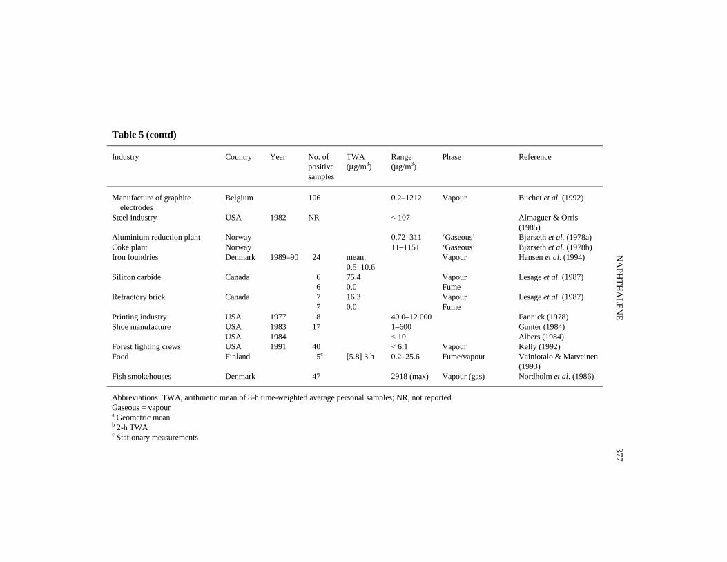
Naphthalene has been measured in a wide variety of workplaces for many years. Onthe basis of the major results summarized in Table 5, the following ranking of respiratoryexposure to naphthalene in the major industries can be made: creosote impregnation> coke manufacturing > asphalt industry > other industries. Exposure to naphthalene hasalso been extensively measured in Germany. Samples collected between 1991 and 1995were predominantly from wood manufacturing (50%), construction (16%) and metal-working and machine construction (12%). Ninety-five per cent of the available 183measurements from 94 factories were below the analytical limit of detection (1.0 mg/m3
for a 2-h sampling time). Only in the manufacture of repellents and perfumed disin-fectants were concentrations above the limit of detection (Bock et al., 1999).
Naphthalene is the most abundant component of creosote vapour and constitutes10–16 wt% of creosote oils (Nylund et al., 1992). Dermal wipe samples were taken in astudy among asphalt workers; less than 10% of the samples showed detectable concen-trations ranging from 5.5 to 520 ng/cm2 (Hicks, 1995).
Urinary naphthols have been measured as biomarkers of occupational exposure tonaphthalene (Bieniek, 1997) (see Section 4.1.1(c)).
1.4.3 Environmental occurrence
The extensive use of naphthalene as an intermediate in the production of plasticizers,resins, insecticides and surface active agents, its presence as a major component of coaltar and coal-tar products such as creosote and its inclusion in a wide variety of consumerproducts (e.g., moth-repellents) has led to its frequent occurrence in industrial effluentsand outdoor and indoor environments (Agency for Toxic Substances and DiseaseRegistry, 1995a,b; Environmental Protection Agency, 2001).
Naphthalene has been identified in the USA by the Environmental Protection Agency(2001) and the Agency for Toxic Substances and Disease Registry (1995b) as one of themost commonly found substances at hazardous waste sites on the ‘National PrioritiesList’. In the USA, naphthalene is listed as one of 189 hazardous air pollutants under theClean Air Act Amendments of 1990 (Title III) of the Environmental Protection Agencywhich mandates reduction of its emissions (Environmental Protection Agency, 1990b;Kelly et al., 1994). Naphthalene features in the Canadian Priority List of hazardoussubstances (Fellin & Otson, 1994).
The general population is exposed to naphthalene principally by inhalation ofambient and indoor air, with naphthalene-containing moth-repellents and tobacco smokeas the main contributors. Another source is the use of kerosene heaters (Traynor et al.,1990). Assuming an urban/suburban average air concentration of 0.95 μg/m3 and aninhalation rate of 20 m3 per day, it has been estimated that the average daily intake ofnaphthalene from ambient air in the USA is 19 μg (Howard, 1989; Agency for ToxicSubstances and Disease Registry, 1995a). Much lower exposure to naphthalene mayoccur from ingestion of drinking-water and/or food. Estimated exposure from drinking-
NAPHTHALENE 375

IARC M
ON
OG
RAPH
S VO
LUM
E 82376
Table 5. Occupational exposure to naphthalene in various industries
Industry Country Year No. ofpositivesamples
TWA(μg/m3)
Range(μg/m3)
Phase Reference
Hot mix plants USA – 8 2.3 Fume/vapour Hicks (1995)Paving USA – 9 6.5 Fume/vapour Hicks (1995)Paving, roofing, steel and silicon carbide industries
Canada – 51 11.4 Vapour (particulate) Lesage et al. (1987)
0.1 Fume (gaseous)Roofing/waterproofing USA 1985 11 0–1.9 Zey & Stephenson
(1986)Roofing manufacturing USA – 7 7.5 Fume/vapour Hicks (1995)Roofing 11 5.2 Fume/vapourEnhanced oil recovery USA 1986 5.0–11.0 Daniels & Gunter
(1988)Refineries/terminals USA – 9 5.5 Fume/vapour Hicks (1995)Coke manufacturing Belgium 16 0.7–959 Vapour Buchet et al. (1992)Coke plant Finland 1988–90 90b 44–500 111–1989 Vapour Yrjänheikki et al.
(1995)(modern technology) Poland 66 0–6000 Vapour Bieniek (1994)Coke plant, tar distillation Poland 69 773a – Vapour Bieniek (1997)Coke plant, naphthalene oil distillation
Poland 33 867a – Vapour Bieniek (1997)
Coke plant Poland 1997 48 170–1210 10–3280 Vapour Bieniek (1998)Creosote impregnation (wood) Finland 18 2200 Vapour Heikkilä et al. (1987)Switch assembly (wood) Finland 8 2600 Vapour Heikkilä et al. (1987)Creosote impregnation (wood) Finland 30 1540 400–4200 Vapour Heikkilä et al. (1997)
Finland 15 1000 22–1960 Vapour Heikkilä et al. (1995)Construction Finland 1 160 Vapour Heikkilä et al. (1995)Aluminium refinery Canada 7 1111 Vapour Lesage et al. (1987)
7 0.5 Fume
NA
PHTH
ALEN
E377
Table 5 (contd)
Industry Country Year No. ofpositivesamples
TWA(μg/m3)
Range(μg/m3)
Phase Reference
Manufacture of graphite electrodes
Belgium 106 0.2–1212 Vapour Buchet et al. (1992)
Steel industry USA 1982 NR < 107 Almaguer & Orris(1985)
Aluminium reduction plant Norway 0.72–311 ‘Gaseous’ Bjørseth et al. (1978a)Coke plant Norway 11–1151 ‘Gaseous’ Bjørseth et al. (1978b)Iron foundries Denmark 1989–90 24 mean,
0.5–10.6Vapour Hansen et al. (1994)
Silicon carbide Canada 6 75.4 Vapour Lesage et al. (1987) 6 0.0 Fume
Refractory brick Canada 7 16.3 Vapour Lesage et al. (1987) 7 0.0 Fume
Printing industry USA 1977 8 40.0–12 000 Fannick (1978)Shoe manufacture USA 1983 17 1–600 Gunter (1984)
USA 1984 < 10 Albers (1984)Forest fighting crews USA 1991 40 < 6.1 Vapour Kelly (1992)Food Finland 5c [5.8] 3 h 0.2–25.6 Fume/vapour Vainiotalo & Matveinen
(1993)Fish smokehouses Denmark 47 2918 (max) Vapour (gas) Nordholm et al. (1986)
Abbreviations: TWA, arithmetic mean of 8-h time-weighted average personal samples; NR, not reportedGaseous = vapoura Geometric meanb 2-h TWAc Stationary measurements

water assuming a water concentration of 0.001–2.0 μg/L naphthalene and water con-sumption of 2 L per day is 0.002–4.0 μg per day (Howard, 1989).
(a) AirMost of the naphthalene entering the environment is discharged to the air (92.2%),
the largest releases (more than 50%) resulting from the combustion of wood and fossilfuels and the off-gassing of naphthalene-containing moth-repellents and deodorants. In1989, about 12 million pounds [5.5 million kg] were released from these sources. Thehighest atmospheric concentrations of naphthalene have been found in the immediatevicinity of specific industrial sources and hazardous waste sites (Agency for ToxicSubstances and Disease Registry, 1995a). Air emissions in the USA reported to theEnvironmental Protection Agency decreased from 1598 tonnes for 473 industrial faci-lities in 1989 to 1224 tonnes for 744 industrial facilities in 1999 (Environmental Pro-tection Agency, 2001).
The median ambient air concentration of naphthalene determined at nine locations(84 samples) in the USA was 1.2 μg/m3 (Kelly et al., 1994). In another series of data from1970–87, the average concentration of naphthalene in ambient air at several locations inthe USA was 0.991 ppb [5.19 μg/m3] for 67 samples, 60 of which were from source-dominated locations (Shah & Heyerdahl, 1988).
In the USA, the Environmental Protection Agency (2000) reviewed studies andcalculated summary statistics for concentrations of chemicals in indoor air from selectedsources, including naphthalene. Studies were selected which provided the best availableestimates of ‘typical concentrations’ in indoor environments. These sources included theBuilding Assessment Survey and Evaluation (BASE) study, National Association ofEnergy Service Companies (NAESCO) study and School Intervention Studies (SIS). Thedata are reported in Table 6. [The Working group noted the unusually high outdoor airconcentration in the School Intervention Studies, the fact that it is based on only threepositive observations and that the original data in this review are unpublished.]
IARC MONOGRAPHS VOLUME 82378
Table 6. Typical concentrations of naphthalene in indoor and outdoor airaccording to the Building Assessment Survey and Evaluation (BASE) studyand the School Intervention Studies (SIS)
BASE (μg/m3) SIS (μg/m3)
Indoor Outdoor Indoor Outdoor
Arithmetic mean concentration 0.95 0.31 1.3 29095th percentile upper limit 2.6 0.81 1.7 1500No. of buildings 70 69 10 10No. of observations 209 69 39 10Frequency of detection 83% 58% 21% 30%

A median naphthalene concentration of 0.18 ppb [0.94 μg/m3] has been reported inurban air in 11 cities in the USA (Howard, 1989). An average naphthalene concentrationof 170 μg/m3 in outdoor air in a residential area of Columbus, OH (Chuang et al., 1991)and a concentration of 3.3 μg/m3 naphthalene in ambient air in Torrance, CA, have alsobeen reported (Propper, 1988; Agency for Toxic Substances and Disease Registry, 1995a).
Average naphthalene concentrations in ambient air at five hazardous waste sites andone landfill in New Jersey ranged from 0.42 to 4.6 μg/m3 (range of arithmetic means)(LaRegina et al., 1986). Naphthalene wasfound at concentrations of less than 0.6 ng/m3
in the air above shale-oil wastewaters in Wyoming (Hawthorne & Sievers, 1984).Naphthalene was found in particulate matter in the atmosphere of La Plata,
Argentina, at concentrations ranging from 0.18 ± 1.05 ng/m3 to 13.2 ± 1.30 ng/m3 whensampled on seven occasions between September 1984 and June 1986 (Catoggio et al.,1989).
In a study to compare naphthalene concentrations (among 35 volatile organic com-pounds) in indoor and outdoor air in northern Italy in 1983–84, the mean values foundin indoor and outdoor samples were 11 and 2 μg/m3, respectively, with the medianindoor/outdoor ratio being 4 for 11 samples (De Bortoli et al., 1986).
Average indoor air concentrations in various residential areas in the USA rangedfrom 0.75 to 1600 μg/m3 (Chuang et al., 1991 (measurements done in 1986–87); Wilson& Chung, 1991). A more representative upper limit concentration of naphthalene inindoor air of 32 μg/m3 was recorded in buildings in heavy traffic urban areas of Taiwan(Hung et al., 1992).
In a study of the effect of smoking on polycyclic aromatic hydrocarbon levels,including naphthalene, in eight homes in the USA, naphthalene was found in the livingroom of homes of smokers (with gas heating and electric cooking) and homes of non-smokers (electric heating and cooking) at concentrations of 2.2 μg/m3 and 1.8 μg/m3,respectively, and the respective outdoors concentrations of naphthalene were 0.33 and0.11 μg/m3 (Wilson & Chuang, 1991).
In a summary of concentrations of volatile organic compounds in 230 homes inGermany, naphthalene was found at a mean concentration of 2.3 μg/m3 with a range of< 1.0–14 μg/m3; the 50th and 90th percentiles were 2.1 and 3.9 μg/m3, respectively(Gold et al., 1993). In a study of the relationship between climatic factors and theconcentrations of 26 volatile organic compounds in Canadian homes in 1991, naphtha-lene concentrations in the winter, spring, summer and autumn were 3.24, 1.10, 3.82 and8.10 μg/m3, respectively (Fellin & Otson, 1994). In a comparison of naphthaleneconcentrations in the indoor air of 50 normal and 38 ‘sick’ homes (in which peoplecomplained about the odour or had symptoms) in Finland, naphthalene was found at amedian concentration of 0.31 μg/m3 in normal homes (with 6% of normal houses having1.5–3.1 μg/m3), while in ‘sick’ homes 2.6% had concentrations of 1.5–3.1 μg/m3, 5.3%of 3.1–15 μg/m3 and 5.3% of 15–62 μg/m3 (Kostiainen, 1995).
Naphthalene has been identified in the emissions of diesel light-duty vehicles(2–6 mg naphthalene released per km ‘distance’ driven on a chassis dynamometer)
NAPHTHALENE 379

(Scheepers & Bos, 1992). The average concentration of naphthalene reported insideautomobiles in commuter traffic was about 4.5 μg/m3 (Löfgren et al., 1991).
(b) WaterNaphthalene released to the atmosphere may be transported to surface water and/or
soil by wet or dry deposition. About 2–3% of naphthalene emitted to air is transported toother environmental media by dry deposition (Coons et al., 1982; Agency for ToxicSubstances and Disease Registry, 1995a). Naphthalene is degraded in water by photo-lysis and biological processes. The half-life for photolysis of naphthalene in surfacewater is about 71 h, but in deeper water (5 m) it is estimated to be 550 days (Agency forToxic Substances and Disease Registry, 1995a).
Surface water discharges of naphthalene from 744 industrial facilities in the USA in1999 amounted to 17.7 tonnes, as reported to the Toxics Release Inventory. An additional73 tonnes of naphthalene were discharged though underground injection (EnvironmentalProtection Agency, 2001). About 5% of all naphthalene entering the environment isreleased to water, mostly arising from coal tar production and distillation processes(Agency for Toxic Substances and Disease Registry, 1995a).
Naphthalene was detected in 7% of 630 ambient water samples in the USA at amedian concentration of less than 10 μg/L, as shown in an analysis of 1980–82 data fromthe Environmental Protection Agency STORET (STOrage and RETrieval) database(Staples et al., 1985; Agency for Toxic Substances and Disease Registry, 1995a).Naphthalene was also detected in 11% of 86 urban run-off samples up to 1982 at concen-trations ranging from 0.8 to 2.3 μg/L (Cole et al., 1984).
In the USA, naphthalene was detected in 35% of samples of groundwater at anaverage concentration of 3.3 mg/L at five wood-treatment facilities (Rosenfeld & Plumb,1991) and in leachate or groundwater plume from industrial and municipal landfills atconcentration ranges of < 10–19 mg/L and 0.1–19 mg/L, respectively (Brown & Donnelly,1988). Naphthalene was detected in groundwater samples from three wells at concen-trations of 380, 740 and 1800 μg/L, respectively, near an underground coal gasificationsite in north-western Wyoming (Stuermer et al., 1982). Concentrations of naphthaleneranging from < 0.2 to 63 μg/L were detected at five out of six landfill sites in southernOntario (Barker, 1987). Naphthalene was found at concentrations of 4.3 and 8.8 μg/L intwo groundwater samples collected near an Orange County landfill site in central Florida(and not in surface water) in 1989–90, but not in 1992–93; this was believed to result fromthe decomposition of municipal solid waste (Chen & Zoltek, 1995).
Naphthalene has been infrequently reported in drinking-water (Agency for ToxicSubstances and Disease Registry, 1995a). It was found in four samples of drinking-waterextracts at concentrations ranging from 6 to 16 ng/L in Athens, GA, in 1976 (Thruston,1978) and in another area in the USA at concentrations up to 1.4 μg/L (Coons et al.,1982).
Naphthalene was measured in samples of raw river water from the Adige River, Italy,collected at 19 sampling stations in the Trento province, during two campaigns in 1989,
IARC MONOGRAPHS VOLUME 82380

and detected at average concentrations of 51 ng/L (range, 3–109) and 284 ng/L (range,3–2240), respectively (Benfenati et al., 1992). It was also detected in two polluted rivers,Besós and Llobregat, in Barcelona, Spain, in 1985–86 at mean concentrations of 1300(SD, 150) ng/L and 180 (SD, 130) ng/L, respectively (Gomez-Belinchon et al., 1991).Naphthalene was one of the main aromatic hydrocarbons detected (at a concentration of0.02 μg/L) in surface waters of Admiralty Bay, King George Island, Antarctica, duringthe summers of 1989, 1990, 1992 and 1993 (Bícego et al., 1996).
Naphthalene was found at concentrations of 0.5–35 ng/L (arithmetic mean, 12 ng/L)over 15 months at a coastal site near piers extending into Vineyard Sound, MA, USA. Adominant wintertime source considered was use of space-heating oil, with higherconcentrations of naphthalene found in the winter and lowest concentrations found in thesummer (Gschwend et al., 1982).
(c) Soil and sedimentsReleases of naphthalene to land from 744 industrial facilities in the USA in 1999
amounted to 66 tonnes (Environmental Protection Agency, 2001). In untreated agricultural soils, naphthalene has been found at concentrations ranging
from 0 to 3 μg/kg in 1942–84 (Wild et al., 1990). It has been found at 6.1 (SD, 0.2) mg/kgin coal tar-contaminated soil (Yu et al., 1990), at 16.7 mg/kg in soil from a former tar-oilrefinery (Weissenfels et al., 1992; Agency for Toxic Substances and Disease Registry,1995a) and at up to 66 μg/kg in sludge-treated soils (Wild et al., 1990).
In the USA, naphthalene was reported to be detectable in 7% of 267 sedimentsamples (with the median concentration for all samples of less than 500 μg/kg) enteredinto the Environmental Protection Agency STORET database (1980–82) (Staples et al.,1985).
Naphthalene has been detected in contaminated sediments in Texas, USA, at averageconcentrations of 54.7 and 61.9 μg/kg at 10 m and 25 m from an oil platform and innearby non-contaminated estuarine sediments at 2.1 μg/kg in 1982–85 (Brooks et al.,1990). It was found at 200 mg/kg in a tar-contaminated sediment of the River Warnowat Schwaan near Rostock, Germany, in August 1989 (Randow et al., 1996). Naphthalenewas found in all four Canadian marine sediments analysed (representing varying concen-trations and sources of polycyclic aromatic hydrocarbon contamination) at concen-trations ranging from 0.1 to 115 mg/kg dry sediment (Simpson et al., 1995).
Naphthalene concentrations ranging from < 2 to 20.2 mg/kg dry wt were reported inthree out of four sediments from lakes in the Northwest Territories in Canada (Lockhartet al., 1992). Primarily due to oxygen limitation, naphthalene persists in coal tar-conta-minated surface sediments (Madsen et al., 1996). Naphthalene concentrations in soilsand sewage sludges are usually less than 1 mg/kg in the United Kingdom (Wild & Jones,1993).
NAPHTHALENE 381

(d ) BiodegradationStudies on biodegradation of polycyclic aromatic hydrocarbons in soil suggest that
absorption to organic matter significantly reduces the bioavailability and thus thebiodegradability of naphthalene (Heitzer et al., 1992; Weissenfels et al., 1992; Agencyfor Toxic Substances and Disease Registry, 1995a). Reported naphthalene half-lives insoil vary considerably. The estimated half-life of naphthalene reported for a solid wastesite was 3.6 months, while in typical soils more rapid biodegradation is expected to occur(Heitkamp et al., 1987; Howard, 1989).
Biodegradation of naphthalene is accomplished via the action of aerobic micro-organisms and generally declines precipitously when soil conditions become anaerobic(Klecka et al., 1990). Naphthalene biodegrades to carbon dioxide in aerobic soils withsalicylate as an intermediate product (Heitzer et al., 1992; Agency for Toxic Substancesand Disease Registry, 1995a).
Although polycyclic aromatic hydrocarbons are persistent in a strictly anaerobicenvironment, naphthalene can be degraded anaerobically under sulfate-reducingconditions: it was oxidized to carbon dioxide in petroleum-contaminated marine harboursediments in San Diego, CA (Coates et al., 1997).
(e) FoodNaphthalene was detected in only two of 13 980 samples of foods analysed in six
states of the USA in 1988–89 (Minyard & Roberts, 1991). Naphthalene is not generallyreported to be present in fish, but has been detected in shellfish in the USA, with concen-trations ranging from 5 to 176 μg/kg in oysters, from 4 to 10 μg/kg in mussels and from< 1 to 10 μg/kg in clams (Bender & Huggett, 1989).
Naphthalene was found in 1993 in many samples of edible portions of nine types ofshrimp and fish in Kuwaiti seafood at concentrations ranging from 2 to 156 μg/kg dry wt.These elevated concentrations of naphthalene were attributed to the pollution of Kuwait’sterritorial waters with crude oils as a result of oil spillage during the Gulf War or thechronic pollution due to oil production, transportation or natural seepage from theseabed. Naphthalene constituted the highest burden of the 14 polycyclic aromatic hydro-carbons screened (Saeed et al., 1995).
Mean concentrations of naphthalene of 19.5 μg/kg dry wt have been found in ediblemuscle of fish collected from the Red Sea coast of Yemen (DouAbul et al., 1997).Naphthalene has been found at maximum concentrations of 27.7 μg/kg and 137 μg/kg inmuscle and liver tissue, respectively, of burbot fish from lakes in the Northwest Terri-tories in Canada (Lockhart et al., 1992).
Naphthalene has also been detected in various fish species collected from the Gulf ofNaples, Italy, e.g., in muscle samples of anchovy, comber and rock goby at concen-trations of 63, 4 and 20 μg/kg wet wt, respectively, and in razor fish, wart venus andshort-necked clams, at levels of 20, 25 and 32 μg/kg wet wt, respectively (Cocchieriet al., 1990).
IARC MONOGRAPHS VOLUME 82382

Naphthalene was measured in 1987–88 in six species of aquatic organisms (seamullet, bony bream, blue catfish, mud crab, pelican and the silver gull) from the BrisbaneRiver estuarine system in Australia. The mean concentrations (μg/kg, wet wt) (in paren-theses: lipid wt basis) were: bony bream, 14.1 (306; 8 samples); blue catfish, 21.3 (433;8 samples); sea mullet, 37.3 (773; 8 samples); mud crab, 16.5 (407; 8 samples); pelican,21.0 (276; 3 samples) and silver gull, 31.6 (395; 3 samples) (Kayal & Connell, 1995).
Naphthalene was identified in the neutral fraction of roast beef flavour isolate (Minet al., 1979).
Use of a mathematical model of naphthalene migration into milk from an atmospherehaving a relatively high level of naphthalene suggested that naphthalene is first absorbedby the packaging material (low-density polyethylene). It was cautioned that when low-density polyethylene is used as the packaging material, the concentration of naphthalenevapour in the storage area should be kept low to minimize the transfer of naphthalene tomilk (Lau et al., 1995).
(f ) Miscellaneous sourcesIn the USA, naphthalene was found in mainstream cigarette smoke at a concentration
of 2.8 μg per cigarette and at 46 μg per cigarette in the sidestream smoke from onecommercial unfiltered cigarette, and at a concentration of 1.2 μg in the smoke from afiltered ‘little’ cigar (Schmeltz et al., 1976).
Naphthalene has been detected in ash from municipal refuse and hazardous wasteincinerators. It was found in seven of eight municipal refuse ash samples at6–28 000 μg/kg, with higher concentrations detected in bottom ash than in fly ash (Shaneet al., 1990) and in five of 18 ash samples from hazardous waste incinerators at 0.17–41(mean, 4.1) mg/kg (Carroll & Oberacker, 1989).
(g) Human tissues and secretionsNaphthalene was found in 40% of human adipose tissue samples at concentrations
ranging from < 9 to 63 μg/kg in a National Human Adipose Tissue Survey (NHATS) inthe USA in 1982 (Stanley, 1986). Naphthalene was also detected (concentrations notreported) in six of eight selected breast milk samples from women in four cities in theUSA (Pellizzari et al., 1982). It was also released in expired air from three out of eightindividuals at concentrations of 1.5, 2.4 and 0.12 μg/h, respectively (Conkle et al., 1975).
1.5 Regulations and guidelines
Occupational exposure limits and guidelines for naphthalene are presented inTable 7.
NAPHTHALENE 383

IARC MONOGRAPHS VOLUME 82384
Table 7. Occupational exposure limits and guidelines for naphthalenea
Country Year Concentration (mg/m3) Interpretationb
Argentina 1991 5075
TWASTEL (15 min)
Australia 1993 5075
TWASTEL (15 min)
Belgium 1993 5075
TWASTEL (15 min)
Canada 1994 5075
TWASTEL (15 min)
Denmark 1993 50 TWAFinland 2002 50c
100cTWASTEL (15 min)
France 1993 50 TWAGermany 2001 50 (CAT-2, skin) TRKHungary 1993 40
80TWASTEL (15 min)
Ireland 1997 5075
TWASTEL (15 min)
Netherlands 1999 50 TWAPhilippines 1993 50 TWAPoland 1993 20 TWARussia 1989 20 STEL (15 min)Sweden 1991 0.2 (skin)
0.6TWASTEL (15 min)
Switzerland 1993 50 TWAUnited Kingdom 2000 50
75TWASTEL (15 min)
USA ACGIHc (TLV)
NIOSH (REL)
OSHA (PEL)
2001
2000
2001
10 ppm [50] (A4, skin)15 ppm [75]507550
TWASTEL (15 min)TWASTEL (15 min)TWA
a From International Labour Office (1991); American Conference of Governmental IndustrialHygienists (ACGIH) (2000, 2001); Deutsche Forschungsgemeinschaft (2001); OccupationalSafety and Health Administration (OSHA) (2001); Sosiaali-ja terveysministeriö (2002); UnitedNations Environment Programme (2002)b TWA, 8-h time-weighted average; STEL, short-term exposure limit; A4, not classifiable as ahuman carcinogen; CAT-2, substances that are considered to be carcinogenic for man becausesufficient data from long-term animal studies or limited evidence from animal studies substan-tiated by evidence from epidemiological studies indicate that they can make a significant contri-bution to cancer risk; skin, danger of cutaneous absorption; TRK, technical exposure limit; TLV,threshold limit value: REL, recommended exposure limit; PEL, permissible exposure limitc Values have been rounded.

2. Studies of Cancer in Humans
Case reports
A cluster of cancer cases in a naphthalene purification plant was reported in theformer East Germany (Wolf, 1976, 1978). This plant operated between 1917 and 1968and a total of 15 employees were reported to have worked in this unit of the plant duringthe preceding 20–30 years. Seven employees were diagnosed with cancer, including fourcases of laryngeal cancer. Diagnosis was established between 1964 and 1973 and the ageat diagnosis was 60–71 years. The incidence rate for laryngeal cancer in the former EastGermany in 1970 was given as 6.3 per 100 000. The four workers had been exposed for7–31 years. The limit value for exposure to naphthalene at that time was 20 mg/m3, withpeak values of 50 mg/m3. Concomitant exposure to various tar products was mentioned.All four cases were reported to have been smokers. [The Working Group noted that noinference on the carcinogenicity of naphthalene can be drawn from these observations.]
Ajao et al. (1988) reported on 23 consecutive cases of colorectal carcinoma admittedduring June 1982 and May 1984 to a university college hospital in Nigeria. Eleven ofthese patients were 30 years or younger at diagnosis. Based on family history, procto-sigmoidoscopy, barium enema and autopsy, no indication of familial polyposis amongthese cases was ascertained. Half of the patients mentioned a history of taking Kafura, alocal indigenous treatment for anorectal problems, which contains naphthalene. Theother half of the patients did not know whether they had been given Kafura during earlychildhood. [The Working Group noted that no inference on the carcinogenicity ofnaphthalene can be drawn from these observations.]
3. Studies of Cancer in Experimental Animals
3.1 Oral administration
Rat: A group of 28 BD I and BD III rats [sex and number of each strain not speci-fied], about 100 days old, was fed a diet [not specified] containing naphthalene (spectro-graphically pure) in oil [type unspecified] at a dose of 10–20 mg per day on six days perweek, for 100 weeks. Animals were kept under observation until they died. The averagelife expectancy was 800 days, which was said to be similar to that of control rats [nodetails were provided regarding control animals]. All animals were subjected to necropsywith histopathological examination of abnormal tissues only. No tumours were found inany of the rats examined. (Schmähl, 1955). [The Working Group noted the small numberof animals used and the incomplete reporting of this study.]
NAPHTHALENE 385

3.2 Inhalation exposure
3.2.1 Mouse
Groups of 70 male and 70 female B6C3F1 mice, 10–11 weeks of age, weresubjected to whole-body exposure to 0 or 10 ppm (0 or 52 mg/m3) naphthalene (> 99%pure) and a group of 135 males and 135 females to 30 ppm naphthalene (157 mg/m3) ininhalation chambers for 6 h per day, five days per week, for 104 weeks. During periodsof non-exposure, animals were housed in groups of five. Mean body weight of exposedmice was slightly lower than that of the controls throughout the study. Survival rates atthe end of the study were significantly lower in control male mice than in exposed malesdue to wound trauma and secondary infection related to fighting (survival: controls,26/70 (37%); 10 ppm, 52/69 (75%); and 30 ppm, 118/133 (89%)). Survival in theexposed female mice was similar to that of controls: controls, 59/69 (86%); 10 ppm,57/65 (88%); and 30 ppm, 102/135 (76%). There was a statistically significant increasein the incidence of bronchiolo-alveolar adenomas in high-dose females (controls, 5/69(7%); 10 ppm, 2/65, (3%); 30 ppm, 28/135 (21%) [p = 0.01; logistic regression test]).One bronchiolo-alveolar carcinoma was noted in a high-dose female. Exposed male micealso showed an increased incidence of bronchiolo-alveolar adenomas and carcinomas butthe increases were not statistically significant (adenomas: 7/70 (10%), 15/69 (22%) and27/135 (20%); carcinomas: 0/70, 3/69 (4%) and 7/135 (5%) in controls, 10 ppm and30 ppm dose groups, respectively). Non-neoplastic changes were seen only in the lungsand nose. A dose-related increase in bronchiolo-alveolar inflammation was seen (males:0/70 (0%), 21/69 (30%) and 56/135 (41%); females: 3/69 (4%), 13/65 (30%) and 52/135(39%) in the 0-, 10- and 30-ppm dose groups, respectively). Virtually all exposed animalsbut none of the controls had nasal chronic inflammation, respiratory epithelial hyper-plasia and metaplasia of the olfactory epithelium (National Toxicology Program, 1992).
In a screening assay based on increased multiplicity and incidence of lung tumoursin a strain of mice highly susceptible to the development of this neoplasm, groups of 30female A/J mice, 8–10 weeks of age, were exposed in inhalation chambers to 0, 10 or30 ppm [0, 52 or 157 mg/m3] naphthalene (purity, 98–99%) for 6 h per day, on five daysper week, for six months. Survival was unaffected by treatment. At the end of the experi-mental period, survivors were killed and examined for pulmonary adenomas. Exposureto 10 or 30 ppm did not cause a significant increase in the incidence of lung adenomascompared with concurrent controls, but histopathological evaluation of the lungs showedan increase in numbers of alveolar adenomas per tumour-bearing mouse but not inadenomas per mouse compared with the concurrent controls (controls, 21% (0.21 ± 0.39adenomas per mouse and 1.00 ± 0.00 adenomas per adenoma-bearing mouse); 10 ppm,29% (0.35 ± 0.55 adenomas per mouse and 1.25 ± 0.07 adenomas per adenoma-bearingmouse); 30 ppm, 30% (0.37 ± 0.55 adenomas per mouse and 1.25 ± 0.07 adenomas peradenoma-bearing mouse) (Adkins et al., 1986).
IARC MONOGRAPHS VOLUME 82386

3.2.2 Rat
Groups of 49 male and 49 female Fischer 344/N rats, six weeks of age, were exposedin inhalation chambers to 0, 10, 30 or 60 ppm [0, 52, 157 or 314 mg/m3] naphthalene(> 99% pure) for 6 h per day, on five days per week, for 105 weeks. Mean body weightsof all exposed groups of male rats were less than that of the chamber control groupthroughout the study, but mean body weights of exposed groups of females were similarto that of the chamber control group. Survival rates in all exposed groups were similarto that of the chamber controls. At the end of the study, 24/49, 22/49, 23/49 and 21/49males and 28/49, 21/49, 28/49 and 24/49 females were alive in the 0, 10, 30 and 60 ppmgroups, respectively. Neuroblastomas of the nasal olfactory epithelium were observed in0/49, 0/49, 4/48 (p = 0.056, Poly-3 test) and 3/48 male rats and in 0/49, 2/49, 3/49 and12/49 (p = 0.001, Poly-3 test) females in the 0, 10, 30 and 60 ppm groups, respectively.In addition, adenomas of the nasal respiratory epithelium were observed in 0/49, 6/49(p = 0.013, Poly-3 test), 8/48 (p = 0.003, Poly-3 test) and 15/48 (p < 0.001, Poly-3 test)males and 0/49, 0/49, 4/49 (p = 0.053, Poly-3 test) and 2/49 females in the 0, 10, 30 and60 ppm groups, respectively. These olfactory neuroblastomas and respiratory epitheliumadenomas had not been observed in the larger database of historical controls in NationalToxicology Program two-year inhalation studies in which animals were fed NationalInstitute of Health (NIH)-07 diet or in the smaller National Toxicology Program database[all routes] in which they were fed NTP-2000 diet. In addition to the nasal neoplasms,the incidences of a variety of non-neoplastic lesions of the nasal tract in both male andfemale rats were significantly increased in naphthalene-exposed animals compared withcontrols (see Section 4.2.2(b)) (National Toxicology Program, 2000).
3.3 Intraperitoneal administration
3.3.1 Mouse
A group of 31 male and 16 female CD-1 mice received intraperitoneal injections ofa 0.05-M solution of naphthalene [purity unspecified] in dimethyl sulfoxide (DMSO) ondays 1, 8 and 15 of life. The total dose received was 1.75 μmol per mouse. Groups of 21male and 21 female mice receiving DMSO alone served as vehicle controls. [Thenumber of mice in the above four groups are reported as the effective number of micethat survived at least six months of treatment and not the starting number.] Mice wereweaned at 21 days, separated by gender and maintained until termination at 52 weeks, atwhich time they were necropsied and gross lesions as well as liver sections were exa-mined histologically. There was no increase in the incidence of tumours in the naphtha-lene-treated mice compared with the vehicle controls (LaVoie et al., 1988).
NAPHTHALENE 387

3.3.2 Rat
Ten BD I and BD III rats, 100 days old [sex and number of each strain not specified],received weekly intraperitoneal injections of 20 mg naphthalene (spectrographicallypure) as a 2% solution in ‘specially purified oil’ for 40 weeks and were held under obser-vation until they died. The average age at death was 900 days, which was reported to besimilar to that of controls [no details were provided regarding control animals]. Allanimals were necropsied with histopathological examination of abnormal tissues only.No tumours were found in any of the rats examined (Schmähl, 1955). [The WorkingGroup noted the small number of animals used and the limited reporting of this study.]
3.4 Subcutaneous administration
Rat: Ten BD I and BD III rats, 100 days old, [sex and number of each strain un-specified] received weekly subcutaneous injections of 20 mg naphthalene (spectro-graphically pure) as a 2% solution in ‘specially purified oil’ for 40 weeks and were keptunder observation until they died. The average age at death was 700 days, which wasreported to be similar to that of controls [no details were provided regarding controlanimals]. All animals were necropsied with histopathological examination of abnormaltissues only. No tumours were found in any of the rats examined (Schmähl, 1955). [TheWorking Group noted the small number of animals and the limited reporting of thisstudy.]
Groups of 38 white inbred rats [age, strain and sex unspecified] received seven sub-cutaneous injections of 0 or 50 mg/kg bw naphthalene (purified by chromatography) asa 15% solution in sesame oil at intervals of around 14 days extending over 3.5 months.Survival was poor due to infectious pneumonia [agent unspecified], with 5/38 treated and11/38 vehicle controls alive at 12 months and 0/38 treated and 4/38 vehicle controls aliveat the termination of the study at 18 months. In the test group, a total of five sarcomas(one uterine and four lymphosarcomas) and a single mammary fibroadenoma developedand, in the control group, a single sarcoma and a single mammary fibroadenoma (Knake,1956). [The Working Group noted the small number of animals, the poor survival and thelimited reporting of this study.]
IARC MONOGRAPHS VOLUME 82388

4. Other Data Relevant to an Evaluation of Carcinogenicity and its Mechanisms
4.1 Absorption, distribution, metabolism and excretion
4.1.1 Humans
(a) AbsorptionNo studies were found that quantitatively determined the extent of absorption of
naphthalene in humans following oral or inhalation exposure. Naphthalene can beabsorbed through the skin. Kanikkannan et al. (2001a) examined the permeation ofJP-8 (jet fuel) containing 0.26% (w/w) naphthalene through human skin in vitro. For18 samples of human skin, the steady-state flux was 0.45 μg/cm2 per hour and thepermeability coefficient was 2.17 × 10⁻⁴ cm per hour.
(b) DistributionIn a survey, naphthalene was detected in 40% of the human adipose tissue samples
tested, with concentrations up to 63 ng/g lipid (Stanley, 1986). Naphthalene has also beenidentified in samples of human breast milk [incidence not clear; concentrations notreported] (Pellizzari et al., 1982).
(c) MetabolismThe major metabolic pathways of naphthalene are illustrated in Figure 1. Naphtha-
lene is metabolized first to naphthalene 1,2-oxide (2, see Figure 1), which can yield1-naphthol (3, see Figure 1) or be converted by epoxide hydrolase to trans-1,2-dihydro-1,2-dihydroxynaphthalene (trans-1,2-dihydrodiol) (5, see Figure 1). The hydroxyl groupof 1-naphthol may also be sulfated or glucuronidated. The 1,2-dihydrodiol can also beconverted to 2-naphthol (10, see Figure 1). The epoxide is also a substrate for glutathioneS-transferase, yielding glutathione conjugates which are eventually eliminated asmercapturic acids. Boyland and Sims (1958) showed that trace quantities of a precursorof 1-naphthyl mercapturic acid, tentatively identified as an N-acetyl-L-cysteine deri-vative, are eliminated in human urine after oral administration of 500 mg naphthalene.Tingle et al. (1993) examined the metabolism of naphthalene by human and mouse livermicrosomes. The ratio of the trans-1,2-dihydrodiol to 1-naphthol was 8.6 for humanmicrosomes compared with 0.4 for microsomes from phenobarbital-treated mice, indi-cating the ready detoxification of the epoxide to the diol in humans.
Buckpitt and Bahnson (1986) measured the metabolism of naphthalene by humanlung microsomes derived from two individuals and detected naphthalene dihydrodiol
NAPHTHALENE 389

IARC M
ON
OG
RAPH
S VO
LUM
E 82390Figure 1. Main metabolic pathways of naphthalene and resulting products in mammals
OH
H
OH
OH
H
+ OR
O2/NADPH2
H
HO H
OR1
OH
H
OHA D
E
K
(1) (2) (5) (6) (10)P
OH
OH O
OH
G
(7) (8)J
OH
OR1 OH
OH
O
O(9) (13) (14)
SR2
H
H
O
SR2
+ H+N
(12)
L
M
+ GSH
(3)B
+ R
OR1C
(4)
OH
OH
Q
?
H
(11)

NA
PHTH
ALEN
E391
Based on BUA (1989) and Agency for Toxic Substances and Disease Registry (1995a)
(1) Naphthalene (2) Naphthalene 1,2-oxide (3) 1-Naphthol (α-naphthol) (4) 1-Naphthyl glucuronide or sulfate (5) trans-1,2-Dihydro-1,2-dihydroxynaphthalene (6) trans-1,2-Dihydro-2-hydroxynaphthyl-1-glucuronide (7) 1,2-Dihydroxynaphthalene (8) 1,2-Naphthoquinone (9) 2-Hydroxynaphthyl-1-sulfate or -glucuronide(10) 2-Naphthol (β-naphthol)(11) N-Acetyl-S-(1,2-dihydro-1-hydroxy-2-naphthyl)-L-cysteine(12) N-Acetyl-S-(1-naphthyl)-L-cysteine (1-naphthyl mercapturic acid)(13) 1,4-Dihydroxynaphthalene(14) 1,4-Naphthoquinone
GSH = GlutathioneR1 = Sulfate or glucuronate groupR2 = N-acetyl-L-cysteine residue
A,Q = O2- and NADPH2-dependent monooxygenase (e.g., cytochromeP450-NADP-cytochrome-c-reductase system, microsomal)
B = Spontaneous isomerizationC,E,J = Conjugation reaction with sulfate (sulfotransferase, cytosolic) or
with glucuronic acid (UDP-glucuronyltransferase, microsomal)D = Epoxide hydrolase, synonym: epoxide hydrase (microsomal)F,N,P = Chemical dehydrationG = Dihydrodiol-dehydrogenase (cystosolic); 3,5-cyclohexadiene-1,2-
diol-NADP-oxidoreductaseH = Chemical dehydrationK = Chemical hydrolysis + dehydrationL = Enzymatic reaction with glutathioneM = γ-Glutamyl transferase, peptidase, N-acetylase

and three glutathione conjugates. These metabolites were also identified in animalstudies, as discussed in Section 4.1.2.
Urinary metabolites of naphthalene are useful biomarkers of exposure. Seventy-fiveworkers exposed to naphthalene while distilling naphthalene oil excreted 7.48 mg/L(4.35 mg/g creatinine) 1-naphthol (geometric mean values) at the end of the workshift.For 24 non-occupationally exposed individuals, the mean urinary concentration of1-naphthol was 0.13 mg/L (Bieniek, 1994). 1-Naphthol, 2-naphthol and 1,4-naphtho-quinone (14, see Figure 1) were identified in the urine of 69 coke-plant workers exposedto a geometric mean air concentration of naphthalene of 0.77 mg/m3 during tar distil-lation. The end-of-workshift urinary concentrations of 1-naphthol and 2-naphthol were693 and 264 μmol/mol creatinine. The correlation coefficients between the urinary excre-tion of naphthols and exposure to naphthalene were 0.64–0.75 for 1-naphthol and0.70–0.82 for 2-naphthol. There was a linear relationship between the overall concen-tration of naphthols in urine and the naphthalene concentration in air (Bieniek, 1997). Ina further study of a coke plant, Bieniek (1998) measured the concentrations of 1-naphtholand 2-naphthol in urine from eight workers in coke batteries, 11 workers in the sortingdepartment and 29 workers in the distillation department. The mean urinary con-centrations of 1-naphthol and 2-naphthol were 294 and 89 μmol/mol creatinine for thecoke-battery workers, 345 and 184 μmol/mol creatinine for the sorters and 1100 and630 μmol/mol creatinine for the distillation workers, respectively.
Andreoli et al. (1999) examined 15 urine samples from workers in a naphthalene-producing plant who were exposed to 0.1–0.7 mg/m3 naphthalene. At the end of theworkshift, the median urinary concentrations of 2-naphthyl sulfate, 2-naphthyl glucu-ronide and 1-naphthyl glucuronide were 0.030 (range, 0.014–0.121), 0.086 (range,0.013–0.147) and 0.084 (range, 0.021–0.448) mg/L, respectively.
Since naphthalene is the most abundant component of creosote (Heikkilä et al.,1987), urinary excretion of 1-naphthol was determined in three assembly workershandling creosote-impregnated wood. The average airborne concentration of naphtha-lene in the breathing zone was approximately 1 mg/m3. The average end-of-shift concen-tration of 1-naphthol in urine changed from 254–722 (mean, 556) μmol/mol creatinineon Monday to 1820–2190 (mean, 2060) μmol/mol creatinine on Wednesday and 870–2330 (mean, 1370) μmol/mol creatinine on Friday. The same metabolite was measuredin the urine of six workers exposed to creosote in a plant impregnating railroad ties(Heikkilä et al., 1997). As measured by use of personal air samplers, the mean airborneconcentration of naphthalene in the workers’ breathing zone was 1.5 (range,0.37–4.2) mg/m3. The mean end-of-shift concentration of 1-naphthol was 20.5 (range,3.5–62.1) μmol/L. There was a good correlation (r = 0.745) between concentrations ofairborne naphthalene and urinary 1-naphthol. No 1-naphthol was detected (limit ofdetection < 0.07 μmol/L) in the urine of five non-exposed controls. Hill et al. (1995)measured 1-naphthol and 2-naphthol in the urine of 1000 adults without occupationalexposure — a subset of the National Health and Nutrition Examination Survey III —who may have been exposed to low levels of naphthalene or pesticides that would yield
IARC MONOGRAPHS VOLUME 82392

these naphthols as metabolites. The frequency of detection was 86% for 1-naphthol and81% for 2-naphthol. The mean concentrations were 15 and 5.4 μg/g creatinine, respec-tively. Concentrations of 1-naphthol ranged up to 1400 μg/g creatinine.
Yang et al. (1999) examined the relationship between certain enzyme poly-morphisms and naphthalene metabolism in 119 men who were not occupationallyexposed to polycyclic aromatic hydrocarbons. A polymorphism in exon 7 of the CYP1A1gene was not related to urinary naphthol excretion. Smokers with the c1/c2 or c2/c2genotype in CYP2E1 excreted higher concentrations of 2-naphthol in the urine thansmokers with the c1/c1 genotype. Smokers deficient in glutathione S-transferase M1(GSTM1) showed higher urinary concentrations (without correction for creatinine) ofboth 1-naphthol and 2-naphthol.
Nan et al. (2001) examined the effects of occupation, lifestyle and genetic poly-morphisms of CYP1A1, CYP2E1 and the glutathione S-transferases GSTM1 and GSTT1on the concentrations of 2-naphthol in the urine of 90 coke-oven workers in comparisonwith 128 university students. The urinary excretion of 2-naphthol was higher in the coke-oven workers (7.69 μmol/mol creatinine) than in the students (2.09 μmol/mol creati-nine). In the control group, the excretion was higher in smokers (3.94 μmol/mol creati-nine) than in nonsmokers (1.55 μmol/mol creatinine). Urinary 2-naphthol concentrationswere higher in coke-oven workers with the c1/c2 or c2/c2 genotypes than in those withthe more common c1/c1 genotype of CYP2E1. Urinary 2-naphthol concentrations werealso higher in the urine of GSTM1-null workers than in GSTM1-positive workers.
4.1.2 Experimental systems
(a) Absorption, distribution and excretionEarly studies indicated that in rats naphthalene is well absorbed from the gastro-
intestinal tract (Chang, 1943). When naphthalene was fed to white male rats (weight,about 300 g) [strain unspecified] at a concentration of 1% (w/w) in the diet, none wasdetected in the faeces. Similarly, when it was administered as a single dose by stomachtube (0.1 g), it was not measurable in the faeces.
Eisele (1985) examined the distribution of [14C]naphthalene in laying pullets, swineand dairy cattle following oral administration. In pullets given a dose of 0.44 mg, themajor site of deposition was the kidney followed by fat, lung and liver. Following acuteadministration of 2.46 mg in swine, the major site of deposition was fat, where the levelwas up to 10 times higher than that in liver. After chronic administration (0.112 mg perday for 31 days), the lung, liver and heart were major sites of accumulation. In cows,chronic exposure (5.115 mg per day for 31 days) led to deposition primarily in the liver.
When [14C]naphthalene was applied dermally (3.3 μg/cm2; total dose, 43 μg) to maleSprague-Dawley rats, the plasma half-life for absorption was 2.1 h and that for elimi-nation was 12.8 h. The highest concentration of radioactivity 48 h after dosing was foundin the skin followed by ileum, duodenum and kidney. Seventy per cent of theradioactivity was found in the urine in the first 48 h, with 3.7% appearing in the faeces
NAPHTHALENE 393

and 13.6% in the expired air. The primary urinary metabolites identified were 2,7-di-hydroxynaphthalene (31.1% of the total radioactivity in the first 12 h), 1,2-dihydroxy-naphthalene (7, see Figure 1) (17.2%), 1,2-naphthoquinone (8, see Figure 1) (11.4%),2-naphthol (4.3%) and 1-naphthol (3.4%). The parent compound naphthalene accountedfor 0.3% of the radioactivity (Turkall et al., 1994). [The Working Group noted that2,7-dihydroxynaphthalene has not been identified as a major metabolite in other studies.]
Kilanowicz et al. (1999) studied the distribution, metabolism and excretion oftritiated naphthalene given intraperitoneally at a dose of 20 mg/kg bw to male IMP:Wistrats. Approximately 88% of the radioactivity was excreted in urine (68%) and faeces(20%) in the first 72 h, with maximum blood concentrations observed 2 h after dosing.The elimination of radioactivity from the blood was biphasic with half-lives of 0.8 and99 h. [The Working Group noted that the 99-h half-life component may have been due totritium exchange.] The highest initial tissue concentrations were found in fat, liver andkidneys. Urinary metabolites were identified as primarily the parent naphthalene,1-naphthol and 2-naphthol with smaller amounts of 1,2-dihydro-1,2-dihydroxy-naphthalene (1,2-dihydrodiol) and methylthionaphthalenes.
Sartorelli et al. (1999) investigated the percutaneous penetration of naphthalene fromlubricating oil in vitro using full-thickness monkey skin. The flux for naphthalene was0.274 nmol/cm2 per hour, which was higher than that for acenaphthene, fluorene, anthra-cene, phenanthrene, pyrene and chrysene. Kanikkannan et al. (2001b) examined thepercutaneous permeation of naphthalene in JP-8 + 100 jet fuel, which contained 0.26%(w/w) naphthalene and was spiked with [14C]naphthalene, using a pig ear skin model.The steady-state flux was 0.42 μg/cm2 per hour, similar to that of nonane but less thanfor tridecane.
(b) Metabolism — species comparisonCorner and Young (1954) compared the urinary metabolites of naphthalene in rats,
rabbits, mice and guinea-pigs [strains unspecified] following administration of a singledose of naphthalene (500 mg/kg bw) either orally or by intraperitoneal injection. 1-Naph-thol and its glucuronide and sulfate were identified in the urine of all four species (withthe exception of the glucuronide in guinea-pigs). 2-Naphthol was detected in all fourspecies but no conjugates of this metabolite were found. Although 1,2-dihydro-1,2-di-hydroxynaphthalene (1,2-dihydrodiol) was found in the urine of all four species, 1,2-di-hydroxynaphthalene was present only in urine of guinea-pigs. Rabbits and rats excretedmore 2-naphthol than 1-naphthol, guinea-pigs excreted 1- and 2-naphthol in equalamounts and mice excreted more 1-naphthol than 2-naphthol. As in humans, a precursorof 1-naphthyl mercapturic acid has been detected as a urinary metabolite in all rodentspecies tested (Boyland & Sims, 1958); the amounts of this metabolite present in theurine of mice, rats and hamsters were greater than those observed in guinea-pigs, whichwere greater than those in humans. However, these data did not take into considerationthe widely different doses given: mice, rats, hamsters, guinea-pigs and humans receivedtotal doses of 20, 100, 100, 400 and 500 mg per animal, respectively. Chen and Dorough
IARC MONOGRAPHS VOLUME 82394

(1979) investigated the formation of glutathione conjugates using [14C]naphthalenegiven to female Spague-Dawley rats. After intraperitoneal injection of 100 mg/kg bw[14C]naphthalene, 65% of the water-soluble fraction of the radioactivity in urine wasidentified as glutathione-derived conjugate (premercapturic acid) over a 72-h period.Total recovery of radioactivity was 74% after 72 h, with 60% present in the urine and14% in the faeces. 1,2-Dihydro-1,2-dihydroxynaphthalene (28%) and 1-naphthol (60%)were the major metabolites in the ether-extractable fraction, which accounted for 6% ofthe administered dose. Summer et al. (1979) found a dose-dependent increase in the uri-nary excretion of mercapturic acid conjugates in male Wistar rats given 30, 75 or200 mg/kg bw naphthalene by stomach tube but did not find any such increase inchimpanzees (Pan troglodytes S.). A similar lack of a significant role for glutathione con-jugation in primates was observed in rhesus monkeys (Macaca mulatta) (Rozman et al.,1982). [The Working Group noted that standards of naphthalene mercapturates were notavailable in these studies and the analytical method employed may have underestimatedthe amounts of mercapturates present in the urine samples.]
Horning et al. (1980) gave naphthalene (100 mg/kg bw) intraperitoneally to maleSprague-Dawley rats and identified 21 oxygenated metabolites in the urine, all but onebeing generated via epoxidation. Along with those identified in other studies, the totalnumber of known naphthalene metabolites was 31, excluding mercapturic acids, conju-gates and related compounds. Bakke et al. (1985) gave [14C]naphthalene orally to maleSprague-Dawley rats and found 4.6% of the 14C dose as naphthols or their glucuronidesin the urine by 24 h. In addition, they found 1,2-dihydro-1-hydroxy-2S-(N-acetyl)-cysteinylnaphthalene (11, see Figure 1) (38.1%), 1,2-dihydroxynaphthalene (7, seeFigure 1) (4.9%), 1,2-dihydro-1,2-dihydroxynaphthalene glucuronide (23.9%), 1,2-di-hydro-1-hydroxy-2-methylthionaphthalene glucuronide (4.6%) and uncharacterizedmetabolites (2.4%). Buonarati et al. (1990) showed that a consistent percentage of a doseof either trans-1S-hydroxy-2S-glutathionyl-1,2-dihydronaphthalene or trans-1R-hydroxy-2R-glutathionyl-1,2-dihydronaphthalene administered intravenously to male SwissWebster mice was eliminated as the corresponding diastereomeric mercapturic acid in theurine. In contrast, a significant percentage of a dose of trans-1R-glutathionyl-2R-hydroxy-1,2-dihydroxynaphthalene (14–25%, depending on the dose) was metabolized to(2-hydroxy-1,2-dihydronaphthalenylthio)pyruvic acid. These observations indicate thatmercapturic acids generated by conjugation at the C2 position of the napththalene nucleuscan be used to assess the stereochemistry of naphthalene metabolism in vivo.
Pakenham et al. (2002) showed that 24–35% of an intraperitoneal dose of [14C]-naphthalene was eliminated as mercapturates by both mice and rats at 24 h after dosing.For both species, this percentage was the same over a wide dose range (3.12–200 mg/kgbw). In contrast, after inhalation exposure, the amounts of mercapturic acid in mouseurine were approximately twice those in rat urine at the same level of exposure. Over a24-h period, approximately 100–500 μmol/kg bw mercapturates were eliminated in urineof mice given intraperitoneal injections of 50–200 mg/kg bw naphthalene. In miceexposed by inhalation to 1–100 ppm (5.24–524 mg/m3) naphthalene for 4 h,
NAPHTHALENE 395

1–240 μmol/kg bw total mercapturic acids were eliminated, while rats exposed to thesame concentrations eliminated 0.6–67 μmol/kg bw.
Jerina et al. (1970) used rat [strain unspecified] liver microsomes to examinenaphthalene metabolism in vitro and identified naphthalene 1,2-oxide (2, see Figure 1) asan intermediate in the formation of all major metabolites including glutathione conju-gates. Bock et al. (1976) used hepatocytes from male Sprague-Dawley rats to show that1,2-dihydro-1,2-dihydroxynaphthalene glucuronide was a major metabolite ofnaphthalene.
Usanov et al. (1982) compared the metabolism of naphthalene by microsomal prepa-rations from rat liver and rabbit lung [strains and sex not specified] by measuring theformation of 1-naphthol. The metabolic efficiency, i.e. the rate of hydroxylation per nmolof cytochrome P450, was 7.35 times higher in rabbit lung than in rat liver microsomes.
d’Arcy Doherty et al. (1985) examined the metabolism of 1-naphthol by a reconsti-tuted cytochrome P450 system from male Wistar rats and identified the products as 1,2-and 1,4-naphthoquinones (8 and 14, see Figure 1). Smithgall et al. (1988) examined themetabolism of trans-1,2-dihydro-1,2-dihydroxynaphthalene (trans-1,2-dihydrodiol) tothe ortho-quinone by cytosolic dihydrodiol dehydrogenase from rat liver, and investi-gated the reactivity of the ortho-quinone with the cellular nucleophiles, cysteine andglutathione. The results showed that ortho-quinones formed by enzymatic oxidation ofdihydrodiols may be effectively scavenged and detoxified by nucleophiles. Buckpitt andcoworkers (Buckpitt & Warren, 1983; Buckpitt et al., 1984, 1985) examined the relation-ships among the initial steps in the oxidative metabolism of naphthalene, conjugationwith glutathione and the ability of reactive metabolites of naphthalene to covalently bindto protein in tissues of male Swiss Webster mice given intraperitoneal doses of[14C]naphthalene. Binding of naphthalene in lung, liver and kidney was similar in vivo,but the rate of microsomal metabolic activation of naphthalene was much lower in thekidney than in liver or lung. Phenobarbital pretreatment increased the binding in all threetissues but only at the highest dose (400 mg/kg bw). 1-Naphthol was shown not to be anobligate intermediate in the binding process. The metabolism of naphthalene by mouse,rat and hamster pulmonary, hepatic and renal microsomal preparations was compared byBuckpitt et al. (1987). In all cases, glutathione adducts derived from naphthalene1,2-oxide were formed and overall activity was particularly high in mouse lung, with aparticular preference in this tissue for the formation of the naphthalene 1R,2S-oxideisomer (10:1 ratio with the 1S,2R-isomer).
Lanza et al. (1999) examined the ability of microsomal fractions from humanlymphoblastoid cells expressing recombinant human CYP2F1 enzyme to metabolizenaphthalene to glutathione adducts. The predominant conjugates formed were derivedfrom naphthalene 1S,2R-oxide (see Table 8), in contrast to the findings in mice (Buckpittet al., 1992) (see Figure 2).
In view of the mouse lung as a target tissue, a number of investigators have examinedspecies, tissue and cytochrome P450 (CYP) isozyme specificities in naphthalene meta-bolism. Nagata et al. (1990) identified the principal pulmonary enzyme in the mouse as
IARC MONOGRAPHS VOLUME 82396

P450m50b [CYP2F2], which formed predominantly naphthalene 1R,2S-oxide (seeTable 8 and Figure 2). Ritter et al. (1991) confirmed that the primary isoform responsiblefor naphthalene metabolism in the mouse lung was in the 2F subfamily. It was not indu-cible by phenobarbital, pyrazole, pregnenolone 16α-carbonitrile or 3-methylcholan-threne. Kanekal et al. (1991) examined the relationship between cytotoxicity and meta-bolism of naphthalene oxide using the isolated perfused lung of male CFW mice.
NAPHTHALENE 397
Table 8. Species comparison in the rates of conversion of naphthalene tonaphthalene 1,2-oxides by recombinant enzymes
Recombinantenzyme
Species Rate ofmetabolisma
Stereoselectivityb Reference
CYP2F1 Human 35.5c 0.13:1 Lanza et al. (1999)CYP2F2 Moused 104 000e 66:1 Shultz et al. (1999)
Modified from Buckpitt et al. (2002)a Expressed in pmol/min/nmol enzymeb Expressed as ratio of epoxide stereoisomers (1R,2S):(1S,2R)c Total amount of glutathione conjugates (1 + 2 + 3) (see Figure 2)d Sequence homology with human enzyme, 82%e Amount of glutathione conjugate 2 (see Figure 2)
Figure 2. Metabolism of naphthalene by murine CYP2F2 to reactive epoxides andtheir subsequent trapping as glutathione conjugates
From Shultz et al. (1999)Conjugates are numbered in the order of their elution after separation by reversed-phase HPLC.GSH Tx, glutathione transferases

Perfusion of the lung with naphthalene 1,2-oxide reduced glutathione levels to 40–60%of control. 1,4-Naphthoquinone and naphthyl-glucuronide were the major polar meta-bolites, along with smaller amounts of the dihydrodiol and thioether conjugates. Whenlungs were perfused with naphthalene, the thioethers and the dihydrodiol predominatedas metabolites. Chichester et al. (1991, 1994) demonstrated that Clara cells isolated frommale Swiss Webster mice metabolized naphthalene to the dihydrodiol and glutathioneconjugates. Microsomal preparations from Clara cells (supplemented with glutathioneand glutathione S-transferases) metabolized naphthalene to the dihydrodiol as a minorproduct and formed a single glutathione adduct, derived from the 1R,2S-isomer ofnaphthalene oxide, as the major product, whereas the dihydrodiol predominated in intactcells. Buckpitt et al. (1992) determined the rates of formation and the stereochemistry ofmetabolites of naphthalene in postmitochondrial supernatant (S9) preparations fromnasal mucosa and in microsomes from lung and liver of mice (Swiss Webster), rats(Sprague-Dawley), hamsters (Syrian golden) and rhesus monkeys (Macaca mulatta) (seeTable 9 for details). Metabolism by mouse lung was considerably greater than that by thelungs of the rat, hamster and monkey. Using total diol and conjugates for comparison, theactivity of mouse lung was two orders of magnitude higher than that of the monkey lung.In mouse lung there was preferential formation of the naphthalene 1R,2S-oxide, asjudged from the stereochemistry of the glutathione conjugates. In microdissectedairways, the extent of metabolism of naphthalene to the dihydrodiol and the glutathioneconjugates was much higher in the airways of Swiss Webster mice compared withSprague-Dawley rats or Syrian golden hamsters. In all three species, the rate of meta-bolism was higher in the distal airways than in the trachea (Buckpitt et al., 1995). Inmice, there was a high degree of stereoselectivity, the only glutathione conjugate being
IARC MONOGRAPHS VOLUME 82398
Table 9. Species comparison in the rates of conversion of naphthalene tonaphthalene oxides: pulmonary and nasal tissue
Microsome source Species Rate of metabolism(nmol/min/mgprotein)a
Stereoselectivityratio(1R,2S):(1S,2R)b
Dihydrodiolas % of totalmetabolites
Mouse 13.8 11.1 7.6Pulmonarymicrosomes Rat 1.69 0.48 4.6
Hamster 5.12 0.61 24.6Rhesus macaque 0.15 0.12 20.6
Mouse 87.1 12.7 7.4Rat 43.5 ~ 36 4.1
Post-mitochondrialsupernatant(olfactory) Hamster 3.9 ? 7.8
Modified from Buckpitt et al. (2002)a Total amount of dihydrodiol plus conjugates 1, 2, 3 formed (see Figure 2)
b The stereoselectivity varies with the concentration of substrate; the values given here are derived
from incubations containing 0.5 mM naphthalene.

that derived from the 1R,2S-oxide of naphthalene. Airways of mice formed the dihydro-diol and naphthalene 1R,2S-oxide at rates substantially higher than those of rats.Immunolocalization of CYP2F2 correlated well with the sites of metabolism, inagreement with the findings of Nagata et al. (1990) and Ritter et al. (1991) as to theimportance of this isoenzyme. This was confirmed in later studies (Shultz et al., 1999),in which CYP2F2, expressed in Spodoptera frugiperda and Trichoplusia ni cells by useof a baculovirus expression vector system, metabolized naphthalene with a high degreeof stereoselectivity to naphthalene 1R,2S-oxide (66:1 enantiomeric ratio). Substitutednaphthalenes such as 1-nitronaphthalene and 2-methylnaphthalene are also substrates forpurified CYP2F2 (Shultz et al., 2001).
Willems et al. (2001) developed a physiologically based pharmacokinetic model fornaphthalene administered by inhalation or by intravenous injection to Fischer 344 ratsand B6C3F1 mice. Model simulations for exposure by inhalation indicated that approxi-mately 88–96% of the absorbed naphthalene was metabolized by rats and 96–98% bymice. The overall percentage of naphthalene metabolized by mice exposed to 30 ppm[157 mg/m3] was higher than for rats exposed to 60 ppm [314 mg/m3] because of thehigher ventilation and metabolic rates in mice. The steady-state concentrations in thelungs of the mice and rats were similar at the same level of naphthalene exposure. Cumu-lative metabolism of naphthalene by the lung was markedly higher in the mouse than inthe rat. The rates of metabolism did not increase proportionally with concentration,suggesting saturation of metabolism in this organ. The model indicated that the meta-bolism of naphthalene by the liver was similar in the two species.
4.2 Toxic effects
4.2.1 Humans
The major toxicological responses reported in humans from acute exposure tonaphthalene have been haemolytic anaemia and cataracts (Zuelzer & Apt, 1949; Ghetti& Mariani, 1956; Dawson et al., 1958; Zinkham & Childs, 1958). Poisoning fromnaphthalene has been accidental or suicidal and occurs as a result of either inhalation offumes containing naphthalene or by ingestion of mothballs (Ojwang et al., 1985; Todiscoet al., 1991). Accidental ingestion of household products containing naphthalene, suchas mothballs or deodorant blocks, frequently occurs in children. Twelve cases of haemo-lytic anaemia in children secondary to the ingestion of naphthalene were reportedbetween 1949 and 1959 (Anziulewicz et al., 1959). Each child had either sucked orswallowed mothballs. Haemolytic anaemia was also observed in babies exposed tonaphthalene from mothball-treated diapers, blankets or clothes (Anziulewicz et al., 1959;Valaes et al., 1963). In 1990, 2400 cases of accidental naphthalene ingestion werereported to 72 poison control centres in the USA. Nearly 90% of these cases occurred inchildren under six years of age (Woolf et al., 1993). Haemolytic anaemia has also beenassociated with ingestion of naphthalene-containing anointing oil (Ostlere et al., 1988).
NAPHTHALENE 399

Siegel and Wason (1986) reviewed a number of case studies to examine the haemato-logical properties of naphthalene; one day after exposure, Heinz-body haemolyticanaemia leads to a sharp fall in haemoglobin, haematocrit and red blood cell counts and,in some cases, to concurrent leukocytosis. Reticulocytosis then follows with a gradualrestoration of normal blood levels, except in cases of severe poisoning.
Individuals with decreased glucose-6-phosphate dehydrogenase activity in their ery-throcytes are sensitive to haemolytic anaemia following exposure to naphthalene,although toxic reactions have also been observed in individuals without red cell defects.Four black patients (three male and one female) who had been exposed to naphthalenewere found to have haemolytic anaemia. One of the patients was a newborn whosemother had ingested mothballs. All four patients had red blood cell glucose-6-phosphatedehydrogenase deficiency (Zinkham & Childs, 1958).
[The Working Group noted that in all of these cases the amount of naphthaleneingested was not reported.]
Additional data on the effects of exposure to naphthalene in infants and children arepresented in Section 4.3.1.
4.2.2 Experimental systems
(a) In-vivo studies with single dosesThe oral LD50 of naphthalene in CD-1 mice was 533 mg/kg bw for males and
710 mg/kg bw for females (Shopp et al., 1984).Naphthalene causes cataracts in rats (Rathbun et al., 1990; Tao et al., 1991), rabbits
(van Heyningen, 1979) and mice (Wells et al., 1989). Haemolytic anaemia has not beenreported in experimental animals.
The biochemical pathways that modulate the cataractogenicity of naphthalene wereinvestigated using biochemical probes and naphthalene bioactivation and detoxificationin C57BL/6 and DBA/2 mice (Wells et al., 1989). These mouse strains differ in suscep-tibility to the induction of CYP enzymes and the development of naphthalene-inducedcataracts. Intraperitoneal injection of 500–2000 mg/kg bw naphthalene caused cataractsin C57BL/6 mice in a dose-dependent fashion. 1-Naphthol (56–62 mg/kg bw),1,2-naphthoquinone and 1,4-naphthoquinone (5–250 mg/kg bw), metabolites ofnaphthalene, administered intraperitoneally to these mice were also found to cause cata-racts. Pretreatment of the mice with SKF525A or metyrapone (CYP inhibitors), severaltypes of antioxidants, the glutathione precursor N-acetylcysteine or the free radical-trapping agent, α-phenyl-N-tert-butylnitrone, decreased the incidence of cataracts. Cata-racts were enhanced by pretreatment with phenobarbital and the glutathione depletordiethyl maleate. DBA/2 mice treated intraperitoneally with naphthalene (2000 mg/kgbw), 1,2- or 1,4-naphthoquinone or diethyl maleate followed by naphthalene did notdevelop cataracts. These results support the hypothesis that the cataractogenesis bynaphthalene in C57BL/6 mice requires CYP-catalysed bioactivation to a reactive inter-mediate and that detoxification is dependent upon glutathione. The lack of cataract
IARC MONOGRAPHS VOLUME 82400

induction in DBA/2 mice after treatment with naphthalene or its metabolites 1,2- or 1,4-naphthoquinone suggests that these mice may be unable to convert the latter into freeradical intermediates.
Spiro-(2,7-difluorofluorene-9,4′-imidazoline)-2′,5′-dione (ALØ1576) is an aldosereductase inhibitor that can prevent the development of naphthalene-induced cataracts inrats. This action is believed to involve inhibition of the reduction of naphthalenedihydrodiol to 1,2-dihydroxynaphthalene (Xu et al., 1992; Lou et al., 1996). Male ratsof five strains (4–5 weeks of age), including two pigmented (Long-Evans and BrownNorway) and three albino (Sprague-Dawley, Wistar and Lewis) strains, were givennaphthalene by gavage at 500 mg/kg bw per day for three days, then 1000 mg/kg bw perday for up to six weeks. Each experiment included groups treated with vehicle alone(control), naphthalene, naphthalene plus ALØ1576 or ALØ1576 alone. The aldosereductase inhibitor was given at 10 mg/kg bw per day by gavage one hour before thenaphthalene feeding. Naphthalene induced cataracts in all five strains. During adminis-tration of naphthalene, whole-lens glutathione levels were 20–30% lower than those ofcontrols. After four weeks of administration, an almost 20-fold increase in the content ofprotein–glutathione mixed disulfides was observed in the lenses. ALØ1576 completelyprevented all morphological and chemical changes in the lenses of naphthalene-treatedrats (Xu et al., 1992). To determine whether pigmentation is required for naphthalene-induced cataract formation, Murano et al. (1993) studied the progression of cataracts inBrown Norway and Sprague-Dawley rats that had received an oral dose of naphthalene(1000 mg/kg bw) every other day for six weeks. The changes in the lens were qualita-tively similar in the two strains, but the cataract progressed more uniformly and morerapidly in the Brown Norway rats.
An intraperitoneal injection of 0.05–2.0 mmol/kg bw naphthalene in corn oil inC57BL/6J mice (weighing 15–20 g) caused a specific bronchiolar lesion characterizedby a dilation of Clara cells with loss of apical projections. The Clara cells later becameexfoliated from large areas of the bronchioles. After loss of the Clara cells, abnormalitiesappeared on the surface of the ciliated cells and, within 48 h after administration ofnaphthalene, there was rapid division of the remaining cells. The repopulated Clara cellswere distributed randomly in the bronchioles, with gradual re-establishment of theclassic canal-like pattern (Mahvi et al., 1977).
Intraperitoneal injection of 200–375 mg/kg bw naphthalene in male Swiss Webstermice produced highly selective necrosis of the bronchiolar epithelial cells but no necrosisin the kidney or liver. This pulmonary damage was more severe when the mice werepretreated with diethyl maleate (which depletes glutathione) and less severe after pre-treatment with piperonyl butoxide (which inhibits CYP enzymes). In contrast, treatmentwith SKF 525A (another CYP inhibitor) before treatment with naphthalene had no effecton naphthalene-induced pulmonary damage. Intraperitoneal injection of 25–600 mg/kgbw [14C]naphthalene resulted in covalent binding of naphthalene-derived radioactivity totissue macromolecules, with the highest levels of binding in lung, liver and kidney. Thelevel of binding corresponded with rapid depletion of glutathione in both the lung and
NAPHTHALENE 401

liver. Pretreatment with diethyl maleate increased the binding of radiolabelled material,while piperonyl butoxide and SKF 525A decreased the binding by 75% and 50%, respec-tively (Warren et al., 1982).
O’Brien et al. (1985) studied the species-dependent pulmonary toxicity of naphtha-lene. Male Swiss T.O. mice (weighing 20–25 g) and male Wistar-derived rats (weighing200–225 g) were given intraperitoneal doses of naphthalene at 200–600 and 400–1600mg/kg bw, respectively. The lungs, livers, kidneys and spleen were removed 24 h afterthe injection and prepared for light microscopy. In mice, there was selective damage tothe non-ciliated bronchiolar epithelial (Clara) cells at low doses of naphthalene. At highdoses of naphthalene, vascular and hydropic degeneration of cells in the proximal convo-luted tubule was observed together with protein casts in the collecting ducts. Tissuedamage was not observed in the lung, liver or kidney of rats that received up to600 mg/kg bw naphthalene. Non-protein sulfhydryl was depleted in a time-dependentmanner in the lungs, liver, spleen and kidneys of naphthalene-treated mice, but only inthe lung and liver of treated rats. Administration of 1-naphthol (200 mg/kg bw to miceand 200–250 mg/kg bw to rats) did not lead to depletion of non-protein sulfhydryl levelsor tissue damage in the liver, lung or kidney of either species. Covalent binding andmetabolism of naphthalene were approximately 10-fold greater in mouse lung micro-somes than in rat lung microsomes. The authors attributed the differences in naphthalene-induced toxicity in mice and rats to differences in metabolism between the two species.
Intraperitoneal injection of 1.6–4.7 mmol/kg bw (200–600 mg/kg bw) naphthalene inmale ddY mice resulted in a dose-dependent increase in lung damage mainly in thebronchiolar region, with no damage following a dose of 0.78 mmol/kg (100 mg/kg bw).The response was enhanced by diethyl maleate treatment. Increasing the dose of naphtha-lene from 1 to 3 mmol/kg bw resulted in a decrease in pulmonary glutathione levels.Naphthalene did not affect lipid peroxidation or phospholipid content in the lungs. Inlung slice preparations, the covalent binding of naphthalene was increased or decreasedwhen the mice had been pretreated with inducers or inhibitors of CYP enzymes, respec-tively (Honda et al., 1990).
A single intraperitoneal injection of 2 mmol/kg bw naphthalene in ddY mice resultedin a 50% reduction of carbonyl reductase activity and microsomal mixed-function oxi-dase activities in the Clara cells (Matsuura et al., 1990).
Naphthalene was given by intraperitoneal injection to Swiss Webster mice(0–400 mg/kg bw), Syrian golden hamsters (0–800 mg/kg bw) and Sprague-Dawley rats(0–1600 mg/kg bw). The animals were killed 24 h later for identification of the specificsites of the respiratory tract affected by the treatment (nasal cavity and tracheobronchialairway tree). In mice, the injury to the tracheobronchial epithelium was dose-dependentand Clara cell-specific. At 50 mg/kg bw, naphthalene produced swelling and vacuolationof Clara cells in terminal bronchioles. The number of terminal bronchioles with vacuo-lated Clara cells and the number of Clara cells within the terminal bronchioles thatshowed vacuolation increased after 100 mg/kg bw naphthalene. Following 200 and300 mg/kg bw, almost all of the non-ciliated cells lining the terminal bronchioles in mice
IARC MONOGRAPHS VOLUME 82402

were exfoliated and necrotic. In contrast, no effect was observed on Clara cells or ciliatedcells of terminal bronchioles in rats treated with up to 1600 mg/kg bw naphthalene. Onlyminor changes in Clara cells at some terminal bronchioles were observed in hamstersdosed with 800 mg/kg bw naphthalene. The nasal cavity showed specific injury in theolfactory epithelium in a dose- and species-dependent manner, with rats being the mostsensitive species (Plopper et al., 1992a). A morphometric comparison of changes in theepithelial population of the terminal bronchioles and lobar bronchi showed that Claracells and ciliated cells in mice were affected by treatment with naphthalene, while thebronchiolar epithelium of rats and hamsters was insensitive to this treatment (Plopperet al., 1992b). In a companion study, Buckpitt et al. (1992) demonstrated that the stereo-chemistry of the epoxidation of naphthalene may be important in the target tissue (lung)and species selectivity (mouse) of naphthalene toxicity (see Section 4.1.2(b)).
Female FVB/n mice (2–4 months of age) were given an intraperitoneal injection of0, 50, 100 or 200 mg/kg bw naphthalene in corn oil and killed at various time points toinvestigate the phenotypic changes in airway epithelial cells after acute Clara cell injury.Clara cell cytotoxicity from naphthalene resulted in the exfoliation of epithelial cellscontaining CC10 protein, a Clara cell secretory protein. This exfoliation occurred at thesame time (24 h after treatment) as a reduction in the levels of mRNA for CC10 andCYP2F monooxygenase. The mRNA for cyclin-dependent kinase 1 (CDK1), a marker ofcell cycling, was detected in a large number of cells in and around the bronchioles andterminal bronchioles 48 h after treatment with naphthalene. The airways were re-popu-lated with immature epithelial cells lacking normal levels of CC10 mRNA and over-expressing the mRNA for surfactant protein B. At 72 h after injection of naphthalene, areduction in the number of CDK1 mRNA-positive cells was observed, except at theairway bifurcations, where increased expression of mRNA CDK1 was observed relativeto the 48-h time point. The results suggest that the repair of acute airway epithelial cellinjury induced by naphthalene occurs in overlapping stages, beginning with clearance ofdead cells followed by the proliferative re-population of injured areas and maturation ofnewly re-populated regions (Stripp et al., 1995).
Naphthalene (300 mg/kg bw) was administered intraperitoneally to male FVB/nmice (7–9 weeks of age) 36–72 h before intraperitoneal administration of [3H]thymidine(20 Ci/mmol) at a dose of 2.5 μCi/kg bw. Lungs were removed five days after treatment.Naphthalene toxicity resulted in pulmonary neuroendocrine-cell hyperplasia, which wascharacterized by increased numbers of neuroepithelial bodies without significantchanges in the number of isolated pulmonary neuroendocrine cells and with increased[3H]thymidine labelling of cells that produce calcitonin gene-related peptide, a marker ofneuroendocrine cells. These results suggest a key role of neuroendocrine cells in thereparative process of airway epithelial cell renewal after naphthalene-induced injury inmice (Stevens et al., 1997). Five days after an intraperitoneal injection of naphthalene(300 mg/kg bw) to male FVB/n mice (12–14 weeks of age), an abundance of pulmonaryneuroendocrine cells and neuroepithelial bodies was observed along the main axialpathway of the right middle lobe of the lung. Calcitonin gene-related peptide was used
NAPHTHALENE 403

to identify the location, size and number of these cells in the airways. Neuroepithelialbodies were significantly increased in number and pulmonary neuroendocrine cells weresignificantly enlarged in naphthalene-treated lungs compared with controls (Peake et al.,2000).
Immature Clara cells of neonatal mice are more susceptible to the toxicity ofnaphthalene than are mature Clara cells of adult mice. Vacuolation and exfoliationassociated with cytotoxicity of Clara cells were dose-dependent when 7-day-old, 14-day-old or adult Swiss Webster mice were given a single intraperitoneal injection of 0, 25, 50or 100 mg/kg bw naphthalene in corn oil and killed 24 h later. The range of doses atwhich Clara cell injury occurred varied with age, with the youngest animals being themost susceptible. The 7- and 14-day-old mice were more sensitive to the toxicity ofnaphthalene despite the fact that, at these ages, the airways have lower ability to activatenaphthalene to its reactive intermediates compared with adult mice (Fanucchi et al.,1997).
To define the repair pattern of Clara cells after massive injury, male Swiss Webstermice (2–3 months of age) were given an intraperitoneal injection of naphthalene(200 mg/kg bw) in corn oil and the lungs were evaluated at various times up to 14 daysafter treatment. Clara cells of terminal bronchioles were vacuolated and swollen on day 1after the naphthalene injection, exfoliated on day 2 and resembled those of the controlson day 14. Cell proliferation was increased within the epithelium and interstitium atday 1, reached a maximum at day 2 and was close to the control level at all other timepoints. Markers of Clara cell differentiation were barely detectable in the terminal bron-chiolar epithelium at days 1 and 2, clearly detectable at day 4 and returned to controllevels between days 5 and 14. The results showed that repair of the bronchiolar epi-thelium after naphthalene treatment involved distinct phases of cell proliferation anddifferentiation, including proliferation of cells other than Clara cells, and interaction ofmultiple cell types including non-target cells (Van Winkle et al., 1995).
Swiss-Webster mice (8–10 weeks of age) were given an intraperitoneal injection of0 or 200 mg/kg bw naphthalene and the temporal pattern of intracellular changes wasevaluated up to 6 h following treatment. Whole-lung preparations from these mice werestained with cell-permeant and -impermeant nuclear binding fluorochromes and exa-mined by means of high-resolution light, electron and confocal fluorescence microscopy.These methods allowed the assessment of Clara cell necrosis and cell permeability on thesame samples. After acute exposure to naphthalene in vivo, early stages of injury tobronchiolar Clara cells included swelling of the smooth endoplasmic reticulum and blebformation, followed by increases in cell membrane permeability (Van Winkle et al.,1999).
In a study in which mice were treated similarly, intracellular glutathione content wasmeasured and compared with the degree of cytotoxicity up to 3 h after treatment. Loss ofintracellular glutathione is an early event that precedes initial signs of cellular damage.Once glutathione concentration dropped below 25% of the control, injury was irre-versible (Plopper et al., 2001).
IARC MONOGRAPHS VOLUME 82404

Male Swiss Webster mice and male Sprague-Dawley rats were exposed to naphtha-lene (0–110 ppm) [0–580 mg/m3] for 4 h via whole-body inhalation (West et al., 2001).Other groups of mice were given an intraperitoneal injection of 0, 50, 100, 200 or400 mg/kg bw naphthalene. Inhalation exposure of rats to naphthalene did not result inany detectable changes in the airway epithelial cells. In mice, exposure to naphthalene atconcentrations above 2 ppm [10.5 mg/m3] resulted in a concentration-dependent increasein Clara cell injury. At low concentrations, naphthalene caused injury in the proximalairways, while at high concentrations, there was injury both in proximal airways and inthe more distal conducting airways. Parenteral exposure of mice to naphthalene causedinjury that was limited to the distal airways at low dose (≤ 200 mg/kg bw), while athigher doses (> 300 mg/kg bw), the injury also included the proximal conducting air-ways. The higher sensitivity of the distal airways was initially attributed to higher ratesof naphthalene metabolism (Buckpitt et al., 1995), but the results of West et al. (2001)suggest that cells throughout the airways are equally sensitive and that sensitivity diffe-rences between proximal and distal airways to naphthalene treatment may be due todifferences in the distribution of the compound, although there are no data to support thissuggestion. In conclusion, the pattern of injury after exposure to naphthalene is species-specific and highly dependent on route of exposure.
The ability of naphthalene to cause oxidative stress was assessed in female Sprague-Dawley rats (weighing 160–180 g) given vitamin E succinate for three days and thenadministered 1100 mg/kg bw naphthalene as a single oral dose on day 4. Another groupof rats received naphthalene alone. Naphthalene induced oxidative stress as measured byincreased lipid peroxidation in mitochondria in liver and brain and reduction of gluta-thione concentrations in these organs. The treatment also increased DNA single-strandbreaks in liver tissue, and induced an increase in membrane fluidity in liver and brainmicrosomes, together with increases in the urinary elimination of malonaldehyde,formaldehyde, acetaldehyde and acetone. These indices of oxidative stress were lessstrong in the rats that had been pretreated with vitamin E succinate (Vuchetich et al.,1996). [The Working Group noted the high dose used in this study.]
(b) In-vivo studies with multiple dosesYamauchi et al. (1986) reported that daily administration of oral doses [number of
days not given] of 1 g/kg bw naphthalene to male Wistar rats (weighing 150–170 g)resulted in increases in levels of serum and liver lipid peroxides, suggesting enhancedlipid peroxidation. Oral administration of naphthalene in dose increments up to750 mg/kg bw over nine weeks also enhanced lipid peroxidation and decreased the acti-vity of selenium-dependent glutathione peroxidase in the liver of male Blue-Spruce rats(Germansky & Jamall, 1988).
Male albino rats (weighing 100–125 g) were administered 1 g/kg bw naphthaleneorally in refined groundnut oil daily for 10 days. Biochemical alterations in the liver,kidney and eye were evaluated. Significant changes were observed only in the liver,including increased liver weight, lipid peroxidation and aniline hydroxylase activity.
NAPHTHALENE 405

Alkaline phosphatase activity was slightly increased in the liver and eye. No significantchanges were observed in the kidney (Rao & Pandya, 1981). [The Working Group notedthe high dose used in this study.]
Male and female CD-1 mice were administered daily doses of 0, 27, 53 or 267 mg/kgbw naphthalene orally for 14 consecutive days or a suspension of 0, 5.3, 53 or 133 mg/kgbw naphthalene in corn oil daily for 90 consecutive days. Surviving animals were killed24 h after the final dose. In the 14-day study, naphthalene caused a decrease in bodyweight and thymus weight in male mice and decreases in body and spleen weights andincreases in lung weights in female mice, at the high dose only. In the 90-day study, thespleen weights were also reduced in the female mice at the high dose only. Immunotoxi-city end-points (e.g., humoral immune response, response to mitogens, delayed hyper-sensitivity response, popliteal lymph node response, bone-marrow stem cell number andbone-marrow function) did not show any significant change from controls after either 14days or 90 days of naphthalene administration. Although there was a slight alteration inhaematological parameters, no haemolytic anaemia was observed. Serum enzymes,electrolyte levels and hexobarbital sleep times did not show consistent or dose-dependentchanges after 14 or 90 days of naphthalene treatment. There was no treatment-relatedeffect on the hepatic mixed-function oxidase system or glutathione levels after 90 daysof exposure. A dose-related inhibition of hepatic aryl hydrocarbon hydroxylase activitywas observed in both males and females (Shopp et al., 1984).
Bronchiolar airways from male Swiss Webster mice treated intraperitoneally withnaphthalene daily for seven days (50, 100 or 200 mg/kg bw) differed only slightly fromthose of untreated control mice, with no evidence of necrotic or exfoliated cells. Incontrast, a single intraperitoneal dose of naphthalene (50, 100 or 200 mg/kg bw) causeda dose-dependent increase in the incidence and severity of bronchiolar epithelial cellnecrosis. Also, when mice were treated with intraperitoneal doses of 200 mg/kg bw dailyfor seven days and then challenged on day 8 with a dose of 300 mg/kg bw naphthalene,the bronchiolar injury was less severe than in mice treated with a single dose of300 mg/kg bw naphthalene. To evaluate the mechanism of this tolerance to naphthalene,the rate of formation of naphthalene 1R,2S-oxide was measured in microsomes fromtreated (7 × 200 mg/kg bw) and control mice. In lung microsomes from naphthalene-treated mice, there was a > 60% decrease in the rate of naphthalene metabolism com-pared with lung microsomes from controls. This effect was not seen in liver microsomesfrom these mice. However, there was no difference in the rate of formation of reactive,covalently bound naphthalene metabolites in vivo or in vitro (measured in lung micro-somal preparations) between tolerant and control mice (O’Brien et al., 1989).
The bronchiolar epithelium of male Swiss Webster mice treated orally daily for sevendays with 200 mg/kg bw naphthalene resembled that of control mice with respect to theciliated and non-ciliated cells and nuclear and cytoplasmic volumes. Subsequenttreatment of these mice with higher doses (300 mg/kg bw) did not cause the Clara cellinjury observed previously in untreated mice after a single injection of 300 mg/kg bwnaphthalene. Repeated exposure to naphthalene resulted in lower activities of CYP
IARC MONOGRAPHS VOLUME 82406

monooxygenases in the bronchiolar epithelium. Covalent binding of reactive naphtha-lene metabolites to proteins in lungs of tolerant mice was similar to that in control mice(Lakritz et al., 1996).
Male Swiss Webster mice were made tolerant by seven daily injections of 200 mg/kgbw naphthalene. The concentration of glutathione in the terminal airways, measured 24 hafter the last injection, was 2.7-fold higher than in vehicle control mice. A challenge doseof naphthalene (300 mg/kg bw, given on day 8) did not produce injury. However, tolerantmice that were allowed to recover for 96 h after the seventh injection were again suscep-tible to injury induced by a challenge dose, and the concentration of glutathione in theterminal airways had declined to control values. Tolerant mice treated on day 8 simulta-neously with the challenge dose of naphthalene and buthionine sulfoximine, an inhibitorof γ-glutamylcysteine synthetase, appeared as susceptible to injury as naphthalene-challenged controls. These results showed that increased rates of glutathione synthesiswere critical for resistance to naphthalene toxicity in male Swiss-Webster mice (Westet al., 2000).
A 10% solution (w/v) of naphthalene in corn oil was administered by gavage toBrown Norway rats at a dose of 0.7 g/kg bw per day for 102 days; control rats receivedcorn oil only. Two of the naphthalene-treated groups were given normal diet containingone of two types of aldose reductase inhibitor at concentrations known to inhibit sugarcataract formation in galactose-treated rats. The remaining naphthalene-treated groupsand the controls were given unmodified diet. Gradual, progressive development of zonalopacities with decreased lens glutathione peroxidase and glutathione reductase activitieswas observed in rats given naphthalene or naphthalene plus a carboxylic acid aldosereductase inhibitor, but not naphthalene plus a hydantoin-type aldose reductase inhibitor.These results led the authors to suggest an oxidative mechanism in naphthalene-inducedcataract formation (Tao et al., 1991). Rathbun et al. (1990) also showed the progressivedevelopment of cataracts in Black-Hooded rats given a daily dose of 1 mL of a 10%(w/v) solution of naphthalene in corn oil by gavage for up to 79 days, together with aprogressive loss of lens glutathione peroxidase and glutathione reductase activity, i.e.,impairment of the defence system against oxidative damage. Holmén et al. (1999) laterdemonstrated that 0.5 and 1.0 g/kg bw naphthalene given by gavage twice a week for 10weeks causes cataractous changes in Brown Norway rats.
Male and female B6C3F1 mice (10–11 weeks of age) were exposed by inhalation to0, 10 or 30 ppm [0, 52 or 157 mg/m3] naphthalene for 6 h per day on five days per weekfor 104 weeks (Abdo et al., 1992; National Toxicology Program, 1992). Naphthalenecaused increased incidence and severity of chronic inflammation, metaplasia of theolfactory epithelium and hyperplasia of the respiratory nasal epithelium and chronicinflammation in the lungs of both male and female mice. In another chronic inhalationstudy, male and female Fischer 344 rats were exposed to 0, 10, 30 or 60 ppm [0, 52, 157or 314 mg/m3] naphthalene for 6 h per day on five days per week for 105 weeks(National Toxicology Program, 2000). Non-neoplastic lesions that were observed inexposed rats at incidences greater than those in the chamber controls included atypical
NAPHTHALENE 407

hyperplasia, atrophy, chronic inflammation and hyaline degeneration of the olfactoryepithelium; hyperplasia, squamous metaplasia, hyaline degeneration and goblet cellhyperplasia of the respiratory epithelium; and glandular hyperplasia and squamousmetaplasia. The incidence and severity of these lesions increased with increasingexposure concentration.
(c) In-vitro studiesNaphthalene and its metabolite 1-naphthol (0–100 μM; 2 h, 37°C) were cytotoxic to
mononuclear leukocytes after metabolic activation with human liver microsomes. Othermetabolites of naphthalene, including 1,2-naphthoquinone and 1,4-naphthoquinone(0–100 μM; 2 h, 37 °C), were directly toxic to mononuclear leukocytes and depletedglutathione to 1.0% of control levels. The primary metabolite of naphthalene, the1,2-epoxide, was not cytotoxic at concentrations up to 100 μM and did not deplete gluta-thione, suggesting that the quinones are responsible for the cytotoxicity of naphthalenein human mononuclear leukocytes (Wilson et al., 1996).
Perfusion of the lungs of male Swiss Webster mice (4–5 weeks of age) with naphtha-lene (0.02–2 mM in Waymouth’s medium for a one-hour period followed by 4 h inmedium) resulted in swelling and vacuolation of Clara cells. This was followed byconcentration-dependent losses of Clara cells from the bronchiolar epithelium. Pulmo-nary glutathione levels decreased over a range of 60% (at the 0.2-mM dose) to less than10% (at the 2-mM dose) of the corresponding control level. Following perfusion with[14C]naphthalene (1.67–167 μM for 30 min followed by 4.5 h in medium), reactivemetabolites were covalently bound to protein in the lung and perfusate. Total binding(nanomoles bound) and specific activity (nanomoles per milligram protein) increased inlung tissue with increasing concentrations of naphthalene (Kanekal et al., 1990). A sub-sequent study with this isolated perfused mouse lung system demonstrated that thecirculating epoxides of naphthalene play a significant role in naphthalene-induced lunginjury. Injury to Clara cells in lungs perfused with naphthalene or secondary metabolitessuch as naphthoquinones, 1-naphthol and 1,2-dihydro-1,2-dihydroxynaphthalene wasless dramatic than the effects observed following exposure to naphthalene 1,2-oxide(Kanekal et al., 1991).
The metabolism and cytotoxicity of naphthalene and its metabolites were investi-gated in vitro in Clara cells isolated from male Swiss Webster mice (4–5 weeks of age).The cells were incubated for 2 or 4 h with 0.1, 0.5 and 1 mM naphthalene, 1,4-naphtho-quinone, 1,2-naphthoquinone, 1-naphthol, naphthalene 1,2-oxide and 1,2-dihydroxy-1,2-dihydronaphthalene. The only metabolites that were more toxic to the Clara cellsthan the parent compound were 1,4-naphthoquinone and naphthalene 1,2-oxide, the latterbeing the most potent. Piperonyl butoxide, a CYP monooxygenase inhibitor, blocked thetoxic effect of naphthalene but not that of naphthalene 1,2-oxide, which suggested thatthe epoxide is a key participant in the process leading to the loss of cell viability inisolated Clara cells (Chichester et al., 1994).
IARC MONOGRAPHS VOLUME 82408

Airways microdissected from male Swiss Webster mice by filling the trachea with1% agarose were maintained in culture for 8 h. When these explants were incubated with0.5 mM naphthalene, the cytotoxic response of the bronchial epithelium was identical tothe vacuolation and exfoliation observed in vivo in bronchioles of mice 24 h after intra-peritoneal administration of naphthalene (100 or 300 mg/kg bw). Pre-incubation withpiperonyl butoxide prevented naphthalene-induced cytotoxicity (Plopper et al., 1991).
To determine whether the formation of reactive metabolites of naphthalene indefined target and non-target regions of the lung correlates with the susceptibility ofthese areas to naphthalene toxicity, the binding of metabolites in various cell types andin various subcompartments of the mouse lung was investigated. Binding was greater indistal bronchioles and isolated Clara cells incubated with [3H]naphthalene than inexplants of mouse trachea or bronchus. Binding was also greater in mouse Clara cellsthan in mouse hepatocytes (non-target cells) or rat trachea cells (non-susceptiblespecies). There was a good correlation between cellular susceptibility to toxicity and theamount of reactive metabolite bound in vitro (Cho et al., 1994a,b).
4.3 Reproductive and developmental effects
4.3.1 Humans
Sensorineural hearing loss was reported in an infant with neonatal hyperbilirubi-naemia from haemolysis due to glucose-6-phosphate dehydrogenase deficiency andnaphthalene exposure. The baby had normal hearing at 13 days of age, but had developedprofound bilateral hearing loss by seven months of age. On the day of admittance tohospital, the infant had been dressed in clothes and placed on a blanket that had all beenstored in naphthalene mothballs for five years (Worley et al., 1996).
In a survey of neonatal jaundice in association with household drugs and chemicalsin Nigeria, the overall incidence of jaundice did not differ significantly in neonates fromhouseholds with or without a history of exposure to drugs or chemicals. Severe neonataljaundice, as judged by the need for exchange blood transfusion or death of the infant,was, however, significantly more frequent among neonates from families with a historyof naphthalene exposure than in those without (Familusi & Dawodu, 1985). [TheWorking Group noted that aflatoxin-contaminated food is a possible confounder in thispopulaton.]
Melzer-Lange and Walsh-Kelly (1989) reported naphthalene-induced haemolysis ina black female infant deficient in glucose-6-phosphate dehydrogenase. Fourteen of 24children identified as having glucose-6-phosphatase deficiency were diagnosed withhaemolysis associated with exposure to naphthalene-containing moth-repellents(Santucci & Shah, 2000).
Acute haemolysis with the presence of Heinz bodies and fragmented erythrocytesoccurred following inhalation of naphthalene in 21 newborn Greek infants, 12 of whom
NAPHTHALENE 409

had deficient glucose-6-phosphate dehydrogenase activity in the erythrocytes (Valaeset al., 1963).
Two case reports of haemolytic anaemia in newborn infants secondary to maternalingestion of mothballs or inhalation exposure to naphthalene have been reported. Thisindicates that naphthalene and/or its metabolites can pass the placenta (Anziulewiczet al., 1959; Athanasiou et al., 1997). [The Working Group noted that the exposureconcentration of naphthalene was not reported.]
4.3.2 Experimental systems
(a) Developmental toxicity studies in vivoIn CD-1 mice given 300 mg/kg bw naphthalene per day by gavage on gestation days
7–14, maternal lethality was increased, maternal weight gain was decreased and theaverage number of live offspring per litter was decreased. There was no concomitantincrease in the number of dead pups, suggesting that the smaller litter size was due toearly embryonic resorption (Plasterer et al., 1985).
In a teratogenicity study in New Zealand white rabbits treated by gavage with up to120 mg/kg bw on gestation days 6–19, no signs of developmental or maternal toxicitywere found (Navarro et al., 1992).
When Sprague-Dawley rats were given intraperitoneal injections of 395 mg/kg bwnaphthalene per day on days 1–15 of gestation, no evidence of maternal toxicity ordevelopmental toxicity was found (Hardin et al., 1981).
Some indications of developmental toxicity were observed when Sprague-Dawleyrats were given up to 450 mg/kg bw naphthalene per day by gavage on gestational days6–15. Maternal weight gain was reduced in the groups that received 150 and 450 mg/kgbw per day. There was a significant trend towards decreased fetal body weight andtowards an increased percentage of adversely affected implants per litter (i.e., non-live ormalformed). An increased incidence of visceral malformations was reported, especiallyenlarged ventricles of the brain. The percentage of malformed fetuses per litter seen inthe 450-mg/kg bw group was 2.5 times greater than in controls, but this difference wasnot significant (Navarro et al., 1991).
(b) Developmental toxicity studies in vitroPreimplantation embryos of ICR mice exposed in vivo by intraperitoneal injection of
the mother with 14 or 56 mg/kg bw naphthalene on gestational day 2 were collected ongestational day 3.5. Their subsequent in-vitro growth was markedly reduced during 72 hof culture. The viability and implantation capacity were also significantly inhibited (Iyeret al., 1990).
In a study of the role of biotransformation on the rodent in-vitro preimplantationembryotoxicity of naphthalene, no toxic effects were observed in the absence of a rat S9activation system in the culture medium. In the presence of the S9 system, naphthalenecaused concentration-dependent embryolethality (approximate LC50, 0.18 mM). This
IARC MONOGRAPHS VOLUME 82410

result indicates that the embryotoxicity of naphthalene is dependent on activation toreactive metabolites (Iyer et al., 1991).
(c) Reproductive toxicity in male ratsPerturbation of glutathione levels in the testes, epididymides and liver following a
single intraperitoneal dose of naphthalene (500 mg/kg bw) was studied in adult Sprague-Dawley rats. Naphthalene decreased hepatic and epididymal glutathione, but had littleeffect on the concentration in the testis. Chemical-induced lowering of glutathione levelsin the male reproductive tract may be a mechanism for potentiation of chemicallyinduced germ-cell mutations (Gandy et al., 1990).
4.4 Genetic and related effects
4.4.1 Humans
No data were available to the Working Group.
4.4.2 Experimental systems (see Table 10 for references)
Naphthalene has consistently been found inactive in standard bacterial mutagenicitytests. However, when it was tested in the presence of nitrogen-containing reagents underphoto-oxidizing or photolytic conditions, mutagenicity was observed, probably as aresult of formation of nitronaphthalenes or hydroxynitronaphthalenes (Suzuki et al.,1982; Arey et al., 1992). Naphthalene also increased the mutagenicity of benzo[a]pyrenetowards Salmonella typhimurium in the presence of an exogenous metabolic activationsystem (Hermann, 1981).
Naphthalene induced somatic mutations and recombination in the Drosophilamelanogaster wing-spot test following larval feeding. It also induced sister chromatidexchange and, in the presence of exogenous metabolic activation, chromosomal aberra-tions in Chinese hamster ovary cells in vitro. Naphthalene did not induce gene mutationsat the TK or HPRT locus in human MCL-5B-lymphoblastoid cells; however, an increasein the frequency of CREST-negative micronuclei, indicative of clastogenicity, wasreported in this cell line. The naphthalene metabolites, 1,2-naphthoquinone and 1,4-naphthoquinone also caused an increase in sister chromatid exchange in dividing humanlymphocytes, while naphthalene 1,2-oxide was inactive.
Oral exposure of mice and rats to naphthalene caused enhanced DNA fragmentationin brain and liver tissues, as judged from the presence of fragmented DNA in super-natants of homogenized tissue lysates. Mice were more sensitive to these effects, parti-cularly p53-deficient mice. Micronuclei were not induced in bone-marrow erythrocytesof Swiss mice exposed to naphthalene in vivo. Adducts to haemoglobin, albumin andother proteins were found in liver, lung, kidney, brain tissue and blood cells of CFW and
NAPHTHALENE 411

IARC M
ON
OG
RAPH
S VO
LUM
E 82412
Table 10. Genetic and related effects of naphthalene and its metabolites
ResultaTest system
Withoutexogenousmetabolicsystem
Withexogenousmetabolicsystem
Doseb
(LED or HID)Reference
Escherichia coli K12 envA– uvrB–, prophage induction NT – 500 Ho & Ho (1981)Escherichia coli GY5027 envA– uvrB–, GY40415 ampR, prophage induction
NT – 2000 μg/plate Mamber et al. (1984)
Escherichia coli PQ37, SOS induction (chromotest) – NT NR Mersch-Sundermannet al. (1993)
Salmonella typhimurium TA1535/pSK1002, umu gene expression (SOS-inducing activity)
– – 83 μg/mL Nakamura et al. (1987)
Escherichia coli WP2/WP100 uvrA– recA– assay, differential toxicity NT – 2000 μg/plate Mamber et al. (1984)Salmonella typhimurium TA100, TA1535, TA1537, TA98, reverse mutation
NT – 100 μg/plate McCann et al. (1975)
Salmonella typhimurium TA100, TA98, reverse mutation – – 384 μg/plate Florin et al. (1980)Salmonella typhimurium TA100, TA98, UHT8413, UHT8414, reverse mutation
– – 2000 μg/plate Connor et al. (1985)
Salmonella typhimurium TA100, TA98, TA2637, reverse mutation – – 500 μg/platec Nohmi et al. (1985)Salmonella typhimurium TA100, TA98, TA97, reverse mutation – – 50 μg/plated Sakai et al. (1985)Salmonella typhimurium TA100, TA1535, TA1537, TA98, reverse mutation
– – 33 μg/plated Mortelmans et al. (1986)
Salmonella typhimurium TA1535, reverse mutation NT – 1000 μg/plate Narbonne et al. (1987)Salmonella typhimurium TA1537, reverse mutation NT – 100 μg/plated Gatehouse (1980)Salmonella typhimurium TA1537, reverse mutation NT – 200 μg/plate Seixas et al. (1982)Salmonella typhimurium TA1538, reverse mutation NT – 500 μg/plate Gatehouse (1980)Salmonella typhimurium TA98, reverse mutation NT – 500 μg/plate Ho et al. (1981)Salmonella typhimurium TA98, reverse mutation NT – 100 μg/plated Narbonne et al. (1987)Salmonella typhimurium TM677, reverse mutation NT – 256 μg/plate Kaden et al. (1979)

NA
PHTH
ALEN
E413
Table 10 (contd)
ResultaTest system
Withoutexogenousmetabolicsystem
Withexogenousmetabolicsystem
Doseb
(LED or HID)Reference
Drosophila melanogaster, somatic mutation and recombination + 640 (larvalfeed)
Delgado-Rodriguezet al. (1995)
DNA strand breaks, rat hepatocytes in vitro, alkaline elution – NT 38 Sina et al. (1983)Micronucleus formation, newt larvae (Pleurodeles waltl) erythrocytes (+) 0.25 ppm Djomo et al. (1995)Sister chromatid exchange, Chinese hamster ovary cells in vitro + + 27 Galloway et al., 1987;
National ToxicologyProgram (2000)
Chromosomal aberrations, Chinese hamster ovary cells in vitro – + 30 Galloway et al., 1987;National ToxicologyProgram (2000)
DNA fragmentation, macrophage J774A.1 cells in vitro, centrifugation + NT 26 Bagchi et al. (1998a)Gene mutation, human MCL-5B-lymphoblastoid cells, TK and HPRT loci, in vitro
– NT 40 Sasaki et al. (1997)
Sister chromatid exchange, human lymphocytes in vitro – – 13 Tingle et al. (1993);Wilson et al. (1995)
Micronucleus formation (CREST–), human MCL-5B-lymphoblastoid cells in vitro
+ NT 30 Sasaki et al. (1997)
Cell transformation, BALB/c 3T3 cells – NT 150 Rundell et al. (1983)Cell transformation, RLV-infected Fischer rat embryo cells – NT 0.5 Freeman et al. (1973)DNA fragmentation, female Sprague-Dawley rat liver and brain tissue in vivo
+ 110 po × 30e Bagchi et al. (1998b)
DNA fragmentation, female C57BL/6NTac mouse liver and brain tissue in vivo
+ 220 po × 1f Bagchi et al. (2000)
DNA fragmentation, female C57BL/6TSG-p53 mouse liver and brain tissue in vivo
+ 22 po × 1f Bagchi et al. (2000)

IARC M
ON
OG
RAPH
S VO
LUM
E 82414
Table 10 (contd)
ResultaTest system
Withoutexogenousmetabolicsystem
Withexogenousmetabolicsystem
Doseb
(LED or HID)Reference
Micronucleus formation, male ICR Swiss mouse bone marrow erythrocytes in vivo
– 500 po × 1 Harper et al. (1984)
1,2-NaphthoquinoneSister chromatid exchange, human lymphocytes in vitro + NT 1.6 Wilson et al. (1996)
1,4-NaphthoquinoneSister chromatid exchange, human lymphocytes in vitro + NT 1.6 Wilson et al. (1996)
Naphthalene 1,2-oxideSister chromatid exchange, human lymphocytes in vitro – NT 15 Wilson et al. (1996)
a +, positive; (+), weak positive; –, negative; NT, not tested; NR, not reported
b LED, lowest effective dose; HID, highest ineffective dose; in-vitro tests, μg/mL; in-vivo tests, mg/kg bw/day; po, oral; ip, intraperitoneal
c Toxicity in the absence of metabolic activationd
Toxicity at next higher dosee Rats sacrificed on days 15, 30, 45, 60, 75, 90, 105 and 120; increased DNA fragmentation starting day 30 through day 120
f LED for liver, based on an oral LD50 = 2200 mg/kg; LED for brain is 1100 mg/kg bw.

B6C3F1 mice given a single intraperitoneal injection of naphthalene (Cho et al., 1994b;Tsuruda et al., 1995).
4.5 Mechanistic considerations
Mechanistic studies conducted in experimental animals and tissues using a variety ofapproaches have attempted to determine the modes of action of naphthalene with respectto its toxicity and carcinogenicity. Such studies can provide insights into the relevanceof the rodent tumours (lung tumours in female but not male mice, and nasal tumours inmale and female rats) in predicting the carcinogenic response in humans.
In general, mice appear to be more susceptible to lung tumour induction by epoxidesand epoxide-forming chemicals than rats (Melnick & Sills, 2001). Thus inhalation ofethylene oxide, 1,3-butadiene, isoprene and chloroprene induced lung tumours in micebut not in rats (Lynch et al., 1984; National Toxicology Program, 1984; Snellings et al.,1984; National Toxicology Program, 1987; Owen et al., 1987; Melnick et al., 1994;National Toxicology Program, 1998, 1999; Melnick & Sills, 2001). The determinantsunderlying the susceptibility of the mouse lung towards tumour formation may rely inpart on toxicokinetic considerations, but toxicodynamic determinants probably also playa role. If naphthalene 1,2-oxide is responsible for the lung tumours observed in mice,species differences in response at this organ may be due to a combination of higher ratesof naphthalene 1,2-oxide production in the Clara cells of the mouse lung, and, possibly,a greater susceptibility of the mouse lung to epoxide-induced carcinogenesis (NationalToxicology Program, 2000).
4.5.1 Interspecies differences in toxicokinetics and metabolism of naphthalene
The initial step in naphthalene metabolism involves the formation of a 1,2-epoxideand this process is a key step in the generation of cytotoxic metabolites. Substantial diffe-rences in both the rates of epoxide formation and the stereochemistry of the epoxidesformed are observed between target tissues (mouse lung and olfactory epithelium, ratolfactory epithelium) and non-target tissues (rat and hamster lung, hamster olfactory epi-thelium). The rates of metabolism in lung microsomes from humans and non-humanprimates are very similar and are 10–100-fold lower than in lung microsomes fromrodents. CYP2F in mouse lung is important in the local metabolism of naphthalene andthis is likely to be a critical determinant in naphthalene-induced cytotoxicity in themouse.
4.5.2 Interspecies differences in toxicodynamics and mode of action ofnaphthalene
There is no evidence for mutagenic activity of naphthalene in the most widely usedgenotoxicity assays. For example, naphthalene was not mutagenic in the Salmonella
NAPHTHALENE 415

assay with or without metabolic activation or in metabolically competent human lympho-blastoid cells at either of two loci tested. In contrast, positive results were obtained inassays for micronucleus formation, chromosomal aberrations and chromosomal recom-binations in vitro, consistent with a potential clastogenic mechanism of action. Some, butnot all, of these tests required metabolic activation for induction of genotoxicity. It is notclear, however, which reactive naphthalene metabolite is responsible for the clastogenic,and presumably carcinogenic, effects, as evidence for the reactivity of both naphthalene1,2-oxide and naphthoquinone exists.
Exposure to naphthalene causes cellular injury and increases cell replication rates,suggesting a cytotoxic mode of action. For example, intraperitoneal administration ofnaphthalene produces injury (swelling, vacuolation, exfoliation, necrosis) of the tracheo-bronchial epithelial Clara cells of mice but not of rats (Plopper et al., 1992a,b). In thesame study, naphthalene was also cytotoxic to the olfactory epithelium of both rats andmice, but the effect was seen at much higher doses in mice, suggesting higher sensitivityof the rat nose. These site and species differences in toxicity correlate well with thehigher rates of metabolism by mouse lung tissue and rat nasal tissue; metabolism in vitroin pulmonary tissue fractions from human and non-human primates is 1–2 orders ofmagnitude lower than that in rodents.
Overall, the proposed mechanism of action of naphthalene is that the higher rates ofmetabolism lead to cytotoxic metabolites in mouse lung, causing increased cell turnoverand tumours. The absence of rat lung tumours is entirely consistent with this mechanism.Significantly, the maximal rates of metabolism in human lung microsomes are about twoorders of magnitude lower than those in mice. The high rates of metabolism in rat nasalepithelium similarly lead to tissue damage and nasal tumours; however, the etiology ofthese nasal tumours, particularly the neuroblastomas, is not fully understood.
5. Summary of Data Reported and Evaluation
5.1 Exposure data
Naphthalene is a commercially important aromatic hydrocarbon which is producedfrom coal tar and petroleum. It is used mainly as an intermediate in the production ofphthalic anhydride, naphthalene sulfonates and dyes and to a lesser extent as a moth-repellent. Human exposure to naphthalene can occur during its production, in creosotetreatment of wood, in coal coking operations, during its use as an industrial intermediate,as a result of its use as a moth-repellent, and as a result of cigarette smoking.
5.2 Human carcinogenicity data
The only data available to the Working Group were two case series. No inference onthe carcinogenicity of naphthalene could be drawn from these.
IARC MONOGRAPHS VOLUME 82416

5.3 Animal carcinogenicity data
Naphthalene was tested for carcinogenicity by oral administration in one study inrats, by inhalation in one study in mice and one in rats and in one screening assay inmice, by intraperitoneal administration in newborn mice and in rats, and by subcuta-neous administration in two studies in rats. Exposure of rats by inhalation was associatedwith induction of neuroblastomas of the olfactory epithelium and adenomas of the nasalrespiratory epithelium in males and females. Both of these tumours were considered tobe rare in untreated rats. In the screening assay study by inhalation using only femalemice, there was an increase in lung adenomas per tumour-bearing mouse. In the inhala-tion study in mice, there was an increase in the incidence of bronchiolo-alveolar ade-nomas in female mice. An apparent increase in the incidence of these tumours in malemice was not statistically significant. The studies by oral administration in rats, intra-peritoneal administration in mice and subcutaneous administration in rats were toolimited for an evaluation of the carcinogenicity of naphthalene.
5.4 Other relevant data
Animal studies suggest that naphthalene is readily absorbed following oral or inha-lation exposure. Although no data are available from human studies on absorption ofnaphthalene, the determination of metabolites in the urine of workers indicates thatabsorption does occur, and there is a good correlation between exposure to naphthaleneand the amount of 1-naphthol excreted in the urine. A number of metabolites, includingquinones, naphthols and conjugates (glucuronides, sulfates, glutathione) are derivedfrom the 1,2-epoxide either directly or through multiple metabolic steps.
Naphthalene causes cataracts in humans, rats, rabbits and mice. Humans accidentallyexposed to naphthalene by ingestion develop haemolytic anaemia, but there is no evi-dence of haemolytic anaemia in rodents. Cases of haemolytic anaemia have beenreported in children and infants after oral or inhalation exposure to naphthalene or aftermaternal exposure during pregnancy.
Naphthalene causes lung toxicity in mice, but not rats, following either intraperi-toneal injection or inhalation exposure. In mice, the injury is dose-dependent and Claracell-specific. After repeated administration of naphthalene, mouse Clara cells becometolerant to the naphthalene-induced injury that occurs following a single dose of naphtha-lene. Acute and chronic exposure to naphthalene caused nasal toxicity in both mice andrats.
In isolated mouse Clara cells, 1,4-naphthoquinone and naphthalene 1,2-oxide weremore toxic than naphthalene. Injury to Clara cells in perfused lungs occurred at lowerconcentrations of naphthalene 1,2-oxide compared with naphthalene or its othermetabolites.
NAPHTHALENE 417

There is some evidence of developmental toxicity in rats and mice at dose levels thatcaused clear maternal toxicity. Clara cells of neonatal mice are more sensitive than thoseof adult mice to the cytotoxic effects of naphthalene.
There is little evidence for induction of gene mutations by naphthalene. In contrast,positive results were obtained in assays for micronucleus formation, chromosomalaberrations and chromosomal recombinations in vitro, which are consistent with a clasto-genic potential.
Overall, the proposed mechanism of carcinogenic action is that the higher rates ofmetabolism of naphthalene in mice lead to cytotoxic metabolites in the lung, causingincreased cell turnover and tumours. The absence of lung tumours in rats is entirelyconsistent with this mechanism. The maximal rates of metabolism measured in humanlung microsomes are about 10–100 times lower than those in mice.
5.5 Evaluation
There is inadequate evidence in humans for the carcinogenicity of naphthalene.There is sufficient evidence in experimental animals for the carcinogenicity of
naphthalene.
Overall evaluation
Naphthalene is possibly carcinogenic to humans (Group 2B).
6. References
Abdo, K.M., Eustis, S.L., McDonald, M., Jokinen, M.P., Adkins, B., Jr & Haseman, J.K. (1992)Naphthalene: A respiratory tract toxicant and carcinogen for mice. Inhal. Toxicol., 4, 393–409
Adkins, B., Jr, Van Stee, E.W., Simmons, J.E. & Eustis, S.L. (1986) Oncogenic response of strainA/J mice to inhaled chemicals. J. Toxicol. environ. Health, 17, 311–322
Agency for Toxic Substances and Disease Registry (1995a) Toxicological Profile for Naphtha-lene, 1-Methylnaphthalene and 2-Methylnaphthalene, Atlanta, GA, Department of Health andHuman Services
Agency for Toxic Substances and Disease Registry (1995b) 1995 Priority List of HazardousSubstances, Atlanta, GA, United States Department of Health and Human Services
Ajao, O.G., Adenuga, M.O. & Lapido, J.K. (1988) Colorectal carcinoma in patients under the ageof 30 years: A review of 11 cases. J.R. Coll. Surg. Edinb., 33, 277–279
Albers, A.T. (1984) Butte Sheltered Workshops, Incorporated, Butte, Montana (Health HazardEvaluation Report No. HETA-84-116-1486), Cincinnati, OH, National Institute for Occupa-tional Safety and Health
Almaguer, D. & Orris, P. (1985) Inland Steel, East Chicago, Indiana (Health Hazard EvaluationReport No. HETA-82-309-1630), Cincinnati, OH, National Institute for Occupational Safetyand Health
IARC MONOGRAPHS VOLUME 82418

American Conference of Governmental Industrial Hygienists (2000) TLVs® and Other Occupa-tional Exposure Values — 2000 CD-ROM, Cincinnati, OH
American Conference of Governmental Industrial Hygienists (2001) TLVs® and BEIs® — Thres-hold Limit Values for Chemical Substances and Physical Agents & Biological ExposureIndices, Cincinnati, OH
Andreoli, R., Manini, P., Bergamaschi, E., Mutti, A., Franchini, I. & Niessen, W.M.A. (1999)Determination of naphthalene metabolites in human urine by liquid chromatography–massspectrometry with electrospray ionization. J. Chromatogr. A, 847, 9–17
Anziulewicz, J.A., Dick, H.J. & Chiarulli, E.E. (1959) Transplacental naphthalene poisoning. Am.J. Obstet. Gynecol., 78, 519–521
Arey, J., Harger, W.P., Helmig, D. & Atkinson, R. (1992) Bioassay-directed fractionation of muta-genic PAH atmospheric photooxidation products and ambient particulate extracts. Mutat.Res., 281, 61–76
Athanasiou, M., Tsantali, C. & Trachana, M. (1997) Hemolytic anemia in a female newborninfant whose mother inhaled naphthalene before delivery. J. Pediatr., 130, 680–681
Bagchi, M., Bagchi, D., Balmoori, J., Ye, X. & Stohs, S.J. (1998a) Naphthalene-induced oxidativestress and DNA damage in cultured macrophage J774A.1 cells. Free Radic. Biol. Med., 25,137–43
Bagchi, D., Bagchi, M., Balmoori, J., Vuchetich, P.J. & Stohs, S.J. (1998b) Induction of oxidativestress and DNA damage by chronic administration of naphthalene to rats. Res. Commun. mol.Pathol. Pharmacol., 101, 249–257
Bagchi, D., Balmoori, J., Bagchi, M., Ye, X., Williams, C.B. & Stohs, S.J. (2000) Role of p53tumor suppressor gene in the toxicity of TCDD, endrin, naphthalene, and chromium (VI) inliver and brain tissues of mice. Free Radic. Biol. Med., 28, 895–903
Bakke, J., Struble, C., Gustafsson, J.-Å. & Gustafsson, B. (1985) Catabolism of premercapturicacid pathway metabolites of naphthalene to naphthols and methylthio-containing metabolitesin rats. Biochemistry, 82, 668–671
Barker, J.F. (1987) Volatile aromatic and chlorinated organic contaminants in groundwater at 6Ontario landfills. Water Pollut. Res. J. Canada, 22, 33–48
Bender, M.E. & Huggett, R.J. (1989) Polynuclear aromatic hydrocarbon residues in shellfish:Species variations and apparent intraspecific differences. In: Kaiser, H.E., ed., ComparativeAspects of Tumor Development, Dordrecht, Kluwer-Academic Publishers, pp. 226–234
Benfenati, E., Di Toro, N., Fanelli, R., Lualdi, G., Tridico, R., Stella, G., Buscaini, P. & Stimilli,L. (1992) Characterization of organic and inorganic pollutants in the Adige river (Italy).Chemosphere, 25, 1665–1674
Bícego, M.C., Weber, R.R. & Ito, R.G. (1996) Aromatic hydrocarbons on surface waters of Admi-ralty Bay, King George Island, Antarctica. Marine Pollut. Bull., 32, 549–553
Bieniek, G. (1994) The presence of 1-naphthol in the urine of industrial workers exposed tonaphthalene. Occup. environ. Med., 51, 357–359
Bieniek, G. (1997) Urinary naphthols as an indicator of exposure to naphthalene. Scand. J. WorkEnviron. Health, 23, 414–420
Bieniek, G. (1998) Aromatic and polycyclic hydrocarbons in air and their urinary metabolites incoke plant workers. Am. J. ind. Med., 34, 445–454
NAPHTHALENE 419

Bjørseth, A., Bjørseth, O. & Fjeldstad, P.E. (1978a) Polycyclic aromatic hydrocarbons in the workatmosphere I. Determination in an aluminum reduction plant. Scand. J. Work Environ. Health,4, 212–223
Bjørseth, A., Bjørseth, O. & Fjeldstad, P.E. (1978b) Polycyclic aromatic hydrocarbons in the workenvironment II. Determination in a coke plant. Scand. J. Work Environ. Health, 4, 224–236
Bock, K.W., Van Ackeren, G., Lorch, F. & Birke, F.W. (1976) Metabolism of naphthalene tonaphthalene dihydrodiol glucuronide in isolated hepatocytes and in liver microsomes. Bio-chem. Pharmacol., 25, 2351–2356
Bock, W., Brock, T.H., Stamm, R. & Wittneben, V. (1999) Altstoffe-Expositionen am Arbeitsplatz(BGAA-Report 1/99), Sankt Augustin, Hauptverband der gewerblichen Berufsgenossen-schaften (HVBG)
Boyland, E. & Sims, P. (1958) Metabolism of polycyclic compounds. Biochem. J., 68, 440–447Brooks, J.M., Kennicutt, M.C., Wade, T.L., Hart, A.D., Denoux, G.J. & McDonald, T.J. (1990)
Hydrocarbon distributions around a shallow water multiwell platform. Environ. Sci. Technol.,24, 1079–1085
Brown, K.W. & Donnelly, K.C. (1988) An estimation of the risk associated with the organicconstituents of hazardous and municipal waste landfill leachates. Haz. Waste haz. Mater., 5,1–30
BUA (1989) Naphthalene (BUA (Beratergremium für Umweltrelevante Altstoffe) Report 39, June1989), Weinheim, GDCh-Advisory Committee on Existing Chemicals of EnvironmentalRelevance
Buchet, J.P., Gennart, J.P., Mercado-Calderon, F., Delavignette, J.P., Cupers, L. & Lauwerys, R.(1992) Evaluation of exposure to polycyclic aromatic hydrocarbons in a coke production anda graphite electrode manufacturing plant: Assessment of urinary excretion of 1-hydroxy-pyrene as a biological indicator of exposure. Br. J. ind. Med., 49, 761–768
Buckpitt, A.R. & Bahnson, L.S. (1986) Naphthalene metabolism by human lung microsomalenzymes. Toxicology, 41, 333–341
Buckpitt, A.R. & Warren, D.L. (1983) Evidence for hepatic formation, export and covalentbinding of reactive naphthalene metabolites in extrahepatic tissues in vivo. J. Pharmacol. exp.Ther., 225, 8–16
Buckpitt, A.R., Bahnson, L.S. & Franklin, R.B. (1984) Hepatic and pulmonary microsomal meta-bolism of naphthalene to glutathione adducts: Factors affecting the relative rates of conjugateformation. J. Pharmacol. exp. Ther., 231, 291–300
Buckpitt, A.R., Bahnson, L.S. & Franklin, R.B. (1985) Evidence that 1-naphthol is not an obligateintermediate in the covalent binding and the pulmonary bronchiolar necrosis by naphthalene.Biochem. biophys. Res. Comm., 126, 1097–1103
Buckpitt, A.R., Castagnoli, N., Jr, Nelson, S.D., Jones, A.D. & Bahnson, L.S. (1987) Stereo-selectivity of naphthalene epoxidation by mouse, rat, and hamster pulmonary, hepatic, andrenal microsomal enzymes. Drug Metab. Disp., 15, 491–498
Buckpitt, A., Buonarati, M., Avey, L.B., Chang, A.M., Morin, D. & Plopper, C.G. (1992) Relation-ship of cytochrome P450 activity to Clara cell cytotoxicity. II. Comparison of stereoselecti-vity of naphthalene epoxidation in lung and nasal mucosa of mouse, hamster, rat and rhesusmonkey. J. Pharmacol. exp. Ther., 261, 364–372
Buckpitt, A., Chang, A.-M., Weir, A., Van Winkle, L., Duan, X., Philpot, R. & Plopper, C.G.(1995) Relationship of cytochrome P450 activity to Clara cell cytotoxicity. IV. Metabolism of
IARC MONOGRAPHS VOLUME 82420

naphthalene and naphthalene oxide in microdissected airways from mice, rats, and hamsters.Mol. Pharmacol., 47, 74–81
Buckpitt, A., Boland, B., Isbell, M., Morin, D., Shultz, M., Baldwin, R., Chan, K., Karlsson, A.,Lin, C., Taff, A., West, J., Fanucchi, M., Van Winkle, L. & Plopper, C. (2002) Naphthaleneinduced respiratory tract toxicity: Metabolic mechanisms of toxicity. Drug. Metab. Rev. (inpress)
Budavari, S., ed. (1998) The Merck Index, 12th Ed., Version 12:2, Whitehouse Station, NJ, Merck& Co. [CD-ROM]
Buonarati, M., Jones, A.D. & Buckpitt, A. (1990) In vivo metabolism of isomeric naphthaleneoxide glutathione conjugates. Drug Metab. Disp., 18, 183–189
Carroll, G.J. & Oberacker, D.A. (1989) Characteristics of Pilot- and Full-scale Hazardous WasteIncinerator Ash (EPA/600/D-89/232), Cincinnati, OH, Environmental Protection Agency
Catoggio, J.A., Succar, S.D. & Roca, A.E. (1989) Polynuclear aromatic hydrocarbon content ofparticulate matter suspended in the atmosphere of La Plata, Argentina. Sci. total Environ., 79,43–58
Chang, L.H. (1943) The fecal excretion of polycyclic hydrocarbons following their administrationto the rat. J. biol. Chem., 151, 93–99
Chemical Information Services (2001) Directory of World Chemical Producters, Dallas, TXChen, K.-C. & Dorough, H.W. (1979) Glutathione and mercapturic acid conjugations in the meta-
bolism of naphthalene and 1-naphthyl N-methylcarbamate (carbaryl). Drug chem. Toxicol.,2, 331–354
Chen, C.S. & Zoltek, J., Jr (1995) Organic priority pollutants in wetland-treated leachates at alandfill in central Florida. Chemosphere, 31, 3455–3464
Chichester, C.H., Philpot, R.M., Weir, A.J., Buckpitt, A.R. & Plopper, C.G. (1991) Character-ization of the cytochrome P-450 monooxygenase system in nonciliated bronchiolar epithelial(Clara) cells isolated from mouse lung. Am. J. respir. Cell mol. Biol., 4, 179–186
Chichester, C.H., Buckpitt, A.R., Chang, A. & Plopper, C.G. (1994) Metabolism and cytotoxicityof naphthalene and its metabolites in isolated murine Clara cells. Mol. Pharmacol., 45,664–672
Cho, M., Jedrychowski, R., Hammock, B. & Buckpitt, A. (1994a) Reactive naphthalene meta-bolite binding to hemoglobin and albumin. Fundam. appl. Toxicol., 22, 26–33
Cho, V., Chichester, C., Morin, D., Plopper, C. & Buckpitt, A. (1994b) Covalent interactions ofreactive naphthalene metabolites with proteins. J. Pharmacol. exp. Ther., 269, 881–889
Chuang, J.C., Mack, G.A., Kuhlman, M.R. & Wilson, N.K. (1991) Polycyclic aromatic hydro-carbons and their derivatives in indoor and outdoor air in an eight-home study. Atmos.Environ., 25B, 369–380
Coates, J.D., Woodward, J., Allen, J., Philp, P. & Lovley, D.R. (1997) Anaerobic degradation ofpolycyclic aromatic hydrocarbons and alkanes in petroleum-contaminated marine harborsediments. Appl. environ. Microbiol., 63, 3589–3593
Cocchieri, R.A., Arnese, A. & Minicucci, A.M. (1990) Polycyclic aromatic hydrocarbons inmarine organisms from Italian central Mediterranean coasts. Marine Pollut. Bull., 21, 15–18
Cole, P.H., Frederick, R.E., Healy, R.P. & Rolan, R.G. (1984) Preliminary findings of the prioritypollutant monitoring project of the nationwide urban runoff program. J. Water Pollut. ControlFed., 56, 898–908
NAPHTHALENE 421

Conkle, J.P., Camp, B.J. & Welch, B.E. (1975) Trace composition of human respiratory gas. Arch.environ. Health, 30, 290–298
Connor, T.H., Theiss, J.C., Hanna, H.A., Monteith, D.K. & Matney, T.S. (1985) Genotoxicity oforganic chemicals frequently found in the air of mobile homes. Toxicol. Letters, 25, 33–40
Coons, S., Byrne, M. & Goyer, M. (1982) An Exposure and Risk Assessment for Benzo(a)pyreneand Other Polycyclic Aromatic Hydrocarbons, Vol. II, Naphthalene. Final Draft Report(EPA-440/4-85-020), Washington, DC, Environmental Protection Agency, Office of WaterRegulations and Standards
Corner, E.D.S. & Young, L. (1954) Biochemical studies of toxic agents. 7. The metabolism ofnaphthalene in animals of different species. Biochem. J., 58, 647–655
Daniels, W.J. & Gunter, B. (1988) Surtek, Incorporated, Golden, Colorado (Health Hazard Eva-luation Report No. HETA-86-468-1875), Cincinnati, OH, National Institute for OccupationalSafety and Health
d’Arcy Doherty, M., Makowski, R., Gibson, G.G. & Cohen, G.M. (1985) Cytochrome P-450dependent metabolic activation of 1-naphthol to naphthoquinones and covalent bindingspecies. Biochem. Pharmacol., 34, 2261–2267
Dawson, J.P., Thayer, W.W, & Desforges, J.F. (1958) Acute hemolytic anemia in the newborninfant due to naphthalene poisoning: Report of two cases, with investigations into the mecha-nism of the disease. Blood, 13, 1113–1125
De Bortoli, M., Knöppel, H., Pecchio, E., Peil, A., Rogora, H., Schauenburg, H., Schlitt, H. &Vissers, H. (1986) Concentrations of selected organic pollutants in indoor and outdoor air inNorthern Italy. Environ. int., 12, 343–350
Delgado-Rodriguez, A., Ortíz-Marttelo, R., Graf, U., Villalobos-Pietrini, R. & Gómez-Arroyo, S.(1995) Genotoxic activity of environmentally important polycyclic aromatic hydrocarbonsand their nitro derivatives in the wing spot test of Drosophila melanogaster. Mutat. Res., 341,235–247
Deutsche Forschungsgemeinschaft (2001) List of MAK and BAT Values 2001 (Report No. 37),Weinheim, Wiley-VCH Verlag, pp. 80, 143
Djomo, J.E., Ferrier, V., Gauthier, L., Zoll-Moreux, C. & Marty, J. (1995) Amphibian micro-nucleus test in vivo: Evaluation of the genotoxicity of some major polycyclic aromatic hydro-carbons found in crude oil. Mutagenesis, 10, 223–226
DouAbul, A.A.-Z., Heba, H.M.A. & Fareed, K.H. (1997) Polynuclear aromatic hydrocarbons(PAHs) in fish from the Red Sea coast of Yemen. Hydrobiologia, 352, 251–262
Eisele, G.R. (1985) Naphthalene distribution in tissues of laying pullets, swine, and dairy cattle.Bull. environ. Contam. Toxicol., 34, 549–556
Eller, P.M., ed. (1994) NIOSH Manual of Analytical Methods (DHHS (NIOSH) Publ. No. 94-113),4th Ed., Cincinnati, OH, National Institute for Occupational Safety and Health [Methods1501, 5506, 5515]
Environmental Protection Agency (1990a) Method 550.1. Determination of polycyclic aromatichydrocarbons in drinking water by liquid-solid extraction and HPLC with coupled ultravioletand fluorescence detection. In: Methods for the Determination of Organic Compounds inDrinking Water, Supplement I (EPA Report No. EPA-600/4-90/020; US NTIS PB91-146027),Cincinnati, OH, Environmental Monitoring Systems Laboratory
IARC MONOGRAPHS VOLUME 82422

Environmental Protection Agency (1990b) Clean Air Act Amendments of 1990 Conference Reportto Accompany S.1630 (Report No. 101-952),Washington DC, Office of Pollution Preventionand Toxics
Environmental Protection Agency (1995a) Method 524.2. Measurement of purgeable organiccompounds in water by capillary column gas chromatography/mass spectrometry [Rev. 4.1].In: Methods for the Determination of Organic Compounds in Drinking Water, Supplement III(EPA Report No. EPA-600/R-95/131; US NTIS PB-216616), Cincinnati, OH, EnvironmentalMonitoring Systems Laboratory
Environmental Protection Agency (1995b) Method 502.2. Volatile organic compounds in waterby purge and trap capillary column gas chromatography with photoionization and electrolyticdetectors in series [Rev. 2.1]. In: Methods for the Determination of Organic Compounds inDrinking Water, Supplement III (EPA Report No. EPA-600/R-95/131; US NTIS PB95-261616), Cincinnati, OH, Environmental Monitoring Systems Laboratory
Environmental Protection Agency (1996a) Method 8100. Polynuclear aromatic hydrocarbons. In:Test Methods for Evaluating Solid Waste — Physical/Chemical Methods (US EPA No. SW-846), Washington DC, Office of Solid Waste
Environmental Protection Agency (1996b) Method 8310. Polynuclear aromatic hydrocarbons. In:Test Methods for Evaluating Solid Waste — Physical/Chemical Methods (US EPA No. SW-846), Washington DC, Office of Solid Waste
Environmental Protection Agency (1996c) Method 8021B. Aromatic and halogenated volatiles bygas chromatography using photoionization and/or electrolytic conductivity detectors[Rev. 2]. In: Test Methods for Evaluating Solid Waste — Physical/Chemical Methods (USEPA No. SW-846), Washington DC, Office of Solid Waste
Environmental Protection Agency (1996d) Method 8260B. Volatile organic compounds by gaschromatography/mass spectrometry (GC/MS) [Rev. 2]. In: Test Methods for Evaluating SolidWaste — Physical/Chemical Methods (US EPA No. SW-846), Washington DC, Office ofSolid Waste
Environmental Protection Agency (1996e) Method 8270C. Semivolatile organic compounds bygas chromatography/mass spectrometry (GC/MS) [Rev. 3]. In: Test Methods for EvaluatingSolid Waste — Physical/Chemical Methods (US EPA No. SW-846), Washington DC, Officeof Solid Waste
Environmental Protection Agency (1996f) Method 8275A. Semivolatile organic compounds(PAHs and PCBs) in soils/sludges and solid wastes using thermal extraction/gas chromato-graphy/mass spectrometry (TE/GC/MS) [Rev. 1]. In: Test Methods for Evaluating SolidWaste — Physical/Chemical Methods (US EPA No. SW-846), Washington DC, Office ofSolid Waste
Environmental Protection Agency (1996g) Method 8410. Gas chromatography/Fourier transforminfrared (GC/FT-IR) spectrometry for semivolatile organics: Capillary column. In: TestMethods for Evaluating Solid Waste — Physical/Chemical Methods (US EPA No. SW-846),Washington DC, Office of Solid Waste
Environmental Protection Agency (1999a) Methods for organic chemical analysis of municipaland industrial wastewater. Method 610 — Polynuclear aromatic hydrocarbons. US Code Fed.Regul., Title 40, Part 136, App. A, pp. 145–157
NAPHTHALENE 423

Environmental Protection Agency (1999b) Methods for organic chemical analysis of municipaland industrial wastewater. Method 625 — Base/neutrals and acids. US Code Fed. Regul.,Title 40, Part 136, App. A, pp. 202–228
Environmental Protection Agency (1999c) Methods for organic chemical analysis of municipaland industrial wastewater. Method 1625B — Semivolatile organic compounds by isotopedilution GC/MS. US Code Fed. Regul., Title 40, Part 136, App. A, pp. 286–306
Environmental Protection Agency (2000) Ranking Air Toxics Indoors, Washington DC, IndoorEnvironments Division
Environmental Protection Agency (2001) TRI [Toxics Release Inventory] Explorer (TRI ExplorerReport (USCH), Washington DC
Familusi, J.B. & Dawodu, A.H. (1985) A survey of neonatal jaundice in association with house-hold drugs and chemicals in Nigeria. Ann. trop. Paediatr., 5, 219–222
Fannick, N.L. (1978) The New York Times Co., Inc., Carlstadt, New Jersey, September, 1978(Health Hazard Evaluation Determination, Report No. HHE-78-23-530), Cincinnati, OH,National Institute for Occupational Safety and Health
Fanucchi, M.V., Buckpitt, A.R., Murphy, M.E. & Plopper, C.G. (1997) Naphthalene cytoxicity ofdifferentiating Clara cells in neonatal mice. Toxicol. appl. Pharmacol., 144, 96–104
Fellin, P. & Otson, R. (1994) Assessment of the influence of climatic factors on concentrationlevels of volatile organic compounds (VOCs) in Canadian homes. Atmos. Environ., 28,3581–3586
Florin, I., Rutberg, L., Curvall, M. & Enzell, C.R. (1980) Screening of tobacco smoke constituentsfor mutagenicity using the Ames’ test. Toxicology, 18, 219–232
Freeman, A.E., Weisburger, E.K., Weisburger, J.H., Wolford, R.G., Maryak, J.M. & Huebner, R.J.(1973) Transformation of cell cultures as an indication of the carcinogenic potential ofchemicals. J. natl Cancer Inst., 51, 799–808
Galloway, S.M., Armstrong, M.J., Reuben, C., Colman, S., Brown, B., Cannon, C., Bloom, A.D.,Nakamura, F., Ahmed, M., Duk, S., Rimpo, J., Margolin, B.H., Resnick, M.A., Anderson, B.& Zeiger, E. (1987) Chromosome aberrations and sister chromatid exchanges in Chinesehamster ovary cells: Evaluations of 108 chemicals. Environ. mol. Mutagen., 10 (Suppl. 10),1–175
Gandy, J. Millner, G.C., Bates, H.K., Casciano, D.A. & Harbison, R.D. (1990) Effects of selectedchemicals on the glutathione status in the male reproductive system of rats. J. Toxicol.environ. Health, 29, 45–57
Gatehouse, D. (1980) Mutagenicity of 1,2 ring-fused acenaphthenes against S. typhimuriumTA1537 and TA1538: Structure-activity relationships. Mutat. Res., 78, 121–135
Germansky, M. & Jamall, I.S. (1988) Organ-specific effects of naphthalene on tissue peroxi-dation, glutathione peroxidases and superoxide dismutase in the rat. Arch. Toxicol., 61,480–483
Ghetti, G. & Mariani, L. (1956) Eye changes due to naphthalene. Med. Lav., 47, 533–538Gold, K.W., Naugle, D.F. & Perry, M.A. (1991) Indoor Air-assessment: Indoor Air Concen-
trations of Environmental Carcinogens (EPA 600/8-90/042), Research Triangle Park, NC,Environmental Protection Agency, Environmental Criteria and Assessment Office, Office ofResearch and Development
Gold, K.W., Naugle, D.F. & Berry, M.A. (1993) Indoor concentrations of environmental carci-nogens. In: Seifert, B., Van De Wiel, H.J., Dodet, B. & O’Neil, I.K., eds, Environmental
IARC MONOGRAPHS VOLUME 82424

Carcinogens. Methods of Analysis and Exposure Measurement, Vol. 12, Indoor Air (IARCScientific Publications No. 109), Lyon, IARC, pp. 41–71
Gomez-Belinchon, J.I., Grimalt, J.O. & Albaigès, J. (1991) Volatile organic compounds in twopolluted rivers in Barcelona (Catalonia, Spain). Water Res., 25, 577–589
Gschwend, P. M., Zafiriou, O.C., Mantoura, R.F.C., Schwarzenbach, R.P. & Gagosian, R.B.(1982) Volatile organic compounds at a coastal site. 1. Seasonal variations. Environ. Sci.Technol., 16, 31–38
Gunter, B.J. (1984) Sheltered Workshop, Butte, Montana (Health Hazard Evaluation Report No.HETA-83-421-1446), Cincinnati, OH, National Institute for Occupational Safety and Health
Hansen, A.M., Omland, Ø., Poulsen, O.M., Sherson, D., Sigsgaard, T., Christensen, J.M. &Overgaard, E. (1994) Correlation between work process-related exposure to polycyclic aro-matic hydrocarbons and urinary levels of alpha-naphthol, beta-naphthylamine and 1-hydroxypyrene in iron foundry workers. Int. Arch. occup. environ. Health, 65, 385–394
Hardin, B.D., Bond, G.P., Sikov, M.R., Andrew, F.D., Beliles, R.P. & Niemeier, R.W. (1981)Testing of selected workplace chemicals for teratogenic potential. Scand. J. Work Environ.Health, 7 (Suppl. 4), 66–75
Harper, B.L., Ramanujam, V.M.S., Gad-El-Karim, M.M. & Legator, M.S. (1984) The influenceof simple aromatics on benzene clastogenicity. Mutat. Res., 128, 105–114
Hawthorne, S.B. & Sievers, R.E. (1984) Emission of organic air pollutants from shale oil wastewaters. Environ. Sci. Technol., 18, 483–490
Heikkilä, P.R., Hämeilä, M., Pyy, L. & Raunu, P. (1987) Exposure to creosote in the impregnationand handling of impregnated wood. Scand. J. Work Environ. Health, 13, 431–437
Heikkilä, P., Luotamo, M., Pyy, L. & Riihimäki, V. (1995) Urinary 1-naphthol and 1-pyrenol asindicators of exposure to coal tar products. Int. Arch. occup. environ. Health, 67, 211–217
Heikkilä, P.R., Luotamo, M. & Riihimäki, V. (1997) Urinary 1-naphthol excretion in the assess-ment of exposure to creosote in an impregnation facility. Scand. J. Work Environ. Health, 23,199–205
Heitkamp, M.A., Freeman, J.P. & Cerniglia, C.E. (1987) Naphthalene biodegradation in environ-mental microcosms: Estimates of degradation rates and characterization of metabolites. Appl.environ. Microbiol., 53, 129–136
Heitzer, A., Webb, O.F., Thonnard, J.E. & Sayler, G.S. (1992) Specific and quantitative assess-ment of naphthalene and salicylate bioavailability by using a bioluminescent catabolicreporter bacterium. Appl. environ. Microbiol., 58, 1839–1846
Hermann, M. (1981) Synergistic effects of individual polycyclic aromatic hydrocarbons on themutagenicity of their mixtures. Mutat. Res., 90, 399–409
van Heyningen, R. (1979) Naphthalene cataract in rats and rabbits: A resumé. Exp. Eye Res., 28,435–439
Hicks, J.B. (1995) Asphalt industry cross-sectional exposure assessment study. Appl. occup.environ. Hyg., 10, 840–848
Hill, R.H., Jr, Head, S.L., Baker, S., Gregg, M., Shealy, D.B., Bailey, S.L., Williams, C.C.,Sampson, E.J. & Needham, L.L. (1995) Pesticide residues in urine of adults living in theUnited States: Reference range concentrations. Environ. Res., 71, 99–108
Ho, Y.L. & Ho, S.K. (1981) Screening of carcinogens with the prophage λcIts857 induction test.Cancer Res., 41, 532–536
NAPHTHALENE 425

Ho, C.-H., Clark, B. R., Guerin, M. R., Barkenbus, B. D., Rao, T. K. & Epler, J. L. (1981) Ana-lytical and biological analyses of test materials from the synthetic fuel technologies. IV.Studies of chemical structure–mutagenic activity relationships of aromatic nitrogen com-pounds relevant to synfuels. Mutat. Res., 85, 335–345
Holmén, J.B., Ekesten, B. & Lundgren, B. (1999) Naphthalene-induced cataract model in rats: Acomparative study between slit and retroillumination images, biochemical changes andnaphthalene dose and duration. Curr. Eye Res. 19, 418–425
Honda, T., Kiyozumi, M. & Kojima, S. (1990) Alkylnaphthalene. XI. Pulmonary toxicity ofnaphthalene, 2-methylnaphthalene, and isopropylnaphthalenes in mice. Chem. pharm. Bull.,38, 3130–3135
Horning, M.G., Stillwell, W.G., Griffin, G.W. & Tsang, W.-S. (1980) Epoxide intermediates in themetabolism of naphthalene by the rat. Drug Metab. Disp., 8, 404–414
Howard, P.H. (1989) Handbook of Environmental Fate and Exposure Data for OrganicChemicals, Vol. I, Chelsea, MI, Lewis Publishers, pp. 408–421
Hung, I.-F., Fan, H.-F. & Lee, T.-S. (1992) Aliphatic and aromatic hydrocarbons in indoor air.Bull. environ. Contam. Toxicol., 48, 579–584
IARC (1985) IARC Monographs on the Evaluation of the Carcinogenic Risk of Chemicals toHumans, Vol. 35, Polynuclear Aromatic Compounds, Part 4, Bitumens, Coal-tars andDerived Products, Shale-oils and Soots, Lyon, IARCPress, pp. 83–159
IARC (1989) IARC Monographs on the Evaluation of Carcinogenic Risks to Humans, Vol. 45,Occupational Exposures in Petroleum Refining; Crude Oil and Major Petroleum Fuels,Lyon, IARCPress, pp. 39–117
International Labour Office (1991) Occupational Exposure Limits for Airborne Toxic Substances,3rd Ed. (Occupational Safety and Health Series), Geneva
Iyer, P., Gollahon, L.S., Martin, J.E. & Irvin, T. R. (1990) Evaluation of the in-vitro growth ofrodent preimplantation embryos exposed to naphthalene in vivo (Abstract). Toxicologist, 10,274
Iyer, P., Martin, J.E. & Irvin, T.R. (1991) Role of biotransformation in the in vitro preimplantationembryotoxicity of naphthalene. Toxicology, 66, 257–270
Jerina, D.M., Daly, J.W., Witkop, B., Zaltzman-Nirenberg, Z. & Udenfriend, S. (1970) 1,2-Naphthalene oxide as an intermediate in the microsomal hydroxylation of naphthalene. Bio-chemistry, 9, 147–156
Kaden, D.A., Hites, R.A. & Thilly, W.G. (1979) Mutagenicity of soot and associated polycyclicaromatic hydrocarbons to Salmonella typhimurium. Cancer Res., 39, 4152–4159
Kanekal, S., Plopper, C., Morin, D. & Buckpitt, A. (1990) Metabolic activation and bronchiolarClara cell necrosis from naphthalene in the isolated perfused mouse lung. J. Pharmacol. exp.Ther., 252, 428–437
Kanekal, S., Plopper, C., Morin, D. & Buckpitt, A. (1991) Metabolism and cytotoxicity ofnaphthalene oxide in the isolated perfused mouse lung. J. Pharmacol. exp. Ther., 256,391–401
Kanikkannan, N., Patel, R., Jackson, T., Shaik, M.S. & Singh, M. (2001a) Percutaneous absorp-tion and skin irritation of JP-8 (jet fuel). Toxicology, 161, 1–11
Kanikkannan, N., Burton, S., Patel, R., Jackson, T., Shaik, M.S. & Singh, M. (2001b) Percu-taneous permeation and skin irritation of JP-8 + 100 jet fuel in a porcine model. Toxicol. Lett.,119, 133–142
IARC MONOGRAPHS VOLUME 82426

Kayal, S. & Connell, D.W. (1995) Polycyclic aromatic hydrocarbons in biota from the BrisbaneRiver estuary, Australia. Estuarine coastal Shelf Sci., 40, 475–493
Kelly, J.E. (1992) US Department of the Interior, National Park Service, New River GorgeNational River, West Virginia (Health Hazard Evaluation Report No. HETA-92-045-2260),Cincinnati, OH, National Institute for Occupational Safety and Health
Kelly, T.J., Mukund, R., Spicer, C.W. & Pollack, A.J. (1994) Concentrations and transformationsof hazardous air pollutants. Environ. Sci. Technol., 28, 378A–387A
Kilanowicz, A., Czerski, B. & Sapota, A. (1999) The disposition and metabolism of naphthalenein rats. Int. J. occup. Med. environ. Health, 12, 209–219
Klecka, G.M., Davis, J.W., Gray, D.R. & Madsen, S.S. (1990) Natural bioremediation of organiccontaminants in ground water: Cliff-Dow Superfund site. Ground Water, 28, 534–543
Knake, E. (1956) [Weak carcinogenic activity of naphthalene and benzene.] Virchows Arch.Pathol. Anat. Physiol., 329, 141–176 (in German)
Kostiainen, R. (1995) Volatile organic compounds in the indoor air of normal and sick houses.Atmos. Environ., 29, 693–702
Lacson, J.G. (2000) CEH Product Review — Naphthalene, Menlo Park, CA, Chemical EconomicsHandbook (CEH)-SRI International
Lakritz, J., Chang, A. Weir, A., Nishio, S., Hyde, D., Philpot, R., Buckpitt, A. & Plopper, C.(1996) Cellular and metabolic basis of Clara cell tolerance to multiple doses of cytochromeP450-activated cytotoxicants. I: Bronchiolar epithelial reorganization and expression of cyto-chrome P450 monooxygenases in mice exposed to multiple doses of naphthalene. J. Phar-macol. exp. Ther., 278, 1408–1418
Lanza, D.L., Code, E., Crespi, C.L., Gonzalez, F.J. & Yost, G.S. (1999) Specific dehydrogenationof 3-methylindole and epoxidation of naphthalene by recombinant human CYP2F1 expressedin lymphoblastoid cells. Drug Metab. Disp., 27, 798–803
LaRegina, J., Bozzelli, J.W., Harkov, R. & Gianti, S. (1986) Volatile organic compounds athazardous waste sites and a sanitary landfill in New Jersey: An up-to-date review of thepresent situation. Environ. Prog., 5, 18–28
Lau, O.-W., Wong, S.-K. & Leung, K.-S. (1995) A mathematical model for the migration ofnaphthalene from the atmosphere into milk drink packaged in polyethylene bottles.Packaging Technol. Sci., 8, 261–270
LaVoie, E.J., Dolan, S., Little, P., Wang, C-X., Sugie, S. & Rivenson, A. (1988) Carcinogenicityof quinoline, 4- and 8-methylquinoline and benzoquinolines in newborn mice and rats. Foodchem. Toxicol., 26, 625–629
Lesage, J., Perrault, G. & Durand, P. (1987) Evaluation of worker exposure to polycyclic aromatichydrocarbons. Am. ind. Hyg. Assoc. J., 48, 753–759
Lide, D.R. & Milne, G.W.A. (1996) Properties of Organic Compounds, Version 5.0, Boca Raton,FL, CRC Press [CD-ROM]
Lockhart, W.L., Wagemann, R., Tracey, B., Sutherland, D. & Thomas, D.J. (1992) Presence andimplications of chemical contaminants in the freshwaters of the Canadian Arctic. Sci. totalEnviron., 122, 165–243
Löfgren, L., Persson, K., Strömvall, A.M. & Petersson, G. (1991) Exposure of commuters to vola-tile aromatic hydrocarbons from petrol exhaust. Sci. total Environ., 108, 225–233
Lou, M.F., Xu, G.-T., Zigler, S., Jr & York, B., Jr (1996) Inhibition of naphthalene cataract in ratsby aldose reductase inhibitors. Curr. Eye Res., 15, 423–432
NAPHTHALENE 427

Lynch, D.W., Lewis, T.R., Moorman, W.J., Burg, J.R., Groth, D.H., Khan, A., Ackerman, L.J. &Cockrell, B.Y. (1984) Carcinogenic and toxicologic effects of inhaled ethylene oxide andpropylene oxide in F344 rats. Toxicol. appl. Pharmacol., 76, 69–84
Madsen, E.L., Mann, C.L. & Bilotta, S.E. (1996) Oxygen limitations and aging as explanationsfor the field persistence of naphthalene in coal tar-contaminated surface sediments. Environ.Toxicol. Chem., 15, 1876–1882
Mahvi, D., Bank, H. & Harley, R. (1977) Morphology of a naphthalene-induced bronchiolarlesion. Am. J. Pathol., 86, 559–572
Mamber, S.W., Bryson, V. & Katz, S.E. (1984) Evaluation of the Escherichia coli K12 inductestfor detection of potential chemical carcinogens. Mutat. Res., 130, 141–151
Mason, R.T. (1995) Naphthalene. In: Kroschwitz, J.I. & Howe-Grant, M., eds, Kirk-OthmerEncyclopedia of Chemical Technology, 4th Ed., Vol. 16, New York, John Wiley & Sons,pp. 963–979
Matsuura, K., Hara, A., Sawada, H., Bunai, Y. & Ohya, I. (1990) Localization of pulmonary car-bonyl reductase in guinea pig and mouse: Enzyme histochemical and immunohistochemicalstudies. J. Histochem. Cytochem., 38, 217–223
McCann, J., Choi, E., Yamasaki, E. & Ames, B. N. (1975) Detection of carcinogens as mutagensin the Salmonella/microsome test: Assay of 300 chemicals. Proc. natl Acad. Sci. USA, 72,5135–5139
Melnick, R.L. & Sills, R.C. (2001) Comparative carcinogenicity of 1,3-butadiene, isoprene, andchloroprene in rats and mice. Chem.-biol. Interact., 135–136, 27–42
Melnick, R.L., Sills, R.C., Roycroft, J.H., Chou, B.J., Ragan, H.A. & Miller, R.A. (1994) Iso-prene, an endogenous hydrocarbon and industrial chemical, induces multiple organ neoplasiain rodents after 26 weeks of inhalation exposure. Cancer Res., 54, 5333–5339
Melzer-Lange, M. & Walsh-Kelly, C. (1989) Naphthalene-induced hemolysis in a black femaletoddler deficient in glucose-6-phosphate dehydrogenase. Pediatr. Emerg. Care, 5, 24–26
Mersch-Sundermann, V., Mochayedi, S., Kevekordes, S., Kern, S. & Wintermann, F. (1993) Thegenotoxicity of unsubstituted and nitrated polycyclic aromatic hydrocarbons. AnticancerRes., 13, 2037–2044
Min, D.B.S., Ina, K., Peterson, R.J. & Chang, S.S. (1979) Preliminary identification of volatileflavor compounds in the neutral fraction of roast beef. J. Food Sci., 44, 639–642
Minyard, J.P., Jr & Roberts, W.E. (1991) Chemical contaminants monitoring — State findings onpesticide residues in foods — 1988 and 1989. J. Assoc. off. anal. Chem., 74, 438–452
Mortelmans, K., Haworth, S., Lawlor, T., Speck, W., Tainer, B. & Zeiger, E. (1986) Salmonellamutagenicity tests: II. Results from the testing of 270 chemicals. Environ. Mutag., 8(Suppl. 7), 1–119
Murano, H., Kojima, M. & Sasaki, K. (1993) Differences in naphthalene cataract formationbetween albino and pigmented rat eyes. Ophthalm. Res., 25, 16–22
Nagata, K., Martin, B.M., Gillette, J.R. & Sasame, H.A. (1990) Isozymes of cytochrome P-450that metabolize naphthalene in liver and lung of untreated mice. Drug Metab. Disp., 18,557–564
Nakamura, S.-I., Oda, Y., Shimada, T., Oki, I. & Sugimoto, K. (1987) SOS-inducing activity ofchemical carcinogens and mutagens in Salmonella typhimurium TA1535/pSK1002: Exami-nation with 15 chemicals. Mutat. Res., 192, 239–246
IARC MONOGRAPHS VOLUME 82428

Nan, H.-M., Kim, H., Lim, H.-S., Choi, J.K., Kawamoto, T., Kang, J.-W., Lee, C.-H., Kim, Y.-D.& Kwon, E.H. (2001) Effects of occupation, lifestyle and genetic polymorphisms ofCYP1A1, CYP2E1, GSTM1 and GSTT1 on urinary 1-hydroxypyrene and 2-naphtholconcentrations. Carcinogenesis, 22, 787–793
Narbonne, J.F., Cassand, P., Alzieu, P., Grolier, P., Mrlina, G. & Calmon, J.P. (1987) Structure–activity relationships of the N-methylcarbamate series in Salmonella typhimurium. Mutat.Res., 191, 21–27
National Institute of Occupational Safety and Health (NIOSH) (1990) National OccupationalExposure Survey (NOES) (1981–1983), unpublished provisional data as of July 1, 1990 (citedby National Toxicology Program, 2000)
National Toxicology Program (1984) Toxicology and Carcinogenesis Studies of 1,3-Butadiene(CAS No. 106-99-0) in B6C3F1 Mice (Inhalation Studies) (Technical Report Series No. 288;NIH Publ. No. 84-2544), Research Triangle Park, NC
National Toxicology Program (1987) Toxicology and Carcinogenesis Studies of Ethylene Oxide(CAS No. 75-21-8) in B6C3F1 Mice (Inhalation Studies) (Technical Report Series No. 326;NIH Publ. No. 88-2582), Research Triangle Park, NC
National Toxicology Program (1992) Toxicology and Carcinogenesis Studies of Naphthalene(CAS No. 91-20-3) in B6C3F1 Mice (Inhalation Studies) (NTP Technical Report No. 410;NIH Publ. No. 92-3141), Research Triangle Park, NC
National Toxicology Program (1998) Toxicology and Carcinogenesis Studies of Chloroprene(CAS No. 126-99-8) in F344/N Rats and B6C3F1 Mice (Inhalation Studies) (Technical ReportSeries No. 467; NIH Publ. No. 98-3957), Research Triangle Park, NC
National Toxicology Program (1999) Toxicology and Carcinogenesis Studies of Isoprene (CASNo. 78-79-5) in F344/N Rats (Inhalation Studies) (Technical Report Series No. 486; NIHPubl. No. 99-3976), Research Triangle Park, NC
National Toxicology Program (2000) Toxicology and Carcinogenesis Studies of Naphthalene(CAS No. 91-20-3) in F344/N Rats (Inhalation Studies) (NTP Technical Report No. 500; NIHPubl. No. 01-4434), Research Triangle Park, NC
Navarro, H.A., Price, C.J., Marr, M.C., Myers, C.B., Heindel, J.J. & Schwetz, B.A. (1991) FinalReport on the Developmental Toxicity of Naphthalene (CAS No. 91-20-3) in Sprague-DawleyRats (NTP TER-91006), Research Triangle Park, NC, National Institute of EnvironmentalHealth Sciences
Navarro, H.A., Price, C.J., Marr, M.C., Myers, C.B., Heindel, J.J. & Schwetz, B.A. (1992) FinalReport on the Developmental Toxicity of Naphthalene (CAS No. 91-20-3) in New ZealandWhite Rabbits (NTP TER-91021), Research Triangle Park, NC, National Institute of Environ-mental Health Sciences
Nohmi, T., Miyata, R., Yoshikawa, K. & Ishidate, M., Jr (1985) [Mutagenicity tests on organicchemical contaminants in city water and related compounds. I. Bacterial mutagenicity tests.]Eisei Shikenjo Hokoku, 103, 60–64 (in Japanese)
Nordholm, L., Espensen, I.-M., Jensen, H.S. & Holst, E. (1986) Polycyclic aromatic hydro-carbons in smokehouses. Scand. J. Work Environ. Health, 12, 614–618
Nylund, L., Heikkilä, P., Hämeilä, M., Pyy, L., Linnainmaa, K. & Sorsa M. (1992) Genotoxiceffects and chemical compositions of four creosotes. Mutat. Res., 265, 223–236
O’Brien, K.A.F., Smith, L.L. & Cohen, G.M. (1985) Differences in napthalene-induced toxicityin the mouse and rat. Chem.-biol. Interact., 55, 109–122
NAPHTHALENE 429

O’Brien, K.A.F., Suverkropp, C., Kanekal, S., Plopper, C.G. & Buckpitt, A.R. (1989) Tolerance tomultiple doses of the pulmonary toxicant, naphthalene. Toxicol. appl. Pharmacol., 99,487–500
O’Neil, M.J., Smith, A. & Heckelman, P.E. (2001) The Merck Index, 13th Ed., WhitehouseStation, NJ, Merck & Co.
Occupational Safety and Health Administration (1990) OSHA Analytical Methods Manual, 2ndEd., Part 1 - Organic Substances, Vol. 2, Methods 29-54, Salt Lake City, UT, Department ofLabor [Method 35]
Occupational Safety and Health Administration (2001) Labor. US Code. Fed. Regul., Title 29,Part 1910, Subpart 1910.1000, pp. 7–19
Ojwang, P.J., Ahmed-Jushuf, I.H. & Abdullah, M.S. (1985) Naphthalene poisoning followingingestion of moth balls: Case report. East Afr. med. J., 62, 71–73
Ostlere, L., Amos, R. & Wass, J.A.H. (1988) Haemolytic anaemia associated with ingestion ofnaphthalene-containing anointing oil. Postgrad. med. J., 64, 444–446
Owen, P.E., Glaister, J.R., Gaunt, I.F. & Pullinger, D.H. (1987) Inhalation toxicity studies with1,3-butadiene. 3. Two year toxicity/carcinogenicity study in rats. Am. ind. Hyg. Assoc. J., 48,407–413
Pakenham, G., Lango, J., Buonarati, M., Morin, D. & Buckpitt, A. (2002) Urinary naphthalenemercapturates as biomarkers of exposure and stereoselectivity of naphthalene epoxidation.Drug Metab. Disp., 30, 1–7
Peake, J.L. Reynolds, S.D., Stripp, B.R., Stephens, K.E. & Pinkerton, K.E. (2000) Alteration ofpulmonary neuroendocrine cells during epithelial repair of naphthalene-induced airwayinjury. Am. J. Pathol., 156, 279–286
Pellizzari, E.D., Hartwell, T.D., Harris, B.S.H., III, Waddell, R.D., Whitaker, D.A. & Erickson,M.D. (1982) Purgeable organic compounds in mother’s milk. Bull. environ. Contam. Toxicol.,28, 322–328
Plasterer, M.R., Bradshaw, W.S., Booth, G.M., Carter, M.W., Schuler, R.L. & Hardin, B.D. (1985)Developmental toxicity of nine selected compounds following prenatal exposure in themouse: Naphthalene, p-nitrophenol, sodium selenite, dimethyl phthalate, ethylenethiourea,and four glycol ether derivatives. J. Toxicol. environ. Health, 15, 25–38
Plopper, C.G., Chang, A.M., Pang, A. & Buckpitt, A.R. (1991) Use of microdissected airways todefine metabolism and cytotoxicity in murine bronchiolar epithelium. Exp. Lung Res., 17,197–212
Plopper, C.G., Suverkropp, C., Morin, D., Nishio, S. & Buckpitt, A. (1992a) Relationship of cyto-chrome P-450 activity to Clara cell cytotoxicity. I. Histopathologic comparison of the respi-ratory tract of mice, rats and hamsters after parenteral administration of naphthalene. J. Phar-macol. exp. Ther., 261, 353–363
Plopper, C.G., Macklin, J., Nishio, S.J., Hyde, D.M. & Buckpitt, A.R. (1992b) Relationship ofcytochrome P-450 activity to Clara cell cytotoxicity. III. Morphometric comparison ofchanges in the epithelial populations of terminal bronchioles and lobar bronchi in mice,hamsters, and rats after parenteral administration of naphthalene. Lab. Invest., 67, 553–565
Plopper, C.G., Van Winkle, L.S., Fanucchi, M.V., Malburg, S.R.C, Nishio, S.J., Chang, A. &Buckpitt, A.R. (2001) Early events in naphthalene-induced acute Clara cell toxicity.II. Comparison of glutathione depletion and histopathology by airway location. Am. J. respir.Cell mol. Biol., 24, 272–281
IARC MONOGRAPHS VOLUME 82430

Propper, R. (1988) Polyaromatic Hydrocarbons (PAH), a Candidate Toxic Air Contaminant(Report TR SS-88-01), Springfield, VA, National Technical Information Service, AirResources Board
Randow, F.F.E., Hübener, T. & Merkel, G. (1996) Hazards for the Rostock water supply from atar-contaminated sediment in the River Warnow. Toxicol. Lett., 88, 355–358
Rao, G.S. & Pandya, K.P. (1981) Biochemical changes induced by naphthalene after oral admi-nistration in albino rats. Toxicol. Lett., 8, 311–315
Rathbun, W.B., Holleschau, A.M., Murray, D.L., Buchanan, A., Sawaguchi, S. & Tao, R.V. (1990)Glutathione synthesis and glutathione redox pathways in naphthalene cataract of the rat. Curr.Eye Res., 9, 45–53
Recochem (1995a) Technical Data Sheet No. 2: Crude Naphthalene — CR II, Montreal, Quebec,Canada
Recochem (1995b) Technical Data Sheet No. 5: Refined Naphthalene — RF II, Montreal, Quebec,Canada
Ritter, J.K., Owens, I.S., Negishi, M., Nagata, K., Sheen, Y.Y., Gillette, J.R. & Sasame, H.A.(1991) Mouse pulmonary cytochrome P-450 naphthalene hydroxylase: cDNA cloning,sequence, and expression in Saccharomyces cerevisiae. Biochemistry, 30, 11430–11437
Rosenfeld, J.K. & Plumb, R.H., Jr (1991) Ground water contamination at wood treatment faci-lities. Ground Water Monit. Rev., II, 133–140
Rozman, K., Summer, K.H., Rozman, T. & Greim, H. (1982) Elimination of thioethers followingadministration of naphthalene and diethylmaleate to the rhesus monkey. Drug chem. Toxicol.,5, 265–275
Rundell, J.O., Guntakatta, M. & Matthews, E.J. (1983) Criterion development for the applicationof BALB/c-3T3 cells to routine testing for chemical carcinogenic potential. Environ. Sci.Res., 27, 309–324
Sadtler Research Laboratories (1980) Sadtler Standard Spectra, 1980 Cumulative Index, Phila-delphia, PA, p. 361
Saeed, T., Al-Yakoob, S., Al-Hashash, H. & Al-Bahloul, M. (1995) Preliminary exposure assess-ment for Kuwaiti consumers to polycyclic aromatic hydrocarbons in seafood. Environ. int.,21, 255–263
Sakai, M., Yoshida, D. & Mizusaki, S. (1985) Mutagenicity of polycyclic aromatic hydrocarbonsand quinones on Salmonella typhimurium TA97. Mutat. Res., 156, 61–67
Sangster, J. (1989) Octanol-water partition coefficients of simple organic compounds. J. phys.chem. Ref. Data, 18, 1128
Santucci, K. & Shah, B. (2000) Association of naphthalene with acute hemolytic anemia. Acad.Emerg. Med., 7, 42–47
Sartorelli, P., Cenni, A., Matteucci, G., Montomoli, L., Novelli, M.T. & Palmi, S. (1999) Dermalexposure assessment of polycyclic aromatic hydrocarbons: In vitro percutaneous penetrationfrom lubricating oil. Int. Arch. occup. environ. Health, 72, 528–532
Sasaki, J.C., Arey, J., Eastmond, D.A., Parks, K.K. & Grosovsky, A.J. (1997) Genotoxicityinduced in human lymphoblasts by atmospheric reaction products of naphthalene andphenanthrene. Mutat. Res., 393, 23–35
Sax, N.I. & Lewis, R.J., Sr (1987) Hawley’s Condensed Chemical Dictionary, 11th Ed., NewYork, Van Nostrand Reinhold, p. 806
NAPHTHALENE 431

Scheepers, P.T.J. & Bos, R.P. (1992) Combustion of diesel fuel from a toxicological perspective.Int. Arch. occup. environ. Health, 64, 149–161
Schmähl, D. (1955) [Testing of naphthalene and anthracene for carcinogenic effects in rats.] Z.Krebsforsch., 60, 697–710 ( in German)
Schmeltz, I., Tosk, J. & Hoffmann, D. (1976) Formation and determination of naphthalenes incigarette smoke. Anal. Chem., 48, 645–650
Seixas, G. M., Andon, B. M., Hollingshead, P. G. & Thilly, W. G. (1982) The aza-arenes as muta-gens for Salmonella typhimurium. Mutat. Res., 102, 201–212
Shah, J.J. & Heyerdahl, E.K. (1988) National Ambient Volatile Organic Compounds (VOCs) DataBase Update (EPA/600/3-88/010a), Research Triangle Park, NC, Environmental ProtectionAgency, Atmospheric Sciences Research Laboratory, Office of Research and Development
Shane, B.S., Henry, C.B., Hotchkiss, J.H., Klausner, K.A., Gutenmann, W.H. & Lisk, D.J. (1990)Organic toxicants and mutagens in ashes from eighteen municipal refuse incinerators. Arch.environ. contam. Toxicol., 19, 665–673
Shopp, G.M., White, K.L., Jr, Holsapple, M.P., Barnes, D.W., Duke, S.S., Anderson, A.C., Condie,L.W., Jr, Hayes, J.R. & Borzelleca, J.F. (1984) Naphthalene toxicity in CD-1 mice: Generaltoxicology and immunotoxicology. Fundam. appl. Toxicol., 4, 406–419
Shultz, M.A., Choudary, P.V. & Buckpitt, A.R. (1999) Role of murine cytochrome P-450 2F2 inmetabolic activation of naphthalene and metabolism of other xenobiotics. J. Pharmacol. exp.Ther., 290, 281–288
Shultz, M.A., Morin, D., Chang, A.-M. & Buckpitt, A. (2001) Metabolic capabilities of CYP2F2with various pulmonary toxicants and its relative abundance in mouse lung subcompartments.J. Pharmacol. exp. Ther., 296, 510–519
Siegel, E. & Wason, S. (1986) Mothball toxicity. Pediatr. Toxicol., 33, 369–374Simpson, C.D., Cullen, W.R., Quinlan, K.B. & Reimer, K.J. (1995) Methodology for the determi-
nation of priority pollutant polycyclic aromatic hydrocarbons in marine sediments. Chemo-sphere, 31, 4143–4155
Sina, J.F., Bean, C.L., Dysart, G.R., Taylor, V.I. & Bradley, M.O. (1983) Evaluation of the alkalineelution/rat hepatocyte assay as a predictor of carcinogenic/mutagenic potential. Mutat. Res.,113, 357–391
Smithgall, T.E., Harvey, R.G. & Penning, T.M. (1988) Spectroscopic identification of ortho-quinones as the products of polycyclic aromatic trans-dihydrodiol oxidation catalyzed bydihydrodiol dehydrogenase. J. biol. Chem., 263, 1814–1820
Snellings, W.M., Weil, C.S. & Maronpot, R.R. (1984) A two-year inhalation study of the carcino-genic potential of ethylene oxide in Fischer 344 rats. Toxicol. appl. Pharmacol., 75, 105–117
Sosiaali-ja terveysministeriö [Ministry of Health and Social Affairs] (2002) HTP-arvot 2002[Values not Known to be Harmful], Tampere, Ministry of Health and Social Affairs, Depart-ment of Occupational Health, Kirjapaino Öhrling
Stanley, J.S. (1986) Broad Scan Analysis of the FY82 National Human Adipose Tissue SurveySpecimens, Vol. I, Executive Summary (EPA-560/5-86-035), Washington DC, EnvironmentalProtection Agency, Office of Toxic Substances
Staples, C.A., Werner, A.F. & Hoogheem, T.J. (1985) Assessment of priority pollutant concen-tration in the United States using STORET database. Environ. Toxicol. Chem., 4, 131–142
Stevens, T.P., McBride, J.T., Peake, J.L., Pinkerton, K.E. & Stripp, B.R. (1997) Cell proliferationcontributes to PNEC hyperplasia after acute airway injury. Am. J. Physiol., 272, L486–L493
IARC MONOGRAPHS VOLUME 82432

Stripp, B.R., Maxson, K., Mera, R. & Singh, G. (1995) Plasticity of airway cell proliferation andgene expression after acute naphthalene injury. Am. J. Physiol., 269, L791–L799
Stuermer, D.H., Ng, D.I. & Morris, C.J. (1982) Organic contaminants in groundwater near anunderground coal gasification site in northeastern Wyoming. Environ. Sci. Technol., 16,582–587
Summer, K.H., Rozman, K., Coulston, F. & Greim, H. (1979) Urinary excretion of mercapturicacids in chimpanzees and rats. Toxicol. appl. Pharmacol., 50, 207–212
Suzuki, J., Okazaki, H., Nishi, Y. & Suzuki, S. (1982) Formation of mutagens by photolysis ofaromatic compounds in aqueous nitrate solution. Bull. environ. Contam. Toxicol., 29,511–516
Tao, R.V., Holleschau, A.M. & Rathbun, W.B. (1991) Naphthalene-induced cataract in the rat. II.Contrasting effects of two aldose reductase inhibitors on glutathione and glutathione redoxenzymes. Ophthalm. Res., 23, 272–283
Thruston, A.D., Jr (1978) High pressure liquid chromatography techniques for the isolation andidentification of organics in drinking water extracts. J. chromatogr. Sci., 16, 254–259
Tingle, M.D., Pirmohamed, M., Templeton, E., Wilson, A.S., Madden, S., Kitteringham, N.R. &Park, B.K. (1993) An investigation of the formation of cytotoxic, genotoxic, protein-reactiveand stable metabolites from naphthalene by human liver microsomes. Biochem. Pharmacol.,46, 1529–1538
Todisco, V., Lamour, J. & Finberg, L. (1991) Hemolysis from exposure to naphthalene mothballs.New Engl. J. Med., 325, 1660–1661
Traynor, G.W., Apte, M.G., Sokol, H.A., Chuang, J.C., Tucker, W.G. & Mumford, J.L. (1990)Selected organic pollutant emissions from unvented kerosene space heaters. Environ. Sci.Technol., 24, 1265–1270
Tsuruda, L.S., Lamé, M.W. & Jones, A.D. (1995) Formation of epoxide and quinone proteinadducts in B6C3F1 mice treated with naphthalene, sulfate conjugate of 1,4-dihydroxy-naphthalene and 1,4-naphthoquinone. Arch. Toxicol., 69, 362–367
Turkall, R.M., Skowronski, G.A., Kadry, A.M. & Abdel-Rahman, M.S. (1994) A comparativestudy of the kinetics and bioavailability of pure and soil-absorbed naphthalene in dermallyexposed male rats. Arch. environ. Toxicol., 26, 504–509
United Nations Environment Programme (2002) UNEP Chemicals Data Bank Legal File, GenevaUsanov, S.A., Erjomin, A.N., Tishchenko, I.V. & Metelitza, D.I. (1982) Comparative study on
oxidation of aromatic compounds by rat liver and rabbit lung microsomes. Acta biol. med.germ., 41, 759–769
Vainiotalo, S. & Matveinen, K. (1993) Cooking fumes as a hygienic problem in the food and cate-ring industries. Am. ind. Hyg. Assoc. J., 54, 376–382
Valaes, T., Doxiadis, S.A., & Fessas, P. (1963) Acute hemolysis due to naphthalene inhalation. J.Pediatr., 63, 904–915.
Van Winkle, L.S., Buckpitt, A.R., Nishio, S.J., Isaac, J.M. & Plopper, C.G. (1995) Cellularresponse in naphthalene-induced Clara cell injury and bronchiolar epithelial repair in mice.Am. J. Physiol., 269, L800–L818
Van Winkle, L.S., Johnson, Z.A., Nishio, S.J. Brown, C.D. & Plopper, C.G. (1999) Early eventsin naphthalene-induced acute Clara cell toxicity. Comparison of membrane permeability andultrastructure. Am. J. respir. Cell mol. Biol., 21, 44–53
NAPHTHALENE 433

Verschueren, K. (1996) Handbook of Environmental Data on Organic Chemicals, 3rd Ed., NewYork, Van Nostrand Reinhold, pp. 1349–1359
Vuchetich, P.J., Bagchi, D., Bagchi, M., Hassoun, E.A., Tang, L. & Stohs, S.J. (1996) Naphtha-lene-induced oxidative stress in rats and the protective effects of vitamin E succinate. FreeRadic. Biol. Med., 21, 577–590
Warren, D.L., Brown, D.L., Jr & Buckpitt, A.R. (1982) Evidence for cytochrome P-450 mediatedmetabolism in the bronchiolar damage by naphthalene. Chem.-biol. Interact., 40, 287–303
Weissenfels, W.D., Klewer, H.-J. & Langhoff, J. (1992) Adsorption of polycyclic aromatic hydro-carbons (PAHs) by soil particles: Influence on biodegradability and biotoxicity. Appl. Micr-obiol. Biotechnol., 36, 689–696
Wells, P.G., Wilson, B. & Lubek, B.M. (1989) In vivo murine studies on the biochemical mecha-nism of naphthalene cataractogenesis. Toxicol. appl. Pharmacol., 99, 466–473
West, J.A.A., Buckpitt, A.R. & Plopper, C.G. (2000) Elevated airway GSH resynthesis confersprotection to Clara cells from naphthalene injury in mice made tolerant by repeated expo-sures. J. Pharmacol. exp. Ther., 294, 516–523
West, J.A.A., Pakenham, G., Morin, D., Fleschner, C.A., Buckpitt, A.R. & Plopper, C.G. (2001)Inhaled naphthalene causes dose dependent Clara cell cytotoxicity in mice but not in rats.Toxicol. appl. Pharmacol., 173, 114–119
Wild, S.R. & Jones, K.C. (1993) Biological and abiotic losses of polynuclear aromatic hydro-carbons (PAHs) from soils freshly amended with sewage sludge. Environ. Toxicol. Chem., 12,5–12
Wild, S.R., Waterhouse, K.S., McGrath, S.P. & Jones, K.C. (1990) Organic contaminants in anagricultural soil with a known history of sewage sludge amendments: Polynuclear aromatichydrocarbons. Environ. Sci. Technol., 24, 1706–1711
Willems, B.A.T., Melnick, R.L., Kohn, M.C. & Portier, C.J. (2001) A physiologically basedpharmacokinetic model for inhalation and intravenous administration of naphthalene in ratsand mice. Toxicol. appl. Pharmacol., 176, 81–91
Wilson, N.K. & Chang, J.C. (1991) Indoor air levels of polynuclear aromatic hydrocarbons andrelated compounds in an eight-home pilot study. In: Cooke, M., Loening, K. & Merritt, J.,eds, Proceeding of the Eleventh International Symposium on Polynuclear Aromatic Hydro-carbons, Gaithersburg, MD, September 1987, Columbus, OH, Battelle, pp. 1053–1064
Wilson, A.S., Tingle, M.D., Kelly, M.D. & Park, B.K. (1995) Evaluation of the generation ofgenotoxic and cytotoxic metabolites of benzo[a]pyrene, aflatoxin B1, naphthalene and tamo-xifen using human liver microsomes and human lymphocytes. Hum. exp. Toxicol., 14,507–515
Wilson, A.S., Davis, C.D., Williams, D.P., Buckpitt, A.R., Pirmohamed, M. & Park, B.K. (1996).Characterization of the toxic metabolite(s) of naphthalene. Toxicology, 114, 233–242
Wolf, O. (1976) [Cancers in chemical workers in a former naphthalene purification plant]. Dt.Gesundh. Wesen, 31, 996–999 (in German)
Wolf, O. (1978) [Carcinoma of the larynx in naphthalene purifiers]. Z. ges. Hyg., 24, 737–739 (inGerman)
Woolf, A.D., Saperstein, A., Zawin, J., Cappock, R. & Sue, Y.-J. (1993) Radiopacity of householddeodorizers, air fresheners, and moth repellents. Clin. Toxicol., 31, 415–428
IARC MONOGRAPHS VOLUME 82434

Worley, G., Erwin, C.W., Goldstein, R.F., Provenzale, J.M. & Ware, E.R. (1996) Delayed deve-lopment of sensorineural hearing loss after neonatal hyperbilirubinemia: A case report withbrain magnetic resonance imaging. Dev. Med. Child Neurol., 38, 271–277
Xu, G., Zigler, J.S., Jr & Lou, M.F. (1992) The possible mechanism of naphthalene cataract in ratand its prevention by an aldose reductase inhibitor (ALØ1576). Exp. Eye Res., 54, 63–72
Yamauchi, T., Komura, S. & Yagi, K. (1986) Serum lipid peroxide levels of albino rats admi-nistered naphthalene. Biochem. int., 13, 1–6
Yang, M., Koga, M., Katoh, T. & Kawamoto, T. (1999) A study for the proper application of uri-nary naphthols, new biomarkers for airborne polycyclic aromatic hydrocarbons. Arch.environ. Contam. Toxicol., 36, 99–108
Yrjänheikki, E., Pyy, L., Hakala, E., Lapinlampi, T., Lisko, A. & Vähäkangas, K. (1995) Exposureto polycyclic aromatic hydrocarbons in a new coking plant. Am. ind. Hyg. Assoc. J., 56,782–787
Yu, X., Wang, X., Bartha, R. & Rosen, J.D. (1990) Supercritical fluid extraction of coal tar conta-minated soil. Environ. Sci. Technol., 24, 1732–1738
Zey, J.N. & Stephenson, R. (1986) Roofing and Waterproofing Sites, Chicago, Illinois (HealthHazard Evaluation Report No. HETA-85-416-1742), Cincinnati, OH, National Institute forOccupational Safety and Health
Zinkham, W.H. & Childs, B. (1958) A defect of glutathione metabolism in erythrocytes frompatients with a naphthalene-induced hemolytic anemia. Pediatrics, 22, 461–471
Zuelzer, W.W. & Apt, L. (1949) Acute hemolytic anemia due to naphthalene poisoning. A clinicaland experimental study. J. Am. med. Assoc., 141, 185–190
NAPHTHALENE 435