nanomechanical mechanism for lipid bilayer damage induced ... · nanomechanical mechanism for lipid...
TRANSCRIPT
Nanomechanical mechanism for lipid bilayer damageinduced by carbon nanotubes confined inintracellular vesiclesWenpeng Zhua,b,1, Annette von dem Busschec,1,2, Xin Yia,1, Yang Qiua, Zhongying Wanga, Paula Westonc,Robert H. Hurta,d, Agnes B. Kanec,d, and Huajian Gaoa,d,2
aSchool of Engineering, Brown University, Providence, RI 02912; bDepartment of Engineering Mechanics, Center for Advanced Mechanics and Materials, KeyLaboratory of Applied Mechanics, Tsinghua University, Beijing 100084, China; cDepartment of Pathology and Laboratory Medicine, Brown University,Providence, RI 02912; and dInstitute for Molecular and Nanoscale Innovation, Brown University, Providence, RI 02912
Edited by Howard A. Stone, Princeton University, Princeton, NJ, and approved September 20, 2016 (received for review March 29, 2016)
Understanding the behavior of low-dimensional nanomaterials con-fined in intracellular vesicles has been limited by the resolution ofbioimaging techniques and the complex nature of the problem. Recentstudies report that long, stiff carbon nanotubes aremore cytotoxic thanflexible varieties, but the mechanistic link between stiffness andcytotoxicity is not understood. Here we combine analytical modeling,molecular dynamics simulations, and in vitro intracellular imagingmethods to reveal 1D carbon nanotube behavior within intracellularvesicles. We show that stiff nanotubes beyond a critical length arecompressed by lysosomal membranes causing persistent tip contactwith the inner membrane leaflet, leading to lipid extraction, lysosomalpermeabilization, release of cathepsin B (a lysosomal protease) into thecytoplasm, and cell death. The precise material parameters needed toactivate this unique mechanical pathway of nanomaterials interac-tion with intracellular vesicles were identified through coupledmodeling, simulation, and experimental studies on carbon nano-materials with wide variation in size, shape, and stiffness, leading toa generalized classification diagram for 1D nanocarbons thatdistinguishes pathogenic from biocompatible varieties based on ananomechanical buckling criterion. For a wide variety of other 1Dmaterial classes (metal, oxide, polymer), this generalized classifica-tion diagram shows a critical threshold in length/width space thatrepresents a transition from biologically soft to stiff, and thusidentifies the important subset of all 1D materials with the potentialto induce lysosomal permeability by the nanomechanical mecha-nism under investigation.
one-dimensional nanomaterials | molecular dynamics | lysosomalpermeabilization | biomembrane | lipid extraction
The interactions of low-dimensional materials with the exter-nal or plasma membrane of living cells have been the subject
of prior studies due to their importance in uptake and delivery,antibacterial action, and nanomaterial safety (1–6). Followinguptake, nanomaterials may also interact with internal membraneswhile under confinement in intracellular vesicles (7–10), but thebiophysics of these geometrically constrained systems is poorlyunderstood. Low-dimensional materials interact with biologicalsystems in complex ways dictated by their 1D nanofibrous or 2Dnanosheet geometries (7, 11–20). These interactions typically be-gin when materials encounter the plasma membrane and initiatephenomena that can include adhesion, membrane deformation,penetration, lipid extraction, entry, frustrated uptake, or cytotox-icity (4, 11–14, 19–21). Recent experimental data suggest that thecellular response to some 1D materials is governed by their in-teraction with the internal lipid-bilayer membranes of endosomesand lysosomes following nanomaterial uptake (7–10). The result-ing geometry is fundamentally different in that the fibrous mate-rials are confined within a vesicle, imposing geometric constraintsand introducing mechanical forces that act bidirectionally––i.e., onboth the thin fibrous structure and the inner leaflet of the softmembrane. The fundamental biophysics of this tube-in-vesicle
system is virtually unexplored, yet may be critical for understandingthe cellular response to nanotubes/fibers, where shape and stiffnessare among the known determinants of toxicity (13, 21). The tech-nique of coarse-grained molecular dynamics (MD), demonstrated tobe effective in the study of complex biomolecular systems (22, 23),has been applied to whole lipid-bilayer patches to reveal a bio-physical mechanism for carbon nanotube interaction with the plasmamembranes leading to tip entry and uptake (4, 19, 20). The sametechnique may also provide insight relevant to internal membraneinteractions, although whole vesicle MD is a significant challenge.Here we use a complement of techniques including coarse-grainedMD, all-atom MD, in vitro bioimaging, and carbon nanotube lengthmodification to reveal the behavior of vesicle-encapsulated carbonnanotubes and identify the conditions and carbon nanotube (CNT)types that lead to mechanical stress and membrane damage fol-lowing cellular uptake and packaging in lysosomes (8).
CNTs Confined in Vesicles: Nanomechanics and Coarse-Grained Molecular DynamicsWe began by exploring the basic nanomechanics of CNTs con-fined in lipid-bilayer vesicles, focusing on possible physical and
Significance
Recent experimental studies report correlations between carbonnanotube toxicity and tube length and stiffness. Very little isknown, however, about the actual behavior of these fibrousnanomaterials inside living cells following uptake, and the fun-damental mechanistic link between stiffness and toxicity is un-clear. Here we reveal a nanomechanical mechanism by whichsufficiently long and stiff carbon nanotubes damage lysosomes, aclass of membrane-enclosed organelles found inside cells that areresponsible for breaking down diverse biomolecules and debris.The precise material parameters needed to activate this uniquemechanical toxicity pathway are identified through coupled the-oretical modeling, molecular dynamics simulations, and experi-mental studies, leading to a predictive pathogenicity classificationdiagram that distinguishes toxic from biocompatible nanomaterialsbased on their geometry and stiffness.
Author contributions: W.Z., A.v.d.B., X.Y., R.H.H., A.B.K., and H.G. designed research; W.Z.,A.v.d.B., X.Y., Y.Q., and Z.W. performed research; P.W. contributed new reagents/analytictools; W.Z., A.v.d.B., X.Y., R.H.H., A.B.K., and H.G. analyzed data; and W.Z., A.v.d.B., X.Y.,Y.Q., Z.W., R.H.H., A.B.K., and H.G. wrote the paper.
The authors declare no conflict of interest.
This article is a PNAS Direct Submission.
Freely available online through the PNAS open access option.1W.Z., A.v.d.B., and X.Y. contributed equally to this work.2To whom correspondence may be addressed. Email: [email protected] or [email protected].
This article contains supporting information online at www.pnas.org/lookup/suppl/doi:10.1073/pnas.1605030113/-/DCSupplemental.
12374–12379 | PNAS | November 1, 2016 | vol. 113 | no. 44 www.pnas.org/cgi/doi/10.1073/pnas.1605030113
Dow
nloa
ded
by g
uest
on
Mar
ch 3
1, 2
020
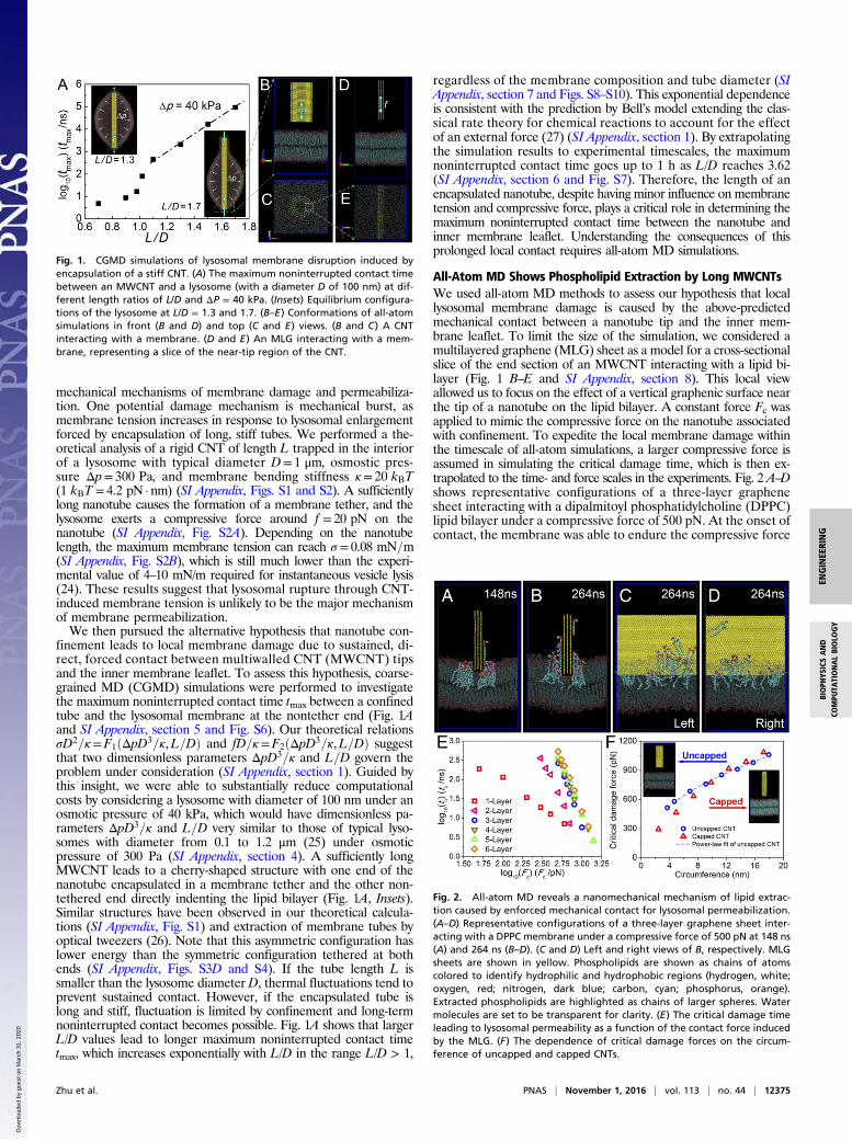
mechanical mechanisms of membrane damage and permeabiliza-tion. One potential damage mechanism is mechanical burst, asmembrane tension increases in response to lysosomal enlargementforced by encapsulation of long, stiff tubes. We performed a the-oretical analysis of a rigid CNT of length L trapped in the interiorof a lysosome with typical diameter D= 1 μm, osmostic pres-sure Δp= 300 Pa, and membrane bending stiffness κ= 20 kBT(1 kBT = 4.2 pN · nm) (SI Appendix, Figs. S1 and S2). A sufficientlylong nanotube causes the formation of a membrane tether, and thelysosome exerts a compressive force around f = 20 pN on thenanotube (SI Appendix, Fig. S2A). Depending on the nanotubelength, the maximum membrane tension can reach σ = 0.08 mN=m(SI Appendix, Fig. S2B), which is still much lower than the experi-mental value of 4–10 mN/m required for instantaneous vesicle lysis(24). These results suggest that lysosomal rupture through CNT-induced membrane tension is unlikely to be the major mechanismof membrane permeabilization.We then pursued the alternative hypothesis that nanotube con-
finement leads to local membrane damage due to sustained, di-rect, forced contact between multiwalled CNT (MWCNT) tipsand the inner membrane leaflet. To assess this hypothesis, coarse-grained MD (CGMD) simulations were performed to investigatethe maximum noninterrupted contact time tmax between a confinedtube and the lysosomal membrane at the nontether end (Fig. 1Aand SI Appendix, section 5 and Fig. S6). Our theoretical relationsσD2=κ=F1ðΔpD3=κ,L=DÞ and fD=κ=F2ðΔpD3=κ,L=DÞ suggestthat two dimensionless parameters ΔpD3=κ and L=D govern theproblem under consideration (SI Appendix, section 1). Guided bythis insight, we were able to substantially reduce computationalcosts by considering a lysosome with diameter of 100 nm under anosmotic pressure of 40 kPa, which would have dimensionless pa-rameters ΔpD3=κ and L=D very similar to those of typical lyso-somes with diameter from 0.1 to 1.2 μm (25) under osmoticpressure of 300 Pa (SI Appendix, section 4). A sufficiently longMWCNT leads to a cherry-shaped structure with one end of thenanotube encapsulated in a membrane tether and the other non-tethered end directly indenting the lipid bilayer (Fig. 1A, Insets).Similar structures have been observed in our theoretical calcula-tions (SI Appendix, Fig. S1) and extraction of membrane tubes byoptical tweezers (26). Note that this asymmetric configuration haslower energy than the symmetric configuration tethered at bothends (SI Appendix, Figs. S3D and S4). If the tube length L issmaller than the lysosome diameter D, thermal fluctuations tend toprevent sustained contact. However, if the encapsulated tube islong and stiff, fluctuation is limited by confinement and long-termnoninterrupted contact becomes possible. Fig. 1A shows that largerL/D values lead to longer maximum noninterrupted contact timetmax, which increases exponentially with L/D in the range L/D > 1,
regardless of the membrane composition and tube diameter (SIAppendix, section 7 and Figs. S8–S10). This exponential dependenceis consistent with the prediction by Bell’s model extending the clas-sical rate theory for chemical reactions to account for the effectof an external force (27) (SI Appendix, section 1). By extrapolatingthe simulation results to experimental timescales, the maximumnoninterrupted contact time goes up to 1 h as L/D reaches 3.62(SI Appendix, section 6 and Fig. S7). Therefore, the length of anencapsulated nanotube, despite having minor influence on membranetension and compressive force, plays a critical role in determining themaximum noninterrupted contact time between the nanotube andinner membrane leaflet. Understanding the consequences of thisprolonged local contact requires all-atom MD simulations.
All-Atom MD Shows Phospholipid Extraction by Long MWCNTsWe used all-atom MD methods to assess our hypothesis that locallysosomal membrane damage is caused by the above-predictedmechanical contact between a nanotube tip and the inner mem-brane leaflet. To limit the size of the simulation, we considered amultilayered graphene (MLG) sheet as a model for a cross-sectionalslice of the end section of an MWCNT interacting with a lipid bi-layer (Fig. 1 B–E and SI Appendix, section 8). This local viewallowed us to focus on the effect of a vertical graphenic surface nearthe tip of a nanotube on the lipid bilayer. A constant force Fc wasapplied to mimic the compressive force on the nanotube associatedwith confinement. To expedite the local membrane damage withinthe timescale of all-atom simulations, a larger compressive force isassumed in simulating the critical damage time, which is then ex-trapolated to the time- and force scales in the experiments. Fig. 2 A–Dshows representative configurations of a three-layer graphenesheet interacting with a dipalmitoyl phosphatidylcholine (DPPC)lipid bilayer under a compressive force of 500 pN. At the onset ofcontact, the membrane was able to endure the compressive force
Fig. 1. CGMD simulations of lysosomal membrane disruption induced byencapsulation of a stiff CNT. (A) The maximum noninterrupted contact timebetween an MWCNT and a lysosome (with a diameter D of 100 nm) at dif-ferent length ratios of L/D and ΔP = 40 kPa. (Insets) Equilibrium configura-tions of the lysosome at L/D = 1.3 and 1.7. (B–E) Conformations of all-atomsimulations in front (B and D) and top (C and E) views. (B and C) A CNTinteracting with a membrane. (D and E) An MLG interacting with a mem-brane, representing a slice of the near-tip region of the CNT.
Fig. 2. All-atom MD reveals a nanomechanical mechanism of lipid extrac-tion caused by enforced mechanical contact for lysosomal permeabilization.(A–D) Representative configurations of a three-layer graphene sheet inter-acting with a DPPC membrane under a compressive force of 500 pN at 148 ns(A) and 264 ns (B–D). (C and D) Left and right views of B, respectively. MLGsheets are shown in yellow. Phospholipids are shown as chains of atomscolored to identify hydrophilic and hydrophobic regions (hydrogen, white;oxygen, red; nitrogen, dark blue; carbon, cyan; phosphorus, orange).Extracted phospholipids are highlighted as chains of larger spheres. Watermolecules are set to be transparent for clarity. (E) The critical damage timeleading to lysosomal permeability as a function of the contact force inducedby the MLG. (F) The dependence of critical damage forces on the circum-ference of uncapped and capped CNTs.
Zhu et al. PNAS | November 1, 2016 | vol. 113 | no. 44 | 12375
ENGINEE
RING
BIOPH
YSICSAND
COMPU
TATIONALBIOLO
GY
Dow
nloa
ded
by g
uest
on
Mar
ch 3
1, 2
020
from the vertical graphene surface without disruption, but lipidextraction by graphenic surface that is energetically favorable (SIAppendix, section 15 and Fig. S15) was observed in the snapshot at148 ns. The early lipid extraction events were slow and steadydespite some loss of membrane integrity. However, at 264 ns, thelipid extraction process suddenly became faster and disruptivewith clusters of lipids climbing the vertical graphene surface. Theinstability of lipid extraction results in a permeable membrane thatwould allow leakage of lysosomal contents into the cytoplasm. Thecritical damage time tc for the membrane to become permeable isdefined as the onset of the burst of lipid extraction, correspondingto the inflection points in the curves of center-of-mass distances inSI Appendix, section 10, Fig. S11. Similar lipid extraction wasshown by Tu et al. in modeling the interaction between graphenenanosheets and bacterial membranes in the absence of a com-pressive force (1). Note that the interaction between finite-sizedgraphene nanosheets and lipid bilayers could be strongly facili-tated by the corners of the nanosheets (2). In our present simu-lations, the MLGs represent a slice of an MWCNT and hencehave no corners. In this case, as spontaneous lipid extraction ishindered by a high energy barrier (2), a compressive force on thenanotube is required and its magnitude can be correlated to thecritical damage time tc. Fig. 2E shows that the critical condition toinduce lysosomal permeabilization can be expressed as a power-law relationship between the contact force and the critical damagetime, with lower contact force postponing membrane damage,which is insensitive to membrane composition (SI Appendix, sec-tion 14 and Fig. S14). Interestingly, tc only has a weak dependenceon the number of graphene layers (Fig. 2E), corresponding to thenumber of nanotube walls. Further investigation revealed that thisweak dependence is due to the localization of contact force dis-tribution at the inner and outer layers of the MLG (SI Appendix,section 11 and Fig. S12). Note that similar lipid extraction pro-cesses have been observed on a small MWCNT (SI Appendix, Fig.S16A) and on an MLG–membrane system of smaller lateral size
(SI Appendix, Fig. S16 B and C). The geometrical effects of thenanotube tip are shown in Fig. 2F, which enables us to calculate tcby an MWCNT (see details in SI Appendix, sections 12 and 13 andFig. S13).Combining the results of CGMD and all-atom simulations allows a
prediction of the link between material properties and lysosomaldamage. The CGMD simulations show that the maximum non-interrupted contact time between the nanotube and lysosomeincreases exponentially with the tube length. When the maximumnoninterrupted contact time exceeds the critical damage time,membrane permeability is induced through the lipid extraction in-stability observed in our all-atom simulations. It can thus be predictedthat, at the same radius, longer nanotubes induce longer contact timeand thus lysosomal membrane permeabilization leading to cell tox-icity, in agreement with our experimental results described next.
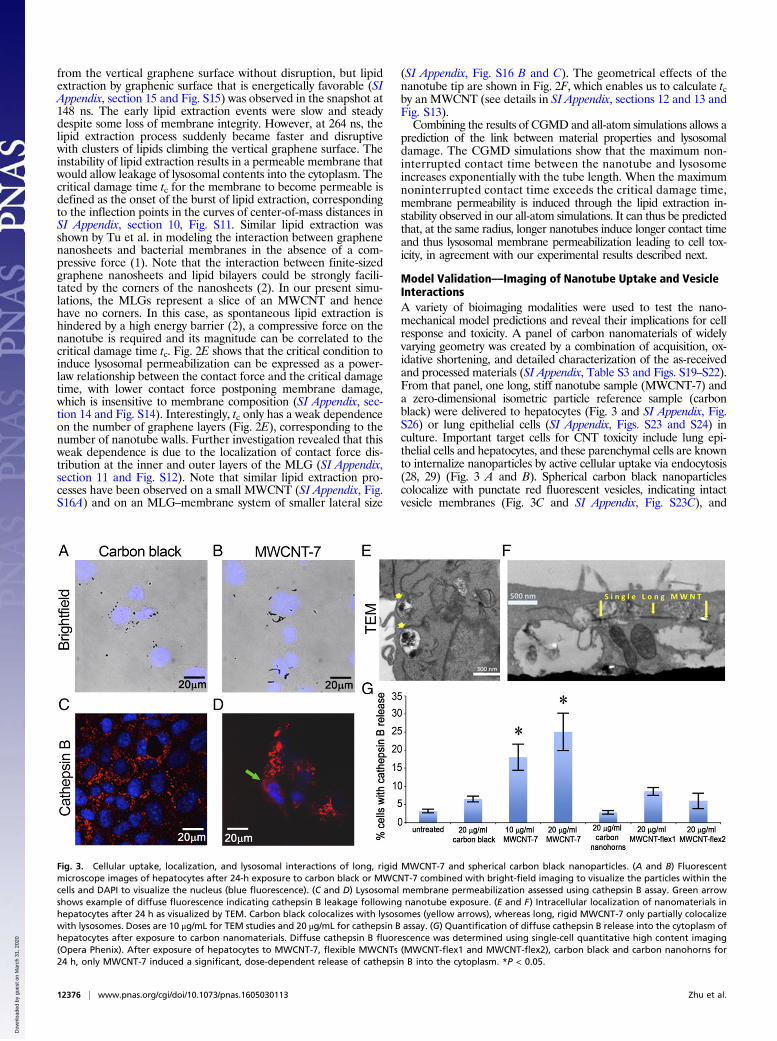
Model Validation––Imaging of Nanotube Uptake and VesicleInteractionsA variety of bioimaging modalities were used to test the nano-mechanical model predictions and reveal their implications for cellresponse and toxicity. A panel of carbon nanomaterials of widelyvarying geometry was created by a combination of acquisition, ox-idative shortening, and detailed characterization of the as-receivedand processed materials (SI Appendix, Table S3 and Figs. S19–S22).From that panel, one long, stiff nanotube sample (MWCNT-7) anda zero-dimensional isometric particle reference sample (carbonblack) were delivered to hepatocytes (Fig. 3 and SI Appendix, Fig.S26) or lung epithelial cells (SI Appendix, Figs. S23 and S24) inculture. Important target cells for CNT toxicity include lung epi-thelial cells and hepatocytes, and these parenchymal cells are knownto internalize nanoparticles by active cellular uptake via endocytosis(28, 29) (Fig. 3 A and B). Spherical carbon black nanoparticlescolocalize with punctate red fluorescent vesicles, indicating intactvesicle membranes (Fig. 3C and SI Appendix, Fig. S23C), and
Fig. 3. Cellular uptake, localization, and lysosomal interactions of long, rigid MWCNT-7 and spherical carbon black nanoparticles. (A and B) Fluorescentmicroscope images of hepatocytes after 24-h exposure to carbon black or MWCNT-7 combined with bright-field imaging to visualize the particles within thecells and DAPI to visualize the nucleus (blue fluorescence). (C and D) Lysosomal membrane permeabilization assessed using cathepsin B assay. Green arrowshows example of diffuse fluorescence indicating cathepsin B leakage following nanotube exposure. (E and F) Intracellular localization of nanomaterials inhepatocytes after 24 h as visualized by TEM. Carbon black colocalizes with lysosomes (yellow arrows), whereas long, rigid MWCNT-7 only partially colocalizewith lysosomes. Doses are 10 μg/mL for TEM studies and 20 μg/mL for cathepsin B assay. (G) Quantification of diffuse cathepsin B release into the cytoplasm ofhepatocytes after exposure to carbon nanomaterials. Diffuse cathepsin B fluorescence was determined using single-cell quantitative high content imaging(Opera Phenix). After exposure of hepatocytes to MWCNT-7, flexible MWCNTs (MWCNT-flex1 and MWCNT-flex2), carbon black and carbon nanohorns for24 h, only MWCNT-7 induced a significant, dose-dependent release of cathepsin B into the cytoplasm. *P < 0.05.
12376 | www.pnas.org/cgi/doi/10.1073/pnas.1605030113 Zhu et al.
Dow
nloa
ded
by g
uest
on
Mar
ch 3
1, 2
020
nanoparticle localization in membrane-bound cytoplasmic vesicleswas confirmed using transmission electron microscopy (TEM)(Fig. 3E and SI Appendix, Fig. S30). The MWCNTs were partiallycolocalized with punctate cytoplasmic vesicles, and longer CNTswere seen penetrating through the vesicle membrane into thecytoplasm by TEM (Fig. 3F). Immunogold labeling confirmed thatthese membrane-bound vesicles are lysosomes (SI Appendix, Fig.S30). Lysosomal membrane permeabilization was assessed using afluorescence assay for a lysosomal protease, cathepsin B, whichretains activity following release into the cytoplasm (30). Confocalfluorescence imaging revealed that exposure to CNTs inducedlow-intensity, diffuse fluorescence reflecting focal release of ca-thepsin B into the cytoplasm (Fig. 3D). Image analysis was usedto further distinguish intact lysosomes from permeable ones basedon object identification and sizing. Intact lysosomes show narrowsize distribution and distinct edges (SI Appendix, Fig. S31A; highthreshold in SI Appendix, Fig. S31B), whereas permeable lyso-somes show weaker, diffuse fluorescence over larger areas in thecytoplasm [SI Appendix, Fig. S31B (Right) and low threshold sizedistributions] associated with cathepsin B leakage followingMWCNT-7 exposure. Additional verification of cathepsin B releaseand lysosomal damage due to long, stiff MWCNT-7 was obtainedusing confocal fluorescence microscopy and quantitative high con-tent imaging (Fig. 3G). The release of cathepsin B initiates a pro-teolytic cascade culminating in activation of caspases leading to celldeath by apoptosis (31) (shown in Fig. 4A and SI Appendix, Figs.S28A, S24 C–E, and S27 C, E, and F). Colocalization of cathepsin Brelease and caspase activation was demonstrated in lung epithelialcells 24 h after exposure to MWCNT-7 (SI Appendix, Fig. S24E),suggesting a link between lysosomal damage induced by exposureto long, rigid MWCNT-7 and cell death. The causal link between
cathepsin B release and cell toxicity was assessed using a selectivecathepsin B inhibitor, methyl ester CA-074. Coexposure of hepa-tocytes to long, rigid MWCNT-7 and the cathepsin B inhibitormethyl ester CA-074 partially prevented cell death as well as cas-pase activation (SI Appendix, Fig. S18 B and C). These observationsconfirm the basic molecular dynamics predictions (vide supra) thatrigid MWCNTs disrupt lysosomal membrane integrity in a length-dependent manner.
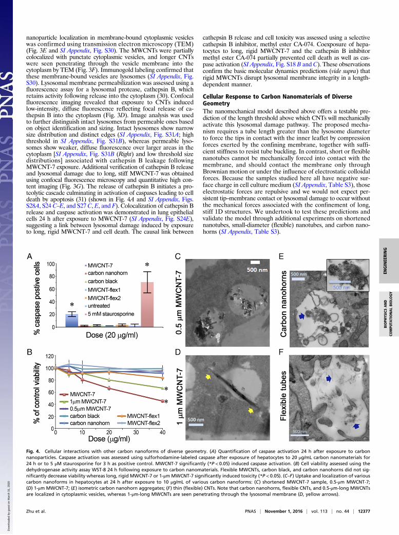
Cellular Response to Carbon Nanomaterials of DiverseGeometryThe nanomechanical model described above offers a testable pre-diction of the length threshold above which CNTs will mechanicallyactivate this lysosomal damage pathway. The proposed mecha-nism requires a tube length greater than the lysosome diameterto force the tips in contact with the inner leaflet by compressionforces exerted by the confining membrane, together with suffi-cient stiffness to resist tube buckling. In contrast, short or flexiblenanotubes cannot be mechanically forced into contact with themembrane, and should contact the membrane only throughBrownian motion or under the influence of electrostatic colloidalforces. Because the samples studied here all have negative sur-face charge in cell culture medium (SI Appendix, Table S3), thoseelectrostatic forces are repulsive and we would not expect per-sistent tip–membrane contact or lysosomal damage to occur withoutthe mechanical forces associated with the confinement of long,stiff 1D structures. We undertook to test these predictions andvalidate the model through additional experiments on shortenednanotubes, small-diameter (flexible) nanotubes, and carbon nano-horns (SI Appendix, Table S3).
Fig. 4. Cellular interactions with other carbon nanoforms of diverse geometry. (A) Quantification of caspase activation 24 h after exposure to carbonnanoparticles. Caspase activation was assessed using sulforhodamine-labeled caspase after exposure of hepatocytes to 20 μg/mL carbon nanomaterials for24 h or to 5 μM staurosporine for 3 h as positive control. MWCNT-7 significantly (*P < 0.05) induced caspase activation. (B) Cell viability assessed using thedehydrogenase activity assay WST-8 24 h following exposure to carbon nanomaterials. Flexible MWCNTs, carbon black, and carbon nanohorns did not sig-nificantly decrease viability whereas long, rigid MWCNT-7 or 1-μmMWCNT-7 significantly induced toxicity (*P < 0.05). (C–F) Uptake and localization of variouscarbon nanoforms in hepatocytes at 24 h after exposure to 10 μg/mL of various carbon nanoforms: (C) shortened MWCNT-7 sample, 0.5-μm MWCNT-7;(D) 1-μm MWCNT-7; (E) isometric carbon nanohorn aggregates; (F) thin (flexible) CNTs. Note that carbon nanohorns, flexible CNTs, and 0.5-μm-long MWCNTsare localized in cytoplasmic vesicles, whereas 1-μm-long MWCNTs are seen penetrating through the lysosomal membrane (D, yellow arrows).
Zhu et al. PNAS | November 1, 2016 | vol. 113 | no. 44 | 12377
ENGINEE
RING
BIOPH
YSICSAND
COMPU
TATIONALBIOLO
GY
Dow
nloa
ded
by g
uest
on
Mar
ch 3
1, 2
020
Uptake and Lysosomal Interactions of Shortened CNTsThe MWCNT-7 sample was progressively shortened using oxida-tive treatment and ultrasonication followed by thermal annealingto restore the original graphenic surfaces (SI Appendix, Table S3and section 17). The original MWCNT-7 sample (mean length11.7 μm) induced cathepsin B release (Fig. 3 D and G and SIAppendix, Fig. S31B), while the same sample shortened to 0.5 μmwas successfully compartmentalized in cytoplasmic vesicles (Fig.4C) and did not induce cathepsin B release from hepatocytes (Fig.3G) or toxicity (Fig. 4B). MWCNTs longer than ∼1 μm wereobserved in contact with, or piercing membranes of cytoplasmicvesicles (Fig. 4D) and this sample induced intermediate toxicity(Fig. 4B and SI Appendix, Fig. S25). The MWCNT lengththreshold for the onset of toxicity (0.5–1 μm) is in the range of themeasured mean lysosome diameter of ∼0.6–1 μm (SI Appendix,Fig. S31), which is in agreement with the predictions of the model.
Uptake and Lysosomal Interactions of Carbon Nanohornsand Flexible CNTsWe next investigated whether thin (more flexible) MWCNTs couldinitiate lysosomal membrane permeabilization and cathepsin B re-lease into the cytoplasm. Two samples of thinner MWCNTs,MWCNT-flex1 and MWCNT-flex2 (SI Appendix, Table S3), werecompartmentalized into cytoplasmic vesicles in hepatocytes and didnot induce cathepsin B release (Figs. 3 G and 4F and SI Appendix,Fig. S27 H and I). Finally, nanomaterials with ultrasharp featureshave been reported to pierce lysosomal membranes (9). To in-vestigate the role of ultrafine-scale surface roughness in this sampleset, hepatocytes were also exposed to carbon nanohorns, which donot have an overall 1D geometry, but rather are aggregated single-walled nanotube cones with outward protruding high-curvature tips(∼1–3 nm). Agglomerates of carbon nanohorns were observedcompartmentalized in cytoplasmic vesicles (Fig. 4E) with no releaseof cathepsin B (SI Appendix, Fig. S27G). Both the carbon nano-horns and the thinner CNTs have higher surface area than the long,rigid MWCNTs or the carbon black nanoparticles; however, theydid not induce cell death at equivalent mass (Fig. 4B) or surfacearea doses (SI Appendix, Fig. S25) after 24 or 48 h. Lysosomaldamage in this system is not related to high surface area or sharpnanoscale surface features, but rather to length and stiffness.
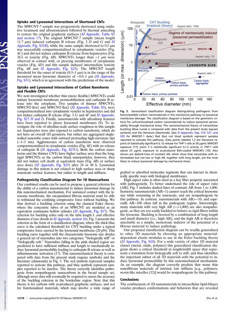
Pathogenicity Classification Diagram for 1D NanocarbonsOur combined results can be used to propose a general criterion forthe ability of a carbon nanomaterial to induce lysosomal damage bythis nanomechanical mechanism. For sustained contact with the ly-sosome membrane, the encapsulated fibrous material must be ableto withstand the confining compressive force without buckling. Wethus derived a buckling criterion using the classical Euler theory,where the concentric tubes of an MWCNT are modeled as anequivalent continuum hollow cylinder (SI Appendix, Fig. S17). Thecriterion for buckling relies only on the tube length L and effectivediameter d (see details in SI Appendix, section 16). Fig. 5 presents thecriterion in the form of a classification diagram, where the thick bluecurve is the calculated threshold for CNT buckling under a typicalcompressive force exerted by the lysosomal membrane (20 pN). Thisbuckling curve together with the characteristic lysosome size dividesa general set of nanotubes into two categories: “biologically stiff” or“biologically soft.” Nanotubes falling in the pink shaded region arepredicted to have sufficient stiffness and length to mechanically in-duce lysosomal permeability leading to cathepsin B release as well asinflammasome activation (17). This nanomechanical theory is com-pared with data from the present study (square symbols) and theliterature (diamonds) in Fig. 5. The red symbols represent samplesreported to activate this pathway, and blue symbols represent sam-ples reported to be inactive. The theory correctly identifies patho-genic from nonpathogenic nanocarbons in the broad sample set,although more data will eventually be required to assess the accuracyof the buckling criterion in the borderline region. Note that thistheory is for carbons with as-produced graphenic surfaces, and notfor functionalized materials, which may involve a wide range of
grafted or adsorbed molecular segments that can interact in chem-ically specific ways with biological membranes.High aspect ratio is often cited as a key fiber property associated
with pathogenicity. To better understand the role of aspect ratio(AR), Fig. 5 includes dashed lines of constant AR from 1 to 1,000.Isometric nanomaterials (AR∼1) cannot reach the critical lysosomesize while remaining at the nanoscale, and are nonpathogenic bythis pathway. In contrast, nanomaterials with AR>∼10, and espe-cially AR∼100 often fall in the pathogenic regime. Interestingly,many materials with very high AR (∼>1,000) are also nonpatho-genic, as they are too easily buckled to behave as rigid fibers withinthe lysosome. Buckling is favored by a combination of long lengthand small diameter (i.e., high AR), and the high AR is thereforeunreliable as a simple, monotonic indictor of the potential of afibrous material to induce pathology.Our proposed classification diagram can be readily generalized
to other 1D materials by choosing an appropriate material-dependent elastic modulus to use in the Euler buckling theory(SI Appendix, Fig. S18). For a wide variety of other 1D materialclasses (metal, oxide, polymer) this generalized classification dia-gram shows a critical threshold in length/width space that repre-sents a transition from biologically soft to stiff, and thus identifiesthe important subset of all 1D materials with the potential to in-duce lysosomal permeability by this nanomechanical mechanism.As an example, the diagram correctly predicts that many thinnanofibrous materials of intrinsic low stiffness [e.g., polymers,worm-like micelles (32)] would be nonpathogenic by this pathway.
ConclusionsThe confinement of 1D nanomaterials in intracellular lipid-bilayervesicles produces conformations and behaviors that are revealed
Fig. 5. Generalized classification diagram distinguishing pathogenic frombiocompatible carbon nanomaterials in the mechanical pathway to lysosomalmembrane damage. The classification diagram is based on the geometric cri-teria for unfunctionalized carbon nanomaterials to induce lysosomal perme-ability through mechanical stress. The nanomechanical theory of intracellularbuckling (blue curve) is compared with data from the present study (squaresymbols) and the literature (diamonds). (See SI Appendix, Figs. S19, S27, andS29 for MWCNT-1 data.) Red (but not blue) symbols represent samplesreported to activate this pathway. Data points labeled 1,2 are literature re-ports of statistically significant IL-1β release for THP-1 cells at 50 μg/mLMWCNTexposure (17); point 3 is statistically significant IL1-β activity in THP-1 cellsabove 25 μg/mL exposure to as-produced BSA-coated MWCNTs (10). Alsoshown are dashed lines of constant AR, which show that nanotubes with in-termediate but not low or high AR, together with long length, are the mostlikely to induce lysosomal damage by mechanical stress.
12378 | www.pnas.org/cgi/doi/10.1073/pnas.1605030113 Zhu et al.
Dow
nloa
ded
by g
uest
on
Mar
ch 3
1, 2
020
by MD and in vitro bioimaging. Cellular attempts to package long1D nanomaterials in spherical vesicles leads to material com-pression that forces persistent mechanical contact between thetube tip and inner membrane leaflet, which for CNTs causes lipidextraction, membrane permeabilization, release of cathepsin B,and cell death by apoptosis. In contrast, this mechanism predictsintact lysosomes and lower toxicity for nanotubes that are short(<∼1 μm), or of very high L=D that easily buckle in the presence ofthe lysosomal compression force (∼20 pN), or for other materialswith similar graphenic chemistry but isometric shape (e.g., carbonblack, carbon nanohorns). These predictions are validated by invitro experiments using hepatocytes as well as lung epithelial cellsexposed to a diverse panel of synthetic nanocarbons. A quantitativematerial classification diagram distinguishes pathogenic from bio-compatible nanotube varieties based on a buckling criterion thatrelies only on minimum and maximum dimension. The presentresults also suggest that the observed low pathogenicity of tangledtubes relative to straight tubes (3, 21) may be usefully understood asan effect of intrinsic biological softness, defined as the resistanceto buckling under lysosomal compressive forces. Unlike tangling,which is an agglomeration or deformation state subject to changethrough material handling or processing, biological softness is afundamental nanomaterial property directly related to diameter andlength. This mechanistic understanding provides guidance for safedesign and material selection of 1D nanomaterials for both bio-medical and nonbiomedical applications.
MethodsCGMD Simulations. CGMD simulations of a CNT encapsulated inside a lyso-some were performed to investigate the maximum noninterrupted contacttime between the CNT and a membrane patch built from solvent-free CGlipids (4, 22). Further details of the CG models of the membrane and CNT canbe found in SI Appendix, sections 2 and 3.
All-Atom MD Simulations. All-atom MD simulations were performed to in-vestigate how an MLG, corresponding to a near-tip slice of an MWCNT, or anMWCNT interactswith amembranepatch. Themembraneswere constructed from
bilayers of the Berger lipids. The Berger lipid force field was used for lipidscombined with an Optimized Potentials for Liquid Simulations representation ofMLGs and MWCNTs. DPPC was adopted to build the lipid bilayer. Further detailsare provided in SI Appendix, section 9.
Nanomaterial Panel and Characterization. A panel of carbon nanomaterials ofdiverse geometry and stiffness was assembled and characterized, includingcommercial MWCNTs fromMitsui & Co. (MWCNT-7) and NanoLab, Inc., whichwere synthesized using catalytic chemical vapor deposition. The process ofshortening and surface restoration used to create variants of the MWCNT-7sample is described in SI Appendix, section 17. The panel of carbon nano-materials included zero-dimensional reference materials including carbonblack M120 (Cabot Corporation) and carbon nanohorns, courtesy of DavidGeohegan, Oak Ridge National Laboratory, Oak Ridge, TN, as isometric (low-AR) reference carbon materials.
Cell Culture and Exposure Conditions. Experiments were conducted on two celltypes: hepatocytes (AML12 cells; American Type Culture Collection; CRL-2254)and lung epithelial cells (H460 cells; American Type Culture Collection; HTB-177).The cells were exposed to commercial MWCNTs, carbon nanohorns, and carbonblack and assessed for viability as described in SI Appendix, Figs. S19 and S20.
Lysosomal Permeabilization. After exposure of cells to carbon nanomaterials,the integrity of lysosomes is determined using a cathepsin B target peptidesequence conjugated to a red fluorophore, which is cleaved by active ca-thepsin B enzyme. Diffuse red cytoplasmic fluorescence indicates lysosomalmembrane permeabilization whereas punctate cytoplasmic fluorescenceindicates intact lysosomes. Lysosomal permeabilization was quantified usingcell segmentation and analysis and quantitative high content fluorescenceimaging as described in SI Appendix, section 17.
ACKNOWLEDGMENTS. The authors acknowledge a generous gift from DonnaMcGraw Weiss and Jason Weiss. This work was supported by the NationalScience Foundation (Grants CBET-1344097 and CMMI-1562904) and the Super-fund Research Program of the National Institute of Environmental HealthSciences (Grant P42 ES013660). The simulations reported were performed onresources provided by the Extreme Science and Engineering Discovery Environ-ment (XSEDE) through Grant MS090046.
1. Tu Y, et al. (2013) Destructive extraction of phospholipids from Escherichia colimembranes by graphene nanosheets. Nat Nanotechnol 8(8):594–601.
2. Li Y, et al. (2013) Graphene microsheets enter cells through spontaneous membrane pen-etration at edge asperities and corner sites. Proc Natl Acad Sci USA 110(30):12295–12300.
3. Nagai H, Toyokuni S (2012) Differences and similarities between carbon nanotubesand asbestos fibers during mesothelial carcinogenesis: Shedding light on fiber entrymechanism. Cancer Sci 103(8):1378–1390.
4. Shi X, von dem Bussche A, Hurt RH, Kane AB, Gao H (2011) Cell entry of one-dimensional nanomaterials occurs by tip recognition and rotation. Nat Nanotechnol6(11):714–719.
5. Vecitis CD, Zodrow KR, Kang S, Elimelech M (2010) Electronic-structure-dependentbacterial cytotoxicity of single-walled carbon nanotubes. ACS Nano 4(9):5471–5479.
6. Zhang S, Li J, Lykotrafitis G, Bao G, Suresh S (2009) Size-dependent endocytosis ofnanoparticles. Adv Mater 21(4):419–424.
7. Li R, et al. (2013) Surface charge and cellular processing of covalently functionalizedmultiwall carbon nanotubes determine pulmonary toxicity. ACS Nano 7(3):2352–2368.
8. Stern ST, Adiseshaiah PP, Crist RM (2012) Autophagy and lysosomal dysfunction asemerging mechanisms of nanomaterial toxicity. Part Fibre Toxicol 9(1):20.
9. Chu Z, et al. (2014) Unambiguous observation of shape effects on cellular fate ofnanoparticles. Sci Rep 4:4495.
10. Wang X, et al. (2012) Pluronic F108 coating decreases the lung fibrosis potential ofmultiwall carbon nanotubes by reducing lysosomal injury. Nano Lett 12(6):3050–3061.
11. Elbaum M, Kuchnir Fygenson D, Libchaber A (1996) Buckling microtubules in vesicles.Phys Rev Lett 76(21):4078–4081.
12. Fygenson DK, Elbaum M, Shraiman B, Libchaber A (1997) Microtubules and vesiclesunder controlled tension. Phys Rev E Stat Phys Plasmas Fluids Relat Interdiscip Topics55(1):850–859.
13. Donaldson K, et al. (2013) Pulmonary toxicity of carbon nanotubes and asbestos -similarities and differences. Adv Drug Deliv Rev 65(15):2078–2086.
14. Hamilton RF, et al. (2009) Particle length-dependent titanium dioxide nanomaterialstoxicity and bioactivity. Part Fibre Toxicol 6:35.
15. Bussy C, et al. (2012) Critical role of surface chemical modifications induced by lengthshortening on multi-walled carbon nanotubes-induced toxicity. Part Fibre Toxicol 9(1):46.
16. Wang X, et al. (2011) Dispersal state of multiwalled carbon nanotubes elicits profi-brogenic cellular responses that correlate with fibrogenesis biomarkers and fibrosis inthe murine lung. ACS Nano 5(12):9772–9787.
17. Hamilton RF, Jr, Wu Z, Mitra S, Shaw PK, Holian A (2013) Effect of MWCNT size,carboxylation, and purification on in vitro and in vivo toxicity, inflammation and lungpathology. Part Fibre Toxicol 10(1):57.
18. Yang M, et al. (2013) Functionalization of carbon nanoparticles modulates inflam-matory cell recruitment and NLRP3 inflammasome activation. Small 9(24):4194–4206.
19. Kraszewski S, Bianco A, Tarek M, Ramseyer C (2012) Insertion of short amino-func-tionalized single-walled carbon nanotubes into phospholipid bilayer occurs by passivediffusion. PLoS One 7(7):e40703.
20. Lacerda L, et al. (2013) How do functionalized carbon nanotubes land on, bind to andpierce through model and plasma membranes. Nanoscale 5(21):10242–10250.
21. Poland CA, et al. (2008) Carbon nanotubes introduced into the abdominal cavityof mice show asbestos-like pathogenicity in a pilot study. Nat Nanotechnol 3(7):423–428.
22. Reynwar BJ, et al. (2007) Aggregation and vesiculation of membrane proteins bycurvature-mediated interactions. Nature 447(7143):461–464.
23. Keten S, Xu Z, Ihle B, Buehler MJ (2010) Nanoconfinement controls stiffness, strengthand mechanical toughness of β-sheet crystals in silk. Nat Mater 9(4):359–367.
24. Hategan A, Law R, Kahn S, Discher DE (2003) Adhesively-tensed cell membranes: lysiskinetics and atomic force microscopy probing. Biophys J 85(4):2746–2759.
25. Kühnel W (2003) Color Atlas of Cytology, Histology, and Microscopic Anatomy(Thieme, New York), 4th Ed, p 34.
26. Lee HJ, Peterson EL, Phillips R, Klug WS, Wiggins PA (2008) Membrane shape as areporter for applied forces. Proc Natl Acad Sci USA 105(49):19253–19257.
27. Bell GI (1978) Models for the specific adhesion of cells to cells. Science 200(4342):618–627.
28. Ji Z, et al. (2009) The hepatotoxicity of multi-walled carbon nanotubes in mice.Nanotechnology 20(44):445101.
29. Kettiger H, Schipanski A, Wick P, Huwyler J (2013) Engineered nanomaterial uptakeand tissue distribution: From cell to organism. Int J Nanomedicine 8:3255–3269.
30. Jordans S, et al. (2009) Monitoring compartment-specific substrate cleavage by ca-thepsins B, K, L, and S at physiological pH and redox conditions. BMC Biochem 10(1):23.
31. Reiners JJ, Jr, et al. (2002) Release of cytochrome c and activation of pro-caspase-9following lysosomal photodamage involves Bid cleavage. Cell Death Differ 9(9):934–944.
32. Geng Y, et al. (2007) Shape effects of filaments versus spherical particles in flow anddrug delivery. Nat Nanotechnol 2(4):249–255.
Zhu et al. PNAS | November 1, 2016 | vol. 113 | no. 44 | 12379
ENGINEE
RING
BIOPH
YSICSAND
COMPU
TATIONALBIOLO
GY
Dow
nloa
ded
by g
uest
on
Mar
ch 3
1, 2
020