nail fungal infections and treatment
TRANSCRIPT
Nail fungal infections and treatmentRobert Baran, MD*
Nail Disease Centre, 42 rue des Serbes, 06400, Cannes, France
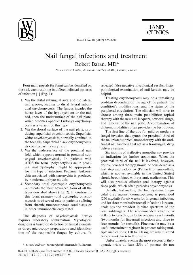
Four main portals for fungi can be identified on
the nail, each resulting in different clinical patterns
of infection [1] (Fig. 1):
1. Via the distal subungual area and the lateral
nail groove, leading to distal lateral subun-
gual onychomycosis. The fungus invades the
horny layer of the hyponychium or the nail
bed, then the undersurface of the nail plate,
which becomes opaque. Endonyx onychomy-
cosis is a variant of this type.
2. Via the dorsal surface of the nail plate, pro-
ducing superficial onychomycosis. Superficial
white onychomycosis is normally confined to
the toenails. Superficial black onychomycosis,
its counterpart, is very rare.
3. Via the undersurface of the proximal nail
fold, which appears normal in proximal sub-
ungual onychomycosis. In patients with
AIDS the term ‘‘polydactylous acute proxi-
mal nail dystrophy’’ might be appropriate
for this type of infection. Proximal leukony-
chia associated with paronychia is produced
by nondermatophyte-moulds.
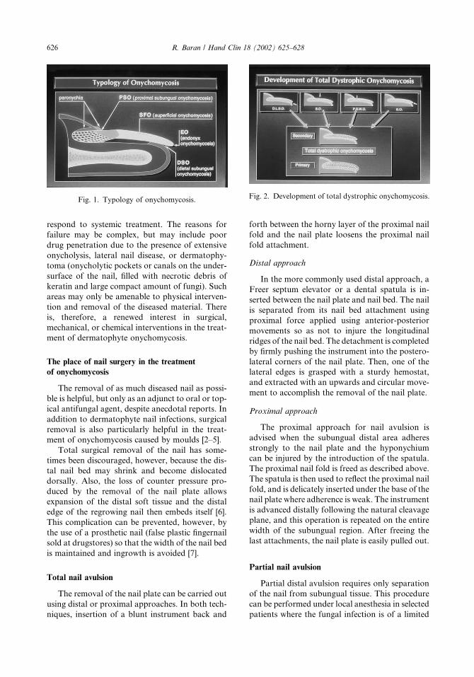
4. Secondary total dystrophic onychomycosis
represents the most advanced form of all the
types described above (Fig. 2). In contrast to
this form, primary total dystrophic onycho-
mycosis is observed only in patients suffering
from chronic mucocutaneous candidiasis or
in other immunodeficiency states.
The diagnosis of onychomycosis always
requires laboratory confirmation. Mycological
diagnosis is based on detection of fungal elements
in direct microscopy preparations and identifica-
tion of the responsible fungus by culture. In
repeated false negative mycological results, histo-
pathological examination of nail keratin may be
helpful.
Treating onychomycosis may be a tantalizing
problem depending on the age of the patient, the
condition’s modifications, and the status of the
peripheral circulation. The clinician will have to
choose among three main possibilities: topical
therapy with the new nail lacquers, new oral drugs,
and removal of the nail plate. A combination of
different modalities often provides the best option.
The first line of therapy for mild or moderate
fungal invasion that spares the proximal third of
the nail plate is topical monotherapy with the anti-
fungal nail lacquers that act as a transungual drug
delivery system.
Six months of ineffective monotherapy provide
an indication for further treatments. When the
proximal third of the nail is involved, however,
double pronged therapy should be considered as a
first step and ciclopirox (Penlac� or amorolfine,
which is not yet available in the United States)
should be combinedwith systemicmedication. This
will also produce effective oral therapy against
tinea pedis, which often precedes onychomycosis.
Usually, terbinafine, the first systemic fungi-
cidal drug against dermatophytes, is prescribed
(250 mg/daily for six weeks for fingernail infection,
and for threemonths for toenail infection). Itracon-
azole has the broadest in vitro spectrum of the
oral antifungals. The recommended schedule is
200 mg twice a day, daily for one week each month
(two months for fingernail infections and three to
four months for toenails). Fluconazole may be a
useful intermittent regimen in patients taking mul-
tiple medications; 150 to 300 mg are administered
once a week for 6 to 9 months.
Unfortunately, even in the most successful ther-
apeutic trials at least 25% of patients do not* E-mail address: [email protected] (R. Baran).
0749-0712/02/$ - see front matter � 2002, Elsevier Science (USA). All rights reserved.
PII: S 0 7 4 9 - 0 7 1 2 ( 0 2 ) 0 0 0 3 7 - 9
Hand Clin 18 (2002) 625–628
respond to systemic treatment. The reasons for
failure may be complex, but may include poor
drug penetration due to the presence of extensive
onycholysis, lateral nail disease, or dermatophy-
toma (onycholytic pockets or canals on the under-
surface of the nail, filled with necrotic debris of
keratin and large compact amount of fungi). Such
areas may only be amenable to physical interven-
tion and removal of the diseased material. There
is, therefore, a renewed interest in surgical,
mechanical, or chemical interventions in the treat-
ment of dermatophyte onychomycosis.
The place of nail surgery in the treatment
of onychomycosis
The removal of as much diseased nail as possi-
ble is helpful, but only as an adjunct to oral or top-
ical antifungal agent, despite anecdotal reports. In
addition to dermatophyte nail infections, surgical
removal is also particularly helpful in the treat-
ment of onychomycosis caused by moulds [2–5].
Total surgical removal of the nail has some-
times been discouraged, however, because the dis-
tal nail bed may shrink and become dislocated
dorsally. Also, the loss of counter pressure pro-
duced by the removal of the nail plate allows
expansion of the distal soft tissue and the distal
edge of the regrowing nail then embeds itself [6].
This complication can be prevented, however, by
the use of a prosthetic nail (false plastic fingernail
sold at drugstores) so that the width of the nail bed
is maintained and ingrowth is avoided [7].
Total nail avulsion
The removal of the nail plate can be carried out
using distal or proximal approaches. In both tech-
niques, insertion of a blunt instrument back and
forth between the horny layer of the proximal nail
fold and the nail plate loosens the proximal nail
fold attachment.
Distal approach
In the more commonly used distal approach, a
Freer septum elevator or a dental spatula is in-
serted between the nail plate and nail bed. The nail
is separated from its nail bed attachment using
proximal force applied using anterior-posterior
movements so as not to injure the longitudinal
ridges of the nail bed. The detachment is completed
by firmly pushing the instrument into the postero-
lateral corners of the nail plate. Then, one of the
lateral edges is grasped with a sturdy hemostat,
and extracted with an upwards and circular move-
ment to accomplish the removal of the nail plate.
Proximal approach
The proximal approach for nail avulsion is
advised when the subungual distal area adheres
strongly to the nail plate and the hyponychium
can be injured by the introduction of the spatula.
The proximal nail fold is freed as described above.
The spatula is then used to reflect the proximal nail
fold, and is delicately inserted under the base of the
nail plate where adherence is weak. The instrument
is advanced distally following the natural cleavage
plane, and this operation is repeated on the entire
width of the subungual region. After freeing the
last attachments, the nail plate is easily pulled out.
Partial nail avulsion
Partial distal avulsion requires only separation
of the nail from subungual tissue. This procedure
can be performed under local anesthesia in selected
patients where the fungal infection is of a limited
Fig. 2. Development of total dystrophic onychomycosis.Fig. 1. Typology of onychomycosis.
626 R. Baran / Hand Clin 18 (2002) 625–628

extent. An affected portion of the nail plate may
be removed in one session, even when the disease
has reached the deeper regions of the subungual
tissue beneath the proximal nail fold (Fig. 3A,B).
Commonly, an English anvil nail splitter or a
double action bone rongeur are used for this
procedure.
Partial surgical section of the lateral or med-
ial segment of the nail plate may be sufficient
for the treatment of distal lateral subungual
Fig. 3. (A) Lateral and proximal onychomycosis. (B) Partial surgical avulsion.
Fig. 4. (A) Recalcitrant chronic Candida paronychia of the thumb with nail plate invasion. (B) Surgical excision of a
crescent of thickened nail fold, followed by nail avulsion.
627R. Baran / Hand Clin 18 (2002) 625–628

onychomycosis. Therefore, enough normal nail is
left on the toe to counteract the upward forces
exerted on the distal soft tissue when walking, and
this will prevent the appearance of a distal nail wall.
In proximal subungual onychomycosis, re-
moval of the nonadherent base of the nail plate,
cut transversely, leaves the distal portion of the
nail in place, which decreases discomfort.
Where Candida or, more rarely, dermatophytes
result in onycholysis, the detached portion of the
nail plate should be thoroughly clipped away. This
facilitates the daily application of a topical anti-
fungal drug until nail regrowth is complete. In
paronychia with Candida lateral nail-edge involve-
ment or mixed infection, the affected nail keratin
should be surgically removed. Recalcitrant Can-
dida paronychia with secondary nail-plate inva-
sion may be treated by surgical excision of a
crescent of thickened nail fold followed by nail
avulsion (Fig. 4A,B).
In any type of surgically treated onychomyco-
sis, the avulsed nail segment must always include
a margin of normal nail and the nail bed should
be treated with the conventional antifungal oint-
ments.
It has been shown that partial surgical avulsion
of the nail as a treatment of onychomycosis, in
combination with griseofulvin or ketoconazole
therapy, reduced the duration of the oral antifun-
gal treatment by 50% [7]. Recently, good results
have been obtained by combining surgical tech-
niques with either intermittent [8] or short dura-
tion use of new oral antifungal drugs [9]. This
combination may be of particular use in limited
dermatophyte toenail infection or in cases of
treatment failure following conventional oral
treatment. The addition of the antifungal nail
lacquers on the remaining nail keratin allows a
‘‘triple therapy’’ model offering the best results.
Since the introduction of the newer oral antifungal
agents, there is no longer room for matricectomy
in the treatment of severe onychomycosis, despite
some isolated reports [10].
The place of chemical avulsion
In patients at risk (immunosuppressive condi-
tions, immunosuppressive therapy, peripheral vas-
cular disease), chemical avulsion is a painless
method that has superseded partial surgical avul-
sion [11]. It may be repeated as often as necessary.
Urea ointment appears to focus its action on the
bond between the nail keratin and the diseased nail
bed; it spares the normal nail tissue.
Forty percent urea ointment is applied to the
nail plate after protecting the surrounding skin,
with adhesive dressing for example. The entire dis-
tal digit is then wrapped for a week.
Blunt dissection, using a nail elevator and nail
clipper, leaves the remaining portion of normal
nail plate intact. Following removal of the dis-
eased part of the nail, topical antifungal agents
should be applied for several months under occlu-
sion, especially if there is no associated systemic
therapy.
Despite its efficacy, such a treatment is difficult
to apply in the elderly, tedious when several digits
are affected, or ineffective when the proximal por-
tion of the nail plate is invaded by fungal organ-
isms beneath the nail fold.
References
[1] Baran R, Hay RJ, Tosti A, Haneke E. In: A new
classification of onychomycosis. Br J Dermatol
1998;119:567–71.
[2] Baran R, Hay R, Haneke E, Tosti A. In: Onycho-
mycosis: the current approach to diagnosis and ther-
apy. London: Martin Dunitz Ltd.; 1999 Chapter 7.
[3] Mc Innes BD, Dockery GL. Surgical treatment of
mycotic toenails. J Am Podiatr Med Assoc 1997;
87:557–64.
[4] Baden HP. Treatment of distal onychomycosis with
avulsion and topical antifungal agents under
occlusion. Arch Dermatol 1994;130:558–9.
[5] Cohen PR, Scher RK. Topical and surgical treat-
ment of onycomycosis. J Am Acad Dermatol
1994;31:S47–77.
[6] Fowler AW. Excision of the germinal matrix: a
unified treatment for toenail and onychogryphosis.
Br J Surg 1958;45:382.
[7] Baran R, Hay R. Partial surgical avulsion of the
nail in onychomycosis. Clin Exp Dermatol 1985;10:
413–8.
[8] Dominguez-Cherit J, Teixera F, Arenas R. Com-
bined surgical and systemic treatment of onycho-
mycosis. Br J Dermatol 1999;140:778–80.
[9] Goodfield MJD, Evans EGV. Combined treatment
with surgery and short duration oral antifungal
therapy in patients with limited dermatophyte toe-
nail infection. J of Dermatol Treat 2000;11:259–62.
[10] Nahabedian MY. Multiple-digit onychomycosis: a
simple surgical cure. Ann Plast Surg 2000;45:
446–50.
[11] South DA, Farber E. Urea ointment in non surgical
avulsion of nail dystrophies. Reappraisal. Cutis
1980;26:609–12.
628 R. Baran / Hand Clin 18 (2002) 625–628