n-alkylation of highly quaternized chitosan derivatives affects the paracellular permeation...
TRANSCRIPT

European Journal of Pharmaceutics and Biopharmaceutics xxx (2013) xxx–xxx
Contents lists available at SciVerse ScienceDirect
European Journal of Pharmaceutics and Biopharmaceutics
journal homepage: www.elsevier .com/locate /e jpb
Research paper
N-alkylation of highly quaternized chitosan derivatives affects theparacellular permeation enhancement in bronchial epithelia in vitro
0939-6411/$ - see front matter � 2013 Elsevier B.V. All rights reserved.http://dx.doi.org/10.1016/j.ejpb.2013.04.002
Abbreviations: A, surface area (cm2); ALI, air–liquid interface; CSLM, confocalscanning laser microscopy; DA, degree of acetylation; DQ, degree of quaternization;DS, degree of substitution; ER, permeation enhancement ratio; FBS, fetal bovineserum; FCS, fetal calf serum; FD4, fluorescein isothiocyanate labeled dextran 4 kDa;FITC, fluorescein isothiocyanate; HBSS, Hanks balanced salt solution; MTT, (4,5-dimethylthiazol-2-yl)-2,5-diphenyltetrazolium bromide; Papp, apparent permeabil-ity; QuatPropyl, N-propyl-N,N-dimethyl chitosan; QuatButyl, N-butyl-N,N-dimethylchitosan; QuatHexyl, N-hexyl-N,N-dimethyl chitosan; TER, transepithelial electricalresistance; TJ, tight junctions; TMC, N,N,N-trimethyl chitosan.⇑ Corresponding author. Faculty of Pharmaceutical Sciences, School of Health
Sciences, University of Iceland, Hofsvallagata 53, IS-107 Reykjavik, Iceland. Tel.:+354 525 4463; fax: +354 525 4071.
E-mail address: [email protected] (M. Másson).
Please cite this article in press as: B.E. Benediktsdóttir et al., N-alkylation of highly quaternized chitosan derivatives affects the paracellular permenhancement in bronchial epithelia in vitro, Eur. J. Pharm. Biopharm. (2013), http://dx.doi.org/10.1016/j.ejpb.2013.04.002
Berglind Eva Benediktsdóttir a, Thórarinn Gudjónsson b, Ólafur Baldursson c, Már Másson a,⇑a Faculty of Pharmaceutical Sciences, School of Health Sciences, University of Iceland, Reykjavik, Icelandb Biomedical Center, School of Health Sciences, University of Iceland, Reykjavík, Icelandc Department of Pulmonary Medicine, Landspitali – The National University Hospital of Iceland, Reykjavík, Iceland
a r t i c l e i n f o
Article history:Received 9 November 2012Accepted in revised form 5 April 2013Available online xxxx
Keywords:Airway drug deliveryChitosanN-quaternary derivativesTMCPermeation enhancementTight junctions
a b s t r a c t
This study describes the structure–activity relationship for carefully characterized N-alkyl-N-quaternarychitosan derivatives as permeation enhancers for drugs that are mainly absorbed through the paracellu-lar pathway, such as macromolecular drugs and hydrophilic drugs, in a well defined bronchial epithelialcell line. The O-methyl free derivatives used in the study were fully trimethylated (100%) N,N,N-trimethylchitosan (TMC) and N-propyl-(QuatPropyl), N-butyl-(QuatButyl) and N-hexyl (QuatHexyl)-N,N-dimethylchitosan, with 85–91% degree of quaternization. The fully trimethylated TMC, from 0.25 mg/ml,decreased transepithelial electrical resistance (TER) in a reversible manner and enhanced the permeationof the macromolecule FITC–dextran 4 kDa (FD4) 2–5 fold. TMC did not cause any alterations in the tightjunction (TJ) protein claudin-4 or in F-actin architecture. QuatHexyl was the most effective polymer toproduce enhanced permeation and decreased TER from 0.016 mg/ml. Nevertheless, this enhanced perme-ation was accompanied by reduced viability and dissociation of F-actin and claudin-4 proteins. The struc-ture–activity relationship suggests that more lipophilic derivatives show more permeation enhancement,TJ disassembly, and less viability in the order of hexyl � butyl > propyl > methyl and demonstrates thatthe permeation effect is not only mediated by permanent positive charge but also by the extent of N-alkylation. These results are relevant to elucidate the structural factors contributing to the permeationenhancement of chitosan derivatives and for potential use in pulmonary applications.
� 2013 Elsevier B.V. All rights reserved.
1. Introduction
Biotechnological medicines, such as vaccines and monoclonalantibodies, are an expanding field within the pharmaceuticalindustry, with currently around 1200 medicines in developmentin Europe [1]. Due to their macromolecular nature and lack of sta-bility, these medicines are usually given in parenteral dosageforms, such as injections, that can result in reduced patient compli-ance. This necessitates the development of novel delivery systems
that can deliver therapeutic agents in a more accessible dosageform. Inhalation of macromolecular drug candidates for systemicdelivery results in higher systemic bioavailability than any othernoninvasive route of delivery [2,3] and can therefore be consideredan interesting alternative to the conventional oral route. The in-creased systemic bioavailability can be attributed to large surfacearea, small aqueous volume at the epithelial surface and relativelylow enzymatic activity [2], attractive for compounds that havephysicochemical properties unfavorable to the conventional oralroute.
A valid model must be used to study the permeation of thesepotential drugs through the lungs. The VA10 human bronchial epi-thelial cell line was recently established as an in vitro drug perme-ation model [4], having permeability properties similar to thewidely used bronchial epithelial cell lines Calu-3 and 16HBE14o-.The VA10 cell line is derived from a normal human bronchus, ex-presses tight junctions (TJs) and has ciliated, 2–3 cell layer mor-phology in air–liquid interface (ALI) culture [4,29], therebyshowing in vivo-like phenotype and providing a valid alternativeto Calu-3 and 16HBE14o-. The passage of hydrophilic and macro-molecular drugs between epithelial cells is limited by the TJ
eation
2 B.E. Benediktsdóttir et al. / European Journal of Pharmaceutics and Biopharmaceutics xxx (2013) xxx–xxx
proteins that serve as a fence mechanism, controlling whichmolecules pass [5]. Absorption enhancers have been studied tocircumvent this gating mechanism to enable the efficient paracel-lular permeation of these drugs.
Chitosan, a linear polysaccharide derived from chitin by N-deacetylation, has been evaluated as a potential permeation en-hancer, mainly to increase absorption in the intestinal mucosain vitro and in vivo [6–8] with disruption of TJ or related proteinsas the proposed mechanism [9,10]. The feasibility of chitosan per-meation enhancement studies on the intestinal mucosa can beattributed to its mucoadhesiveness [11], biodegradability [12],and safety in non-parental dosage formulations [13]. This polymeris only soluble under acidic pH, forming a polycation, at similar pHas is present in some parts of the intestine. However, studies usingchitosan in airway epithelia [14,15] are scarce due to limited solu-bility of chitosan above its pKa of 6.6 [16].
Introducing a permanent positive charge, such as with N,N,N-trimethyl chitosan (TMC), is one way of increasing physiologicalaqueous solubility of the polymer, thereby enabling effective per-meation enhancing studies on other mucosal surfaces such as thebronchial epithelia. TMC has gained increased attention as a poly-
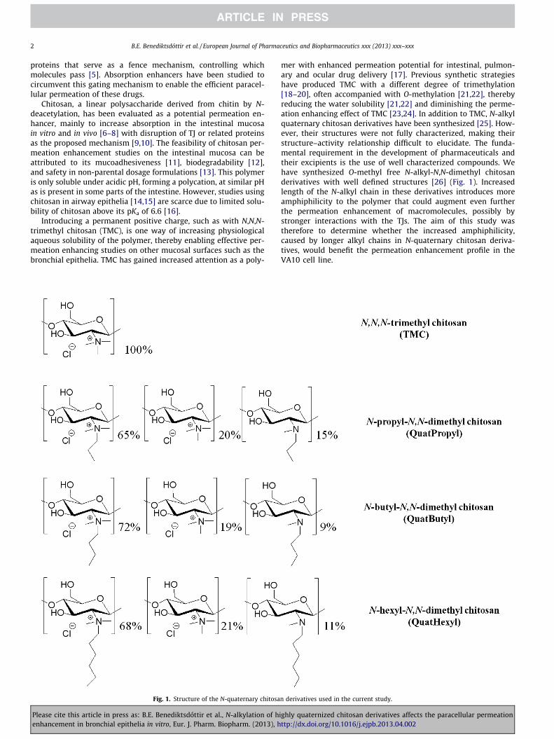
Fig. 1. Structure of the N-quaternary chitosa
Please cite this article in press as: B.E. Benediktsdóttir et al., N-alkylation of henhancement in bronchial epithelia in vitro, Eur. J. Pharm. Biopharm. (2013), h
mer with enhanced permeation potential for intestinal, pulmon-ary and ocular drug delivery [17]. Previous synthetic strategieshave produced TMC with a different degree of trimethylation[18–20], often accompanied with O-methylation [21,22], therebyreducing the water solubility [21,22] and diminishing the perme-ation enhancing effect of TMC [23,24]. In addition to TMC, N-alkylquaternary chitosan derivatives have been synthesized [25]. How-ever, their structures were not fully characterized, making theirstructure–activity relationship difficult to elucidate. The funda-mental requirement in the development of pharmaceuticals andtheir excipients is the use of well characterized compounds. Wehave synthesized O-methyl free N-alkyl-N,N-dimethyl chitosanderivatives with well defined structures [26] (Fig. 1). Increasedlength of the N-alkyl chain in these derivatives introduces moreamphiphilicity to the polymer that could augment even furtherthe permeation enhancement of macromolecules, possibly bystronger interactions with the TJs. The aim of this study wastherefore to determine whether the increased amphiphilicity,caused by longer alkyl chains in N-quaternary chitosan deriva-tives, would benefit the permeation enhancement profile in theVA10 cell line.
n derivatives used in the current study.
ighly quaternized chitosan derivatives affects the paracellular permeationttp://dx.doi.org/10.1016/j.ejpb.2013.04.002

B.E. Benediktsdóttir et al. / European Journal of Pharmaceutics and Biopharmaceutics xxx (2013) xxx–xxx 3
2. Materials and methods
2.1. Chemicals
The chitosan derivatives N,N,N-trimethyl chitosan (TMC), N-propyl-N,N-dimethyl chitosan (QuatPropyl), N-butyl-N,N-dimethylchitosan (QuatButyl), and N-hexyl-N,N-dimethyl chitosan (Quat-Hexyl) were synthesized using highly deacetylated chitosan asstarting material (Mw 8.5 kDa according to a published intrinsicviscometric method [27] and 3% degree of acetylation as deter-mined with 1H NMR analysis [26]). These derivatives were charac-terized with 1H NMR, 13C NMR, 1H–1H COSY, and 1H–13C HSQCusing either Bruker Avance 400 or a Bruker Avance 300 with D2Oas NMR solvent. The characterization has been described in detailin our previous publication [26]. Additionally, reducing-end label-ing of TMC with a fluorophore has enabled the determination of itsMw of 7.7 kDa using 1H NMR, using Bruker Avance 400 with D2O asNMR solvent, and fluorescence spectroscopy [28]. All the deriva-tives were O-methyl free and highly quaternized (85–91%), TMCwas fully trimethylated (100%) [26] as Fig. 1 shows. Prior to use,all the chitosan derivatives were filtered (0.4 lm) under laminarflow and freeze-dried to give semi-aseptic material. FITC–dextran4 kDa (FD4) was obtained from Sigma–Aldrich (St. Louis, USA).Hanks balanced salt solution (HBSS) was prepared as before [4].Mouse monoclonal anti-claudin-4 (329400, clone 3E2C1 IgG1)was obtained from Life Technologies, and mouse anti-b-actin(ab3280, clone ACTN05 (C4), IgG1) was obtained from Abcam(Cambridge, UK). Infrared secondary antibodies for Western blotwere acquired from LiCOR Biosystems. F-actin phallotoxin, To-Pro-3, isotype specific Alexa Fluor secondary antibodies, and 10%SDS Bis–Tris gels were purchased from Life Technologies. Cell cul-ture plastics were obtained from Becton Dickinson (NJ, USA), andTranswell cell culture filters (pore size 0.4 lm, 12 mm diameter,polyester membrane) were obtained from Corning Costar Corpora-tion (through Sigma–Aldrich). Black 96 well microplates were pur-chased from Eppendorf (Hamburg, Germany). The cell culturemedium LHC-9 and Dulbecco’s minimum essential medium: Ham’sF12 1:1 (DMEM/F-12) medium were obtained from Gibco (Burling-ton, Canada) and supplemented with 50 IU/ml penicillin, 50 lg/mlstreptomycin, and 40 lg/ml azithromycin (Pfizer). Ultroser-G ser-um substitute was purchased from Pall Life Sciences (Cergy-Saint-Christophe, France), and bovine serum albumin (BSA) wasobtained from Applichem. TACS� MTT cell proliferation assay,including (4,5-dimethylthiazol-2-yl)-2,5-diphenyltetrazolium bro-mide (MTT) reagent and detergent, was purchased from Trevigen(Gaithersburg, USA).
2.2. Culture of the VA10 bronchial epithelial cell line
The newly established bronchial epithelial cell line, VA10 [29],was used between the passages 15–21 and cultured at air–liquidinterface (ALI) as before [4] and used when the transepithelial elec-trical resistance (TER) was at least 800 X cm2.
2.3. Immunofluorescent staining
After treatment with either HBSS control, TMC, or QuatHexyl(both at 0.25 mg/ml), the ALI cell layers were fixed for 10 min with3.7% (v/v) formaldehyde, permeabilized with 0.1% (v/v) Triton X-100 for 7 min and then blocked (5% v/v goat serum, 0.3% v/v TritonX-100 in PBS) for 10 min. The following primary antibodies wereused: mouse antihuman claudin-4 (IgG1, 1:125) and Alexa Fluor488 phallotoxin for F-actin staining (1:40), diluted in a buffer con-sisting of 0.2% (v/v) Triton X-100, 0.1% (w/v) BSA and 0.05% (v/v)Tween�20 in PBS. Cell layers were incubated with primary anti-
Please cite this article in press as: B.E. Benediktsdóttir et al., N-alkylation of henhancement in bronchial epithelia in vitro, Eur. J. Pharm. Biopharm. (2013), h
bodies overnight at 4 �C. Then, the cells were incubated with iso-type specific Alexa Fluor secondary antibodies (1:1000) andToPro-3 for nuclear staining (1:500), diluted with the same solu-tion as for the primary antibodies, for 30 min.
2.4. Confocal microscopy
Immunofluorescent images were obtained using Zeiss LSM 5Pascal confocal laser scanning microscope (CLSM, Carl Zeiss AG,Munich, Germany) with Plan-Neofluar 40� and Plan-Apochromat63� oil immersion lenses. VA10 cells cultured under ALI conditionswere mounted with coverslips, and Fluoromount-G was added be-fore visualization.
2.5. Western blotting
Cell lysates were obtained after treatment with either HBSScontrol or fully quaternized TMC (0.25 mg/ml), by gently washingthe Transwell filters with icecold PBS that was then completely re-moved. RIPA lysis buffer (20 ll) was then applied to each well andfilters placed on ice for 10 min. The cells were then scraped fromthe filters and sonicated for 2 min followed by centrifugation at12,000g for 20 min at 4 �C. The supernatant was collected andthe protein concentration determined with the Bradford assay.Equal amounts of proteins were separated with 10% SDS Bis–Trisgel followed by transfer onto nitrocellulose membranes. The mem-branes were then blocked with 5% BSA in Tris buffered saline con-taining 0.1% (v/v) Tween�20 for 1 h. Primary antibodies (b-actin at1:2000 or claudin-4 at 1:250) were added to the blocking solutionand incubated overnight at 4 �C. Then, the membrane was rinsedthree times with PBS and subsequently blotted with infrared sec-ondary antibodies (anti-rabbit or anti-mouse at 1:20,000) for 1 hat room temperature and rinsed with PBS before visualization withOdyssey Infrared Image Scanner and the corresponding Image Stu-dio software (LiCOR Biosystems).
2.6. Transepithelial electrical resistance (TER) measurements
The TER of VA10 cells was measured with Millicell-ERS volth-ometer (Millipore, MA, USA) for ALI cultured VA10 cell layers. Priorto the TER studies, the cell layers were washed twice with HBSS(37 �C) and then allowed to equilibrate in the incubator with HBSS(0.5 ml apical and 1.5 ml basolateral) for 30 min followed by base-line TER measurement every 20 min for 1 h. At t = 0, the HBSS waseither replaced with 0.5 ml of the chitosan derivative test solutionand 1.5 ml basolateral HBSS or by 0.5 ml apical and 1.5 ml basolat-eral HBSS as a control. The TER was measured at 0, 20, 40, 60, 80,100, and 120 min after administration. Thereafter, the test solutionwas aspirated, the epithelium rinsed three times with HBSS andthen incubated in culture medium for 24 h to monitor the recoveryof the epithelium. The TER value for every epithelial layer att = �20 min was normalized to 100% to minimize any inter-groupvariation present in the initial TER since t = 0 min was measureddirectly after the addition of the test solutions. Corrected TER valuewas obtained after subtraction of the background from the cell-freeculture insert.
2.7. Transport studies for FD4
Prior to the transport studies, the ALI cultured cell layers werewashed twice with HBSS (37 �C) and then allowed to equilibratein the incubator with HBSS for 1 h. Test solutions were either asep-tic solution of FD4 dissolved in HBSS buffer to a final concentrationof 1.5 mg/ml, as control, or FD4 co-dissolved at 1.5 mg/ml with thechitosan derivative that had the final concentration of 1.0, 0.25,0.063, or 0.016 mg/ml. Before the FD4 transport studies were
ighly quaternized chitosan derivatives affects the paracellular permeationttp://dx.doi.org/10.1016/j.ejpb.2013.04.002
4 B.E. Benediktsdóttir et al. / European Journal of Pharmaceutics and Biopharmaceutics xxx (2013) xxx–xxx
started, the HBSS buffer was aspirated and replaced by 0.52 ml ofprewarmed test solution and 1.5 ml of HBSS at the basolateral side.Immediately, a sample (20 ll) of the test solution was removed todetermine the initial concentration (C0). The cells were incubatedat 37 �C and agitated on an orbital shaker at 80 rpm. Sampling fromthe basolateral compartment (100 ll) was done after 20, 40, 60, 80,and 120 min and replaced with equal volume of fresh HBSS buffer.TER measurements were done at the same time points as thesampling.
2.8. MTT viability assay
For the general cell viability assay, cells were seeded on 96 wellplates at the density of 1.4 � 104 cells per well and maintained inLHC-9 media for 24 h. Then, the medium was aspirated and cellsrinsed with HBSS before incubation with the chitosan derivativesTMC, QuatPropyl, QuatButyl, and QuatHexyl at 1.0, 0.25, 0.063,0.016, 0.004, 0.001, and 0.00025 mg/ml for 2 h at 37 �C. After incu-bation, the test solutions were aspirated and the cells rinsed twicewith HBSS. Directly thereafter, 100 ll of HBSS were introduced toeach well along with 10 ll of the MTT reagent and incubated for4 h, to measure instant effects on viability. To measure late effectson cell viability or cell recovery, the cells were incubated for 24 hwith LHC-9 after the 2 h treatment with the chitosan derivatives,before the addition of MTT reagent as above. Then, the MTT solu-tion was aspired followed by addition of 80 ll of detergentsolution.
For ALI MTT viability assay, confluent VA10 cells cultured onTranswell filters for 30 days under ALI conditions (as described inSection 2.2) were used. The assay was done as previously described[30]. Briefly, the medium was aspirated and cells rinsed twice withHBSS before incubation with the TMC derivative (1.0, 0.25, 0.063,and 0.016 mg/ml) for 2 h at 37 �C. After incubation, the test solu-tions were aspirated and the epithelium rinsed twice with HBSS.Then, 15 ll of MTT solution was introduced to the apical side(0.5 ml) and 45 ll to the basolateral side (1.5 ml) and incubated
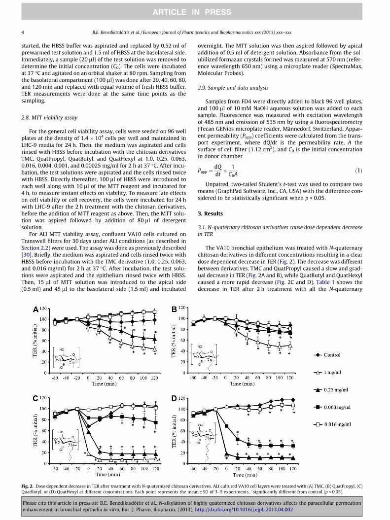
Fig. 2. Dose dependent decrease in TER after treatment with N-quaternized chitosan deriQuatButyl, or (D) QuatHexyl at different concentrations. Each point represents the mean
Please cite this article in press as: B.E. Benediktsdóttir et al., N-alkylation of henhancement in bronchial epithelia in vitro, Eur. J. Pharm. Biopharm. (2013), h
overnight. The MTT solution was then aspired followed by apicaladdition of 0.5 ml of detergent solution. Absorbance from the sol-ubilized formazan crystals formed was measured at 570 nm (refer-ence wavelength 650 nm) using a microplate reader (SpectraMax,Molecular Probes).
2.9. Sample and data analysis
Samples from FD4 were directly added to black 96 well plates,and 100 ll of 10 mM NaOH aqueous solution was added to eachsample. Fluorescence was measured with excitation wavelengthof 485 nm and emission of 535 nm by using a fluorospectrometry(Tecan GENios microplate reader, Männedorf, Switzerland. Appar-ent permeability (Papp) coefficients were calculated from the trans-port experiment, where dQ/dt is the permeability rate, A thesurface of cell filter (1.12 cm2), and C0 is the initial concentrationin donor chamber
Papp ¼dQdt� 1
C0Að1Þ
Unpaired, two-tailed Student’s t-test was used to compare twomeans (GraphPad Software, Inc., CA, USA) with the difference con-sidered to be statistically significant when p < 0.05.
3. Results
3.1. N-quaternary chitosan derivatives cause dose dependent decreasein TER
The VA10 bronchial epithelium was treated with N-quaternarychitosan derivatives in different concentrations resulting in a cleardose dependent decrease in TER (Fig. 2). The decrease was differentbetween derivatives. TMC and QuatPropyl caused a slow and grad-ual decrease in TER (Fig. 2A and B), while QuatButyl and QuatHexylcaused a more rapid decrease (Fig. 2C and D). Table 1 shows thedecrease in TER after 2 h treatment with all the N-quaternary
vatives. ALI cultured VA10 cell layers were treated with (A) TMC, (B) QuatPropyl, (C)± SD of 3–5 experiments, �significantly different from control (p < 0.05).
ighly quaternized chitosan derivatives affects the paracellular permeationttp://dx.doi.org/10.1016/j.ejpb.2013.04.002
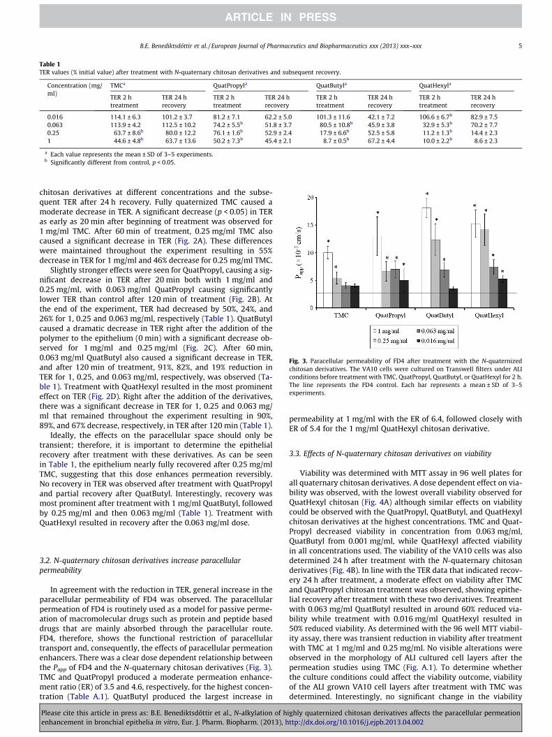
Table 1TER values (% initial value) after treatment with N-quaternary chitosan derivatives and subsequent recovery.
Concentration (mg/ml)
TMCa QuatPropyla QuatButyla QuatHexyla
TER 2 htreatment
TER 24 hrecovery
TER 2 htreatment
TER 24 hrecovery
TER 2 htreatment
TER 24 hrecovery
TER 2 htreatment
TER 24 hrecovery
0.016 114.1 ± 6.3 101.2 ± 3.7 81.2 ± 7.1 62.2 ± 5.0 101.3 ± 11.6 42.1 ± 7.2 106.6 ± 6.7b 82.9 ± 7.50.063 113.9 ± 4.2 112.5 ± 10.2 74.2 ± 5.5b 51.8 ± 3.7 80.5 ± 10.8b 45.9 ± 3.8 32.9 ± 5.3b 70.2 ± 7.70.25 63.7 ± 8.6b 80.0 ± 12.2 76.1 ± 1.6b 52.9 ± 2.4 17.9 ± 6.6b 52.5 ± 5.8 11.2 ± 1.3b 14.4 ± 2.31 44.6 ± 4.8b 63.7 ± 13.6 50.2 ± 7.3b 45.4 ± 2.1 8.7 ± 0.5b 67.2 ± 4.4 10.0 ± 2.2b 8.6 ± 2.3
a Each value represents the mean ± SD of 3–5 experiments.b Significantly different from control, p < 0.05.
Fig. 3. Paracellular permeability of FD4 after treatment with the N-quaternizedchitosan derivatives. The VA10 cells were cultured on Transwell filters under ALIconditions before treatment with TMC, QuatPropyl, QuatButyl, or QuatHexyl for 2 h.The line represents the FD4 control. Each bar represents a mean ± SD of 3–5experiments.
B.E. Benediktsdóttir et al. / European Journal of Pharmaceutics and Biopharmaceutics xxx (2013) xxx–xxx 5
chitosan derivatives at different concentrations and the subse-quent TER after 24 h recovery. Fully quaternized TMC caused amoderate decrease in TER. A significant decrease (p < 0.05) in TERas early as 20 min after beginning of treatment was observed for1 mg/ml TMC. After 60 min of treatment, 0.25 mg/ml TMC alsocaused a significant decrease in TER (Fig. 2A). These differenceswere maintained throughout the experiment resulting in 55%decrease in TER for 1 mg/ml and 46% decrease for 0.25 mg/ml TMC.
Slightly stronger effects were seen for QuatPropyl, causing a sig-nificant decrease in TER after 20 min both with 1 mg/ml and0.25 mg/ml, with 0.063 mg/ml QuatPropyl causing significantlylower TER than control after 120 min of treatment (Fig. 2B). Atthe end of the experiment, TER had decreased by 50%, 24%, and26% for 1, 0.25 and 0.063 mg/ml, respectively (Table 1). QuatButylcaused a dramatic decrease in TER right after the addition of thepolymer to the epithelium (0 min) with a significant decrease ob-served for 1 mg/ml and 0.25 mg/ml (Fig. 2C). After 60 min,0.063 mg/ml QuatButyl also caused a significant decrease in TER,and after 120 min of treatment, 91%, 82%, and 19% reduction inTER for 1, 0.25, and 0.063 mg/ml, respectively, was observed (Ta-ble 1). Treatment with QuatHexyl resulted in the most prominenteffect on TER (Fig. 2D). Right after the addition of the derivatives,there was a significant decrease in TER for 1, 0.25 and 0.063 mg/ml that remained throughout the experiment resulting in 90%,89%, and 67% decrease, respectively, in TER after 120 min (Table 1).
Ideally, the effects on the paracellular space should only betransient; therefore, it is important to determine the epithelialrecovery after treatment with these derivatives. As can be seenin Table 1, the epithelium nearly fully recovered after 0.25 mg/mlTMC, suggesting that this dose enhances permeation reversibly.No recovery in TER was observed after treatment with QuatPropyland partial recovery after QuatButyl. Interestingly, recovery wasmost prominent after treatment with 1 mg/ml QuatButyl, followedby 0.25 mg/ml and then 0.063 mg/ml (Table 1). Treatment withQuatHexyl resulted in recovery after the 0.063 mg/ml dose.
3.2. N-quaternary chitosan derivatives increase paracellularpermeability
In agreement with the reduction in TER, general increase in theparacellular permeability of FD4 was observed. The paracellularpermeation of FD4 is routinely used as a model for passive perme-ation of macromolecular drugs such as protein and peptide baseddrugs that are mainly absorbed through the paracellular route.FD4, therefore, shows the functional restriction of paracellulartransport and, consequently, the effects of paracellular permeationenhancers. There was a clear dose dependent relationship betweenthe Papp of FD4 and the N-quaternary chitosan derivatives (Fig. 3).TMC and QuatPropyl produced a moderate permeation enhance-ment ratio (ER) of 3.5 and 4.6, respectively, for the highest concen-tration (Table A.1). QuatButyl produced the largest increase in
Please cite this article in press as: B.E. Benediktsdóttir et al., N-alkylation of henhancement in bronchial epithelia in vitro, Eur. J. Pharm. Biopharm. (2013), h
permeability at 1 mg/ml with the ER of 6.4, followed closely withER of 5.4 for the 1 mg/ml QuatHexyl chitosan derivative.
3.3. Effects of N-quaternary chitosan derivatives on viability
Viability was determined with MTT assay in 96 well plates forall quaternary chitosan derivatives. A dose dependent effect on via-bility was observed, with the lowest overall viability observed forQuatHexyl chitosan (Fig. 4A) although similar effects on viabilitycould be observed with the QuatPropyl, QuatButyl, and QuatHexylchitosan derivatives at the highest concentrations. TMC and Quat-Propyl decreased viability in concentration from 0.063 mg/ml,QuatButyl from 0.001 mg/ml, while QuatHexyl affected viabilityin all concentrations used. The viability of the VA10 cells was alsodetermined 24 h after treatment with the N-quaternary chitosanderivatives (Fig. 4B). In line with the TER data that indicated recov-ery 24 h after treatment, a moderate effect on viability after TMCand QuatPropyl chitosan treatment was observed, showing epithe-lial recovery after treatment with these two derivatives. Treatmentwith 0.063 mg/ml QuatButyl resulted in around 60% reduced via-bility while treatment with 0.016 mg/ml QuatHexyl resulted in50% reduced viability. As determined with the 96 well MTT viabil-ity assay, there was transient reduction in viability after treatmentwith TMC at 1 mg/ml and 0.25 mg/ml. No visible alterations wereobserved in the morphology of ALI cultured cell layers after thepermeation studies using TMC (Fig. A.1). To determine whetherthe culture conditions could affect the viability outcome, viabilityof the ALI grown VA10 cell layers after treatment with TMC wasdetermined. Interestingly, no significant change in the viability
ighly quaternized chitosan derivatives affects the paracellular permeationttp://dx.doi.org/10.1016/j.ejpb.2013.04.002
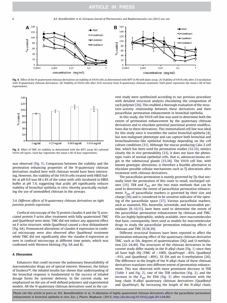
Fig. 4. Effect of the N-quaternized chitosan derivatives on viability in VA10 cells as determined with MTT in 96 well plate assay. (A) Viability of VA10 cells after 2 h incubationwith N-quaternary chitosan derivatives. (B) Viability of VA10 cells after 24 h recovery from N-quaternary chitosan treatment. Each point represents the mean ± SD of fourexperiments.
Fig. 5. Effect of TMC on viability as determined with the MTT assay ALI culturedVA10 cell layers. Each bar represents the mean ± SD of four experiments.
6 B.E. Benediktsdóttir et al. / European Journal of Pharmaceutics and Biopharmaceutics xxx (2013) xxx–xxx
was observed (Fig. 5). Comparison between the viability and thepermeation enhancing properties of the N-quaternary chitosanderivatives studied here with chitosan would have been interest-ing. However, the viability of the VA10 cells treated with HBSS buf-fer at pH 6.0 was 68 ± 8% of the value with cells incubated in HBSSbuffer at pH 7.4, suggesting that acidic pH significantly reducesviability of bronchial epithelia in vitro, thereby practically exclud-ing the use of unmodified chitosan in the airways.
3.4. Different effects of N-quaternary chitosan derivatives on tightjunction protein expression
Confocal microscopy of the TJ protein claudin-4 and the TJ asso-ciated protein F-actin after treatment with fully quaternized TMCand QuatHexyl were done. TMC did not induce any apparent effecton F-actin structure, while QuatHexyl caused a clear disbandment(Fig. 6A). Pronounced alterations of claudin-4 expression in confo-cal microscopy were also observed after QuatHexyl treatmentwhile TMC did not significantly alter the claudin-4 expression asseen in confocal microscopy at different time points, which wasconfirmed with Western blotting (Fig. 6A and B).
4. Discussion
Enhancers that could increase the pulmonary bioavailability ofmacromolecular drugs are of special interest. However, the failureof Exubera™, the inhaled insulin has shown that understanding ofthe bronchial response is fundamental to the success of inhaleddosage forms for systemic delivery. In the current study, weemphasized on the use of well defined polymers and experimentalmodels. All the N-quaternary chitosan derivatives used in the cur-
Please cite this article in press as: B.E. Benediktsdóttir et al., N-alkylation of henhancement in bronchial epithelia in vitro, Eur. J. Pharm. Biopharm. (2013), h
rent study were synthesized according to our previous procedurewith detailed structural analysis elucidating the composition ofeach polymer [26]. This enabled a thorough evaluation of the struc-ture–activity relationship between these derivatives and theirparacellular permeation enhancement in bronchial epithelia.
In this study, the VA10 cell line was used to determine both theextent of permeation enhancement by the quaternary chitosanderivatives and to elucidate potential junctional protein modifica-tions due to these derivatives. This immortalized cell line was idealfor this study since it resembles the native bronchial epithelia [4],has non-malignant phenotype and can capture both bronchial andbronchioalveolar-like epithelial histology depending on the cellculture conditions [31]. Although the mucus producing Calu-3 cellline, which has been used for permeation studies [32,33], mimicsclosely the in vivo permeability [33], it does not have the pheno-typic traits of normal epithelial cells, that is, adenocarcinoma ori-gin in the submucosal glands [33,34]. The VA10 cell line, withknown genotypic alterations, is therefore a feasible alternative toelucidate possible cellular mechanisms such as TJ alterations aftertreatment with chitosan derivatives.
The paracellular permeation is mainly governed by TJs that nor-mally limit the permeation of this route to small, uncharged sol-utes [35]. TER and Papp are the two main methods that can beused to determine the extent of paracellular permeation enhance-ment. Papp of paracellular markers is governed by their size andcharge [36] and is considered to be sensitive indicator of the open-ing of the paracellular space [37]. Various paracellular markers,such as mannitol, FDs, buserelin, octreotide, and horseradish per-oxidases [8–10,15], have been used to determine the extent ofthe paracellular permeation enhancement by chitosan and TMC.FDs are highly hydrophilic, widely available, inert macromoleculesthat have, consequently, been used as models for macromoleculardrugs to study the paracellular permeation enhancing effects ofchitosan and TMC [9,38,39].
Different structural features have been reported to affect thepermeation enhancing effect of the quaternary chitosan derivativeTMC, such as DA, degrees of quaternization (DQ) and O-methyla-tion [22–24,40]. The structures of the chitosan derivatives in thecurrent study differ mainly in the N-alkyl chain (Fig. 1) since theyall have high DQ (TMC of �100%, QuatPropyl �85%, QuatButyl�91%, and QuatHexyl �89%), 3% DA and no O-methylation [26].The difference in the length of the N-alkyl chain of these chitosanderivatives translates into different extent of permeation enhance-ment. This was observed with more prominent decrease in TER(Table 1 and Fig. 2), rate of the TER reduction (Fig. 2), and theincrease in the Papp for FD4 (Fig. 3) after treatment with thelong chain N-alkyl quaternary chitosan derivatives (QuatButyland QuatHexyl). By increasing the length of the N-alkyl chain,
ighly quaternized chitosan derivatives affects the paracellular permeationttp://dx.doi.org/10.1016/j.ejpb.2013.04.002

Fig. 6. Expression of the TJ protein claudin-4 and the TJ associated protein F-actin in ALI cultured VA10 cell layers after treatment with TMC and QuatHexyl. (A) Confocalfluorescent expression of claudin-4 and F-actin after treatment with 0.25 mg/ml TMC and 0.25 mg/ml QuatHexyl. (B) Western blots of claudin-4 after treatment with0.25 mg/ml TMC, with b-actin as loading control. (For interpretation of the references to color in this figure legend, the reader is referred to the web version of this article.)
B.E. Benediktsdóttir et al. / European Journal of Pharmaceutics and Biopharmaceutics xxx (2013) xxx–xxx 7
the polymer becomes more amphiphilic, thereby resembling qua-ternary ammonium surfactants such as cetyltrimethylammoniumbromide, a known permeation enhancer in transdermal drugdelivery [41,42]. The increased amphiphilicity of QuatButyl andQuatHexyl clearly aided in their permeation enhancing properties,since they were as follows: QuatHexyl � QuatButyl > QuatPropyl >TMC at pH 7.4. The TER recovery for QuatButyl was in reverseorder, that is, the epithelia treated with the highest concentrationof QuatButyl (1 mg/ml) showed the highest recovery. The effect ofthe longer N-alkylated chitosan derivatives might be different fromthat of TMC, since concomitant decrease in viability was observedwith increase in the N-alkyl chain length.
Reduced viability can cause increased permeability and de-creased TER and should consequently be studied. Viability assayssuch as MTT, lactate dehydrogenase (LDH), and trypan blue are fre-quently used to evaluate potential toxicity of a new permeation en-hancer. However, these assays are not in agreement when reportingthe effects of chitosan and its derivatives on viability. All trypanblue staining for cytotoxicity of TMC are negative, indicating thatviability is not affected [39,43–45]. Trypan blue only assess if thecell is dead or alive and might not be the appropriate marker to as-sess reduced viability. MTT is a convenient, quantitative high
Please cite this article in press as: B.E. Benediktsdóttir et al., N-alkylation of henhancement in bronchial epithelia in vitro, Eur. J. Pharm. Biopharm. (2013), h
throughput assay that measures metabolic activity of the cell andhas been reported to be more sensitive than neutral red assay andmore reliable than LDH for evaluating acute toxicity of excipientsin the Calu-3 cell line [30]. As opposed to trypan blue staining, thereare disparities in the MTT viability assays for TMC. While TMC (DQ43%) reduced the viability at 0.1 mg/ml in the Caco-2 cell line [24],another study demonstrated that treatment with TMC (DQ 20% and60%) did not result in reduced viability in Calu-3 cells after treat-ment with 15 mg/ml TMC [15]. Interestingly, that study reportedthat acidic buffer (pH 5.5) did not decrease the viability of Calu-3cells, in contrast to our observation and previous studies in Calu-3 [30] and 16HBE14o- [46] bronchial epithelial cells.
The data indicate that fully quaternized TMC at the two highestconcentrations (1 mg/ml and 0.25 mg/ml) reduced viability, withrecovery observed 24 h after treatment in the 96 well plates. Of no-tice, is the fact that no reduction in viability after TMC treatmentwas observed when using the MTT assay on ALI cultured cells. Thisis in contrast to previous observations with Calu-3 cells thatshowed more effect on viability in Transwell filters than in the96 well plates after chitosan treatment [30]. There is, however,marked difference between these two cell lines. The Calu-3 is amucus producing cell line derived from adenocarcinoma [34],
ighly quaternized chitosan derivatives affects the paracellular permeationttp://dx.doi.org/10.1016/j.ejpb.2013.04.002
8 B.E. Benediktsdóttir et al. / European Journal of Pharmaceutics and Biopharmaceutics xxx (2013) xxx–xxx
while the progenitor cell line VA10 was isolated from normal hu-man bronchi [29], does not produce mucus under the current reac-tion conditions, and differentiates toward ciliated cells whencultured under ALI [4]. Consequently, testing the viability in 96well format where the cells are in a more basal cell phenotypecould explain their sensitivity to the quaternary chitosan deriva-tives compared to the ALI culture. The N-quaternary chitosanderivatives decreased viability with increased N-alkyl chain length.This indicates that longer alkyl chain produces both higher degreeof permeation and irreversible adverse effects to the epithelialmembrane.
A part of the aim in the current study was to determine whateffects these chitosan derivatives have on the paracellular space.TJs form a barrier between cells, thereby regulating the paracellu-lar permeability across the cell layer [5]. Studies disagree whetherthe mechanism behind the permeation enhancement lies in thealterations of TJs or TJ associated proteins. F-actin disbandment[9,47] or redistribution [48] has been introduced as a part of thepermeation enhancing properties of chitosan. However, researchfocusing on the effects of TMC on TJ is scarce. No effects on theF-actin structure after TMC treatment were observed in our study,concurring with previous chitosan [38] and TMC studies [49]. Clau-din-4 has been reported to be internalized into lysosomes as theresult of chitosan permeation enhancement, followed by synthesisof claudin-4 after chitosan removal [50]. The expression of claudin-4 in the current study was considered not to be markedly differentfrom control after TMC treatment and co-staining of claudin-4with the early endosomal marker EEA-1 at different time pointsdid not show any co-localization (Fig. A.2). Further observationsup to 24 h after TMC treatment with confocal imaging did notshow any TJ related alterations for F-actin and claudin-4(Figs. A.2 and A.3). Loss in ZO-1 staining and occludin expression[10,48,51] and their possible relocalization to the cytoskeleton[10] has also been attributed to permeation enhancement by chito-san. However, no signs of altered expression of ZO-1, investigatedup to 4 h post TMC treatment in confocal microscopy, were found(Fig. A.3). Expression of occludin or claudin-1, as determined withWestern blotting (Fig. A.4), did not show any marked differencecompared to control.
The gradual decrease in TER accompanied with no apparent ef-fect on TJ relocalization after treatment with TMC compared to therapid decrease in TER and complete dissociation of the TJ proteininvestigated for QuatHexyl also indicates a different mode of ac-tion. It is known that the TJ protrude into the paracellular space,are highly hydrated and contain fixed negative sites [52]. Conse-quently, changes in the ionic concentration in these pores, as withthe cationic TMC, could lead to the delicately modulated displace-ment of the TJ structures leading to their transient ‘‘opening’’ tolarger solutes such as FDs without significantly altering the TJarchitecture. Larger quantities of TMC could, therefore, cause in-creased dissociation of the TJ and other adhesion proteins suchas integrins, leading to cell death due to loss in adhesion (anoikis)[53]. Fluorescently labeled TMC has been reported to be internal-ized into epithelial cells [28], while none or limited internalizationof solubilized chitosan has been reported [9,54,55] inviting furtherspeculations regarding different effects of chitosan and TMC. In-creased N-alkyl chain length in quaternary chitosan could exertits effect both in the paracellular space as with TMC but also onthe lipophilic cell membrane itself, thereby causing severe de-crease in viability.
5. Conclusion
All the N-quaternary chitosan derivatives caused a dose depen-dent decrease in TER and an increase in paracellular permeability
Please cite this article in press as: B.E. Benediktsdóttir et al., N-alkylation of henhancement in bronchial epithelia in vitro, Eur. J. Pharm. Biopharm. (2013), h
of the macromolecular inert marker FD4. Consequently, thesechitosan derivatives, TMC in particular, could be used to increasethe paracellular permeation of other hydrophilic macromoleculessuch as peptide and protein based drugs. The mechanism behindthe permeation enhancing properties of these N-quaternized chito-san derivatives appears to be different, with the fully trimethylatedTMC delicately modulating the effect through the paracellularspace, while the amphiphilic QuatHexyl induced strong effectspossibly mediated through the complete dissociation of all junc-tional complexes in the paracellular space. Our study showed thatincreased amphiphilicity of the longer N-alkyl-N,N-quaternarychitosan derivatives caused increased permeation and TJ disassem-bly, and decreased viability in the order of QuatHexyl � QuatBu-tyl > QuatPropyl > TMC at pH 7.4. These different effects could bemediated by different mechanism; TMC could delicately modulatethe TJ architecture in the paracellular space while the longer N-al-kyl quaternary chitosan derivatives could also perturb the cellmembrane, causing adverse effects.
Acknowledgements
Financial support from the Eimskip Fund of University of Ice-land, the University of Iceland Research Fund and the LandspitaliUniversity Hospital Science Fund is gratefully acknowledged.
Appendix A. Supplementary material
Supplementary data associated with this article can be found, inthe online version, at http://dx.doi.org/10.1016/j.ejpb.2013.04.002.
References
[1] Beyond Borders. Global Biotechnology Report 2012, Ernst & Young. Availablefrom: <http://www.ey.com/Publication/vwLUAssets/Beyond_borders_2012/$FILE/Beyond_borders_2012.pdf>.
[2] J.S. Patton, Mechanisms of macromolecule absorption by the lungs, Adv. DrugDeliv. Rev. 19 (1996) 3–36.
[3] A. Tronde, B. Nordén, H. Marchner, A.-K. Wendel, H. Lennernäs, U.H. Bengtsson,Pulmonary absorption rate and bioavailability of drugs in vivo in rats:structure–absorption relationships and physicochemical profiling of inhaleddrugs, J. Pharm. Sci. 92 (2003) 1216–1233.
[4] B. Benediktsdóttir, A. Arason, S. Halldórsson, T. Gudjónsson, M. Másson, Ó.Baldursson, Drug delivery characteristics of the progenitor bronchial epithelialcell line VA10, Pharm. Res. 30 (2013) 781–791.
[5] K. Shin, V.C. Fogg, B. Margolis, Tight junctions and cell polarity, Annu. Rev. CellDev. Biol. 22 (2006) 207–235.
[6] N.G.M. Schipper, K.M. Vårum, P. Artursson, Chitosans as absorption enhancersfor poorly absorbable drugs. 1: Influence of molecular weight and degree ofacetylation on drug transport across human intestinal epithelial (Caco-2) cells,Pharm. Res. 13 (1996) 1686–1692.
[7] A.F. Kotze, H.L. Luessen, B.J. de Leeuw, B.G. de Boer, J.C. Verhoef, H.E. Junginger,Comparison of the effect of different chitosan salts and N-trimethyl chitosanchloride on the permeability of intestinal epithelial cells (Caco-2), J. Control.Release 51 (1998) 35–46.
[8] M. Thanou, B.I. Florea, M.W.E. Langemeyer, J.C. Verhoef, H.E. Junginger, N-trimethylated chitosan chloride (TMC) improves the intestinal permeation ofthe peptide drug buserelin in vitro (Caco-2 cells) and in vivo (rats), Pharm. Res.17 (2000) 27–31.
[9] N.G.M. Schipper, S. Olsson, J.A. Hoogstraate, A.G. deBoer, K.M. Vårum, P.Artursson, Chitosans as absorption enhancers for poorly absorbable drugs 2:mechanism of absorption enhancement, Pharm. Res. 14 (1997) 923–929.
[10] J. Smith, E. Wood, M. Dornish, Effect of chitosan on epithelial cell tightjunctions, Pharm. Res. 21 (2004) 43–49.
[11] C.-M. Lehr, J.A. Bouwstra, E.H. Schacht, H.E. Junginger, In vitro evaluation ofmucoadhesive properties of chitosan and some other natural polymers, Int. J.Pharm. 78 (1992) 43–48.
[12] H. Onishi, Y. Machida, Biodegradation and distribution of water-solublechitosan in mice, Biomaterials 20 (1999) 175–182.
[13] P. Baldrick, The safety of chitosan as a pharmaceutical excipient, Regul.Toxicol. Pharmacol. 56 (2010) 290–299.
[14] L. Illum, N.F. Farraj, S.S. Davis, Chitosan as a novel nasal delivery system forpeptide drugs, Pharm. Res. 11 (1994) 1186–1189.
[15] B.I. Florea, M. Thanou, H.E. Junginger, G. Borchard, Enhancement of bronchialoctreotide absorption by chitosan and N-trimethyl chitosan shows linearin vitro/in vivo correlation, J. Control. Release 110 (2006) 353–361.
ighly quaternized chitosan derivatives affects the paracellular permeationttp://dx.doi.org/10.1016/j.ejpb.2013.04.002

B.E. Benediktsdóttir et al. / European Journal of Pharmaceutics and Biopharmaceutics xxx (2013) xxx–xxx 9
[16] M.W. Anthonsen, O. Smidsrod, Hydrogen-ion titration of chitosans withvarying degrees of N-acetylation by monitoring induced H-1-NMR chemical-shifts, Carbohydr. Polym. 26 (1995) 303–305.
[17] J.K. Sahni, S. Chopra, F.J. Ahmad, R.K. Khar, Potential prospects of chitosanderivative trimethyl chitosan chloride (TMC) as a polymeric absorptionenhancer: synthesis, characterization and applications, J. Pharm. Pharmacol.60 (2008) 1111–1119.
[18] R.A.A. Muzzarelli, F. Tanfani, The N-permethylation of chitosan and thepreparation of N-trimethyl chitosan iodide, Carbohydr. Polym. 5 (1985) 297–307.
[19] C.H. Kim, J.W. Choi, H.J. Chun, K.S. Choi, Synthesis of chitosan derivatives withquaternary ammonium salt and their antibacterial activity, Polym. Bull. 38(1997) 387–393.
[20] P. Ledung, M. Milas, M. Rinaudo, J. Desbrieres, Water-soluble derivativesobtained by controlled chemical modifications of chitosan, Carbohydr. Polym.24 (1994) 209–214.
[21] A.B. Sieval, M. Thanou, A.F. Kotze, J.E. Verhoef, J. Brussee, H.E. Junginger,Preparation and NMR characterization of highly substituted N-trimethylchitosan chloride, Carbohydr. Polym. 36 (1998) 157–165.
[22] A. Polnok, G. Borchard, J.C. Verhoef, N. Sarisuta, H.E. Junginger, Influence ofmethylation process on the degree of quaternization of N-trimethyl chitosanchloride, Eur. J. Pharm. Biopharm. 57 (2004) 77–83.
[23] R.J. Verheul, M. Amidi, S. van der Wal, E. van Riet, W. Jiskoot, W.E. Hennink,Synthesis, characterization and in vitro biological properties of O-methyl freeN,N,N-trimethylated chitosan, Biomaterials 29 (2008) 3642–3649.
[24] R.J. Verheul, M. Amidi, M.J. van Steenbergen, E. van Riet, W. Jiskoot, W.E.Hennink, Influence of the degree of acetylation on the enzymatic degradationand in vitro biological properties of trimethylated chitosans, Biomaterials 30(2009) 3129–3135.
[25] A.M.M. Sadeghi, F.A. Dorkoosh, M.R. Avadi, M. Weinhold, A. Bayat, F. Delie, R.Gurny, B. Larijani, M. Rafiee-Tehrani, H.E. Junginger, Permeation enhancereffect of chitosan and chitosan derivatives: comparison of formulations assoluble polymers and nanoparticulate systems on insulin absorption in Caco-2cells, Eur. J. Pharm. Biopharm. 70 (2008) 270–278.
[26] B.E. Benediktsdottir, V.S. Gaware, O.V. Runarsson, S. Jonsdottir, K.J. Jensen, M.Masson, Synthesis of N, N, N-trimethyl chitosan homopolymer and highlysubstituted N-alkyl-N, N-dimethyl chitosan derivatives with the aid of di–tert-butyldimethylsilyl chitosan, Carbohydr. Polym. 86 (2011) 1451–1460.
[27] M.H. Ottoy, K.M. Varum, O. Smidsrod, Compositional heterogeneity ofheterogeneously deacetylated chitosans, Carbohydr. Polym. 29 (1996) 17–24.
[28] B.E. Benediktsdóttir, K.K. Sørensen, M.B. Thygesen, K.J. Jensen, T. Gudjónsson,Ó. Baldursson, M. Másson, Regioselective fluorescent labeling of N, N, N-trimethyl chitosan via oxime formation, Carbohydr. Polym. 90 (2012) 1273–1280.
[29] S. Halldorsson, V. Asgrimsson, I. Axelsson, G.H. Gudmundsson, M.Steinarsdottir, O. Baldursson, T. Gudjonsson, Differentiation potential of abasal epithelial cell line established from human bronchial explant, In VitroCell. Dev. – Anim. 43 (2007) 283–289.
[30] R. Scherließ, The MTT assay as tool to evaluate and compare excipient toxicityin vitro on respiratory epithelial cells, Int. J. Pharm. 411 (2011) 98–105.
[31] S. Franzdottir, I. Axelsson, A. Arason, O. Baldursson, T. Gudjonsson, M.Magnusson, Airway branching morphogenesis in three dimensional culture,Respir. Res. 11 (2010) 162.
[32] K.A. Foster, M.L. Avery, M. Yazdanian, K.L. Audus, Characterization of the Calu-3 cell line as a tool to screen pulmonary drug delivery, Int. J. Pharm. 208 (2000)1–11.
[33] N.R. Mathias, J. Timoszyk, P.I. Stetsko, J.R. Megill, R.L. Smith, D.A. Wall,Permeability characteristics of Calu-3 human bronchial epithelial cells:in vitro–in vivo correlation to predict lung absorption in rats, J. Drug Target.10 (2002) 31–40.
[34] J. Fogh, J.M. Fogh, T. Orfeo, 127 Cultured human tumor-cell lines producingtumors in nude mice, J. Natl. Cancer I (59) (1977) 221–226.
[35] C.J. Watson, M. Rowland, G. Warhurst, Functional modeling of tight junctionsin intestinal cell monolayers using polyethylene glycol oligomers, Am. J.Physiol. – Cell Physiol. 281 (2001) C388–C397.
Please cite this article in press as: B.E. Benediktsdóttir et al., N-alkylation of henhancement in bronchial epithelia in vitro, Eur. J. Pharm. Biopharm. (2013), h
[36] S. Lu, A.W. Gough, W.F. Bobrowski, B.H. Stewart, Transport properties are notaltered across Caco-2 cells with heightened TEER despite underlyingphysiological and ultrastructural changes, J. Pharm. Sci. 85 (1996) 270–273.
[37] S.Y. Yee, In vitro permeability across Caco3 cells (colonic) can predict in vivo(small intestinal) absorption in man – fact or myth, Pharm. Res. 14 (1997)763–766.
[38] R. Rosenthal, D. Günzel, C. Finger, S.M. Krug, J.F. Richter, J.-D. Schulzke, M.Fromm, S. Amasheh, The effect of chitosan on transcellular and paracellularmechanisms in the intestinal epithelial barrier, Biomaterials 33 (2012) 2791–2800.
[39] A.F. Kotze, M.M. Thanou, H.L. Luessen, B.G. de Boer, J.C. Verhoef, H.E. Junginger,Effect of the degree of quaternization of N-trimethyl chitosan chloride on thepermeability of intestinal epithelial cells (Caco-2), Eur. J. Pharm. Biopharm. 47(1999) 269–274.
[40] C. Jonker, J.H. Hamman, A.F. Kotze, Intestinal paracellular permeationenhancement with quaternised chitosan: in situ and in vitro evaluation, Int.J. Pharm. 238 (2002) 205–213.
[41] P. Ashton, K.A. Walters, K.R. Brain, J. Hadgraft, Surfactant effects inpercutaneous absorption I. Effects on the transdermal flux of methylnicotinate, Int. J. Pharm. 87 (1992) 261–264.
[42] A. Nokhodchi, J. Shokri, A. Dashbolaghi, D. Hassan-Zadeh, T. Ghafourian, M.Barzegar-Jalali, The enhancement effect of surfactants on the penetration oflorazepam through rat skin, Int. J. Pharm. 250 (2003) 359–369.
[43] A.F. Kotze, M.M. Thanou, H.L. Luebetaen, A.G. de Boer, J.C. Verhoef, H.E.Junginger, Enhancement of paracellular drug transport with highlyquaternized N-trimethyl chitosan chloride in neutral environments: in vitroevaluation in intestinal epithelial cells (Caco-2), J. Pharm. Sci. 88 (1999) 253–257.
[44] A.F. Kotze, H.L. Luessen, B.J. deLeeuw, B.G. deBoer, J.C. Verhoef, H.E. Junginger,N-trimethyl chitosan chloride as a potential absorption enhancer acrossmucosal surfaces: In vitro evaluation in intestinal epithelial cells (Caco-2),Pharm. Res. 14 (1997) 1197–1202.
[45] M.M. Thanou, A.F. Kotze, T. Scharringhausen, H.L. Luessen, A.G. de Boer, J.C.Verhoef, H.E. Junginger, Effect of degree of quaternization of N-trimethylchitosan chloride for enhanced transport of hydrophilic compounds acrossintestinal Caco-2 cell monolayers, J. Control. Release 64 (2000) 15–25.
[46] L. Kudsiova, M.J. Lawrence, A comparison of the effect of chitosan andchitosan-coated vesicles on monolayer integrity and permeability across Caco-2 and 16HBE14o-cells, J. Pharm. Sci. 97 (2008) 3998–4010.
[47] P. Artursson, T. Lindmark, S.S. Davis, L. Illum, Effect of chitosan on thepermeability of monolayers of intestinal epithelial-cells (Caco-2), Pharm. Res.11 (1994) 1358–1361.
[48] V. Dodane, M. Amin Khan, J.R. Merwin, Effect of chitosan on epithelialpermeability and structure, Int. J. Pharm. 182 (1999) 21–32.
[49] F.A. Dorkoosh, C.A.N. Broekhuizen, G. Borchard, M. Rafiee-Tehrani, J.C. Verhoef,H.E. Junginger, Transport of octreotide and evaluation of mechanism ofopening the paracellular tight junctions using superporous hydrogel polymersin Caco-2 cell monolayers, J. Pharm. Sci. 93 (2004) 743–752.
[50] T.-H. Yeh, L.-W. Hsu, M.T. Tseng, P.-L. Lee, K. Sonjae, Y.-C. Ho, H.-W. Sung,Mechanism and consequence of chitosan-mediated reversible epithelial tightjunction opening, Biomaterials 32 (2011) 6164–6173.
[51] D. Vllasaliu, R. Exposito-Harris, A. Heras, L. Casettari, M. Garnett, L. Illum, S.Stolnik, Tight junction modulation by chitosan nanoparticles: comparisonwith chitosan solution, Int. J. Pharm. 400 (2010) 183–193.
[52] J.L. Madara, Intestinal absorptive cell tight junctions are linked to cytoskeleton,Am. J. Physiol. – Cell Physiol. 253 (1987) C171–C175.
[53] S. Frisch, H. Francis, Disruption of epithelial cell–matrix interactions inducesapoptosis, J. Cell Biol. 124 (1994) 619–626.
[54] X. Jia, X. Chen, Y. Xu, X. Han, Z. Xu, Tracing transport of chitosan nanoparticlesand molecules in Caco-2 cells by fluorescent labeling, Carbohydr. Polym. 78(2009) 323–329.
[55] M. Huang, E. Khor, L.-Y. Lim, Uptake and cytotoxicity of chitosan moleculesand nanoparticles: effects of molecular weight and degree of deacetylation,Pharm. Res. 21 (2004) 344–353.
ighly quaternized chitosan derivatives affects the paracellular permeationttp://dx.doi.org/10.1016/j.ejpb.2013.04.002