mycobacteria || antibiotics and antibiotic resistance in mycobacteria
TRANSCRIPT
1 Introduction, 2872 The adversaries, 2873 The weapons and how they act, 288
3.1 Antitubercular agents: thefront-line drugs, 288
3.2 Other antibiotics used fortreatment of mycobacterialinfections, 297
4 Other factors leading to resistance, 301
287
Chapter 15 / Antibiotics* and antibiotic resistance in mycobacteria
VERA WEBB & JULIAN DAVIES
4.1 Hypermutability, 3014.2 Compensatory mutations, 302
5 Conclusions and afterthoughts, 3026 Acknowledgements, 3037 References, 303
1 Introduction
Mycobacteria have, unquestionably, played a signifi-cant role in human disease throughout history andsuch infections have been a considerable force in the social history of humankind. The availability ofeffective antibiotic therapy in the late 1940s and early1950s had a significant impact on morbidity and mor-tality due to Mycobacterium tuberculosis infection andhad dramatic social implications in the industrializedand developing worlds. Regrettably, mycobacteriahave presented unusual problems during and sincethe antibiotic era (since 1945). The early and dra-matic success of streptomycin in the treatment oftuberculosis, followed by the introduction of other highly effective drugs (isoniazid, pyrazinamide,ethambutol, rifampin) suggested that effective,worldwide control of mycobacterial infections wouldbe guaranteed. However, the development of anti-biotic resistance intervened in this anticipatedsuccess. The result has been initial encouragementfollowed by subsequent (and recent) treatmentfailure on a widespread basis with the unanticipatedre-emergence of tuberculosis and other mycobacter-ial diseases in industrialized nations. Resistance to
antimycobacterial drugs has played a key role in thisrevanche although other factors such as changes insocial behaviour, and failures in the delivery andmaintenance of effective health care have con-tributed. At present, there is no antibiotic treatmentthat is free of the problem of resistance development.Recent reviews of antibiotic resistance in mycobacte-ria have been published by Jarlier and Nikaido(1994), Musser (1995), Blanchard (1996), Nolan(1997) and Rattan et al. (1998).
In this review, we will focus primarily on antitu-bercular agents but will include information on treat-ments for leprosy, M. avium complex (MAC) diseaseand other ailments caused by mycobacterial speciesin humans. While we have tried to put the problem of antibiotic resistance in mycobacteria in historicalcontext, we have devoted most of our attention toreports published between 1994 and 1998.
2 The adversaries
Other chapters in this volume discuss the structureand physiology of mycobacteria and the ways inwhich these unique characteristics affect antibioticsusceptibility. The mycobacteria are divided into twotypes, namely the fast-growers and the slow-growers.These two groups have different virulence/patho-genicity profiles and different susceptibilities to a
*In this review, ‘antibiotic’ refers to any ‘antimicrobial’agent, whether it be naturally occurring or synthetic.
Mycobacteria: Molecular Biology and Virulence Edited by colin Ratledge and Jeremy Dale
© 1999 Blackwell Science Ltd. ISBN: 978-0-632-05304-9
288 Chapter 15
range of antimicrobial agents; hence, different drugtherapies are recommended and different types ofresistance have developed.
A frequently used minimal definition of multidrug-resistant (Mdr) M. tuberculosis, is resistance to, at theleast, both rifampicin and isoniazid. Strains havebeen isolated from many sites in the United States(Bifani et al. 1996) that are resistant to rifampicin andisoniazid plus pyrazinamide and streptomycin. In arecent study of new cases of tuberculosis in theUnited States, it was found that about 13% wereresistant to at least one of the five front-line drugs(Bloch et al. 1994). Outbreaks of Mdr tuberculosishave been reported in Italy, France, England, Spain,Argentina and Mozambique (Caugant et al. 1995;Nolan 1997).
In general, mycobacteria are intrinsically resistantto many of the commonly used antibiotics; however,there is considerable variation in the susceptibility ofmycobacteria to different antibiotics. For example, M. tuberculosis is quite resistant to many drugs that are active against other mycobacterial strains, butshows almost unique and exquisite susceptibility to isoniazid, an agent which is ineffective againstother mycobacteria, such as M. smegmatis. Similarly,M. tuberculosis and M. bovis are susceptible to anotherfront-line drug, rifampicin, while M. smegmatis andMAC are recalcitrant. The genetic basis for this varia-tion is unclear, although it has been the subject ofmuch discussion (Deretic et al. 1996). These differ-ences in susceptibility have several biochemicalexplanations, such as:1 the composition and structure of the mycobacterialcell wall preventing the uptake of many types of molecules;2 the existence of multisubstrate efflux pumps;3 unique metabolic functions that create specificdrug susceptibilities; and4 chromosomally encoded resistance to certainclasses of antibiotics (b-lactams, aminoglycosides).
While M. tuberculosis and other mycobacteria dopossess chromosomal genes that encode resistance by drug inactivation to certain classes of antibiotics,there is no evidence to suggest that antibiotic resis-tance in mycobacteria is the result of anything otherthan mutational alteration of the appropriate chro-
mosomal gene. Multidrug resistance is the result of the accumulation of independent mutations andmany such mutations have been isolated and exam-ined (see below). Furthermore, the fact that there isno evidence for acquired, plasmid-mediated, antibi-otic resistance in mycobacteria sets them apart fromall other bacterial pathogens.
3 The weapons and how they act
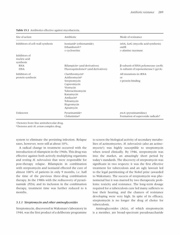
Table 15.1 summarizes the drugs that are cur-rently available for the treatment of mycobacterial infections.
3.1 Antitubercular agents: the front-line drugs
Today, the standard ‘short-course’ therapy for tuber-culosis involves the treatment of patients with a four-drug combination of rifampicin, isoniazid,ethambutol, and pyrazinamide for 2 months, fol-lowed by treatment with a combination of rifampicinand isoniazid for an additional 4 months. Whilemultidrug therapy appears unusual when compared to the treatment of other infectious diseases such as ‘strep throat’ or gonorrhea, this approach has been the rule for tuberculosis since the 1950s.Streptomycin is not part of this ‘short course’, but it isstill widely used and considered one of the front-linedrugs. When first used against tuberculosis in the1940s, streptomycin was administered alone, but it was commonly found that within 3 months 80% of patients were harbouring streptomycin-resistantorganisms. Grosset has recently reviewed the historyof chemotherapy for cavitary pulmonary tuberculosis(Grosset 1996) and notes that, given the rapid devel-opment of streptomycin resistance, there was nohope of curing tuberculosis with any single anti-microbial agent. Subsequent treatment schemes indicated that suppression of resistant mutants waspossible if patients were cotreated with p-aminosali-cylic acid (PAS) together with isoniazid, and so by themid-1950s combination therapy became the rule.Because a small number of drug-sensitive organismssurvived even this three-drug regimen, 18–24months of treatment were necessary for the immune
Antibiotic resistance 289
system to eliminate the persisting infection. Relapserates, however, were still at about 10%.
A radical change in treatment occurred with theintroduction of rifampicin in the 1960s. This drug waseffective against both actively multiplying organismsand resting M. tuberculosis that were responsible forpost-therapy relapse. Rifampicin in combinationwith streptomycin and isoniazid effected the cure ofalmost 100% of patients in only 9 months, i.e. halfthe time of the previous three-drug combinationtherapy. In the 1980s with the rediscovery of pyrazi-namide (PZA) and its inclusion in the combinationtherapy, treatment time was further reduced to 6months.
3.1.1 Streptomycin and other aminoglycosides
Streptomycin, discovered in Waksman’s laboratory in1944, was the first product of a deliberate programme
to screen the biological activity of secondary metabo-lites of actinomycetes. M. tuberculosis (also an actino-mycete!) was highly susceptible to streptomycinwhen tested clinically. By 1946, streptomycin wasinto the market, an amazingly short period by today’s standards. The discovery of streptomycin wassignificant in two respects: it was the first effectivetreatment for tuberculosis and an ugly lawsuit led to the legal partitioning of the Nobel prize (awardedto Waksman). The success of streptomycin was phe-nomenal but it was marred by two therapeutic prob-lems: toxicity and resistance. The long-term dosagerequired for a tuberculosis cure led many sufferers tolose their hearing, and the chances of resistancedeveloping were very high. In spite of its efficacy,streptomycin is no longer the drug of choice fortuberculosis.
Aminoglycosides (AGs), of which streptomycin is a member, are broad-spectrum pseudosaccharide
Table 15.1 Antibiotics effective against mycobacteria.
Site of action Antibiotic Mode of resistance
Inhibitors of cell-wall synthesis Isoniazida (ethionamide) inhA, katG (mycolic acid synthesis)Ethambutola,b embBD-cycloserine D-alanine racemase
Inhibitors ofnucleic acidsynthesis
RNA Rifampicina (and derivatives) b-subunit of RNA polymerase (rpoB)DNA Fluoroquinolonesb (and derivatives) A-subunit of topoisomerase I (gyrA)
Inhibitors of Clarithromycinb All mutations in rRNAprotein synthesis Azithromycinb or
Streptomycin r-protein bindingCapreomycinViomycinTuberactinomycinKanamycinAmikacinb
TobramycinHygromycinApramycin
Unknown Pyrazinamidea pncA (pyrazinamidase)Clofazimineb Formation of superoxide radicals?
a Denotes front-line antitubercular drug.b Denotes anti-M. avium complex drug.
290 Chapter 15
antibiotics, composed of three or more cyclitol units.They bind to the 30S subunit of the ribosome andinterfere with the transition from the initiationcomplex to the elongation complex; they also disruptthe decoding process. In most bacterial pathogens,resistance to aminoglycosides is due to the acquisi-tion of aminoglycoside-inactivating enzymes (Shawet al. 1993), and target-site mutations are rare. Mostfast-growing bacteria have multiple copies of therRNA genes, and because resistance is geneticallyrecessive to antibiotic sensitivity, only rare mutations,in the gene for protein S12, have been isolated undernormal situations with such bacteria. DominantrRNA mutants can be established in strains carryingmultiple copies of the resistant allele (on a plasmid).Mutations altering one or more ribosomal proteinshave been identified in laboratory studies and rarelyin clinical isolates of pathogens such as Neisseria gonorrhoeae. However, because the slow-growingmycobacteria possess only single copies of the rRNAgenes, resistance to streptomycin and other AGs canarise by mutational alteration of either 16S rRNA orribosomal protein S12. Mutations in these ribosomalcomponents are the only types of aminoglycosideresistance found clinically in mycobacteria (Finken
et al. 1993). Recent work by Puglisi and coworkers(Fourmy et al. 1996) has defined the interactionbetween paromomycin (an aminoglycoside that hada brief period as a potential antitubercular agent) andits rRNA target in precise molecular terms. Furtheranalysis of this interaction in concert with increasingknowledge of the working of the ribosome in transla-tion should define the action of this class of drugs.
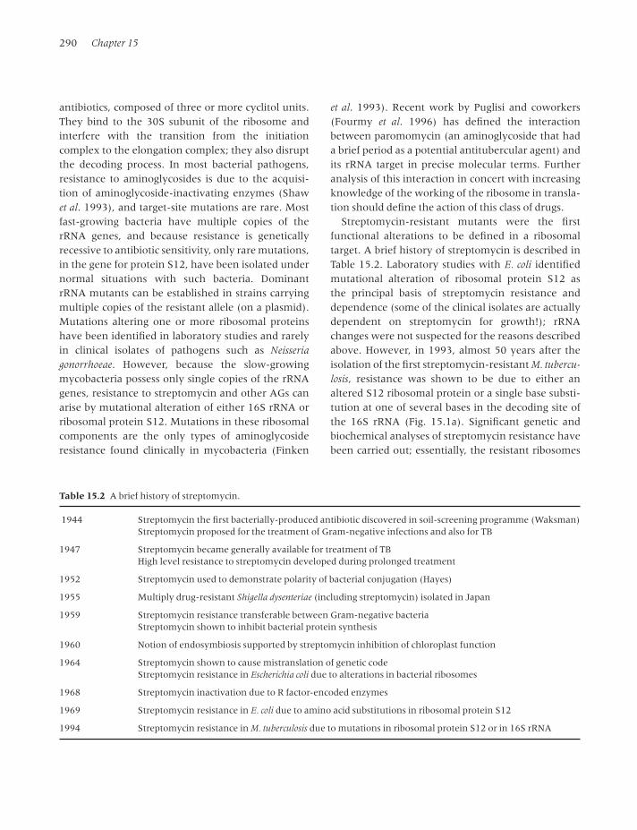
Streptomycin-resistant mutants were the firstfunctional alterations to be defined in a ribosomaltarget. A brief history of streptomycin is described inTable 15.2. Laboratory studies with E. coli identifiedmutational alteration of ribosomal protein S12 as the principal basis of streptomycin resistance anddependence (some of the clinical isolates are actuallydependent on streptomycin for growth!); rRNAchanges were not suspected for the reasons describedabove. However, in 1993, almost 50 years after theisolation of the first streptomycin-resistant M. tubercu-losis, resistance was shown to be due to either analtered S12 ribosomal protein or a single base substi-tution at one of several bases in the decoding site ofthe 16S rRNA (Fig. 15.1a). Significant genetic andbiochemical analyses of streptomycin resistance havebeen carried out; essentially, the resistant ribosomes
Table 15.2 A brief history of streptomycin.
1944 Streptomycin the first bacterially-produced antibiotic discovered in soil-screening programme (Waksman)Streptomycin proposed for the treatment of Gram-negative infections and also for TB
1947 Streptomycin became generally available for treatment of TBHigh level resistance to streptomycin developed during prolonged treatment
1952 Streptomycin used to demonstrate polarity of bacterial conjugation (Hayes)
1955 Multiply drug-resistant Shigella dysenteriae (including streptomycin) isolated in Japan
1959 Streptomycin resistance transferable between Gram-negative bacteriaStreptomycin shown to inhibit bacterial protein synthesis
1960 Notion of endosymbiosis supported by streptomycin inhibition of chloroplast function
1964 Streptomycin shown to cause mistranslation of genetic codeStreptomycin resistance in Escherichia coli due to alterations in bacterial ribosomes
1968 Streptomycin inactivation due to R factor-encoded enzymes
1969 Streptomycin resistance in E. coli due to amino acid substitutions in ribosomal protein S12
1994 Streptomycin resistance in M. tuberculosis due to mutations in ribosomal protein S12 or in 16S rRNA
Antibiotic resistance 291
· ·
UACGGAGGGUGCAA
GGCGGA GACCGCCU CU
AU C
A C AAAAC GG
20592058
20572611
C GC GC GG CG CG CA CC UC GU G
A
ACAUAA
GCUGAUCUUCCCG
CGGUUGGUAGGGU
2500U
UU·
GG
A
A
CC
CC
GG
G
GG
UUU
U
A
G CU GC GG CA U
·2580
2590
UAU
A
CA G
UU
UG
GC
C
CC
A A
AAA
GCCCGUCACGUCAU
CA
GCU
AGCAGGG
AU 1480
1473 vicB (E. coli ; 1491)(M. tbc ; 1483)
AG
1470
1395
1385
nek 1387
nek 1389(E. coli ; 1408)(M. tbc ; 1400)
AGUUUGAGAA
5‘
10
G
U
AG
CA
C
A U U G
920GCUCA
UC
GU
AA
UA
UU
AU
AG
CC
GG
CU
30G
GUGCCUCCCACG
540AA
U
GGAA
UAU
AA
AC
GG/A
915region
G A
U
G
C
U
A
G
A AA
A C890
G
U
G
U
C
G
C
G
G
AAC
900
GCCUGG
CGGGUC
880570
AAUG
GC
GCCG
A
A
GC
CC
530520
560
U
U/C U530loop
(a)
··
(b) (c)
···
·
·
G/C/UC
UCGG
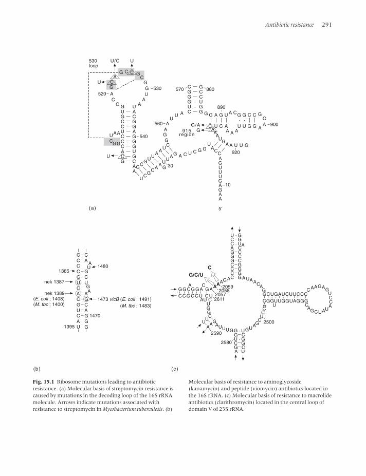
Fig. 15.1 Ribosome mutations leading to antibioticresistance. (a) Molecular basis of streptomycin resistance iscaused by mutations in the decoding loop of the 16S rRNAmolecule. Arrows indicate mutations associated withresistance to streptomycin in Mycobacterium tuberculosis. (b)
Molecular basis of resistance to aminoglycoside(kanamycin) and peptide (viomycin) antibiotics located inthe 16S rRNA. (c) Molecular basis of resistance to macrolideantibiotics (clarithromycin) located in the central loop ofdomain V of 23S rRNA.
292 Chapter 15
either fail to bind or bind the drug weakly (Sander et al. 1996).
In general, the aminoglycosides (apart fromstreptomycin) are rarely used in tuberculosis treat-ment because the (necessarily) long treatment periodleads to significant side-effects. An exception to this isfound in Japan where kanamycin has been used forthe treatment of tuberculosis. Interestingly, muta-tional alteration of the ribosome rarely gives rise tokanamycin resistance in the laboratory. However,resistance in M. smegmatis is due to an AÆG transitionat position 1389 (equivalent to 1408 of Escherichiacoli) in the 16S rRNA; high-level resistant M. tubercu-losis strains had an AÆG transition at position 1400(equivalent to A1389 in M. smegmatis; see Fig. 15.1b)(Taniguchi et al. 1997). The AÆG transition at position 1400 has been reported to confer high-level resistance in a majority of clinical isolates of M.tuberculosis, M. abscessus and M. chelonae resistant toamikacin, kanamycin and other 2-deoxystreptamineaminoglycoside antibiotics (Alangaden et al. 1998;Prammananan et al. 1998).
While clinical resistance to streptomycin in M.tuberculosis is mediated by target-site mutation (seeabove), the presence of aminoglycoside-modifyingenzymes in the genus as a whole has been widelyreported (Mitsuhashi et al. 1977; Udou et al. 1986,1987). More recently Aínsa et al. (1996, 1997), usingDNA hybridization, have identified genes encodingaminoglycoside 2¢-N-acetyltransferase aac(2¢) in allmycobacterial species tested, including M. tuberculosis.The aac2¢ enzyme confers resistance to gentamicin,tobramycin, dibekacin, netilmicin, 2¢-N-ethyl-netilmicin and 6¢-N-ethyl-netilmicin. When M. smegmatis mc2155 was transformed with plasmidscarrying the aac(2¢)-lb gene from M. fortuitum or theaac(2¢)-ld from M. smegmatis mc2155, the minimuminhibitory concentrations (MICs) for these aminogly-cosides increased by two- to 60-fold. Aínsa et al.(1997) concluded from their phylogenetic analysis ofthe aac(2¢) genes, that every mycobacterial speciesappears to have a specific aminoglycoside 2¢-N-acetyltransferase. Nonetheless, the function of theseenzymes remains unclear. When the aac(2¢) gene wasdisrupted in M. smegmatis mc2155, the culture became
more susceptible to lysozyme treatment, leading tothe suggestion that the aac(2¢) enzyme may beinvolved with the acetylation of cell-wall compo-nents. This idea is supported by an earlier observation(Udou et al. 1989) that amino sugars and derivativesof coenzyme A inhibited aminoglycoside acetyl trans-ferase activity from cultures of M. fortuitum.
3.1.2 Rifamycin
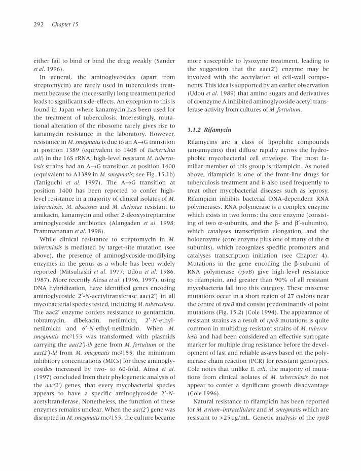
Rifamycins are a class of lipophilic compounds (ansamycins) that diffuse rapidly across the hydro-phobic mycobacterial cell envelope. The most fa-miliar member of this group is rifampicin. As noted above, rifampicin is one of the front-line drugs fortuberculosis treatment and is also used frequently totreat other mycobacterial diseases such as leprosy.Rifampicin inhibits bacterial DNA-dependent RNApolymerases. RNA polymerase is a complex enzymewhich exists in two forms: the core enzyme (consist-ing of two a-subunits, and the b- and b¢-subunits),which catalyses transcription elongation, and theholoenzyme (core enzyme plus one of many of the ssubunits), which recognizes specific promoters andcatalyses transcription initiation (see Chapter 4).Mutations in the gene encoding the b-subunit ofRNA polymerase (rpoB) give high-level resistance to rifampicin, and greater than 90% of all resistantmycobacteria fall into this category. These missensemutations occur in a short region of 27 codons nearthe centre of rpoB and consist predominantly of pointmutations (Fig. 15.2) (Cole 1994). The appearance ofresistant strains as a result of rpoB mutations is quitecommon in multidrug-resistant strains of M. tubercu-losis and had been considered an effective surrogatemarker for multiple drug resistance before the devel-opment of fast and reliable assays based on the poly-merase chain reaction (PCR) for resistant genotypes.Cole notes that unlike E. coli, the majority of muta-tions from clinical isolates of M. tuberculosis do notappear to confer a significant growth disadvantage(Cole 1996).
Natural resistance to rifampicin has been reportedfor M. avium–intracellulare and M. smegmatis which areresistant to >25µg/mL. Genetic analysis of the rpoB
Antibiotic resistance 293
genes from these organisms indicates that they are of the susceptible type. This intrinsic resistance isthought to be due to a property of the cell envelope ofthese species, since inclusion of the detergent Tween80 in the growth media renders these organisms sus-ceptible to rifampicin. However, other explanationsfor natural resistance among fast-growing mycobac-teria are possible. Dabbs et al. (1995) have reportedthe inactivation of rifampicin by ribosylation at the C23 position or removal of a side chain at the C3position of the drug in several species of fast-growing mycobacteria. Furthermore, Cole (1996) has reported that about 5% of clinically isolatedrifampicin resistant M. tuberculosis strains have nomutation in their rpoB gene and notes that some typeof inactivation mechanism in these strains cannot beruled out.
The semisynthetic rifampicin analogue, rifabutin,has been used alone as a prophylactic for MACdisease in acquired immune deficiency syndrome(AIDS) patients. While it appears to have the same
therapeutic and pharmacokinetic advantages as itsparent, the MICs against MAC for this compound aretwo to 16 times lower than rifampicin (Kunin 1996).There have been reports that rifabutin has someactivity against certain rifampicin-resistant M. tuber-culosis strains. Cole (1996) reported that a subset ofrifampicin-resistant mutants with the substitutionsLeu511Pro, Asp516Tyr, Asp516Val or Ser522Leu,remained slightly susceptible to rifabutin. However,since cross-resistance to the rifamycins has beendemonstrated, it is essential to exclude the possibilityof M. tuberculosis infection before prescribing rifabutinas a prophylactic for MAC disease.
3.1.3 Isoniazid
Isoniazid (isonicotinic acid hydrazide, INH) was firstdescribed in 1912, but not until 1952 was it known tobe effective in the treatment of M. tuberculosis. Todayit is the most commonly prescribed antimycobacterialdrug for prophylaxis and treatment. Both M. tubercu-
M. tuberculosisG T S Q L S Q F M D Q N N P L S G L T H K R R L S A L -
E. coliG S S Q L S Q F M D Q N N P L S E I T H K R R I S A L -507-
L N F Y C F PP V S
16% 19% 6% 9% 16% 9% 3%
Δ6%
ins13%
***
P T L V L Y L PR R K Y D Q
P E Q WN YL CERP
3%1%2% 9% 1% 1%
1% 5% 34% 43%Δ Δ/ins
Δ3%
Coldsensitive
Temperaturesensitive
Slow growth
Termination defects
Slow growth
Fig. 15.2. Structure of the rifampicin-resistance locus from the rpoB genesof Escherichia coli and M. tuberculosis,showing the positions of individualmutations, their frequencies, and forE. coli, the associated phenotypes.Amino acid differences between E. coli and M. tuberculosis are shown asasterisks and insertions/deletions(ins/D) are represented by horizontalbars. (From Cole, 1994.)
294 Chapter 15
losis and M. bovis are extremely susceptible to the drug(MICs 0.02–0.2µg/mL) while other mycobacterialspecies, such as M. leprae and M. avium, are less sus-ceptible (MICs 5–50µg/mL) and enteric bacteria like E. coli are resistant to at least 500µg/mL. Re-cent studies of the oxidative stress pathways of themycobacteria have uncovered an explanation for theexquisite sensitivity of M. tuberculosis to isoniazid. Anumber of groups have shown that M. tuberculosis,unlike most mycobacteria, lacks a functional oxyRgene which encodes a transcriptional regulator that stimulates expression of the ahpC gene duringtimes of oxidative stress (Deretic et al. 1995, 1996,1997; Sherman et al. 1996; Zhang et al. 1996;Dhandayuthapani et al. 1997; see also Chapter 4).The ahpC gene encodes the small, 22-kDa subunit ofalkyl hydroperoxide reductase that is part of theoxygen detoxification system.
Shortly after the inclusion of INH in the treatmentfor tuberculosis, studies on specimens taken frompatients treated with it reported the loss of the char-acteristic acid-fast property. This observation led tothe suggestion that the drug may interfere with the synthesis or incorporation of mycolic acids orother cell surface components. Subsequent studies by Takayama and coworkers demonstrated that INHspecifically inhibits the biosynthesis of unsaturatedfatty acids greater than 26 carbons in length(Takayama et al. 1975; Davidson and Takayama1979). However, the story of the antitubercular activ-ity of isoniazid was far from solved.
As with other antibiotics, resistance to isoniaziddeveloped soon after its introduction into clinical use. The earliest reports of resistance recorded theloss of catalase activity (reviewed in Sacchettini &Blanchard 1996). At first glance it is difficult to seehow this mechanism of resistance is related to theinhibition of fatty acid synthases, but recent geneticand biochemical studies of INH-resistant mycobacte-ria have reported how the two biochemical effectsmight be linked and eventually lead to INH resis-tance. Making use of a spontaneous INH-resistantmutant of M. smegmatis, Jacobs and coworkers havecloned and sequenced the inhA locus which confersINH resistance to M. bovis when transferred on a mul-
ticopy plasmid. A single amino acid change at residue94 converting a serine to an alanine is sufficient toconfer INH resistance in M. smegmatis. Amino acidsequence alignment analysis indicated that the 29-kDa InhA protein had significant homology to theEnvM protein of E. coli, a protein thought to beinvolved in fatty acid biosynthesis (Banerjee et al.1994). PCR amplification of genomic DNA allowedthe cloning and overexpression of the inhA gene from M. tuberculosis in E. coli. The InhA protein hasbeen characterized biochemically and found to be anenoyl-acyl carrier protein (ACP) reductase, whichcatalyses the reduced nicotinamide adenine dinu-cleotide (NADH)-dependent reduction of fatty 2-trans-enoyl thioesters of either CoA or ACP involvedin fatty acid synthesis (Quemard et al. 1995). A recentsurvey of spontaneous M. smegmatis INH-resistantmutants showed that at least half are defective inNADH dehydrogenase (Miesel et al. 1998). X-raycrystallographic and mass spectrographic studies ofthe M. tuberculosis enoyl-ACP reductase (Rozwarski et al. 1998) have demonstrated the covalent attach-ment of the activated form of isoniazid to the nicoti-namide ring of NAD bound within the active site ofthe enzyme.
In other studies, Cole and coworkers have charac-terized a series of clinically isolated INH-resistant M.tuberculosis which have either deletion or missensemutations in the katG gene encoding catalase–peroxidase (Zhang et al. 1992; Heym et al. 1995).Recent epidemiological studies indicate that 50–75%of INH-resistant isolates are katG mutants (Musser etal. 1996; Haas et al. 1997). The current hypothesis isthat isoniazid is a prodrug which is oxidized by thecatalase–peroxidase to an active compound whichthen binds to the enoyl-ACP reductase (Sacchettiniand Blanchard 1996). Thus, resistance to isoniazidarises either when mutations in the katG geneprevent the conversion of INH to the active drug orwhen mutations occur in the target structural gene,inhA. However, there are some problems with thistheory. First, hundreds of isolates of isoniazid-resis-tant M. tuberculosis have been screened: polymor-phisms have been identified in the inhA structuralgene, yet none has conferred resistance when moved
Antibiotic resistance 295
to a susceptible host (Mdluli et al. 1996). Second, howdoes M. tuberculosis survive the oxidatively stressfulenvironment of the macrophage without a functionalcatalase–peroxidase?
Biochemical and genetic analyses reported from anumber of groups begin to address these questions. Inexperiments comparing the fatty acid profiles of iso-niazid-treated cultures of M. tuberculosis and M. smeg-matis, it was found that fatty acid chains of 24- and26-carbons accumulated on a 12-kDa ACP (AcpM) inM. tuberculosis, while in the M. smegmatis cultures onlyshort-chain fatty acids accumulated (Mdluli et al.1996; Mdluli et al. 1998), suggesting that isoniazidaffects a target further along in mycolic acid biosyn-thesis in M. tuberculosis. In addition, a protein speciesisolated from isoniazid-treated M. tuberculosis wasshown to consist of a covalent complex of INH, AcpMand a b-ketoacyl ACP synthase (KasA) (Mdluli et al.1998). Amino acid-altering mutations in the KasAprotein have been identified from INH-resistant clini-cal isolates (Mdluli et al. 1998). These results suggestthat activated isoniazid may target other enzymes aswell as enoyl-ACP reductase in M. tuberculosis (Mdluliet al. 1996). This hypothesis is further supported bygenetic experiments in which transformation withmulticopy plasmids containing the inhA genes con-ferred INH resistance on M. smegmatis but not on M. tuberculosis (Mdluli et al. 1996).
Nucleotide sequence analysis of isoniazid-resistantkatG mutants of M. tuberculosis by a number of differ-ent groups has revealed mutations in the promoterregion of the ahpC gene which give compensatorygene expression (Dhandayuthapani et al. 1996;Sherman et al. 1996; Wilson & Collins 1996; Kelley et al. 1997) (see section 4.2). Thus, katG mutantsappear to counteract the loss/reduction of catalase–peroxidase activity by hyperexpression of the alkylhydroperoxidase encoded by ahpC. The mutationsare seen only in katG mutants and are unable toconfer INH resistance when transferred into sensitiveM. bovis strains (Sherman et al. 1996). These observa-tions are confirmed in a recent study investigatingthe relationship between overexpression of alkylhydroperoxide reductase and virulence (Heym et al.1997). However, others suggest that mutations in the
upstream region of ahpC alone can contribute to INHresistance (Dhandayuthapani et al. 1996; Wilson &Collins 1996; Sreevatsan et al. 1997a).
The relationship between katG mutants and com-pensatory mutations in the ahpC has been shown tobe quite complex. An analysis of the ahpC regionfrom clinical isolates of isoniazid-resistant M. tubercu-losis found a paucity of ahpC promoter mutations inkatG codon 315 mutants (Sreevatsan et al. 1997a).The most common katG mutations occur withincodon 315 (Haas et al. 1997) and have been reportedto confer high-level INH resistance plus a 20-folddecrease in catalase–peroxidase activity (Heym et al.1995). One explanation might be that differentamino acid substitutions at codon 315 have differenteffects on either the catalase or the peroxidase activity of the enzyme and would thus have dif-ferent requirements for compensatory mutations(Sreevatsan et al. 1997a). Finally, M. tuberculosisstrains producing catalase–peroxidase are more virulent than isogenic katG-deficient mutants, inimmunocompetent mice; however, the loss of catalase–peroxidase activity is less important whenimmunodeficient mice, unable to produce activatedmacrophages, are infected (Heym et al. 1997).
3.1.4 Ethambutol
Ethambutol (EMB), also a synthetic drug, has beenknown for its antimycobacterial activity since it wasinitially described in 1961. The critical target forethambutol lies in the pathway for the biosynthesis of cell-wall components. Mycobacterial cell walls arecomprised of a framework of covalently attachedmycolic acids, arabinan, galactan and peptidoglycan,known as the mycolylarabinogalactan–peptidoglycancomplex (reviewed by Brennan &Nikaido 1995; seealso Chapters 12 and 13). Studies in the late 1980sdefined the synthesis of the arabinan component ofarabinogalactan as the target. It was further demon-strated that treatment of ethambutol-susceptible cul-tures of M. smegmatis inhibited arabinogalactan andarabinomannan synthesis and resulted in the accu-mulation of decaprenyl phosphoarabinose (DPA), anintermediate in arabinan biosynthesis (Wolucka et al.
296 Chapter 15
1994). Subsequently, a cell-free assay using [14C]-labelled DPA was developed to demonstrate thatethambutol specifically inhibits arabinosyl trans-ferase activity (Lee et al. 1995).
Utilizing the DPA assay and a recombinant strainthat was drug resistant due to target overexpressionon a plasmid vector, a 9.8-kb region of M. avium DNAencoding the embAB and embR genes was cloned andcharacterized (Belanger et al. 1996). The emb operonof M. smegmatis, M. tuberculosis and M. leprae has alsobeen characterized recently (Telenti et al. 1997). Aplasmid containing a 9-kb DNA fragment from ahigh-level EMB-resistant strain conferred resistancewhen transformed into M. smegmatis mc2155. The M.smegmatis genes were then used to identify the M.tuberculosis homologue. The emb operon of M. smeg-matis, M. tuberculosis, and M. leprae is composed ofthree structural genes, embC, embA, embB, each ª3200bp in length. These genes represent examples ofgene duplication, encoding proteins with amino acidsimilarities of 61–68%. Telenti and coworkers notedthat the topology of the EmbCAB proteins suggeststhat they are integral membrane proteins with 12transmembrane domains (Telenti et al. 1997). In con-trast, the emb operon of M. avium comprises only twostructural genes, embA and embB, homologues of the genes found in the other mycobacterial species. In addition, the M. avium operon includes a regula-tory gene embR, which has N-terminal amino acidsequence homology to AfsR and Dnr I, transcrip-tional activators for operons encoding the biosyn-thesis of secondary metabolites from Streptomyces(Belanger et al. 1996). Using the biosynthesis of sec-ondary metabolites as a model, these authors notethat the synthesis of these complex molecules alwaysoccurs from the condensation of single identicalstarter units and that mycobacterial arabinan may be considered in a similar fashion, since thishomopolysaccharide is generated from multiple D-arabinofuranose units. Interestingly, Telenti andcoworkers reported no sequence similarities amongthe embR genes of M. smegmatis, M. tuberculosis, and M. leprae.
Recently, the embB genes from 85 EMB-resistantand 33 EMB-susceptible strains of M. tuberculosis were
analysed and mutations at codon 306 were found to have a critical role in resistance to ethambutol(Sreevatsan et al. 1997c). Over 60% of the EMB-resistant organisms had a Met306Leu, Met306Val orMet306Ile replacement. About 30% of the EMB-resistant isolates had no change in the embB gene sug-gesting that there are probably other targets forethambutol activity. Several reports from Brennan’sgroup indicate that ethambutol has a differentialeffect on arabinogalactan and lipoarabinomannansynthesis in M. smegmatis (Deng et al. 1995; Mikusovaet al. 1995). It has been suggested that EMB may exertits effect by inhibiting any one of a number of arabi-nosyltransferases involved in mycobacteria cell-wallbiosynthesis (Khoo et al. 1996).
3.1.5 Pyrazinamide
Pyrazinamide (PZA), a synthetic analogue of nicoti-namide, was discovered in 1952, but its unique effectin accelerating antimycobacterial therapy when usedin combination with isoniazid and rifampicin was dis-covered only in the 1980s. These observations led to a reduction in the duration of tuberculosis therapyfrom 9 to 6 months and made PZA one of the front-line drugs in the treatment of tuberculosis.Pyrazinamide is unique among antimycobacterialdrugs because it is effective against the semidormantbacterial population persisting in low-pH environ-ments (pH4.8–5.6). It is also effective in acute-inflammation sites and within the phagosomes ofinfected macrophages (Mitchison 1985). However,despite almost 50 years of use, its mode of action isstill not completely understood. Resistance to PZAhas been correlated with the loss of pyrazinamidase(PZAase) activity. The current model is that pyrazin-amide is a prodrug that must be activated or converted by bacterial PZAase to pyrazinoic acid,which then attacks a target not yet identified in themycobacterial cell. PZAase is a nicotinamidase, anenzyme involved in the conversion of nicotinamideto nicotinic acid and found in most species of bacteria.The PZAase gene (pncA) of M. tuberculosis has beencloned by PCR, using degenerate primers based onthe sequence of the pncA gene from E. coli (Scorpio &
Antibiotic resistance 297
Zhang 1996). Sequence analysis revealed that the M. tuberculosis pncA gene (558 bases) encoded aprotein of 186 amino acids with a 35% identity to theE. coli nicotinamidase.
The conventional PZA-susceptibility tests are bothtime-consuming (2–6 weeks) and unreliable. In tworecent studies 10–17% of the PZA-resistant isolatesanalysed were false or misidentified, i.e. they had no mutations in the pncA structural gene and whenretested proved to be sensitive to PZA (Scorpio &Zhang 1996; Scorpio et al. 1997). With the nucleotidesequence now available, DNA-based tests to identifya PZA-sensitive phenotype have been developed tostudy the epidemiology of resistance and the struc-ture–function mechanisms of resistance. Prior to thecharacterization of the pncA gene, susceptibility toPZA was rarely included in epidemiological studies ofmultidrug-resistant M. tuberculosis. For example, theanalysis of PZA resistance was not included in theanalysis of the origin and spread of an Mdr clone of M. tuberculosis in New York City (Bifani et al. 1996).With the pncA sequence available, Sreevatsan andcoworkers reanalysed the Bifani Mdr strains anddemonstrated that PZA resistance arose once in a Wstrain that was already resistant to rifampicin, isoni-azid and streptomycin (Sreevatsan et al. 1997b).Unlike mutations in the rpoB, rspL and katG genes,which are confined to a small region of the gene,mutations in pncA have been found dispersedthroughout the gene (Scorpio et al. 1997; Sreevatsanet al. 1997b). These studies analysed over 80 PZA-resistant isolates and located mutations fromnucleotide –11 to nucleotide +518, which includedamino acid substitutions in residue 5 through residue171. Clearly the crystal structure of the enzymewould be useful in understanding the interactionsbetween the enzyme and its substrate.
These studies of PZA-resistant M. tuberculosissuggest that mutations in the pncA gene are theprimary mechanism of resistance to this drug.However, it is still unclear what the target of pyrazi-noic acid, the metabolite of PZA, might be. Aftereliminating false-resistants, Scorpio and coworkersidentified only one strain that had no mutation in thepncA gene and which had low-level resistance to PZA
(MIC 200–300µg/mL as opposed to >900µg/mL for high-level resistance). They also reported thatafter repeated attempts they were unable to obtainmutants resistant to pyrazinoic acid on 7H11 agar.They suggest that mycobacteria with mutations inthis target may not be viable in vitro in normalmedium or in vivo in patients (Scorpio et al. 1997).The target must be unique to mycobacteria, since theconversion of PZA to pyrazinoic acid by pyrazinami-dase/nicotinamidase does not appear to have delete-rious effects in other bacteria.
3.2 Other antibiotics used for treatment of mycobacterial infections
3.2.1 Inhibitors of nucleic acid biosynthesis: the fluoroquinolones
Nucleic acid metabolism has attracted much at-tention as a potential target for antimicrobial drugs;the strategy of hitting at the ‘heart’ of the microbeseemed the most likely to lead to effective bactericidalagents. Unfortunately, the ubiquity of DNA and RNAand the failure to identify discriminating target differ-ences in the biosynthetic enzymes made this searchunproductive until recently, when the fluoro-quinolones (FQs, a class of synthetic drugs with no known natural analogues) were introduced.However, resistance mechanisms were not long inappearing, and the FQs, instead of being the ‘super-drug’ needed, are already limited in their efficacy bywidespread resistance.
The molecular basis of FQ activity has beenrecently reviewed (Drlica & Zhao 1997). Nalidixicacid, the prototype quinolone antibiotic, was dis-covered in 1962 and had limited therapeutic use for urinary tract infections caused by Gram-negativebacteria. Resistance developed by mutation, and theNalR phenotype proved to be the first useful markerfor gyrA, the gene encoding the A subunit of DNAgyrase (topoisomerase II). The development of high-level resistance to nalidixic acid occurred solely bythis type of mutation, and plasmid-determined resis-tance has never been reliably identified. This is notsurprising, given that nalidixic acid is a purely syn-
298 Chapter 15
thetic chemical and no natural analogue has beenidentified.
In the late 1970s the first FQ antimicrobials wereintroduced; these proved to be vastly superior tonalidixic acid and remain among the most potentantimicrobial agents known. A number FQ antibi-otics have now been introduced as anti-infectives andmost show good broad-spectrum activity. The FQsshow moderate in vitro activity (MIC 0.25–0.5µg/mL)against M. tuberculosis (Garcia-Rodriguez & Garcia1993), and sparfloxacin has been shown to be themost effective (Lalande et al. 1993), followed byciprofloxacin and ofloxacin. Clinically, FQs haveexcellent bactericidal activity and achieve effectiveserum, tissue and intracellular levels following oraladministration (reviewed by Alangaden & Lerner1997). In addition FQs produce few adverse effects.
In laboratory studies, mutations to high-level resistance occurred at relatively low frequency, andgenetic studies identified a number of different targetsassociated with DNA replication. As with nalidixicacid, the A subunit of DNA gyrase (topoisomerase II)is the principal target, but other targets involved inDNA replication (such as topoisomerase IV) whichgive the FQ-resistant phenotype have been identifiedin different bacterial species. In clinical use, resistanceto the newer FQs has been found to develop quiterapidly as a result of one or more mutations. The gyrAgene of M. tuberculosis has been cloned, sequenced andused to characterize FQ-resistant mutants (Takiff et al. 1994). The mutations within codons analogous to those described in other FQ-resistant bacterial spe-cies were found in all strains resistant to ciprofloxa-cin concentrations greater than 2µg/mL (Takiff et al. 1994). Similar results were found when a well-characterized collection of multi-drug resistant (Mdr)tuberculosis strains was examined for FQ resistance.Eleven of the 13 isolates possessed gyrA alleles nor-mally associated with FQ resistance in other bacteria(Xu et al. 1996). Recently Alangaden and Lerner(1997) reviewed clinical reports that examine the useof FQs in the treatment of tuberculosis, leprosy andMAC disease. They conclude that when a FQ is thesole active agent in a multidrug therapy, resistance toFQs emerges in the same manner as during mono-
therapy. They further suggest that FQs be used onlywhen effective alternative drugs are not available.
Since mycobacterial resistance to the FQs occurs bygyrA mutations, it can be assumed that DNA gyrase isthe primary target. However, in the absence of addi-tional biochemical studies, one cannot rule out thepossibility that FQs interact with other componentsof the DNA replication system to inhibit mycobacte-ria. The biochemical mechanism of action of FQs is not completely resolved: the drugs have beenshown to interact with the DNA–topoisomerasecomplex. Novobiocin and coumermycin, well-known inhibitors of the topoisomerase B subunit(encoded by the gyrB gene), are active against myco-bacteria, but to our knowledge they have not beenused clinically for mycobacterial disease treatment.
Active efflux of the FQs has also been found to bean important mechanism leading to resistance inmany bacterial genera. For example, the norAmutation identifies a multidrug-resistance system inS. aureus. A gene lfrA, encoding a putative protonantiporter efflux pump from M. smegmatis, has beencloned. When overexpressed on a multicopy plasmid,ciprofloxacin resistance is conferred on an FQ-sensitive strain of M. smegmatis (Takiff et al. 1996). ThelfrA gene is homologous to the qacA gene of Staphylo-coccus aureus. Interestingly, Takiff and coworkersfound that increased expression of lfrA augments theappearance of subsequent mutations to higher levelsof FQ resistance. Thus, it is likely that resistance to FQ antibiotics is due to multiple mutations leading toboth increased efflux and alteration of components ofthe DNA synthetic apparatus. Recently, Doran et al.(1997) reported the presence in M. tuberculosis of theefpA gene that encodes a putative efflux protein withhomology to the QacA transporters family.
3.2.2 Other protein-synthesis inhibitors
Peptide antibiotics
The peptide antibiotics tuberactinomycin, viomycinand capreomycin have been employed for tuberculo-sis treatment, and the latter is a reserve drug in NorthAmerica. These translation inhibitors interfere with
Antibiotic resistance 299
the functions of both ribosomal subunits during earlystages of peptide bond formation. It has been foundthat single and double mutations in 16S or 23S rRNAgenerate a resistance phenotype in M. smegmatis.Once again, such mutations are difficult to obtain inthe laboratory with E. coli, usually at low frequency(10–9); however, in M. tuberculosis they are isolated assingle-step mutants to resistance and are often cross-resistant to the aminoglycosides (see Fig. 15.1b)(Taniguchi et al. 1997). Nucleic acid sequence analysisof PCR-amplified rDNA fragments from resistantstrains of M. smegmatis showed that a limited numberof base substitutions were involved. Similar alter-ations were found in resistant clinical isolates of M. tuberculosis. The nephrotoxicity of the tuberactino-mycin class of antibiotics restricts their use, but ingeneral, the frequency of mutation(s) to resistance isrelatively rare.
Tetracyclines
Tetracyclines inhibit protein synthesis by blocking thebinding of aminoacyl tRNAs to the A-site of the ribo-some. The two most common mechanisms leading totetracycline resistance are active efflux of the drugfrom the cell or the protection of the ribosomal target. Tetracyclines have been rarely applied to thetreatment of diseases caused by the slow-growingmycobacteria (M. tuberculosis and M. leprae); however,they have occasionally been used to treat soft tissue orbone infections caused by the fast-growing mycobac-teria M. fortuitum and M. chelonae. Despite this infre-quent use, resistance has been described. Pang et al.(1994) analysed human mycobacterial isolates thatwere found in mixed culture with streptomycetes.Many of the strains carried the genes for either theTetL or TetK determinants (active efflux) or the OtrA(ribosome protection) and OtrB (active efflux) resis-tance alleles. A surprising finding was the presence ofthe otr genes (typically found in streptomycete strainswhich produce oxytetracycline) in mycobacteria and,coincidentally, of TetL or TetK (typically found in bacterial pathogens) in streptomycetes. These resultssuggest that tetracycline resistance determinants arereadily exchanged with mycobacteria by an unknown
mechanism (Pang et al. 1994). However, E. Paget andJ. E. Davies (unpublished observations 1998) foundthat M. smegmatis strains often possess a gene for the TetM determinant (ribosome protection 1998),apparently included in an element of the Tn916 type.More recently, the presence of the genes encoding theTetL and TetK determinants has been demonstrated ina number of clinical isolates M. avium and M. intracel-luare (Doran et al. 1997).
Macrolides
The newer macrolides (azithromycin and clarithro-mycin) have good activity against mycobacteria andare recommended for treatment of disseminatedMAC disease. The use of clarithromycin against M.avium infections has recently been reviewed (Heifets1996). Briefly, monotherapy with clarithromycinresulted in elimination of bacteremia in almost allpatients with disseminated infection; however, arelapse of bacteremia in patients who survive longenough to reach this event inevitably followed. Therelapses of bacteremia were caused by the multiplica-tion of pre-existing mutants. Resistance occurs bymutations at A2058 in the central loop of domain Vof the 23S rRNA (see Fig. 15.1c). Surprisingly, multi-ple-resistance mutations at the same site were isolated from single patients (Meier et al. 1996). Thus, although the frequency of spontaneous clar-ithromycin resistance is of the order of 10–9, themicrobial load during infection allows for the possi-bility of such rare mutations in a species with only asingle copy of the rRNA gene. Therefore, a majorproblem in the treatment of MAC disease is the selection of companion drugs to be used in combina-tion with clarithromycin (or azithromycin; cross-resistance to azithromycin has been confirmed). Arecent randomized open-label clinical trial found thatthe inclusion of ethambutol with clarithromycin andclofazimine reduced relapses and the emergence ofclarithromycin resistance (Dubé et al. 1997).
Clarithromycin has been tested against M. tubercu-losis in in vitro assays and in mouse models by anumber of groups (Luna-Herrera et al. 1995; Truffot-Pernot et al. 1995; Rastogi et al. 1996) with the same
300 Chapter 15
conclusion: it is inactive against this organism!However, a recent study on M. tuberculosis infection of human macrophages reported a synergistic effecton reducing the MIC when clarithromycin was usedin combination with pyrazinamide (Mor & Esfandiari1997).
3.2.3 Other cell-wall synthesis inhibitors
D -Cycloserine
The antimycobacterial activity of D-cycloserine (DCS)has been known for many years but it has not beenroutinely incorporated into standard therapeutic reg-imens because of its neurotoxic effects. Nonetheless,DCS, an analogue of alanine and a potent inhibitor ofD-alanine racemase and ligase, is recommended fortuberculosis therapy under certain circumstances.Early studies showed that DCS-resistant mutants ofM. tuberculosis could be obtained, but the biochemicalmechanism was not elucidated. Recently, in detailedstudies using M. smegmatis, Cáceres et al. (1997)demonstrated that one mechanism of resistance wasdue to overexpression of the D-alanine racemase geneand that such a mechanism was likely to be operativein M. tuberculosis. It will be of interest to see if otherclasses of DCS-resistant mutants can be obtained. Theauthors emphasize the specificity of the bacterialtarget of DCS and noted that additional studies toobtain DCS analogues could lead to the developmentof useful antimycobacterial drugs.
b-Lactams
As mentioned above, b-lactam antibiotics are ineffec-tive against most mycobacteria, including M. tubercu-losis. This intrinsic resistance is due to the combinedeffects of the permeability barrier of the mycobacter-ial cell wall and to a lesser degree to chromosomallyencoded b-lactamases (reviewed in Jarlier & Nikaido1994: see Chapter 12). M. tuberculosis possesses atleast four penicillin-binding proteins (PBPs) of ª94,82, 52 and 37kDa, and they bind therapeuticallyachievable concentrations of ampicillin, amoxicillin
and imipenem. The three largest of these are the critical targets (Chambers et al. 1995). Permeabilitystudies indicated that the rate of penetration of b-lactam antibiotics to these targets was not sufficientto account for resistance. Furthermore, the inclusionof clavulanic acid (a b-lactamase inhibitor) in theassay could reverse resistance (Chambers et al. 1995).An endogenous b-lactamase of M. tuberculosis hasbeen cloned and characterized (Hackbarth et al.1997). Substrate profiles and amino acid sequenceanalysis have shown it to be a typical class A enzymewith 60% homology to class A b-lactamases fromother actinomycetes. This study also identified thenucleotide sequence of a gene from the M. tuberculosisgenome project database encoding a class C b-lactamase. More recently, Voladri and coworkersreport the expression and characterization of theclass A b-lactamase of M. tuberculosis which accountsfor almost 90% of the postchromatofocusing b-lacta-mase activity (Voladri et al. 1998).
3.2.4 Other targets
Folic acid biosynthesis
Folic acid is involved in the transfer of one-carbongroups utilized in the synthesis and metabolism ofamino acids such as methionine and glycine and in nucleotide precursors adenine, guanine andthymine. Folic acid is converted in two reductionsteps to tetrahydrofolate (THF), which serves as theintermediate carrier of hydroxymethyl, formyl ormethyl groups in a large number of enzymatic reac-tions in which one-carbon groups are transferredfrom one metabolite to another or are inter-converted. The synthetic antibacterial agents,sulphonamides and trimethoprim, inhibit specificsteps in the biosynthesis of THF. The current state of resistance to sulphonamides and trimethoprim inmajor bacterial pathogens and the mechanismsinvolved have recently been reviewed (Huovinen et al. 1995).
The sulphonamides were first discovered in 1932and introduced into clinical practice in the late 1930s.
Antibiotic resistance 301
They have a wide spectrum of antibacterial activityand have been used principally in urinary tract infec-tions due to the Enterobacteriaceae, in respiratorytract infections due to Streptococcus pneumonia andHaemophilus influenzae, in skin infections due toStaphylococcus aureus, and in gastrointestinal tractinfections due to E. coli and Shigella spp. The enzymedihydropteroate synthase (DHPS) catalyses the for-mation of dihydropteric acid, the immediate pre-cursor of dihydrofolic acid, and is the target ofsulphonamides. These drugs are structural analoguesof p-aminobenzoic acid, the normal substrate ofDHPS, and act as competitive inhibitors for thisenzyme.
Sulphonamides have been little used againstmycobacterial infections in general, although dap-sone and its derivatives remain important drugs forthe amelioration of leprosy. Thus, one would notexpect to find sulphonamide resistance in this groupof bacteria. The mechanisms of resistance to this class of synthetic drug commonly involve mutationto overproduction of the target enzyme, dihydro-pteroate synthase, or modification of the enzymestructure. In Gram-negative bacteria, sulphonamideresistance is usually plasmid determined in the formof a novel dihydropteroate synthase gene thatencodes an enzyme refractory to the inhibitor. It wasall the more surprising therefore to find a sulR gene(sul3) in M. fortuitum associated with a defective inte-gron (Martin et al. 1990). Integrons are the naturallyoccurring expression systems that have been shownto be responsible for much of the acquired antibioticresistance in the Enterobacteriaceae (Recchia & Hall1995). The demonstration of such a structure (albeitlacking a complete integrase gene) in mycobacteria issurprising and may indicate that the mycobacteriaparticipate in horizontal acquisition and dissemina-tion of antibiotic-resistance genes in natural populations.
Trimethoprim (TMP) was first introduced in 1962and since 1968 has been used (often in combinationwith sulphonamides for a supposed synergistic activity) in numerous clinical situations. Like thesulphonamides, trimethoprim has a wide spectrum
of activity and low cost. The enzyme dihydrofolatereductase (DHFR) is essential in all living cells.Trimethoprim is a structural analogue of dihydrofolicacid and acts as a competitive inhibitor of the reduc-tase. The human DHFR is naturally resistant totrimethoprim, which is the basis for its use as anantibacterial agent. Mycobacteria are poorly inhib-ited by TMP. A new DHFR inhibitor, epiroprim, whenused alone has somewhat better activity than TMPagainst MAC; however, a synergistic effect was seenwhen epiroprim was used in combination withdapsone (Locher et al. 1996).
Clofazimine
Clofazimine, a phenazine compound used initially to treat leprosy, has been useful in treating MACdisease. Its antimicrobial activity results from thegeneration of superoxide radicals. Resistance is probably due to induction of protective mechanismsagainst superoxide damage, such as superoxide dis-mutase, catalase and carotenoid pigments (Warek &Falkinham 1996).
4 Other factors leading to resistance
4.1 Hypermutability
At this point it is worth considering the generation of antibiotic-resistance mutations in mycobacteria.For many resistance alleles, the frequency of sponta-neous mutations in laboratory studies is relativelylow, especially with reference to functions involvedin replication (gyrA), transcription (rpoB) or transla-tion (rpsL, rrnA, rrnB); such mutations occur gener-ally at 10–7 per generation (or less) in bacteria.However, given the rapidity with which mycobacter-ial infections become recalcitrant to antibiotics, it issuggested that mutations to antibiotic resistanceoccur at a more significant rate in vivo (Davies 1998).The DNA repair systems of mycobacteria have notbeen well studied; the mutT locus encodes a hydrolasethat removes the mutagenic base 8-oxo-dGTP(Taddei et al. 1997a,b). Are successful intracellular
302 Chapter 15
bacterial pathogens more likely to be deficient in thisfunction?
4.2 Compensatory mutations
The development of compensatory mutations leadingto isoniazid resistance in clinical isolates of M. tubercu-losis has been observed (see section 3.1.3). However,compensatory mutations in targets for other antibi-otics are also possible and have been identified inSalmonella typhimurium. When components of essen-tial macromolecular biosynthetic complexes (such asribosomes) are altered by mutation, as in antibiotic-resistant strains, the mutants are frequently growthdefective, which may lead to reduction in virulenceof the antibiotic-resistant strains as compared to thedrug-sensitive wild type. It has been shown recently(Schrag et al. 1997; Björkman et al. 1998) that com-pensatory mutations appear and restore the ‘fitness’of the pathogen. Björkman et al. (1998) analysed the appearance of virulent streptomycin-resistant S. typhimurium in mouse infection studies. While themajority of the infecting organisms did not survive,rapid selection of fast-growing (surviving) strainstook place: these strains had acquired additionalmutations (sometimes intracodon) compensating thegrowth defect without altering the resistant pheno-type. Interestingly, the compensatory mutations,when isolated from their cognate resistance muta-tion, reduce the survival fitness of the host. This sug-gests that antibiotic-resistant microbial pathogens areprobably multiple mutants: the initial mutation togenerate the resistance phenotype, accompanied bythe compensating mutation(s) that maintain full virulence characteristics.
5 Conclusions and afterthoughts
Whether one is considering antibiotic susceptibilityor resistance, the mycobacteria are a special case.Given the nature of tuberculosis, the extended dura-tion of treatment leads to the inevitable appearanceof resistant strains during the course of infection andis the major problem of tuberculosis therapy. Thus, inthe half-century since streptomycin was discovered,
effective treatment has been complicated by the fre-quent appearance of multiple drug resistance. Thetreatment of antibiotic-resistant tuberculosis is prob-ably going to be the principal consideration for theforeseeable future; this means that multidrug ther-apy and complete compliance must be the norm.Unfortunately, afflicted persons often have a pri-mary defect in their immune response due to AIDS,drug abuse, malnutrition or some other deficiency.Seventy years ago tuberculosis was considered adeath sentence; and still today, an immunosup-pressed person infected with multidrug-resistant M. tuberculosis has virtually no chance of reprieve.
Because of the unique physiology and biochem-istry of the mycobacteria, there is no shortage ofpotential drug targets. What is surprising is that noeffective new agent for tuberculosis treatment hasbeen discovered for more than 25 years! Perhaps thisis because in the industrialized world tuberculosis isno longer a major economic and social concern, inspite of the advent of Mdr tuberculosis. Is it too muchto expect the development of a new generation ofantitubercular drugs that are rapid-acting and effec-tive on the intracellular pathogen in its dormantstate? We would like to argue for more concertedresearch efforts on the treatment of dormant intracel-lular pathogens. This may not be the dominant typeof infection at the moment, but as the populationages steadily, treatments for this disease in personswith reduced immunocompetence are likely tobecome more and more needed in the next century.
It is indeed fortunate that the mycobacteria aredefective (or reduced) in sexual activity, since thenon-appearance of transmissible drug resistance intuberculosis and related mycobacterial diseases hasmade treatment uncomplicated by the promiscuoustransmission of antibiotic-resistance genes and viru-lence factors. In retrospect, the pharmaceuticalindustry should not have given up the search forantimycobacterial drugs, and in prospect, the searchshould begin again. In spite of the difficulties in dealing with mycobacterial drugs, diagnosticmethodology is now reaching the point where a fewinfecting microbes can be identified reliably andrapidly, thus permitting the early institution of treat-
Antibiotic resistance 303
ment. However, in addition to new methods of diag-nosis and surveillance, increased studies of mycobac-terial infection and the ways in which they becomerefractory to drug treatment are essential to main-taining some form of therapeutic truce or parity withever-present mycobacterial infection.
6 Acknowledgements
We thank the Canadian Bacterial Diseases Networkand the National Science and Engineering Council ofCanada for support.
7 References
Aínsa, J.A., Martín, C., Gicquel, B. & Gómez-Lus, R. (1996)Characterization of the chromosomal aminoglycoside 2¢-N-acetyltransferase gene from Mycobacterium fortuitum. Antimicrobial Agents and Chemotherapy 40,2350–2355.
Aínsa, J., Pérez, E., Pelicic, V., Berthet, F.-X., Gicquel, B. &Martín, C. (1997) Aminoglycoside 2¢-N-acetyltransferasegenes are universally present in mycobacteria:characterization of the aac (2¢)-Ic gene from Mycobacteriumtuberculosis and the aac (2¢) -Id gene from Mycobacteriumsmegmatis. Molecular Microbiology 24, 431–441.
Alangaden, G.J., Kreiswirth, B.N., Aouad, A. et al. (1998)Mechanism of resistance to amikacin and kanamycin inMycobacterium tuberculosis. Antimicrobial Agents andChemotherapy 42, 1295–1297.
Alangaden, G.J. & Lerner, S.A. (1997) The clinical use offluoroquinolones for the treatment of mycobacterialdiseases. Clinical Infection and Disease 25, 1213–1221.
Banerjee, A., Dubnau, E., Quemard, A. et al. (1994) inhA, agene encoding a target for isoniazid and ethionamide inMycobacterium tuberculosis. Science 263, 227–230.
Belanger, A.E., Besra, G.S., Ford, M.E. et al. (1996) TheembAB genes of Mycobacterium avium encode anarabinosyl transferase involved in cell wall arabinanbiosynthesis that is the target for the antimycobacterialdrug ethambutol. Proceedings of the National Academy ofSciences of the USA 93, 11919–11924.
Bifani, P.J., Plikaytis, B.B., Kappur, V. et al. (1996) Originand interstate spread of a New York City multidrug-resistant Mycobacterium tuberculosis clone family.Journal of the American Medicical Association 275, 452–457.
Björkman, J., Hughes, D. & Andersson, D.I. (1998)Virulence of antibiotic resistant Salmonella typhimurium.Proceedings of the National Academy of Sciences of the USA 95,3949–3953.
Blanchard, J.S. (1996) Molecular mechanisms of drugresistance in Mycobacterium tuberculosis. Annual Review ofBiochemistry 65, 215–239.
Bloch, A.B., Cauthen, G.M., Onorato, I.M. et al. (1994)Nationwide survey of drug-resistant tuberculosis in theUnited States. Journal of the American Medical Association271, 665–671.
Brennan, P.J. & Nikaido, H. (1995) The envelope ofmycobacteria. Annual Review of Biochemistry 64, 29–63.
Cáceres, N.E., Harris, N.B., Wellehan, J.F., Feng, Z., Kapur,V. & Garletta, R.G. (1997) Overexpression of the D-alanine racemase gene confers resistance to D-cycloserinein Mycobacterium smegmatis. Journal of Bacteriology 179,5049–5055.
Caugant, D.A., Sandven, P., Eng, J., Jeque, J.T. & Tønjm, T.(1995) Detection of rifampin resistance among isolates ofMycobacterium tuberculosis from Mozambique. MicrobialDrug Resistance 1, 321–326.
Chambers, H.F., Moreau, D., Yajko, D. et al. (1995) Canpenicillins and other b-lactam antibiotics be used to treattuberculosis? Antimicrobial Agents and Chemotherapy 39,2620–2624.
Cole, S.T. (1994) Mycobacterium tuberculosis: drug-resistancemechanisms. Trends in Microbiology 2, 411–415.
Cole, S.T. (1996) Rifamycin resistance in mycobacteria.Research Microbiology 147, 48–52.
Dabbs, E.R., Yazawa, K., Mikami, Y., Miyaji, M., Morisaki,N., Iwasaki, S. & Furihata, K. (1995) Ribosylation bymycobacterial strains as a new mechanism of rifampininactivation. Antimicrobial Agents and Chemotherapy 39,1007–1009.
Davidson, L.A. & Takayama, K. (1979) Isoniazid inhibitionof the synthesis of monosaturated long-chain fatty acidsin Mycobacterium tuberculosis. Antimicrobial Agents andChemotherapy 16, 104–105.
Davies, J. (1998) Antibiotic resistance in mycobacteria. In:Genetics and Tuberculosis (eds D. J. Chadwick & G.Cardew), Novartis Foundation Symposium 217.Chichester: John Wiley & Sons, pp. 195–208.
Deng, L., Mikusova, K., Robuck, K.G., Scherman, M.,Brennan, P.J. & McNeil, M.R. (1995) Recognition ofmultiple effects of ethambutol on metabolism ofmycobacterial cell envelope. Antimicrobial Agents andChemotherapy 39, 694–701.
Deretic, V., Pagán-Ramos, E., Zhang, Y., Dhandayuthapani,S. & Via, L.E. (1996) The extreme sensitivity ofMycobacterium tuberculosis to the front-lineantituberculosis drug isoniazid. Nature Biotechnology 14,1557–1561.
Deretic, V., Philipp, W., Dhandayuthapani, S. et al. (1995)Mycobacterium tuberculosis is a natural mutant with aninactivated oxidative-stress regulatory gene: implicationsfor sensitivity to isoniazid. Molecular Microbiology 17,889–900.
304 Chapter 15
Deretic, V., Song, J. & Pagán-Ramos, E. (1997) Loss of oxyRin Mycobacterium tuberculosis. Trends in Microbiology 5,367–371.
Dhandayuthapani, S., Mudd, M. & Deretic, V. (1997)Interactions of OxyR with the promoter region of theoxyR and ahpC genes from Mycobacterium leprae andMycobacterium tuberculosis. Journal of Bacteriology 179,2401–2409.
Dhandayuthapani, S., Zhang, Y., Mudd, M.H. & Deretic, V.(1996) Oxidative stress response and its role in sensitivityto isoniazid in mycobacteria: characterization andinducibility of ahpC by peroxides in Mycobacteriumsmegmatis and lack of expression in M. aurum and M.tuberculosis. Journal of Bacteriology 178, 3641–3649.
Doran, J.L., Pang, Y., Mduli, K.E. et al. (1997) Mycobacteriumtuberculosis efpA encodes an efflux protein of the QacAtransporter family. Clinical and Diagnostic LaboratoryImmunology 4, 23–32.
Drlica, K. & Zhao, X. (1997) DNA gyrase, topoisomerase IV,and the 4-quinolones. Microbiological Molecular BiologicalReview 61, 377–392.
Dubé, M.P., Sattler, F.R., Torriani, F.J. et al. (1997) Arandomized evaluation of ethambutol for prevention ofrelapse and drug resistance during treatment ofMycobacterium avium complex bacteremia withclarithromycin-based combination therapy. Journal ofInfectious Diseases 176, 1225–1232.
Finken, M., Kirschner, P., Meier, A., Wrede, A. & Böttger,E.C. (1993) Molecular basis of streptomycin resistance inMycobacterium tuberculosis: alterations of the ribosomalprotein S12 gene and point mutations within a functional16S ribosomal RNA pseudoknot. Molecular Microbiology 9,1239–1246.
Fourmy, D., Recht, M.I., Blanchard, S.C. & Puglisi, J.D.(1996) Structure of the A site of Escherichia coli 16Sribosomal RNA complexed with an aminoglycosideantibiotic. Science 274, 1367–1371.
Garcia-Rodriguez, J.A. & Garcia, A.C.G. (1993) In-vitroactivities of quinolones against mycobacteria.Antimicrobial Agents and Chemotherapy 32, 797–808.
Grosset, J. (1996) Current problems with tuberculosistreatment. Research Microbiology 147, 10–16.
Haas, W.H., Schilke, K., Brand, J. et al. (1997) Molecularanalysis of katG gene mutations in strains ofMycobacterium tuberculosis complex from Africa.Antimicrobial Agents and Chemotherapy 41, 1601–1603.
Hackbarth, C.J., Unsal, I. & Chambers, H.F. (1997) Cloningand sequence analysis of a class A b-lactamase fromMycobacterium tuberculosis H37Ra. Antimicrobial Agents andChemotherapy 41, 1182–1185.
Heifets, L.B. (1996) Clarithromycin against Mycobacteriumavium complex infections. Tubercle and Lung Disease 77,19–26.
Heym, B., Alzari, P.M., Honoré, N. & Cole, S.T. (1995)
Missense mutations in the catalase-peroxidase gene,katG, are associated with isoniazid resistance inMycobacterium tuberculosis. Molecular Microbiology 15,235–245.
Heym, B., Stavropuolos, E., Honoré, N. et al. (1997) Effectsof overexpression of the alkyl hydroperoxide reductaseAhpC on the virulence and isoniazid resistance ofMycobacterium tuberculosis. Infection and Immunity 65,1395–1401.
Huovinen, P., Sundström, L., Swedberg, G. & Sköld, O.(1995) Trimethoprim and sulfonamide resistance.Antimicrobial Agents and Chemotherapy 39, 279–289.
Jarlier, V. & Nikaido, H. (1994) Mycobacterial cell wall:structure and role in natural resistance to antibiotics.FEMS Microbiological Letters 123, 11–18.
Kelley, C.L., Rouse, D.A. & Morris, S.L. (1997) Analysis ofahpC gene mutations in isoniazid-resistant clinicalisolates of Mycobacterium tuberculosis. Antimicrobial Agentsand Chemotherapy 41, 2057–2058.
Khoo, K.-H., Douglas, E., Asadi, P. et al. (1996) Truncatedstructural variants of lipoarabinomannan in ethambutoldrug-resistant strains of Mycobacterium smegmatis:inhibition of arabinan biosynthesis by ethambutol.Journal of Biological Chemistry 271, 28682–28690.
Kunin, C.M. (1996) Antimicrobial activity of rifabutin.Clinical Infection and Disease 22 (Suppl. 1), S3–S14.
Lalande, V., Truffot-Pernot, A., Paccaly-Moulin, A. &Grosset, J. (1993) Powerful activity of sparfloxacin (AT 4140) against Mycobacterium tuberculosis in mice.Antimicrobial Agents and Chemotherapy 37, 407–413.
Lee, R.L., Mikusova, K., Brennan, P.J. & Besra, G.S. (1995)Synthesis of the mycobacterial arabinose donor b-D-arabinofuranosyl-1-monophosphonyldecaprenol,development of a basic arabinosyl-transferase assay, andidentification of ethambutol as an arabinosyl transferaseinhibitor. Journal of the American Chemical Society 117,11829–11832.
Locher, H.H., Schlunegger, H., Hartman, P.G., Angehrn, P. &Then, R.L. (1996) Antibacterial activities of epiroprim, anew dihydrofolate reductase inhibitor, alone and incombination with dapsone. Antimicrobial Agents andChemotherapy 40, 1376–1381.
Luna-Herrera, J., Reddy, V.M., Daneluzzi, D. &Gangadharam, P.R. (1995) Antituberculosis activity ofclarithromycin. Antimicrobial Agents and Chemotherapy 39,2692–2695.
Martin, C., Timm, J., Rauzier, J., Gomez-Lus, R., Davies, J.& Gicquel, B. (1990) Transposition of an antibioticresistance element in mycobacteria. Nature 345, 739–743.
Mdluli, K., Sherman, D.R., Hickey, M.J., et al. (1996)Biochemical and genetic data suggest that InhA is not theprimary target for activated isoniazid in Mycobacteriumtuberculosis. Journal of Infection and Disease 174,1085–1090.
Antibiotic resistance 305
Mdluli, K., Slayden, R.A., Zhu, Y. et al. (1998) Inhibition ofa Mycobacterium tuberculosis b-ketoacyl ACP synthase byisoniazid. Science 280, 1607–1610.
Meier, A., Heifets, L., Wallace, R.J. Jr, Zhang, Y., Brown,B.A., Sander, P. & Böttger, E.C. (1996) Molecularmechanisms of clarithromycin resistance inMycobacterium avium: observation of multiple 23S rRNAmutations in a clonal population. Journal of Infection andDisease 174, 354–360.
Miesel, L., Weisbrod, T.R., Marcinkeviciene, J.A., Bittman,R. & Jacobs, W.R. Jr (1998) NADH dehydrogenase defectsconfer isoniazid resistance and conditional lethality inMycobacterium smegmatis. Journal of Bacteriology 180,2459–2467.
Mikusova, K., Slayden, R.A., Besra, G.S. & Brennan, P.J.(1995) Biogenesis of the mycobacterial cell wall and thesite of action of ethambutol. Antimicrobial Agents andChemotherapy 39, 2484–2489.
Mitchison, D.A. (1985) The action of antituberculosis drugsin short-course chemotherapy. Tubercle 66, 219–225.
Mitsuhashi, S., Tanaka, T., Kawabe, H. & Umezawa, H.(1977) Biochemical mechanism of kanamycin resistancein Mycobacterium tuberculosis. Microbiological Immunology21, 325–327.
Mor, N. & Esfandiari, A. (1997) Synergistic activities ofclarithromycin and pyrazinamide against Mycobacteriumtuberculosis in human macrophages. Antimicrobial Agentsand Chemotherapy 41, 2035–2036.
Musser, J.M. (1995) Antimicrobial agent resistance inmycobacteria: molecular genetic insights. ClinicalMicrobiological Review 8, 469–514.
Musser, J.M., Kapur, V., Williams, D.L., Kreiswirth, B.N.,van Soolingen, D. & van Embden, J.D.A. (1996)Characterization of the catalase-peroxidase gene (katG)and inhA locus in isoniazid-resistant and -susceptiblestrains of Mycobacterium tuberculosis by automated DNAsequencing: restricted array of mutations associated withdrug resistance. Journal of Infection and Disease 173,196–202.
Nolan, C.M. (1997) Nosocomial multidrug-resistanttuberculosis —global spread of the third epidemic. Journalof Infection and Disease 176, 748–751.
Pang, Y., Brown, B.A., Steingrube, V.A., Wallace, R.J., Jr &Roberts, M.C. (1994) Tetracycline resistancedeterminants in Mycobacterium and Streptomyces species.Antimicrobial Agents and Chemotherapy 38, 1408–1412.
Prammananan, T., Sander, P., Brown, B.A. et al. (1998) Asingle 16S RNA substitution is responsible for resistanceto amikacin and other 2-deoxystreptamineaminoglycosides in Mycobacterium abscessus andMycobacterium chelonae. Journal of Infection and Disease 177,1573–1581.
Quemard, A., Sacchettini, J.C., Dessen, A., Jacobs, W.R. Jr& Blanchard, J.S. (1995) Enzymatic characterization of
the target for isoniazid in Mycobacterium tuberculosis.Biochemistry 34, 8235–8241.
Rastogi, N., Labrousse, V. & Goh, K.S. (1996) In vitroactivities of fourteen antimicrobial agents against drugsusceptible and resistant clinical isolates of Mycobacteriumtuberculosis and comparative intracellular activitiesagainst the virulent H37Rv strain in humanmacrophages. Current Microbiology 33, 167–175.
Rattan, A., Kalia, A. & Ahmad, N. (1998) Multidrug-resistant Mycobacterium tuberculosis: molecularperspectives. Emerging Infectious Diseases 4, 195–209.
Recchia, G.D. & Hall, R.M. (1995) Gene cassettes: a newclass of mobile element. Microbiology 141, 3015–3027.
Rozwarski, D.A., Grant, G.A., Barton, D.H.R., Jacobs, W.R.Jr & Sacchettini, J.C. (1998) Modification of the NADH ofthe isoniazid target (InhA) from Mycobacteriumtuberculosis. Science 279, 98–102.
Sacchettini, J.C. & Blanchard, J.S. (1996) The structure andfunction of the isoniazid target in M. tuberculosis. ResearchMicrobiology 147, 36–43.
Sander, P., Meier, A. & Böttger, E.C. (1996) Ribosomal drugresistance in mycobacteria. Research Microbiology 147,59–67.
Schrag, S.J., Perrot, V. & Levin, B.R. (1997) Adaptation to the fitness costs of antibiotic resistance in Escherichiacoli. Proceedings of the Royal Society of London B 264,1287–1291.
Scorpio, A., Lindholm-Levy, P., Heifets, L. et al. (1997)Characterization of pncA mutations in pyrazinamide-resistant Mycobacterium tuberculosis. Antimicrobial Agentsand Chemotherapy 41, 540–543.
Scorpio, A. & Zhang, Y. (1996) Mutations in pncA, a geneencoding pyrazinamidase/nicotinamidase, causeresistance to the antituberculous drug pyrazinamide intubercle bacillus. Nature Medicine 2, 662–667.
Shaw, K.J., Rather, P.N., Hare, R.S. & Miller, G.H. (1993)Molecular genetics of aminoglycoside resistance genesand familial relationships of the aminoglycoside-modifying enzymes. Microbiological Reviews 57, 138–163.
Sherman, D.R., Mdluli, K., Hickey, M.J. et al. (1996)Compensatory ahpC gene expression in isoniazid-resistant Mycobacterium tuberculosis. Science 272,1641–1643.
Sreevatsan, S., Pan, X., Zhang, Y., Deretic, V. & Musser, J.M.(1997a) Analysis of the oxyR-ahpC region in isoniazid-resistant and -susceptible Mycobacterium tuberculosiscomplex organisms recovered from diseased humans andanimals in diverse localities. Antimicrobial Agents andChemotherapy 41, 600–606.
Sreevatsan, S., Pan, X., Zhang, Y., Kreiswirth, B.N. &Musser, J.M. (1997b) Mutations associated withpyrazinamide resistance in pncA of Mycobacteriumtuberculosis complex organisms. Antimicrobial Agents andChemotherapy 41, 636–640.
306 Chapter 15
Sreevatsan, S., Stockbauer, K.E., Pan, X. et al. (1997c)Ethambutol resistance in Mycobacterium tuberculosis:critical role of embB mutations. Antimicrobial Agents andChemotherapy 41, 1677–1681.
Taddei, F., Hayakawa, H., Bouton, M.-F. et al. (1997a)Counteraction by MutT protein of transcriptional errorscaused by oxidative damage. Science 278, 128–130.
Taddei, F., Radman, M., Maynard-Smith, J., Toupance, B.,Gouyon, P.H. & Godelle, B. (1997b) Role of mutatoralleles in adaptive evolution. Nature 387, 700–702.
Takayama, K., Schnoes, H.K., Armstrong, E.L. & Boyle,R.W. (1975) Site of inhibitory action of isoniazid in thesynthesis of mycolic acids in Mycobacterium tuberculosis.Journal of Lipid Research 16, 308–317.
Takiff, H.E., Cimino, M., Musso, M.C. et al. (1996) Effluxpump of the proton antiporter family confers low-levelfluoroquinolone resistance in Mycobacterium smegmatis.Proceedings of the National Academy of Sciences of the USA 93,362–366.
Takiff, H.E., Salazar, L., Guerrero, C. et al. (1994) Cloningand nucleotide sequence of Mycobacterium tuberculosisgyrA and gyrB genes and detection of quinoloneresistance mutations. Antimicrobial Agents andChemotherapy 38, 773–780.
Taniguchi, H., Chang, B., Abe, C., Nikaido, Y., Mizuguchi, Y.& Yoshida, S.-I. (1997) Molecular analysis of kanamycinand viomycin resistance in Mycobacterium smegmatis byuse of the conjugation system. Journal of Bacteriology 179,4795–4801.
Telenti, A., Philipp, W.J., Sreevatsan, S. et al. (1997) The emboperon, a gene cluster of Mycobacterium tuberculosisinvolved in resistance to ethambutol. Nature Medicine 3,567–570.
Truffot-Pernot, C., Lounis, N., Grosset, J.H. & Ji, B. (1995)Clarithromycin is inactive against Mycobacteriumtuberculosis. Antimicrobial Agents and Chemotherapy 39,2827–2828.
Udou, T., Mizuguchi, Y. & Wallace, R.J. (1987) Patterns anddistribution of aminoglycoside-acetylating enzymes inrapidly growing mycobacteria. American Review ofRespiratory Diseases 136, 338–343.
Udou, T., Mizuguchi, Y. & Wallace, R.J. Jr (1989) Does
aminoglycoside-acetyltransferase in rapidly growingmycobacteria have a metabolic function in addition toaminoglycoside inactivation? FEMS Microbiological Letters57, 227–230.
Udou, T., Mizuguchi, Y. & Yamada, T. (1986) Biochemicalmechanisms of antibiotic resistance in a clinical isolate of Mycobacterium fortuitum. Presence of beta-lactamase and aminoglycoside-acetyltransferase and possibleparticipation of altered drug transport on resistancemechanism. American Review of Respiratory Diseases 133,653–657.
Voladri, R.K.R., Lakey, D.L., Hennigan, S.H., Menzies, B.E.,Edwards, K.M. & Kernodle, D.S. (1998) Recombinantexpression and characterization of the major b-lactamaseof Mycobacterium tuberculosis. Antimicrobial Agents andChemotherapy 42, 1375–1381.
Warek, U. & Falkinham, J.O. (1996) Action of clofazimineon the Mycobacterium avium complex. Research Microbiology147, 43–48.
Wilson, T.M. & Collins, D.M. (1996) ahpC, a gene involved in isoniazid resistance of the Mycobacteriumtuberculosis complex. Molecular Microbiology 19,1025–1034.
Wolucka, B.A., McNeil, M.R., Hoffmann, E.D., Chojnacki,T. & Brennan, P.J. (1994) Recognition of the lipidintermediate for arabinogalactan/arabinomannanbiosynthesis and its relation to the mode of action ofethambutol on mycobacteria. Journal of BiologicalChemistry 269, 23328–23335.
Xu, C., Kreiswirth, B.N., Sreevatsan, S., Musser, J.M. &Drlica, K. (1996) Fluoroquinolone resistance associatedwith specific gyrase mutations in clinical isolates ofmultidrug-resistant Mycobacterium tuberculosis. Journal ofInfection and Disease 174, 1127–1130.
Zhang, Y., Dhandayuthapani, S. & Deretic, V. (1996)Molecular basis for the exquisite sensitivity ofMycobacterium tuberculosis to isoniazid. Proceedings of the National Academy of Sciences of the USA 93, 13212–13216.
Zhang, Y., Heym, B., Allen, B., Young, D. & Cole, S. (1992)The catalase-peroxidase gene and isoniazid resistance ofMycobacterium tuberculosis. Nature 358, 591–593.