myasthenia gravis: subgroup classification and therapeutic ... g.pdfmyasthenia gravis: subgroup...
TRANSCRIPT

www.thelancet.com/neurology Vol 14 October 2015 1023
Review
Lancet Neurol 2015; 14: 1023–36
Department of Clinical Medicine, University of Bergen, Bergen, Norway (Prof N E Gilhus MD); Department of Neurology, Haukeland University Hospital, Bergen, Norway (Prof N E Gilhus); and Department of Neurology, Leiden University Medical Center, Leiden, Netherlands (Prof J J Verschuuren MD)
Correspondence to:Prof Nils Erik Gilhus, Department of Neurology, Haukeland University Hospital, 5021 Bergen, [email protected]
Myasthenia gravis: subgroup classifi cation and therapeutic strategiesNils Erik Gilhus, Jan J Verschuuren
Myasthenia gravis is an autoimmune disease that is characterised by muscle weakness and fatigue, is B-cell mediated, and is associated with antibodies directed against the acetylcholine receptor, muscle-specifi c kinase (MUSK), lipoprotein-related protein 4 (LRP4), or agrin in the postsynaptic membrane at the neuromuscular junction. Patients with myasthenia gravis should be classifi ed into subgroups to help with therapeutic decisions and prognosis. Subgroups based on serum antibodies and clinical features include early-onset, late-onset, thymoma, MUSK, LRP4, antibody-negative, and ocular forms of myasthenia gravis. Agrin-associated myasthenia gravis might emerge as a new entity. The prognosis is good with optimum symptomatic, immunosuppressive, and supportive treatment. Pyridostigmine is the preferred symptomatic treatment, and for patients who do not adequately respond to symptomatic therapy, corticosteroids, azathioprine, and thymectomy are fi rst-line immunosuppressive treatments. Additional immunomodulatory drugs are emerging, but therapeutic decisions are hampered by the scarcity of controlled studies. Long-term drug treatment is essential for most patients and must be tailored to the particular form of myasthenia gravis.
IntroductionDysfunction at the neuromuscular junction underlies several disorders that are characterised by skeletal muscle weakness usually involving some but not all muscle groups. Genetic forms of these disorders are termed congenital myasthenic syndromes. Some toxins, like botulinum toxin and curare, can cause neuromuscular dysfunction; acquired antibody-mediated forms include autoimmune and neonatal myasthenia gravis, Lambert–Eaton myasthenic syndrome, and neuromyotonia.
Myasthenia gravis forms the largest disease group of neuromuscular junction disorders and is caused by pathogenic autoantibodies to components of the postsynaptic muscle endplate (fi gure 1).1–4 Fluctuations in severity of muscle weakness are typical. Some, but not all, muscles are aff ected and not necessarily symmetrically. Increased weakness with continued muscle activity represents a diagnostic clue for myasthenia gravis, but these clinical features can vary. Patients with myasthenia gravis should be classifi ed into subgroups, with implications for diagnosis, optimum therapy, and prognosis. In myasthenia gravis guidelines and consensus reports, subgrouping is recommended,1–5 but exact defi nitions vary and new subgroups are emerging as a result of increased knowledge. As this subgrouping takes into account myasthenia gravis autoantibodies, epidemiology, clinical presentation, and comorbidities, the subgroups are discussed after these sections in this Review. For a few patients, subgrouping is not possible owing to insuffi cient precise information, including suboptimum autoantibody testing and pathological changes of the thymus below the detection threshold of imaging.
Autoantibodies against the acetylcholine receptor (AChR), muscle-specifi c kinase (MUSK), and lipoprotein-related protein 4 (LRP4) are well established as sensitive and specifi c diagnostic markers and pathogenic factors, and these autoantibodies are instrumental for subgrouping patients with myasthenia gravis. A
prerequisite for optimum diagnosis and treatment, therefore, is access to autoantibody testing.1–5
With modern immunosuppressive, symptomatic, and supportive treatments, the prognosis for patients with myasthenia gravis is good. Most patients with mild-to-moderate symptoms will obtain full remission or substantial improvement. Full remission is rare in severe cases, some variation over time is common, and steady progression is unusual. Daily life functions of individuals with myasthenia gravis are not, or only modestly, aff ected and life expectancy is not reduced.6 Long-term drug treatment is necessary for nearly all patients with myasthenia gravis.2,7 In 10–15% of these patients, full control of the disease is not possible or is only at the cost of severe side-eff ects of immunosuppressive therapy.8
Treatment protocols at leading centres are not based purely on results from well controlled studies or guidelines based on such studies, because well controlled studies are sparse for this disease, and do not take into account the variation in therapeutic response among the diagnostic subgroups. Myasthenia gravis is a rare disease, and most patients do well on existing treatments, both aspects that are a challenge for new trials. We will combine information from controlled studies, consensus reports, and expert views with insights from theoretical and experimental studies relevant for myasthenia gravis subgroups, with the aim of assessing the evidence base for the use of treatments, including interventions directed at the patho-physiological process.
Autoantibodies in myasthenia gravisAChR antibodies are highly specifi c for myasthenia gravis, and their presence combined with muscle weakness confi rms the disease. Further diagnostic investigation is necessary only to defi ne the subgroup and disease severity. The value of repeated AChR antibody testing in patients with this disorder is debated,

1024 www.thelancet.com/neurology Vol 14 October 2015
Review
but changes in antibody concentration might predict disease severity in patients given immunosuppressive drugs and therefore can support therapeutic decisions. No correlation has been shown between AChR antibody concentration and disease severity. AChR antibodies are directly pathogenic through crosslinking of AChRs leading to accelerated degradation of these receptors, through complement binding and activation, and by inducing AChR conformational changes or blocking acetylcholine binding.1–4 Radioimmuno precipitation is the standard commercial test and gives a quantitative AChR antibody measure. Cell-based assays can have an even higher sensitivity than radio immuno precipitation, but are not yet commercially available and standardised.9 Tests avoiding radioactive ligands are also in use such as ELISA and fl uorescence tests based on immuno-precipitation,10 but they tend to be less sensitive than assays with radioactive ligands.
Standard tests for MUSK antibodies use radio-immunoprecipitation or an ELISA. Cell-based assays used for research can increase sensitivity. MUSK antibodies are directly pathogenic in experimental animal models,11–13 even if the predominant IgG4 antibodies do not bind complement. Any value of repeated tests in the follow-up of patients has not been established because prospective, high-quality studies have not been done.
LRP4 antibodies bind to the membrane protein in vivo, block the agrin–LRP4 interaction and thereby also inhibit AChR clustering in the membrane. Interference with the LRP4–MUSK interaction might also be a relevant disease mechanism for this subgroup. Mice immunised with LRP4 develop typical myasthenia gravis.14 Thus, LRP4 antibodies are directly pathogenic through interference with AChR function.
Agrin antibodies have been detected in a few patients with myasthenia gravis and AChR, MUSK, or LRP4 antibodies.15,16 Agrin is essential for AChR function, but whether these antibodies contribute to the muscle weakness in this disease is still unclear. Similarly, cortactin autoantibodies have been reported in patients with myasthenia gravis, both with and without other neuromuscular autoantibodies.17
Titin and ryanodine receptor antibodies occur in some patients with AChR-associated myasthenia gravis. Titin maintains the fl exibility of the cell structure, whereas the ryanodine receptor is a sarcoplasmic reticulum calcium channel that mediates contraction of the muscle cell. Titin and ryanodine receptor antibodies probably do not enter the muscle cell in vivo and might not mediate any muscle weakness, but rather could be disease markers.18 These antibodies are present with a high frequency in thymoma-associated myasthenia gravis, with an intermediate frequency in late-onset myasthenia gravis, and very rarely in early-onset and ocular myasthenia gravis; they are not detected by standard testing in MUSK, LRP4, or antibody-negative myasthenia gravis.7,19 Titin and ryanodine receptor antibodies can be used to diagnose a thymoma in patients younger than 50 years.19 These antibodies have been proposed as markers for severe myasthenia gravis with a need for long-term immunosuppression and no response to thymectomy. Commercial tests with ELISA are available for titin but not for ryanodine receptor antibodies.
EpidemiologyAutoimmune myasthenia gravis has a reported worldwide prevalence of 40–180 per million people, and an annual incidence of 4–12 per million people.20–23 Recently collected fi gures of prevalence and incidence tend to be higher than older ones, especially for late-onset myasthenia gravis, partly explained by increased case fi nding and more widespread autoantibody testing. Population demographics with an increased number of elderly people and reduced myasthenia gravis mortality aff ect incidence and prevalence. AChR-associated
Figure 1: The neuromuscular junction(A) The AChR and MUSK are expressed at the top of the junctional folds. (B) Trophic signal: binding of agrin to the LRP4–MUSK complex activates aggregation of AChRs and promotes transition from the plaque to pretzel form of the neuromuscular junction. (C) Activation signal: binding of acetylcholine to the AChR induces a brief opening of the central ion channel causing membrane depolarisation, which in turn elicits a muscle action potential that leads to contraction of the muscle fi bre. AChR=acetylcholine receptor. MUSK=muscle-specifi c kinase. LRP4=lipoprotein-related protein 4.
Nerve
Muscle
Nerve
Synapse
Muscle
AcetylcholineAChR
Nerve
Synapse
Muscle
AgrinLRP4–MUSK complex
Schwann cell
ACh
Vesicles
A
CB

www.thelancet.com/neurology Vol 14 October 2015 1025
Review
myasthenia gravis has a bimodal age pattern of incidence, with a peak in young adults aged about 30 years and then a steady increase in incidence with increasing age older than 50 years.20,21 The incidence peak in young adults is mainly because of the high frequency in women, typical for many autoimmune disorders, although late-onset myasthenia gravis is slightly more frequent in men. No evidence suggests that the occurrence of this disease is increasing as a result of a change in external causative factors such as infections or diet.24
Overall, myasthenia gravis incidence and prevalence shows little geographical variation; however, this distribution is not the case for all subgroups of the disease. Juvenile myasthenia gravis, a subtype of early-onset disease, has a high frequency in east Asia, in which up to 50% of all cases have onset before age 15 years, many of them with ocular symptoms only.22,25 Myasthenia gravis incidence in children (aged <15 years) in a mixed population from Canada was 1–2 per million per year, and highest in those of Asian ethnicity, especially for the ocular subgroup. LRP4 antibodies were recorded in 19% of patients without AChR antibodies,5 and MUSK antibodies in a third of patients without AChR antibodies.3,4,26 Epidemiological data suggest that LRP4-associated myasthenia gravis is half as frequent as the MUSK form of the disease. MUSK-associated myasthenia gravis incidence is estimated at 0·3 patients per million per year, with a prevalence of 2·9 per million people, and is more common in southern than northern Europe.27 Genetic predisposition and external factors linked to infections or diet are potential explanations for some geographical variation in this disease and its subtypes.
Clinical presentationMuscle weakness is a major symptom and sign in myasthenia gravis. The combination of weakness localisation, variation in weakness over time, and exercise-induced weakness usually gives strong clues to the diagnosis of the disease for all subgroups. In older individuals with eye muscle weakness and bulbar symptoms, cerebrovascular disease of the brainstem is sometimes suspected. In younger individuals, unspecifi c fatigue disorders can be part of the diff erential diagnoses.1,3,7
Weakness in myasthenia gravis arises in the extraocular, bulbar, limb, and axial muscles (fi gure 2). 60% of patients present with ptosis or diplopia, or both, and in 20% of patients, the disease is restricted to ocular myasthenia gravis.1–4 Weakness of external eye muscles is nearly always asymmetrical (fi gure 3), whereas limb weakness is symmetrical and more proximal than distal (fi gure 2).28 The variability in symptoms in skeletal muscles is surprising because they all express the autoimmune target protein. This variation results from many subtle factors aff ecting neuromuscular transmission, muscle cell depolarisation or contraction, resistance to an immunological attack, and regenerative capacity of muscle structures.2,17
ComorbiditiesPatients with early-onset and ocular subgroups of myasthenia gravis have increased frequency of organ-specifi c and general autoimmune disorders, especially thyroiditis.29 Patients with thymoma-associated myasthenia gravis are at an increased risk of developing haematological autoimmune disorders. Thymectomies have not been shown to increase the risk of infections, autoimmune disease, or cancer. Myasthenia gravis muscle weakness might increase the risk of respiratory infections and osteoporosis, becoming overweight, and developing other complications. A widespread autoimmune infl ammatory myopathy can occur in myasthenia gravis.30 AChR antibodies and myasthenia gravis-like features have been described occasionally in patients with amyotrophic lateral sclerosis.31
Several studies32–34 have investigated the cancer risk in patients with myasthenia gravis and its subgroups. Methodological challenges due to myasthenia gravis patient selection, sensitivity in cancer detection, follow-up time, and types of control groups have led to varying conclusions. Thymomas in general seem to confer a moderately increased risk for other cancer types,32 whereas myasthenia gravis and its immunoactive treatment, according to a Danish population-based study33 with a long-term follow-up and relevant controls, was not associated with a signifi cantly increased risk, perhaps with the exception of non-melanoma skin cancer.34
AChR, MUSK, and LRP4 antibodies do not cross-react with the heart muscle. In population studies,6 no increased mortality or morbidity related to cardiac factors have been established. However, cardio-physiological function can be marginally aff ected by these antibodies.35 Many case reports of severe cardiomyositis and heart conduction abnormalities in thymoma-associated myasthenia gravis and late-onset myasthenia gravis have been noted, most probably induced by heart muscle autoimmunity.36,37 Heart function monitoring is recommended during severe myasthenia gravis exacerbations, especially in patients with various antimuscle antibodies.38
Figure 2: Distribution of weakness and relative prevalence of subtypes of myasthenia gravisAChR=acetylcholine receptor. MUSK=muscle-specifi c kinase. LRP4=lipoprotein-related protein 4. LEMS=Lambert–Eaton myasthenic syndrome.
Relative prevalence 80% 4% 2% 5% 4%
Subtype AChR myasthenia gravis
MUSK myasthenia gravis
LRP4 myasthenia gravis
Seronegative myasthenia gravis
LEMS

1026 www.thelancet.com/neurology Vol 14 October 2015
Review
Myasthenia gravis subgroupsEarly-onset myasthenia gravis with AChR antibodiesPatients with early-onset myasthenia gravis have, by defi nition, onset of their fi rst symptom before age 50 years (table 1).1,7,39 Serum AChR antibodies are detected by standard diagnostic testing. Patients with a thymoma detected on imaging or during surgery are excluded from this myasthenia gravis subgroup. Thymic follicular hyperplasia occurs often but is not a prerequisite, and this group responds to thymectomy. Female cases outnumber male cases by three to one.20,22 Early-onset myasthenia gravis has an association with HLA-DR3, HLA-B8, and other autoimmune risk genes (table 1),40,41 and all autoimmune disorders are more widely reported in relatives of patients in this myasthenia gravis subgroup.42 These fi ndings suggest subgroup diff erences in the pathogenesis of myasthenia gravis.
Late-onset myasthenia gravis with AChR antibodiesPatients with late-onset myasthenia gravis are defi ned as having their fi rst onset of symptoms after age 50 years. In this group, serum AChR antibodies are present, thymoma is not evident on imaging or during surgery, and thymic hyperplasia occurs only rarely; these patients most often will not respond to thymectomy. The disease is slightly more frequently reported in males than females, and weak HLA associations occur with HLA-DR2, HLA-B7, and HLA-DRB1*15:01.43
Thymoma-associated myasthenia gravisThymoma-associated myasthenia gravis is a para-neoplastic disease. Myasthenia gravis is by far the most widely reported autoimmune disease associated with a thymoma, although pure red aplasia and neuromyotonia are also associated with thymoma; this association does not occur in other autoimmune disorders. A thymoma is recorded in 10–15% of all patients with myasthenia gravis. Nearly all have detectable AChR antibodies and generalised disease. About 30% of patients with a
thymoma develop myasthenia gravis, and even more have AChR antibodies without myasthenia gravis.44
MUSK-associated myasthenia gravisMUSK is a protein expressed in the postsynaptic muscle membrane that is functionally linked to AChR and necessary to maintain AChR function. Overall, 1–4% of patients with myasthenia gravis have serum MUSK antibodies, but more cases will probably be identifi ed with increasingly sensitive test assays. MUSK and AChR antibodies rarely coexist in the same patient. MUSK-associated myasthenia gravis is usually reported in adults, and rarely in the very old or in children.45 No thymus pathological changes are reported and patients usually have no response to thymectomy. IgG4 antibodies have an important role in the pathogenesis, and there is an HLA association with HLA-DQ5,46–48 unlike in other myasthenia gravis subgroups.
MUSK-associated myasthenia gravis shows pre-dominant involvement of cranial and bulbar muscles. About a third of the patients present with ptosis and diplopia.27 In more than 40% of patients with MUSK-associated myasthenia gravis, bulbar weakness is a fi rst symptom, with facial, pharyngeal, and tongue weakness, often associated with neck and respiratory involvement. Limb weakness is not common, and ocular muscles are often unaff ected.27 Little variation in muscle strength is reported during the day, and muscle atrophy might occur.
LRP4-associated myasthenia gravisLRP4 is expressed in the postsynaptic muscle membrane; it is a receptor for nerve-derived agrin and an activator of MUSK, and is necessary to maintain AChR function. LRP4 antibodies have been detected in 2–27% of patients with myasthenia gravis without AChR and MUSK antibodies, with a female pre-ponderance.49,50 Most of these patients present with ocular or generalised mild myasthenia gravis, and about 20% of patients have only ocular weakness for
Figure 3: Two patients with AChR-associated myasthenia gravisFemale patient with ophthalmoplegia (note adduction of right eye) and ptosis of the left eye (A). Male patient with opthalmoplegia (note the upward position of the left eye) and ptosis of the right eye before treatment (B) and 1 year after immunosuppressive treatment (C).
A CB

www.thelancet.com/neurology Vol 14 October 2015 1027
Review
more than 2 years. Respiratory insuffi ciency occurs very rarely, except in a subgroup with additional MUSK antibodies. In two-thirds of patients with LRP4-associated myasthenia gravis, the thymus is atrophic and normal for age, but hyperplasia has been reported.5 Commercial tests are not yet available for LRP4 antibody testing, meaning that this group can be identifi ed only by a few institutions.
Antibody-negative generalised myasthenia gravisMyasthenia gravis without detectable AChR, MUSK, or LRP4 antibodies represents a heterogeneous group pathogenically. Some patients have low-affi nity anti-bodies or low concentration of antibodies to AChR, MUSK, or LRP4 antigen targets, identifi ed by cell-based methods only, that are not detectable in routine assays.51,52 Low-affi nity antibodies are pathogenic in vivo, and the disease in patients with such antibodies is probably similar to that in the myasthenia gravis subgroup with detectable antibodies. Low-affi nity antibodies seem to account for 20–50% of patients in the antibody-negative generalised myasthenia gravis subgroup.51,52 Antibodies to agrin and cortactin often occur in combination with other autoantibodies.15,17,52 Their functional relationship to other targeted proteins is not clear. Some patients with myasthenia gravis probably have pathogenic antibodies against yet-undefi ned antigens in the postsynaptic membrane. The diagnosis is more challenging in patients in whom no specifi c autoantibodies are detected. In such patients, non-myasthenia gravis myasthenic syndromes and other muscle and non-muscle disorders should also be considered.3
Ocular myasthenia gravisIn some patients with myasthenia gravis, the weakness is restricted to the ocular muscles. Patients with purely ocular weakness are at risk of developing generalised myasthenia gravis, especially early in the disease. 90% of those who have had the ocular form for more than 2 years will remain in this subgroup.53 Half of patients with ocular myasthenia gravis have detectable AChR antibodies, whereas MUSK antibodies very rarely occur.53
Thymus pathological changesThymoma, but no other thymic tumours, is associated with myasthenia gravis. Thymic hyperplasia is reported in most patients with early-onset myasthenia gravis and in some patients with late-onset, ocular, and antibody-negative disease. CT scanning or MRI of the mediastinum should be undertaken in all patients with myasthenia gravis to assess for a thymoma.1–4,7 Both sensitivity and specifi city are challenges for imaging.
Experimental and clinical evidence strongly suggests that early-onset and thymoma-associated myasthenia gravis are initiated within the thymus.44 Myoid muscle-like cells and professional antigen-presenting cells are
elements of the thymus and are active in early-onset myasthenia gravis, whereas thymoma cells contain muscle-specifi c antigens and have antigen-presenting properties.54 AChR expression can be activated in thymic epithelial cells through cytokine and receptor signalling, potentially triggered by a virus;3,55 however, no specifi c virus has been identifi ed so far. MicroRNAs can mediate immunoregulatory processes, be induced by environ-mental events, and seem to be abnormally expressed in myasthenia gravis.56 Autoreactive T cells, specifi c for AChR, escape the normal intrathymic surveillance and are exported to the periphery where they stimulate B cells to produce antibodies. Diff erences in autoantibody pattern, HLA associations, thymic patho logical changes, cytokine intrathymic pattern, and T-cell subsets and clones all point to diff erences in induction mechanisms for early-onset, late-onset, and thymoma-associated myasthenia gravis.44
Neurophysiological testingNeurophysiological tests are unnecessary in patients with typical myasthenia gravis symptoms because diagnosis can be confi rmed by specifi c antibody tests; these tests are also not helpful for myasthenia gravis subgroup classifi cation. However, they are important for correct diagnosis in patients with myasthenia gravis without detectable autoantibodies.
Repetitive nerve stimulation and single-fi bre electro-myography for an increased jitter are useful tests for patients with myasthenia gravis. Single-fi bre testing is the most sensitive, whereas decrement at repetitive stimulation is the most specifi c.1 Both sensitivity and
Myasthenia gravis subgroup
Age at onset Sex HLA associations
Thymus pathological changes
Active immune response
AChR Early onset <50 years More female than male DR3-B8-A1 Hyperplasia
AChR Late onset >50 years More male than female Diverse Normal or hyperplasia
AChR Thymoma Variable ·· ·· Lymphoepithelioma
MUSK MUSK-myasthenia gravis
Variable Substantially more female than male
DR14, DR16, DQ5
Normal
LRP4 LRP4-myasthenia gravis
Variable ·· ·· Normal
Unknown SNMG Variable ·· ·· Normal or hyperplasia
Passive transfer of antibodies
AChR, or MUSK, or LEMS
Neonatal myasthenia gravis
Neonate Equal proportion of female to male
·· None
AChR=acetylcholine receptor. MUSK=muscle-specifi c kinase. LRP4=lipoprotein-related protein 4. SNMG=seronegative myasthenia gravis. LEMS=Lambert-Eaton myasthenic syndrome.
Table 1: Myasthenia gravis antibody and subgroup characteristics

1028 www.thelancet.com/neurology Vol 14 October 2015
Review
specifi city rely on investigation quality. Even after combined neurophysiological and antibody testing, myasthenia gravis can be diffi cult to rule out. Most
patients for whom some doubt about diagnosis remains after testing, from our experience, do not have auto-immune myasthenia gravis.
Drug Control or comparator Number of participants
Duration Primary outcome measure
ClinicalTrials.gov number
Result
Corticosteroids
Mount (1964)58 Corticotropin Placebo 43 12 weeks Eye movements ·· No signifi cant diff erence
Howard et al (1976)59 Alternate-day prednisone Placebo 13 24 weeks Clinical score ·· No signifi cant diff erence
Lindberg et al (1998)60 Pulse methylprednisone Placebo 19 2 weeks Muscle fatigue test ·· p<0·01
Benatar et al (2015)61 Prednisolone Placebo 11 16 weeks Treatment failure NCT00995722 Completed
Assistance Publique—Hôpitaux de Paris (2009–2015)
Slow decrease of prednisolone plus azathioprine
Rapid decrease of prednisolone plus azathioprine
118 60 weeks Minimal manifestation
NCT00987116 Ongoing
Azathioprine
Bromberg et al (1997)62 Azathioprine Prednisone 10 52 weeks Observational ·· Descriptive
Palace et al (1998)63 Prednisolone and azathioprine
Prednisolone and placebo 34 156 weeks Prednisone dose ·· p=0·02
Ciclosporin
Tindall et al (1987)64 Ciclosporin Placebo 20 52 weeks QMGS, AChR titre ·· Only QMGS signifi cant
Tindal et al (1993)65 Ciclosporin Placebo 39 26 weeks QMGS, AChR titre ·· p=0·004
Tacrolimus (FK506)
Nagane et al (2005)66 FK506 Placebo 34 52 weeks Prednisone dose ·· p<0·05
Yoshikawa et al (2011)67 Tacrolimus Placebo 80 28 weeks Prednisone dose NCT00309088 No signifi cant diff erence
Astellas Pharma Inc (2011–14) Tacrolimus Placebo 83 24 weeks QMGS NCT01325571 Ongoing
Mycophenolate
Meriggioli et al (2003)68 Mycophenolate mofetil Placebo 14 20 weeks QMGS ·· No signifi cant diff erence, except SFEMG (p=0.03)
Hoff mann-La Roche (2004–07)
Mycophenolate mofetil Placebo 136 36 weeks Responder status NCT00683969 Completed
Hoff mann-La Roche (2004–07)
Mycophenolate mofetil Placebo 136 12–52 weeks Adverse events NCT00408213 Completed
FDA Offi ce of Orphan Products Development/Duke University, NC, USA (2008)
Mycophenolate mofetil Placebo 80 12 weeks QMGS NCT00285350 No signifi cant diff erence
Sanders et al (2008)69 Mycophenolate mofetil Placebo 176 36 weeks Myasthenia gravis composite
·· No signifi cant diff erence
Qualitix Clinical Research Co Ltd (2009–11)
Mycophenolate mofetil Azathioprine 40 52 weeks Remission NCT00997412 Completed
Methotrexate
Pasnoor et al (2013)70 Methotrexate Placebo 50 36 weeks Prednisone dose NCT00814138 Ongoing
Immunoglobulin or plasma exchange
Gajdos et al (1997)71 Plasma exchange vs intravenous immunoglobulin
·· 87 15 days Myasthenic muscular score
·· No signifi cant diff erence
Wolfe et al (2002)72 Intravenous immunoglobulin
Placebo 15 6 weeks QMGS ·· No signifi cant diff erence
Gajdos et al (2005)73 Intravenous immunoglobulin (two doses)
Placebo 173 2 weeks Myasthenic muscular score
·· No signifi cant diff erence
Zinman et al (2007)74 Intravenous immunoglobulin
Placebo 50 4 weeks QMGS NCT00306033 Intravenous immunoglobulin eff ective (p<0.047)
Barth et al (2011)75 Intravenous immunoglobulin
Plasma exchange 84 2 weeks QMGS NCT01179893 Equally eff ective
Benesis Corporation (2007–10)
Intravenous immunoglobulin (GB-0998)
Plasma exchange 46 4 weeks QMGS NCT00515450 Completed
(Table 2 continues on next page)
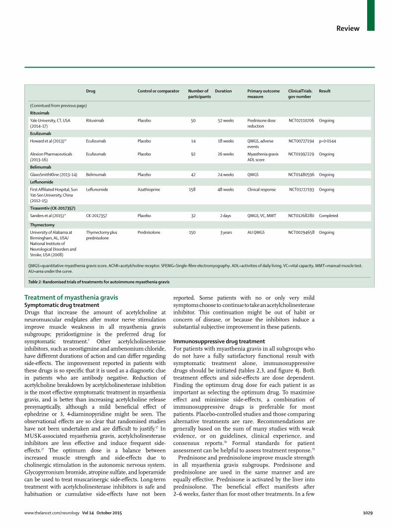
www.thelancet.com/neurology Vol 14 October 2015 1029
Review
Treatment of myasthenia gravisSymptomatic drug treatmentDrugs that increase the amount of acetylcholine at neuromuscular endplates after motor nerve stimulation improve muscle weakness in all myasthenia gravis subgroups; pyridostigmine is the preferred drug for symptomatic treatment.7 Other acetylcholinesterase inhibitors, such as neostigmine and ambenonium chloride, have diff erent durations of action and can diff er regarding side-eff ects. The improvement reported in patients with these drugs is so specifi c that it is used as a diagnostic clue in patients who are antibody negative. Reduction of acetylcholine breakdown by acetyl cholinesterase inhibition is the most eff ective symptomatic treatment in myasthenia gravis, and is better than increasing acetylcholine release presynaptically, although a mild benefi cial eff ect of ephedrine or 3, 4-diaminopyridine might be seen. The observational eff ects are so clear that randomised studies have not been undertaken and are diffi cult to justify.57 In MUSK-associated myasthenia gravis, acetylcholinesterase inhibitors are less eff ective and induce frequent side-eff ects.27 The optimum dose is a balance between increased muscle strength and side-eff ects due to cholinergic stimulation in the autonomic nervous system. Glycopyrronium bromide, atropine sulfate, and loperamide can be used to treat muscarinergic side-eff ects. Long-term treatment with acetyl cholinesterase inhibitors is safe and habituation or cumulative side-eff ects have not been
reported. Some patients with no or only very mild symptoms choose to continue to take an acetylcholinesterase inhibitor. This continuation might be out of habit or concern of disease, or because the inhibitors induce a substantial subjective improvement in these patients.
Immunosuppressive drug treatmentFor patients with myasthenia gravis in all subgroups who do not have a fully satisfactory functional result with symptomatic treatment alone, immunosuppressive drugs should be initiated (tables 2,3, and fi gure 4). Both treatment eff ects and side-eff ects are dose dependent. Finding the optimum drug dose for each patient is as important as selecting the optimum drug. To maximise eff ect and minimise side-eff ects, a combination of immunosuppressive drugs is preferable for most patients. Placebo-controlled studies and those comparing alternative treatments are rare. Recommendations are generally based on the sum of many studies with weak evidence, or on guidelines, clinical experience, and consensus reports.78 Formal standards for patient assessment can be helpful to assess treatment response.79
Prednisone and prednisolone improve muscle strength in all myasthenia gravis subgroups. Prednisone and prednisolone are used in the same manner and are equally eff ective. Prednisone is activated by the liver into prednisolone. The benefi cial eff ect manifests after 2–6 weeks, faster than for most other treatments. In a few
Drug Control or comparator Number of participants
Duration Primary outcome measure
ClinicalTrials.gov number
Result
(Conintued from previous page)
Rituximab
Yale University, CT, USA (2014–17)
Rituximab Placebo 50 52 weeks Prednisone dose reduction
NCT02110706 Ongoing
Eculizumab
Howard et al (2013)76 Eculizumab Placebo 14 18 weeks QMGS, adverse events
NCT00727194 p=0·0144
Alexion Pharmaceuticals (2013–16)
Eculizumab Placebo 92 26 weeks Myasthenia gravis ADL score
NCT01997229 Ongoing
Belimumab
GlaxoSmithKline (2013–14) Belimumab Placebo 42 24 weeks QMGS NCT01480596 Ongoing
Lefl unomide
First Affi liated Hospital, Sun Yat-Sen University, China (2012–15)
Lefl unomide Azathioprine 158 48 weeks Clinical response NCT01727193 Ongoing
Tirasemtiv (CK-2017357)
Sanders et al (2015)77 CK-2017357 Placebo 32 2 days QMGS, VC, MMT NCT01268280 Completed
Thymectomy
University of Alabama at Birmingham, AL, USA/National Institute of Neurological Disorders and Stroke, USA (2008)
Thymectomy plus prednisolone
Prednisolone 150 3 years AU QMGS NCT00294658 Ongoing
QMGS=quantitative myasthenia gravis score. AChR=acetylcholine receptor. SFEMG=Single-fi bre electromyography. ADL=activities of daily living. VC=vital capacity. MMT=manual muscle test. AU=area under the curve.
Table 2: Randomised trials of treatments for autoimmune myasthenia gravis

1030 www.thelancet.com/neurology Vol 14 October 2015
Review
patients, initial deterioration of generalised myasthenia gravis has been reported lasting for up to 3 weeks.7,54,80 The starting dose is most often 0·75–1·0 mg/kg per day1 for prednisone and prednisolone and is gradually increased; alternate-day dosing is thought to reduce side-eff ects and is recommended by some treatment guidelines.2,3,81 After optimum improvement has been induced, the drug dose should be gradually reduced, and continued at the lowest dose necessary to obtain maximum eff ect. Prednisone or
prednisolone should not be given as an alternate-day treatment to patients with diabetes because fl uctuations in glucose concentrations result from this treatment approach. If muscle strength diff ers for off -treatment days and on-treatment days, a low dose (5–10 mg) of prednisone or prednisolone can be added on off -days. For ocular myasthenia gravis, observational studies53,82 suggest that prednisolone treatment reduces the risk of developing generalised myasthenia gravis, although this observation has not been confi rmed. For patients who take long-term corticosteroids, specifi c precautions should be taken to reduce the risks of glucose intolerance, gaining excess bodyweight, hypertension, and development of osteoporosis. A UK registry-based study83 did not report an increased fracture risk in patients with myasthenia gravis.
Azathioprine is an eff ective drug for all myasthenia gravis subgroups, with 2–3 mg/kg being the most eff ective dose in combination with prednisolone.62,63,84 This combination is often recommended as a fi rst-choice treatment for patients with generalised myasthenia gravis who need immunosuppression, and is more benefi cial than corticosteroids alone with fewer side-eff ects. The azathioprine eff ect is delayed and from clinical experience is usually seen after 6–15 months, and might further increase during the subsequent 1–2 years.63 This makes the combination with prednisolone convenient, and prednisolone can be reduced when the azathioprine eff ect has been established. Regular follow-up is necessary because of the risk of leucopenia and hepatotoxic eff ects, especially during the fi rst months of treatment. Low thiopurine methyltransferase activity increases the risk for azathioprine toxic eff ects, and can be tested before the start of treatment. Long-term treatment is also safe and eff ective in young individuals.85 Azathioprine and corticosteroids in combination are eff ective in almost all patients with myasthenia gravis. Patients with ocular myasthenia gravis often respond well to a small dose (10–30 mg on alternate days) of corticosteroids alone.
Mycophenolate mofetil is a prodrug that after conversion blocks purine synthesis and interferes with B-cell and T-cell proliferation. Most guidelines recommend the drug for mild and moderate myasthenia gravis if the initial immunosuppressive therapy fails,2–4 often together with prednisolone. This recommendation is based on retrospective studies1–3,8 and clinical experience. Mycophenolate mofetil is not recommended as fi rst-line treatment. In two prospective and controlled trials,69,86 mycophenolate mofetil did not show additional benefi t when given as initial treatment combined with prednisone. The studies had short durations of only 12 weeks and 9 months. There were no stopping rules for the use of corticosteroids and the lowest prednisone dose was 7·5 mg per day, which might have obscured an eff ect of mycophenolate mofetil. Little is known about myasthenia gravis subgroup responses for this drug.87
Number of participants
Duration Study design ClinicalTrials.gov number
Study period
Bortezomib 18 6 months Open NCT02102594 2014–16
GM-CSF 12 120 days Open NCT01555580 2012–13
Plasmapheresis 10 14 weeks Observational NCT01927692 2013–14
Rituximab 10 12 months Open NCT00619671 2004–09
Rituximab 30 12 months Open NCT00774462 2008–11
Stem-cell therapy 10 5 years Open, phase 1 NCT00424489 2002–16
Subcutaneous intravenous immunoglobulin
25 12 weeks Open, phase 2 NCT02100969 2014–17
Subcutaneous intravenous immunoglobulin
10 6 months Open NCT01828294 2011–15
Tacrolimus 11 28 weeks Open NCT00309101 2006–09
GM-CSF=granulocyte-macrophage colony stimulating factor.
Table 3: Ongoing non-randomised trials of treatments for autoimmune myasthenia gravis registered in ClinicalTrials.gov
Figure 4: Treatment of generalised myasthenia gravis
Myasthenia gravis diagnosis confirmed
Start acetylcholine esterase inhibitor and undertake thymectomy (if early onset or thymoma)
Clinical remission?Continue with acetylcholine esterase inhibitor
Start prednisolone and azathioprine
Very good clinical effect?Continue with prednisolone (lowest possible dose) and azathioprine
Start mycophenolate mofetil (mild, moderatesymptoms) or rituximab (severe symptoms)
Sufficient effect?
A Start other immunosuppressive drugs (methotrexate, ciclosporin, tacrolimus)B Re-evaluate myasthenia gravis
Continue treatment
No
No
No
Yes
Yes
Yes

www.thelancet.com/neurology Vol 14 October 2015 1031
Review
Side-eff ects are rare, with mild headache, nausea, and diarrhoea the most commonly reported.
Rituximab has emerged as a potentially eff ective drug in myasthenia gravis.81,88,89 It is a chimeric IgG1 monoclonal antibody that depletes all types of B lymphocytes through specifi c binding to the transmembrane CD20 antigen. This drug should, in our opinion, be considered in moderate and especially severe myasthenia gravis that does not respond suffi ciently to fi rst-line immuno suppressive treatment. However, controlled studies have not been done, and rituximab is not regarded as a fully established treatment. About two-thirds of patients with severe myasthenia gravis and insuffi cient response to prednisolone and azathioprine have a substantial improvement on this treatment.81,88–91 Open and uncontrolled studies90–92 show that patients with MUSK-associated myasthenia gravis in particular have a favourable response, which is especially important as this myasthenia gravis subgroup often has a lower response to the fi rst-line symptomatic and immuno-suppressive treatment. In most reports, the induction treatment recommended for rheumatological diseases has been used, which is two doses of rituximab 1000 mg, and then another two doses of 1000 mg after 2 weeks.81,88–91 Lower doses have been suggested for myasthenia gravis.88 Most centres would give additional rituximab doses only to patients with deterioration after a substantial and long-lasting response, and then in the lowest eff ective dose.92 Rituximab is most often combined with prednisolone and the combination with prednisolone and azathioprine is also regarded as safe. Severe side-eff ects have been reported as rare events with rituximab for other autoimmune disorders, including JC-virus-related pro-gressive multifocal leukoencephalopathy, and have restricted the use of rituximab in myasthenia gravis. Even in the absence of controlled prospective studies and with high drug costs, rituximab has, in our opinion, a place as an early treatment for an increasing number of patients with MUSK and AChR-associated myasthenia gravis.
Prospective and controlled studies have shown that ciclosporin and methotrexate are eff ective as secondary drugs for myasthenia gravis.65,70,93 The eff ect occurs in all myasthenia gravis subgroups. Although comparative studies have not been undertaken, ciclosporin and methotrexate are thought to be as eff ective as azathioprine.1–4,7 Patients should be monitored for potential side-eff ects, especially nephrotoxic eff ects and hypertension.
Tacrolimus has similarities to ciclosporin. A small (34 patients) randomised but unblinded study66 showed that prednisone could be given at a reduced dose after 52 weeks when combined with tacrolimus. However, a large double-blind study67 comprising of 80 patients did not confi rm this fi nding. The length of this study was only 28 weeks and the therapeutic eff ect of prednisone alone was better than expected.94 A new trial comparing tacrolimus with placebo for patients with an insuffi cient
response to glucocorticoids is in progress (NCT01325571). Tacrolimus has an additional eff ect on ryanodine receptor-mediated calcium release from the sarcoplasmic reticulum, which theoretically could lead to improvements in muscle strength in patients with myasthenia gravis.
ThymectomyMany studies have reported a substantial eff ect of thymectomy in myasthenia gravis. These studies have included control groups, but prospective and randomised studies have not been done.1–3,7,95,96 For early-onset myasthenia gravis, we recommend a thymectomy early after symptom onset. All thymus tissue needs to be removed. Video-assisted thoracoscopic and robotic-assisted methods are well established, used by an increasing number of centres, and are usually preferred by patients.97 Thymectomy can be safe for juvenile myasthenia gravis, down to an age of about 5 years.98 Improvement in response to thymectomy occurs gradually after some months, and according to follow-up studies, continues for up to 2 years postoperatively.95 No other autoimmune disorders have been shown to improve after thymectomy. Thymectomy should be undertaken as an oncological intervention when a thymoma is detected or is strongly suspected to avoid local compression and spread to the thoracic cavity. Any positive eff ect on myasthenia gravis is more unpredictable for the thymoma than for the early-onset subgroup.
Use of thymectomy in late-onset myasthenia gravis is debated. For patients with late-onset disease with an atrophic thymus or onset at age 60–65 years or older, thymectomy is not recommended because no convincing data support surgery for this group. However, some guidelines7 recommend treating young patients (up to age 60–65 years) with late-onset disease who have an enlarged thymus on imaging and no antibodies to muscle titin or the ryanodine receptor, similar to patients with early-onset myasthenia gravis. For younger patients with late-onset myasthenia gravis, the thymus is most probably involved in the pathogenesis and the response to thymectomy would be expected to be similar to that for early-onset disease.
Thymectomy is not recommended for patients with MUSK, LRP4, or ocular forms of myasthenia gravis as no therapeutic eff ect has been shown. For patients with generalised myasthenia gravis and low-affi nity AChR antibodies, thymus hyperplasia is usually impossible to establish by imaging. Such patients would be expected to respond to thymectomy but cannot be distinguished from other patients with myasthenia gravis who are found to be antibody negative.
Thymectomy should be done early, but is never an emergency; patients should be in a stable condition. Intravenous immunoglobulin or plasma exchange immediately before surgery will improve the myasthenia gravis symptoms, reduce the risk of complications, and contribute to a faster recovery.

1032 www.thelancet.com/neurology Vol 14 October 2015
Review
Supportive treatmentPhysical activity and low intensity and medium intensity training provide short-term and long-term benefi ts for patients with myasthenia gravis. Weakness increases with repetitive muscle use, but patients with myasthenia gravis can still fi nd activities for which they can adjust intensity and duration to increase their long-term physical ability. Rest after such exercise is needed. No controlled studies of myasthenia gravis training programmes have been published.
Bodyweight control is important, as for other disorders with muscle weakness. Such control is especially relevant in patients with involvement of respiratory muscles. Infections in patients with myasthenia gravis should be treated early and vigorously because they can lead to myasthenia gravis exacerbation and add to respiratory impairment.1–4,7
Drugs that interfere negatively with neuromuscular transmission should be avoided. D-penicillamine and telithromycin should not be given to patients with myasthenia gravis, and fl uoroquinolones, aminoglycosides, macrolides, and neuromuscular blocking drugs will often cause worsening of the disease. Neuromuscular blockade should be used with care during anaesthesia. Sedatives that could suppress respiration should be avoided in the treatment of patients with severe myasthenia gravis. If a patient deteriorates when given a new drug, this drug should be withdrawn. However, most patients with myasthenia gravis with mild-to-moderate disease, or in stable remission, tolerate drugs that have a relative warning, and most drugs can be used with caution.
Treatment of myasthenia gravis crisisCrisis is defi ned as a need for intubation for respiratory support caused by muscle weakness related to the disease. Treatment includes intensive care with respiratory support, treatment of infections, and monitoring of vital functions and mobilisation (fi gure 5). Intravenous immunoglobulin and plasma exchange are specifi c immunosuppressive treatments with a rapid eff ect occurring after 2–5 days, and either one should be given to patients with severe myasthenia gravis exacerbations and always for crisis.99–103 These two treatment alternatives are equally eff ective, and can be given in sequence if necessary, as patients can respond to one but not to the other. Standard protocols include treatment for 3–6 consecutive days. Intravenous immunoglobulin is often slightly more convenient and with a lower risk of severe side-eff ects, whereas plasma exchange might have a slightly faster eff ect. Catheter placement procedures for plasma exchange can be complex because access to large veins is necessary. The treatment eff ect is usually restricted to 2–3 months, owing to continuing antibody synthesis. Plasma exchange and intravenous immunoglobulin can be repeated when the eff ect tapers off . To secure long-term improvement, this treatment is usually combined with standard immunosuppressive drugs, in higher doses than before the crisis or with add-on drugs. In patients with an acute exacerbation that does not respond to intravenous immunoglobulin or plasma exchange, corticosteroids in high doses can be tried. Myasthenia gravis crisis is a reversible condition. Sometimes the treatment response is delayed, but intensive care and vigorous immunosuppression should be continued for as long as necessary, sometimes for several weeks.
Treatment of myasthenia gravis in pregnancyPregnancy does not aff ect myasthenia gravis in any consistent way, with no increased risk of severe deterioration or myasthenia gravis crisis.85,104,105 During the fi rst weeks and few months post partum, the risk of symptom worsening is moderately increased, mainly because of stress and new demands.
Pyridostigmine and corticosteroids are regarded as safe treatments for pregnant women.85 These drugs do not increase the risk of fetal malformations or delayed fetal development. Plasma exchange and intravenous immunoglobulin can be used safely for exacerbations in pregnancy, and also as preparation for women giving birth. Evidence for potential teratogenic eff ects of other immunosuppressive drugs is sparse. However, caution is recommended for use of these drugs, and the manufacturers of immunosuppressive drugs generally advise against their use in pregnancy. Azathioprine has been widely used for many years by young women with AChR, MUSK, or LRP4 forms of myasthenia gravis. The general view is that this drug has very low, if any, increased teratogenic risk.85 Lactation should be encouraged in
Figure 5: Treatment of severe myasthenia gravis exacerbations*1000 mg a day for 3 days.
No
No
Yes
Yes
Severe, generalised myasthenia gravis
Start intensive care, respirator if necessary, start intravenous immunoglobulin or plasma exchange
Improvement?
Start plasma exchange or intravenous immunoglobulinStart corticosteroids in high-dosemethylprednisolone*
Improvement?
Start rituximab (outside intensive care unit), continue intensive care and treat complications
Start or continue and intensify long-term immunosuppressivetreatment
Continue and intensify long-term immunosuppressivetreatment

www.thelancet.com/neurology Vol 14 October 2015 1033
Review
patients with myasthenia gravis, also for women on immunosuppressive drugs,105 but the passage of some medications into breastmilk should be taken into account. Mycophenolate mofetil and methotrexate have teratogenic potential. Methotrexate might also reduce female fertility. These two drugs should only rarely be used in young women, and not in pregnancy.
Most female patients with myasthenia gravis give birth in an uncomplicated way. Apart from the risk of neonatal myasthenia gravis, no precautions are usually needed. Caesarean section is not recommended as a routine for these women, but should be considered in prolonged births for women with moderate or severe generalised myasthenia gravis because of muscle fatigue.
Treatment of neonatal myasthenia gravisNeonatal myasthenia gravis occurs in 10–15% of babies of mothers with the disease. The cause of this transient muscular weakness in these babies is transfer of the mother’s AChR or MUSK antibodies of the IgG class across the placenta. This weakness usually lasts for only days or a few weeks and is typically mild but can interfere with feeding and respiration. Mothers with myasthenia gravis should always give birth at hospitals experienced in respiratory support treatment for newborn babies. The fact that neonatal myasthenia gravis does not occur in all babies and that occurrence in babies is not correlated with maternal disease severity or AChR antibody concentration might be explained by variation in AChR epitopes, epitope-binding affi nity, and non-AChR factors.105
Transplacental AChR antibodies can, in rare cases, produce arthrogryposis due to severe intrauterine movement inhibition. Such skeletal malformations were reported in three of 127 babies in an unselected national cohort.106 Arthrogryposis, AChR-antibody induced still-births, and repeated spontaneous abortions can be avoided by intravenous immunoglobulin infusions or plasma exchange before and during pregnancy. This treatment should be given in female patients with myasthenia gravis who have already experienced such a pregnancy outcome.
Conclusions and future directionsMost patients with myasthenia gravis do well and have well controlled disease. However, most need long-term and often life-long drug treatment with acetylcholinesterase inhibitors and usually low-dose immunosuppression. Pathogenic autoantibodies are well characterised and myasthenia gravis subgroups are defi ned accordingly. However, treatment is far from antibody specifi c and is not even specifi c to the disease subgroup. Many new and more traditional drugs that have not been tested properly in myasthenia gravis have modes of action that are expected to suppress autoantibody production directly or indirectly, and therefore might benefi t patients with myasthenia gravis. For patients with severe symptoms that do not respond suffi ciently to standard treatment, with a diagnosis confi rmed by the presence of
autoantibodies and no comorbidity as the symptom cause, such drugs could be tried, off -label, and with strict monitoring. These include monoclonal antibody drugs with a proven eff ect for other autoimmune disorders. Complement inhibition is one of several potential strategies,76 with a focus on several factors in the complement system. Eculizumab, belimumab, lefl unomide, and etanercept are drugs that might have the potential to become new myasthenia gravis treatment options,76,107–109 although some immunoactive drugs can precipitate or worsen myasthenia gravis.110 Tirasemtiv (CK-2017357) selectively sensitises fast skeletal muscle to calcium by binding to its troponin complex and amplifi es the muscle response when neural input is diminished secondary to neuromuscular disease.111 A dose-related, short-term improvement was reported in a phase 2a randomised placebo-controlled trial.77 Any functionally relevant long-term benefi t to patients is still to be proven. Several non-antibody factors linked to the immune system and skeletal muscle aff ect the individual’s muscle strength and immune responses, and thereby each patient’s myasthenia gravis manifestations.
The high number of factors associated with muscle function in myasthenia gravis should drive future research towards an individually adapted treatment approach based on biomarker (autoantibody) assessment and monitoring. The aim should be to suppress the anti-AChR, anti-MUSK, or anti-LRP4 immune response without aff ecting other immune reactions. An alternative approach could be treatment that promotes tolerance to the antigens (AChR, MUSK, and LRP4) that induce myasthenia gravis.112 Patients with myasthenia gravis without detectable antibodies probably have pathogenic antibodies against undefi ned antigens in the neuromuscular junction; many proteins aff ect AChR function, synthesis, and maintenance that could potentially underlie antibody-negative disease. Auto-immune myasthenia gravis with a T-cell-mediated and non-antibody mechanism aff ecting neuromuscular transmission could theoretically exist.
When the causes of myasthenia gravis can be identifi ed, they might be possible to avoid or prevent, potentially, for example, by vaccination. Until antigen-specifi c treatment is available, however, research eff orts should target new
Search strategy and selection criteria
We searched MEDLINE and the Cochrane Library with the terms “myasthenia gravis”, “myasthenic syndromes”, and “myasthenia” from January 1995, to April, 2015. Guideline and review papers were assessed in detail, and controlled studies sought for in particular. Papers were selected by title and abstract. Only papers in English were included. Randomised trials on established and emerging therapies for myasthenia gravis are often scarce, so our recommendations are based on the best available evidence or clinical experience, where stated.

1034 www.thelancet.com/neurology Vol 14 October 2015
Review
immunosuppressive drugs and drug combinations for the myasthenia gravis subgroups. Prospective and controlled studies should be encouraged and supported. Severe myasthenia gravis is a reversible disorder that should be treated with intensity and optimism.ContributorsNEG planned the Review and wrote the fi rst draft. JJV edited and rewrote the fi rst draft. Both authors searched primary sources for information, produced tables and fi gures, and fi nalised the text.
Declaration of interestsNEG has received speaker’s honorarium from Octapharma, Baxter, and Merck Serono. JJV is a partner in an FP7 European grant that is associated with Curavac. The Department of Neurology at Leiden University Medical Center has received fees from BioMarin for JJV’s consultancy work and royalties from antibody tests. JJV received research grants from Prinses Beatrix Spierfonds and National Institutes of Health.
References1 Meriggioli MN, Sanders DB. Autoimmune myasthenia gravis:
emerging clinical and biological heterogeneity. Lancet Neurol 2009; 8: 475–99.
2 Gilhus NE. Myasthenia and neuromuscular junction. Curr Opin Neurol 2012; 25: 523–29.
3 Querol L, Illa I. Myasthenia and the neuromuscular junction. Curr Opin Neurol 2013: 26: 459–65.
4 Verschuuren JJ, Huijbers MG, Plomp JJ, et al. Pathophysiology of myasthenia gravis with antibodies to the acetylcholine receptor, muscle-specifi c kinase and low-density lipoprotein receptor-related protein 4. Autoimmun Rev 2013; 12: 918–23.
5 Zisimopoulou P, Brenner T, Trakas N, Tzartos SJ. Serological diagnostics in myasthenia gravis based on novel assays and recently identifi ed antigens. Autoimmun Rev 2013; 12: 924–30.
6 Owe JF, Daltveit AK, Gilhus NE. Causes of death among patients with myasthenia gravis in Norway between 1951 and 2001. J Neurol Neurosurg Psychiatry 2006; 77: 203–07.
7 Skeie GO, Apostolski S, Evoli A, et al. Guidelines for treatment of autoimmune neuromuscular transmission disorders. Eur J Neurol 2010; 17: 893–902.
8 Suh J, Goldstein JM, Nowak RJ. Clinical characteristics of refractory myasthenia gravis patients. Yale J Biol Med 2013; 86: 255–60.
9 Jacob S, Viega S, Leite MI. Presence and pathogenic relevance of antibodies to clustered acetylcholine receptor in ocular and generalized myasthenia gravis. Arch Neurol 2012; 69: 994–1001.
10 Yang L, Maxwell S, Leite MI, et al. Non-radioactive serological diagnosis of myasthenia gravis and clinical features of patients from Tianjin, China. J Neurol Sci 2011; 301: 71–76.
11 Cole RN, Ghazanfari N, Ngo ST, et al. Patient autoantibodies deplete postsynaptic muscle-specifi c kinase leading to dissembly of the ACh receptor scaff old and myasthenia gravis in mice. J Physiol 2010; 17: 3217–29.
12 Y, Ito M, Hirayama M, et al. Anti-MuSK autoantibodies block binding of collagen Q to MuSK. Neurology 2011; 77: 1819–28.
13 Plomp JJ, Huijbers MG, Maarel SMVD, Verschuuren JJ. Pathogenic IgG4 subclass autoantibodies in MuSK myasthenia gravis. Ann NY Acad Sci 2012; 1275: 114–22.
14 Shen C, Lu Y, Zhang B, et al. Antibodies against low-density lipoprotein receptor-related protein 4 induce myasthenia gravis. J Clin Invest 2013; 123: 5190–202.
15 Gasperi C, Melms A, Schoser B, et al. Anti-agrin autoantibodies in myasthenia gravis. Neurology 2014; 82: 1976–83.
16 Zhang B, Shen C, Bealmear B, et al. Autoantibodies to agrin in myasthenia gravis. PLoS One 2014; 9: e91816.
17 Gallardo E, Martinez-Hernandez E, Titulaer MJ, et al. Cortactin autoantibodies in myasthenia gravis. Autoimmun Rev 2014; 13: 1003–07.
18 Skeie GO, Mygland A, Treves S. Ryanodine receptor antibodies in myasthenia gravis: epitope mapping and eff ect on calcium release in vitro. Muscle Nerve 2003; 27: 81–89.
19 Romi F, Aarli JA, Gilhus NE. Myasthenia gravis patients with ryanodine receptor antibodies have distinctive clinical features. Eur J Neurol 2007; 14: 617–20.
20 Heldal AT, Owe JF, Gilhus NE, et al. Seropositive myasthenia gravis; a nationwide epidemiologic study. Neurology 2009; 73: 150–51.
21 Andersen JB, Engeland A, Owe JF, et al. Myasthenia gravis requiring pyridostigmine treatment in a national population cohort. Eur J Neurol 2010; 17: 1445–50.
22 Carr AS, Cardwell CR, McCarron PO, et al. A systematic review of population based epidemiological studies in myasthenia gravis. BMC Neurol 2010; 10: 46
23 Pakzad Z, Aziz T, Oger J. Increasing incidence of myasthenia gravis among elderly in British Columbia, Canada. Neurology 2011; 76: 1526–28.
24 Pedersen EG, Hallas J, Hansen K, et al. Late-onset myasthenia not on the increase: a nationwide register study in Denmark, 1996–2009. Eur J Neurol 2012; 20: 942–48.
25 VanderPluym, J, Vajsar J, Jacob FD, et al. Clinical characteristics of pediatric myasthenia: a surveillance study. Pediatrics 2013; 132: e939–44.
26 Niks EH, Kuks JBM, Verschuuren JJGM. Epidemiology of myasthenia gravis witht anti-muscle specifi c kinaase antibodies in the Netherlands. J Neurol Neurosurg Psychiatry 2007; 78: 417–18.
27 Guptill JT, Sanders DB, Evoli A. Anti-MuSK antibody myasthenia gravis; clinical fi ndings and response to treatment in two large cohorts. Muscle Nerve 2011; 44: 36–40.
28 Rodolico C, Toscano A, Autunno M, et al. Limb-girdle myasthenia; clinical, electrophysiological and morphological features in familial and autoimmune cases. Neuromuscul Disord 2002; 12: 964–69.
29 Gilhus NE, Nacu A, Andersesn JB, Owe JF. Myasthenia gravis and risks for comorbidity. Eur J Neurol 2015; 22: 17–23.
30 Liewluck T. Immune-mediated rippling muscle disease; another infl ammatory myopathy in myasthenia gravis. Arch Neurol 2010; 67: 896–97.
31 Mehanna R, Patton EL, Phan CL, Harati Y. Amyotrophic lateral sclerosis with positive anti-acetylcholine receptor antibodies; case report and review of the literature. J Clin Neuromuscul Dis 2012; 14: 82–85.
32 Filosso PL, Galassi C, Ruffi ni E, et al. Thymoma and the increased risk of developing extrathymic malignancies: a multicentre study. Eur J Cardiothor Surg 2013; 44: 219–24.
33 Pedersen EG, Pottegard A, Hallas J, et al. Use of azathioprine for non-thymoma myasthenia and risk of cancer: a nationwide case-control study in Denmark. Eur J Neurol 2013; 20: 942–48.
34 Pedersen EG, Pottegard A, Hallas J, et al. Risk of non-melanoma skin cancer in myasthenia patients treated with azathioprine. Eur J Neurol 2014; 20: 942–48.
35 Owe JF, Davidsen ES, Eide GE, et al. Left ventricular long-axis function in myasthenia gravis. J Neurol 2008; 255: 1777–84.
36 Suzuki S, Utsugisawa K, Yoshikawa H, et al. Autoimmune targets of heart and skeletal muscles in myasthenia gravis. Arch Neurol 2009; 66: 1334–38.
37 Kumagai S, Kato T, Ozaki A, et al. Serial measurements of cardiac troponin I in patients with myasthenia gravis-related cardiomyopathy. Int J Cardiol 2013; 168: e79–80.
38 Suzuki S, Utsugisawa K, Suzuki N. Overlooked non-motor symptoms in myasthenia gravis. J Neurol Neurosurg Psychiatry 2013; 84: 989–94.
39 Alkhawajah NM, Oger J. Late-onset myasthenia gravis; a review when incidence in older adults keeps increasing. Muscle Nerve 2013; 48: 705–10
40 Gregersen PK, Kosoy R, Lee AT, et al. Risk for myasthenia gravis maps to a (151) pro-ala change in TNIP1 and human leukocyte antigen-B*08. Ann Neurol 2012; 72: 927–35.
41 Renton AE, Piner HA, Provenzano C, et al. A genome-wide association study of myasthenia gravis. JAMA Neurol 2015; 72: 396–404.
42 Klein R, Marx A, Ströbel P, et al. Autoimmune associations and autoantibody screening show focused recognition in patient subgroups with generalized myasthenia gravis. Hum Immunol 2013; 74: 1184–93.
43 Maniaol AH, Elsais A, Lorentzen ÅR, et al. Late onset myasthenia gravis is associated with HLA DRB1*15:01 in the Norwegian population. PLoS One 2012; 7: e36603.
44 Marx A, Pfi ster P, Schalke B, et al. The diff erent roles of the thymus in the pathogenesis of the various myasthenia gravis subtypes. Autoimmun Rev 2013; 12: 875–84.

www.thelancet.com/neurology Vol 14 October 2015 1035
Review
45 Skjei KL, Lennon VA, Kuntz NL. Muscle specifi c kinase autoimmune myasthenia gravis in children: a case series. Neuromuscul Disord 2013; 23: 874–82.
46 Bartoccioni E, Scuderi F, Augugiaro A, et al. HLA class II allele analysis in MuSK-positive myasthenia gravis suggests a role for DQ5. Neurology 2009; 72: 195–97.
47 Klooster R, Plomp JJ, Huijbers MG, et al. Muscle-specifi c kinase myasthenia gravis IgG4 autoantibodies cause severe neuromuscular junction dysfunction in mice. Brain 2012; 135: 1081–101.
48 Alahgholi-Hajibehzad M, Yilmaz V, Guisen-Parman Y, et al. Association of HLA-DR131*14,-DR131*16 and –DQB1*05 with MuSK-myasthenia gravis in patients from Turkey. Hum Immunol 2013; 74: 1633–35.
49 Higuchi O, Hamuo J, Motomura M, et al. Autoantibodies to low-density lipoprotein receptor-related protein 4 in myasthenia gravis. Ann Neurol 2011; 69: 418–22.
50 Zhang B, Tzartos JS, Viegas S, et al. Autoantibodies to lipoprotein-related protein 4 in patients with double-negative myasthenia gravis. Arch Neurol 2012; 69: 445–51
51 Leite MI, Jacob S, Viegas S, et al. IgG1 antibodies to acetylcholine receptors in ”seronegative” myasthenia gravis. Brain 2008; 131: 1940–52.
52 Cossins J, Belaya K, Zoltowska K, et al. The search for new antigenic targets in myasthenia gravis. Ann N Y Acad Sci 2013; 1275: 123–28.
53 Kerty E, Elsais A, Argov Z, Evoli A, Gilhus NE. EFNS/ENS guidelines for the treatment of ocular myasthenia gravis. Eur J Neurol; 2014: 21: 687–93.
54 Romi F, Bo L, Skeie GO, et al. Titin and ryanodine receptor epitopes are expressed in cortical thymoma along with costimulatory molecules. J Neuroimmunol 2002; 128: 82–89.
55 Cufi P, Dragin N, Weiss JM, et al. Implication of double-stranded RNA signaling in the etiology of autoimmune myasthenia gravis. Ann Neurol 2012; 73: 281–93.
56 Panse RL, Berrih-Aknin S. Autoimmune myasthenia gravis: autoantibody mechanisms and new developments on immune regulation. Curr Opin Neurol 2013; 26: 569–76.
57 Mehndiratta MM, Pandey S, Kuntzer T. Acetylcholinesterase inhibitor treatment for myasthenia gravis. Cochrane Database Syst Rev 2014; CD006986.
58 Mount, FW. Corticotropin in treatment of ocular myasthenia; a controlled clinical trial. Arch Neurol 1964; 11: 114–24.
59 Howard FM Jr, Duane DD, Lambert EH, Daube JR. Alternate-day prednisone: preliminary report of a double-blind controlled study. Ann N Y Acad Sci 1976; 274: 596–607.
60 Lindberg C1, Andersen O, Lefvert AK. Treatment of myasthenia gravis with methylprednisolone pulse: a double blind study. Acta Neurol Scand 1998; 97: 370–73.
61 Benatar M, McDermott MP, Sanders DB, et al, for the Muscle Study Group (MSG). Effi cacy of Prednisone for the Treatment of Ocular Myasthenia (EPITOME): a randomized controlled trial. Muscle Nerve 2015; published online July 14. DOI:10.1002/mus.24769.
62 Bromberg MB, Wald JJ, Forshew DA, et al. Randomized trial of azathioprine or prednisone for initial immunosuppressive treatment of myasthenia gravis. J Neurol Sci 1997; 150: 59–62.
63 Palace J, Newsom-Davis J, Lecky B. A randomized double-blind trial of prednisolone alone or with azathioprine in myasthenia gravis. Neurology 1998; 50: 1778–83.
64 Tindall RS, Rollins JA, Phillips JT, Greenlee RG, Wells L, Belendiuk G. Preliminary results of a double-blind, randomized, placebo-controlled trial of cyclosporine in myasthenia gravis. N Engl J Med 1987; 316: 719–24.
65 Tindall RS, Philips JT, Rollins JA, et al. A clinical therapeutic trial of cyclosporine in myasthenia gravis. Ann N Y Acad Sci 1993; 681: 539–51.
66 Nagane Y, Utsugisawa K, Obara D, et al. Effi cacy of low-dose FK506 in the treatment of myasthenia gravis; a randomized pilot study. Eur Neurol 2005; 53: 146–50.
67 Yoshikawa H, Kiuchi T, Saida T, et al. Randomised, double-blind, placebo-controlled study of tacrolimus in myasthenia gravis. J Neurol Neurosurg Psychiatry 2011; 82: 970–77.
68 Meriggioli MN, Rowin J, Richman JG, Leurgans S. Mycophenolate mofetil for myasthenia gravis: a double-blind, placebo-controlled pilot study. Ann N Y Acad Sci 2003; 998: 494–9.
69 Sanders DB, Hart IK, Mantegazza R, et al. An international, phase III, randomized trial of mycophenolate mofetil in myasthenia gravis. Neurology 2008; 71: 400–06.
70 Pasnoor M, He J, Herbelin L, et al. Phase II trial of methotrexate in myasthenia gravis. Ann N Y Acad Sci 2013; 1275: 23–28.
71 Gajdos P, Chevret S, Clair B, Tranchant C, Chastang C. Clinical trial of plasma exchange and high-dose intravenous immunoglobulin in myasthenia gravis. Myasthenia Gravis Clinical Study Group. Ann Neurol 1997; 41: 789–96.
72 Wolfe GI, Barohn RJ, Foster BM, et al, for the Myasthenia Gravis-IVIG Study Group. Randomized, controlled trial of intravenous immunoglobulin in myasthenia gravis. Muscle Nerve 2002; 26: 549–52.
73 Gajdos P, Tranchant C, Clair B, et al, for the Myasthenia Gravis Clinical Study Group. Treatment of myasthenia gravis exacerbation with intravenous immunoglobulin: a randomized double-blind clinical trial. Arch Neurol 2005; 62: 1689–93.
74 Zinman L, Ng E, Bril V. IV immunoglobulin in patients with myasthenia gravis: a randomized controlled trial. Neurology 2007; 68: 837–41.
75 Barth D, Nabavi Nouri M, Ng E, Nwe P, Bril V. Comparison of IVIg and PLEX in patients with myasthenia gravis. Neurology 2011; 76: 2017-23.
76 Howard JF, Barohn PJ, Cutter GR, et al. A randomized, double-blind, placebo-controlled phase II study of eculizumab in patients with refractory generalized myasthenia gravis. Muscle Nerve 2013; 48: 76–84.
77 Sanders DB, Rosenfeld J, Dimachkie MM, et al. A double-blinded, randomized, placebo-controlled trial to evaluate effi cacy, safety and tolerability of single doses of tirasemtiv in patients with acetylcholine receptor-binding antibody-positive myasthenia gravis. Neurotherapeutics 2015; 12: 455–60.
78 Hart IK, Sthasiam S, Sharshar T. Immunosuppressive agents for myasthenia gravis (review). Cochrane Database Syst Rev 2007; CD005224.
79 Jaretzki III A, Barohn RJ, Ernstoff RM et al. Myasthenia gravis: recommendations for clinical research standards. Ann Thorac Surg 2000; 70: 327–34.
80 Benatar M, Sanders DB, Wolfe GI, McDermott MP, Tawil R. Design of the effi cacy of prednisone in the treatment of ocular myasthenia (EPITOME) trial. Ann N Y Acad Sci 2012; 1275: 17–22.
81 Benveniste O, Hilton-Jones D. The role of rituximab in the treatment of myasthenia gravis. Eur Neurol Rev 2010; 5: 95–100.
82 Benatar M, Kaminski H. Medical and surgical treatment for ocular myasthenia. Cochrane Database Syst Rev 2012; CD005081.
83 Pouwels S, Boer AD, Javaid MK, et al. Fracture rates in patients with myasthenia gravis; the general practice research database. Osteoporos Int 2013; 24: 467–76.
84 Marinkovic G, Kroon J, Hoogenboezem M, et al. Inhibition of GTPase rac1 in endothelium by 6-mercaptpurine results in immunosuppression in nonimmune cells; new target for an old drug. J Immunol 2014; 192: 4370–78.
85 Norwood F, Dhanjal M, Hill M, et al. Myasthenia in pregnancy; best practice guidelines from a UK multispeciality working group. J Neurol Neurosurg Psychiatry 2014; 85: 538–43.
86 The Muscle Study Group. A trial of mycophenolate mofetil with prednisone as initial immunotherapy in myasthenia gravis. Neurology 2008; 71: 394–399.
87 Hehir MK, Burns TM, Alpers J, et al. Mycophenolate mofetil in AChR-antibody-positive myasthenia gravis; outcomes in 102 patients. Muscle Nerve 2010; 41: 593–98.
88 Blum S, Gillis D, Brown H, et al. Use and monitoring of low dose rituximab in myasthenia gravis. J Neurol Neurosurg Psychiatry 2011; 82: 659–63.
89 Maddison P, McConville J, Farrugia ME, et al. The use of rituximab in myasthenia gravis and Lambert-Eaton myasthenic syndrome. J Neurol Neurosurg Psychiatry 2011; 82: 671–73.
90 Diaz-Manera J, Martinez-Hernandez E, Querol L, et al. Long-lasting treatment of rituximab in MuSK myasthenia. Neurology 2012; 78: 189–93.

1036 www.thelancet.com/neurology Vol 14 October 2015
Review
91 Keung B, Robeson KR, DiCapua DB, et al. Long-term benefi t of rituximab in MuSK autoantibody myasthenia gravis patients. J Neurol Neurosurg Psychiatry 2013; 84: 1407–09.
92 Yi JS, DeCroos EC, Sanders DB et al. Prolonged B-cell depletion in MuSK myasthenia gravis following rituximab treatment. Muscle Nerve 2013; 48: 992–93.
93 Heckmann JM, Rawoot A, Bateman K, et al. A single-blinded trial of methotrexate versus azathioprine as steroid-sparing agents in generalized myasthenia gravis. BMC Neurol 2011; 11: 97.
94 Benatar M, Sanders D. The importance of studying history; lessons learnt from a trial of tacrolimus in myasthenia gravis. J Neurol Neurosurg Psychiatry 2011; 82: 945.
95 Gronseth GH, Barohn RJ. Thymectomy for autoimmune myasthenia gravis (an evidence-based review). Neurology 2000; 55: 7–15.
96 Cea G, Benatar M, Verdugo RJ, Salinas RA. Thymectomy for non-thymomatous myasthenia gravis. Cochrane Database Syst Rev 2013; cd008111.
97 Ye B, Tantal J-C, Li W, et al. Video-assisted thoracoscopic surgery versus robotic-assisted thoracoscopic surgery in the surgical treatment of Masaoka stage I thymoma. World J Surg Oncol 2013; 11: 157–62.
98 Liew WKM, Kang PB. Update on juvenile myasthenia gravis. Curr Opin Pediatr 2013; 25: 694–700.
99 Gajdos P, Chevret S, Toyka KV. Plasma exchange for generalised myasthenia gravis. Cochrane Database Syst Rev 2002; CD002275.
100 Mandawat A, Kaminski H, Cutter G, et al. Comparative analysis of therapeutic options used for myasthenia gravis. Ann Neurol 2010; 68: 797–805.
101 Barth D, Nabavi N, Ng E, et al. Comparison of IVIg and PLEX in patients with myasthenia gravis. Neurology 2011; 76: 2017–23.
102 Gilhus NE. Acute treatment for myasthenia gravis. Nat Rev Neurol 2011; 7: 132–34.
103 Gajdos P, Chevret S, Toyka KV. Intravenous immunoglobulin for myasthenia gravis. Cochrane Database Syst Rev 2012; CD002277.
104 Ferrero S, Pretta S, Nicoletti A, et al. Myasthenia gravis: management issues during pregnancy. Eur J Obst Gynecol Reproduct Biol 2005; 121: 129–38.
105 Hoff JM, Daltveit AK, Gilhus NE. Myasthenia gravis in pregnancy and birth: identifying risk factors, optimising care. Eur J Neurol 2007; 14: 38–43.
106 Hoff JM, Loane M, Gilhus NE, et al. Arthrogryposis multiplex congenital: an epidemiologic study of nearly 9 million births in 24 Eurocat registers. Eur J Obst Gynecol Reproduct Biol 2011; 159: 347–50.
107 Ragheb S, Lisak R, Lewis R, et al. A potential role for B-cell activating factor in the pathogenesis of autoimmune myasthenia gravis. Arch Neurol 2008; 65: 1358–62.
108 Gomez AM, Vrolix K, Martinez-Martinez P, et al. Proteasome inhibition with bortezomib depletes plasma cells and autoantibodies in experimental autoimmune myasthenia gravis. J Immunol 2011; 186: 2503–13.
109 Rowin J, Thiruppathi M, Arrebamen E, et al. Granulocyte macrophage colony-stimulating factor treatment of a patient in myasthenic crisis; eff ects on regulatory T cells. Muscle Nerve 2012; 46: 449–53.
110 Fee DB, Kasarskis EJ. Myasthenia gravis associated with etanercept therapy. Muscle Nerve 2009; 39: 866–70.
111 Russell AJ, Hartman JJ, Hinken AC, et al. Activation of fast skeletal muscle troponin as a potential therapeutic approach for treating neuromuscular diseases. Nat Med 2012; 18: 452–56.
112 Steinman L. The road not taken: antigen-specifi c therapy and neuroinfl ammatory disease. JAMA Neurol 2013; 70: 1100–01.