musculoskeletal system trauma
TRANSCRIPT
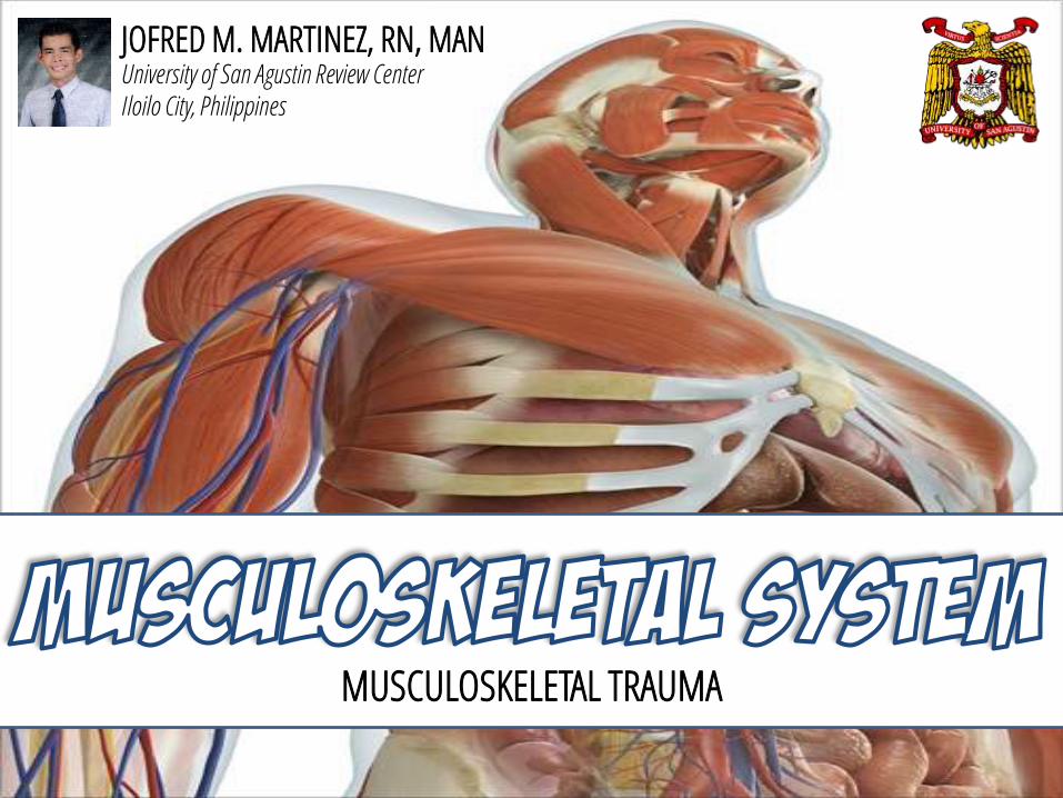
MUSCULOSKELETAL TRAUMA
JOFRED M. MARTINEZ, RN, MANUniversity of San Agustin Review CenterIloilo City, Philippines
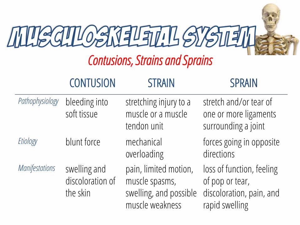
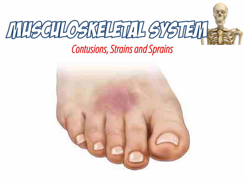
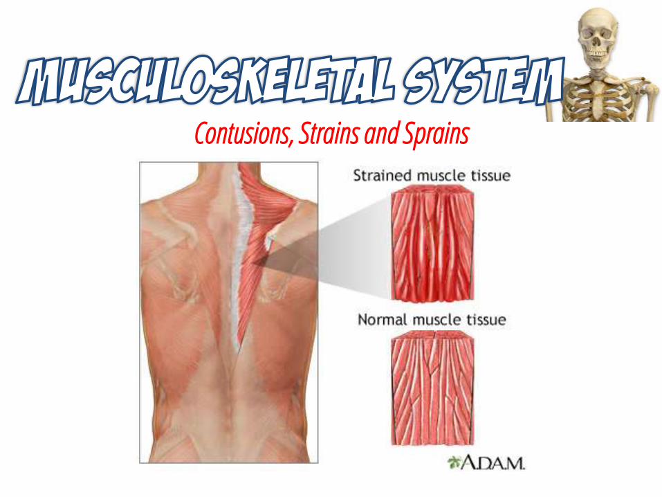
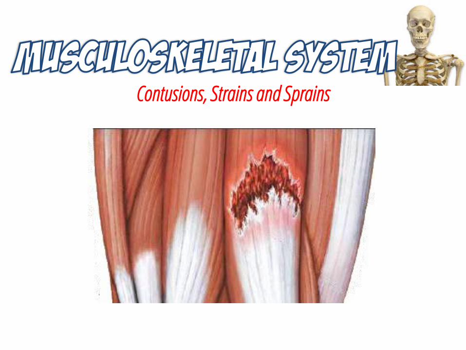
Contusions, Strains and Sprains
CONTUSION STRAIN SPRAIN
Pathophysiology bleeding into soft tissue
stretching injury to a muscle or a muscle tendon unit
stretch and/or tear of one or more ligaments surrounding a joint
Etiology blunt force mechanical overloading
forces going in opposite directions
Manifestations swelling and discoloration of the skin
pain, limited motion, muscle spasms, swelling, and possible muscle weakness
loss of function, feeling of pop or tear, discoloration, pain, and rapid swelling
Contusions, Strains and Sprains
Contusions, Strains and Sprains
Contusions, Strains and Sprains
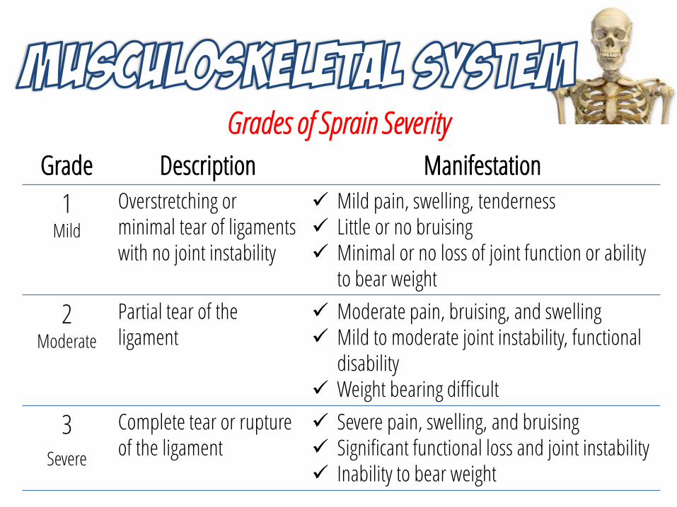
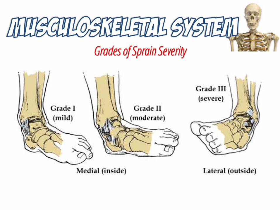
Grades of Sprain Severity
Grade Description Manifestation
1Mild
Overstretching or minimal tear of ligaments with no joint instability
Mild pain, swelling, tenderness Little or no bruising Minimal or no loss of joint function or ability
to bear weight
2Moderate
Partial tear of the ligament
Moderate pain, bruising, and swelling Mild to moderate joint instability, functional
disability Weight bearing difficult
3Severe
Complete tear or rupture of the ligament
Severe pain, swelling, and bruising Significant functional loss and joint instability Inability to bear weight
Grades of Sprain Severity
Contusions, Strains and Sprains
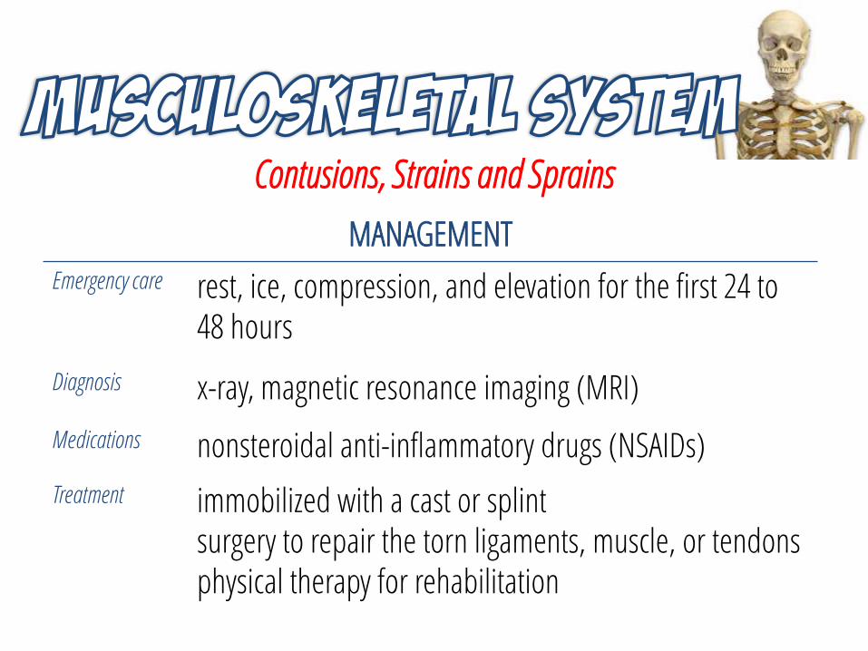
MANAGEMENT
Emergency care rest, ice, compression, and elevation for the first 24 to 48 hours
Diagnosis x-ray, magnetic resonance imaging (MRI)
Medications nonsteroidal anti-inflammatory drugs (NSAIDs)
Treatment immobilized with a cast or splintsurgery to repair the torn ligaments, muscle, or tendonsphysical therapy for rehabilitation
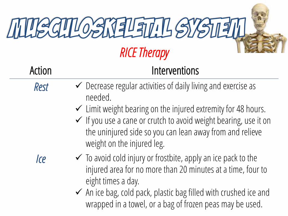
RICE Therapy
Action Interventions
Rest Decrease regular activities of daily living and exercise as needed.
Limit weight bearing on the injured extremity for 48 hours. If you use a cane or crutch to avoid weight bearing, use it on
the uninjured side so you can lean away from and relieve weight on the injured leg.
Ice To avoid cold injury or frostbite, apply an ice pack to the injured area for no more than 20 minutes at a time, four to eight times a day.
An ice bag, cold pack, plastic bag filled with crushed ice and wrapped in a towel, or a bag of frozen peas may be used.
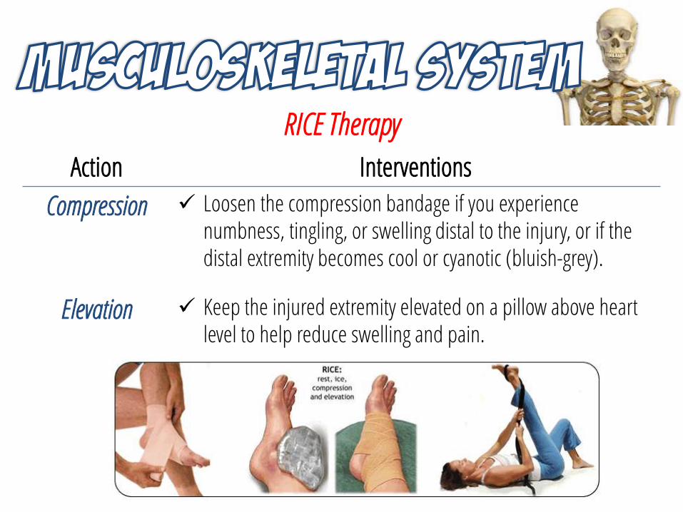
RICE Therapy
Action Interventions
Compression Loosen the compression bandage if you experience numbness, tingling, or swelling distal to the injury, or if the distal extremity becomes cool or cyanotic (bluish-grey).
Elevation Keep the injured extremity elevated on a pillow above heart level to help reduce swelling and pain.
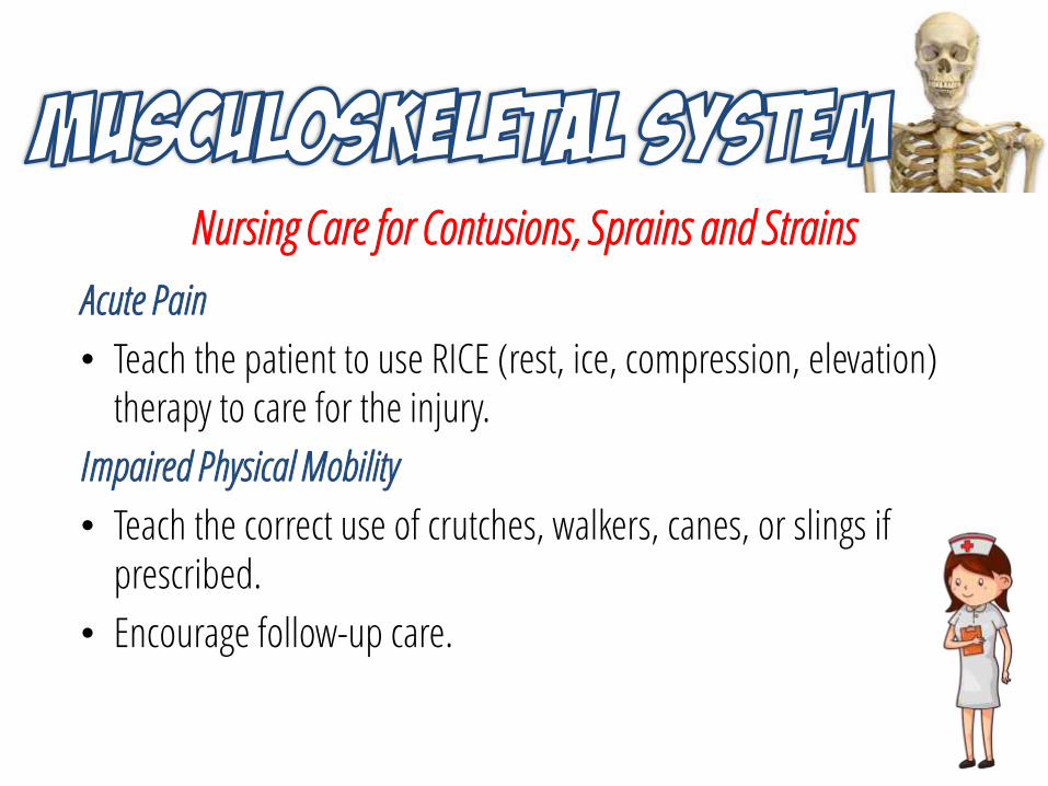
Nursing Care for Contusions, Sprains and Strains
Acute Pain
• Teach the patient to use RICE (rest, ice, compression, elevation) therapy to care for the injury.
Impaired Physical Mobility
• Teach the correct use of crutches, walkers, canes, or slings if prescribed.
• Encourage follow-up care.
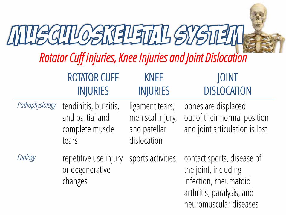
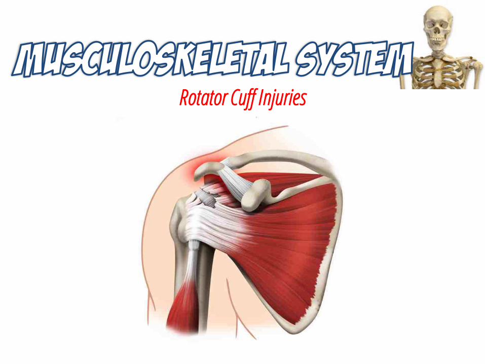
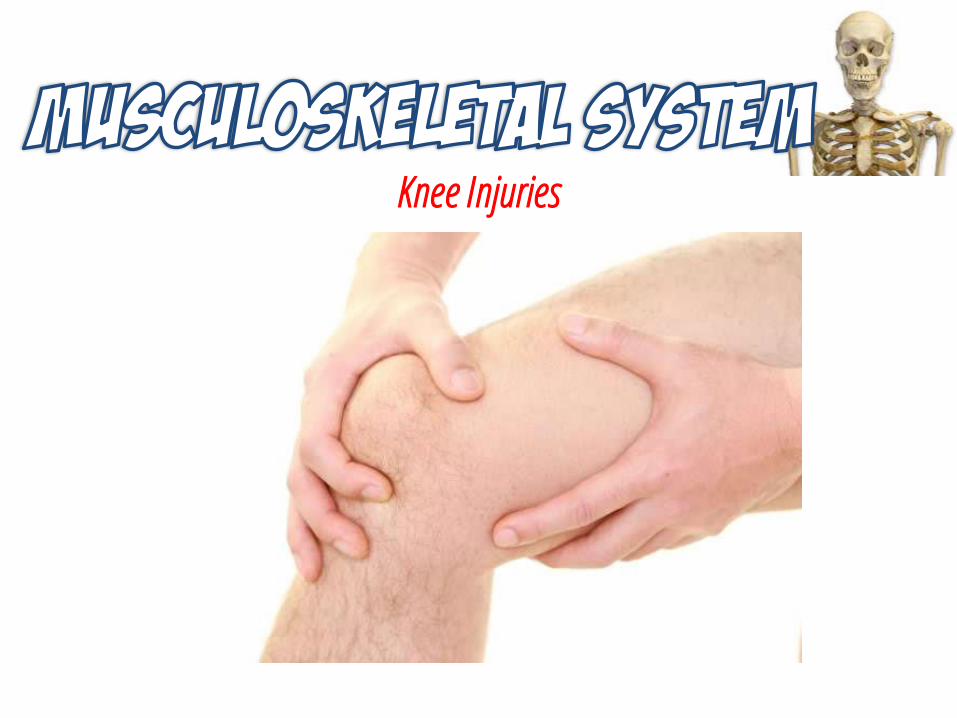
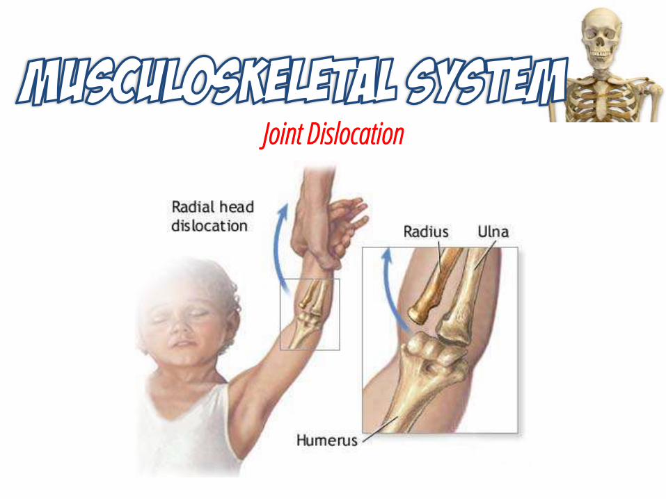
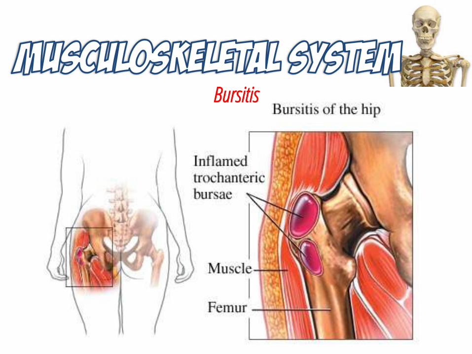
Rotator Cuff Injuries, Knee Injuries and Joint Dislocation
ROTATOR CUFFINJURIES
KNEE INJURIES
JOINT DISLOCATION
Pathophysiology tendinitis, bursitis, and partial and complete muscle tears
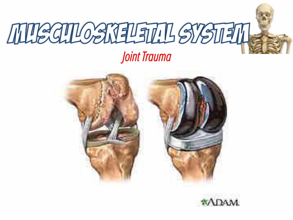
ligament tears, meniscal injury, and patellar dislocation
bones are displacedout of their normal position and joint articulation is lost
Etiology repetitive use injury or degenerative changes
sports activities contact sports, disease of the joint, including infection, rheumatoid arthritis, paralysis, and neuromuscular diseases
Rotator Cuff Injuries
Knee Injuries
Joint Dislocation
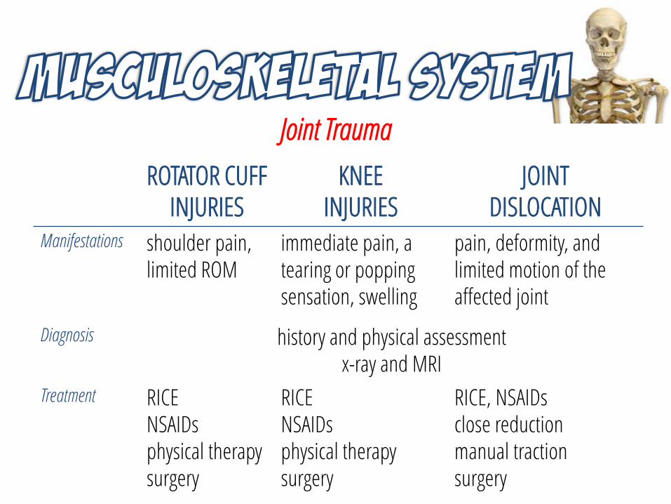
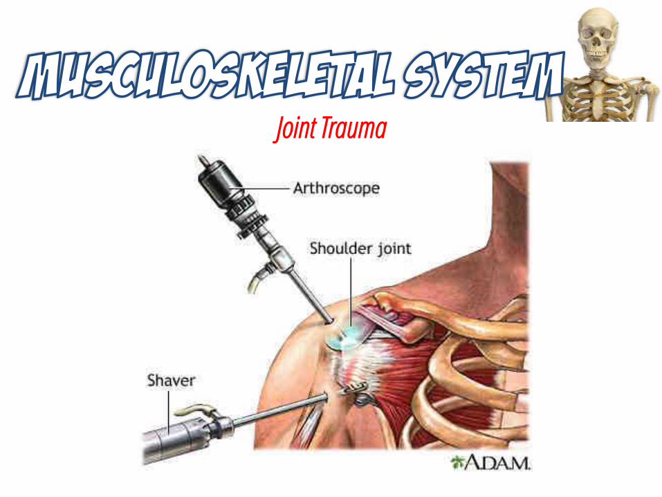
Joint Trauma
ROTATOR CUFFINJURIES
KNEE INJURIES
JOINT DISLOCATION
Manifestations shoulder pain, limited ROM
immediate pain, a tearing or popping sensation, swelling
pain, deformity, and limited motion of the affected joint
Diagnosis history and physical assessmentx-ray and MRI
Treatment RICENSAIDsphysical therapysurgery
RICENSAIDsphysical therapysurgery
RICE, NSAIDsclose reductionmanual tractionsurgery
Joint Trauma
Joint Trauma
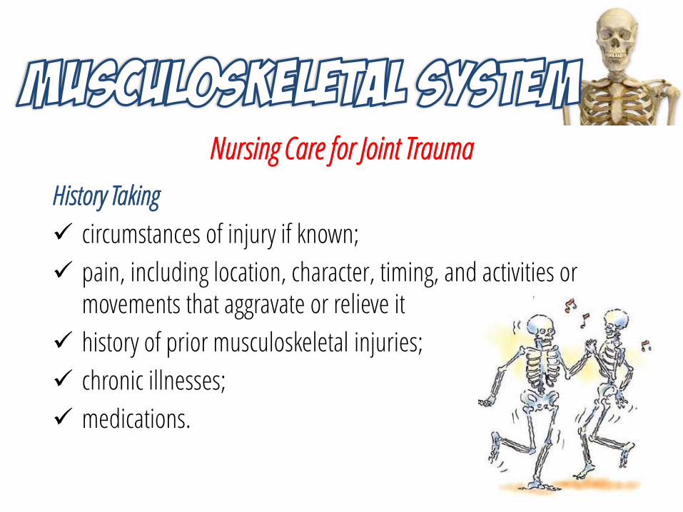
Nursing Care for Joint Trauma
History Taking
circumstances of injury if known;
pain, including location, character, timing, and activities or movements that aggravate or relieve it
history of prior musculoskeletal injuries;
chronic illnesses;
medications.
Nursing Care for Joint Trauma
Physical Assessment
Compare the position, color, size, and temperature of the affected joint to the corresponding unaffected joint.
Palpate for tenderness, crepitus, temperature, and swelling.
Instruct the patient or assist to move the joint through its normal range of motion, stopping and noting where pain is experienced.
When a joint dislocation is suspected, assess color, temperature, pulses, movement, and sensation of the limb distal to the affected joint.
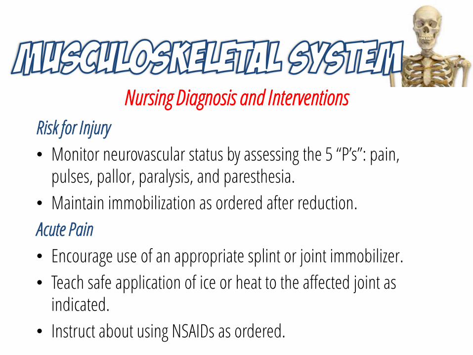
Nursing Diagnosis and Interventions
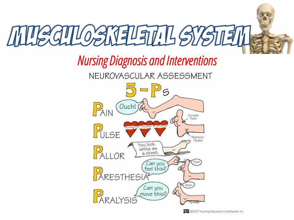
Risk for Injury
• Monitor neurovascular status by assessing the 5 “P’s”: pain, pulses, pallor, paralysis, and paresthesia.
• Maintain immobilization as ordered after reduction.
Acute Pain
• Encourage use of an appropriate splint or joint immobilizer.
• Teach safe application of ice or heat to the affected joint as indicated.
• Instruct about using NSAIDs as ordered.
Nursing Diagnosis and Interventions
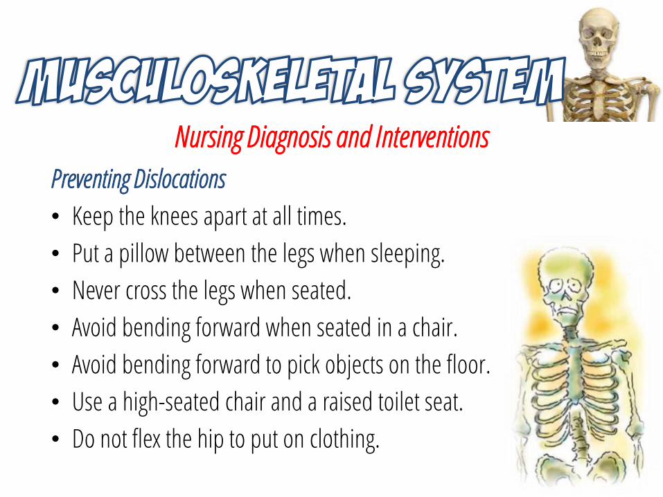
Preventing Dislocations
• Keep the knees apart at all times.
• Put a pillow between the legs when sleeping.
• Never cross the legs when seated.
• Avoid bending forward when seated in a chair.
• Avoid bending forward to pick objects on the floor.
• Use a high-seated chair and a raised toilet seat.
• Do not flex the hip to put on clothing.
Nursing Diagnosis and Interventions
Nursing Diagnosis and Interventions
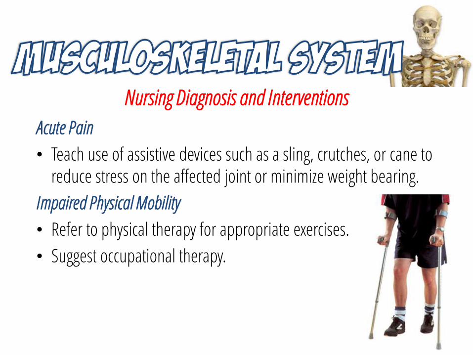
Acute Pain
• Teach use of assistive devices such as a sling, crutches, or cane to reduce stress on the affected joint or minimize weight bearing.
Impaired Physical Mobility
• Refer to physical therapy for appropriate exercises.
• Suggest occupational therapy.
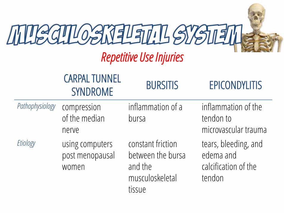
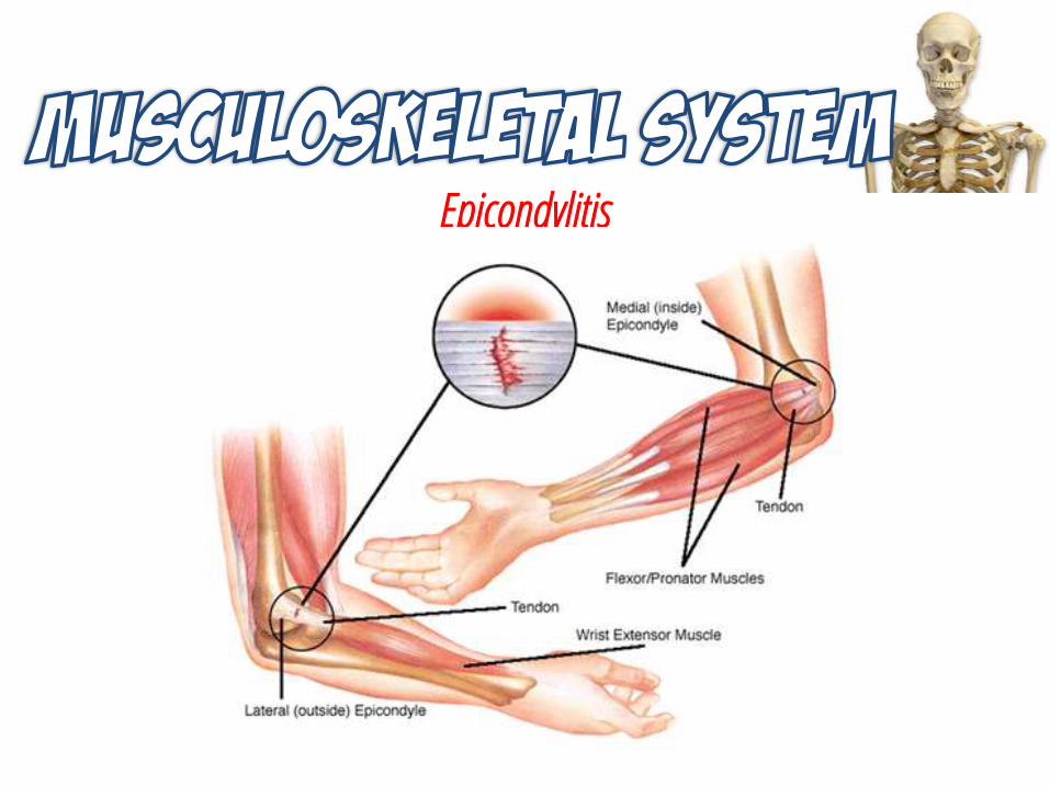
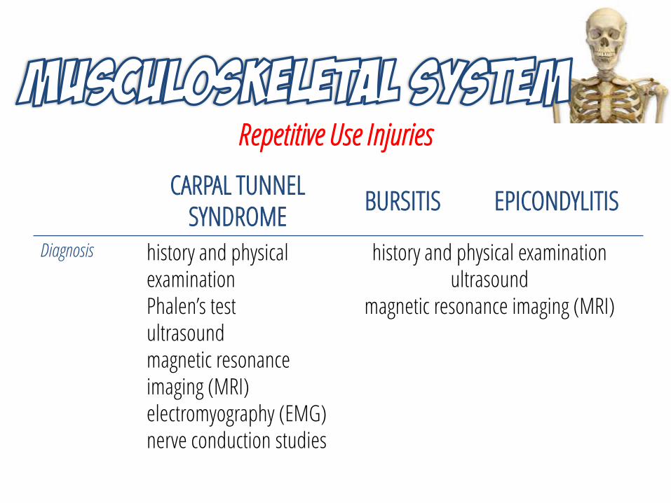
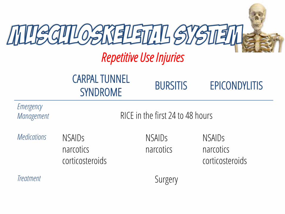
Repetitive Use Injuries
CARPAL TUNNEL SYNDROME
BURSITIS EPICONDYLITIS
Pathophysiology compressionof the median nerve
inflammation of a bursa
inflammation of the tendon to microvascular trauma
Etiology using computerspost menopausal women
constant friction between the bursa and the musculoskeletal tissue
tears, bleeding, andedema and calcification of the tendon
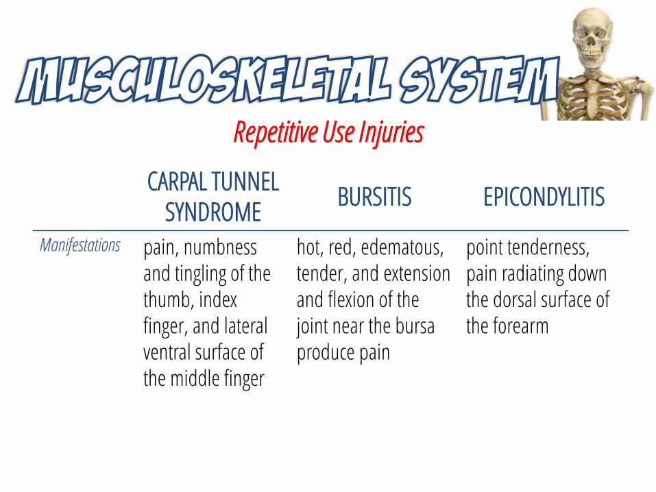
Repetitive Use Injuries
CARPAL TUNNEL SYNDROME
BURSITIS EPICONDYLITIS
Manifestations pain, numbness and tingling of the thumb, index finger, and lateral ventral surface of the middle finger
hot, red, edematous, tender, and extension and flexion of the joint near the bursa produce pain
point tenderness, pain radiating down the dorsal surface of the forearm
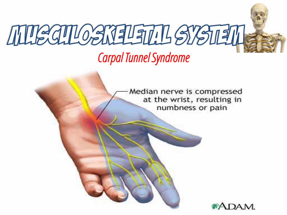
Carpal Tunnel Syndrome
Bursitis
Epicondylitis
Repetitive Use Injuries
CARPAL TUNNEL SYNDROME
BURSITIS EPICONDYLITIS
Diagnosis history and physical examinationPhalen’s testultrasound magnetic resonance imaging (MRI)electromyography (EMG)nerve conduction studies
history and physical examinationultrasound
magnetic resonance imaging (MRI)
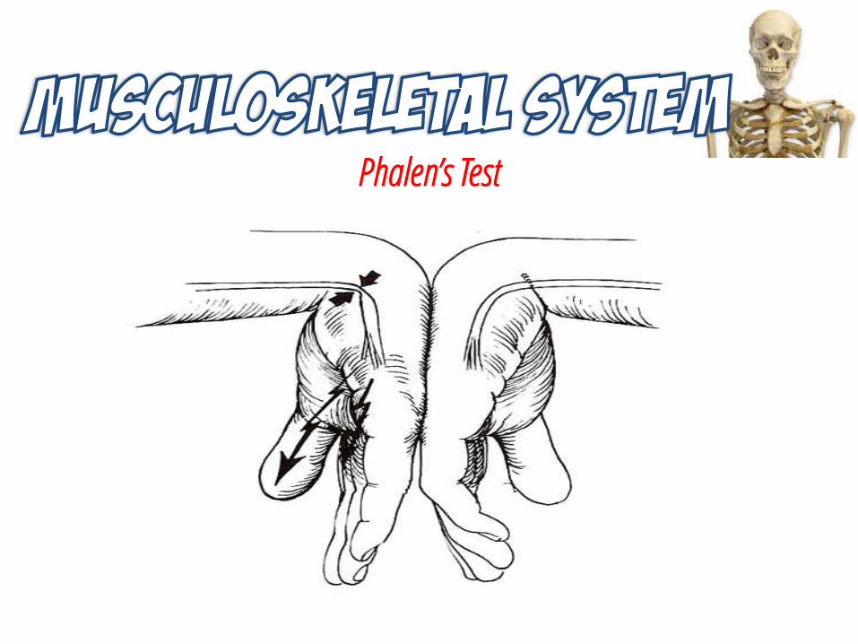
Phalen’s Test
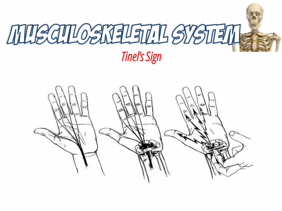
Tinel’s Sign
Repetitive Use Injuries
CARPAL TUNNEL SYNDROME
BURSITIS EPICONDYLITIS
Emergency Management RICE in the first 24 to 48 hours
Medications NSAIDsnarcoticscorticosteroids
NSAIDsnarcotics
NSAIDsnarcoticscorticosteroids
Treatment Surgery
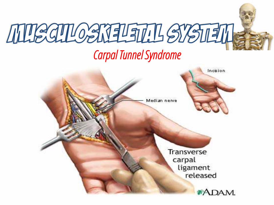
Carpal Tunnel Syndrome
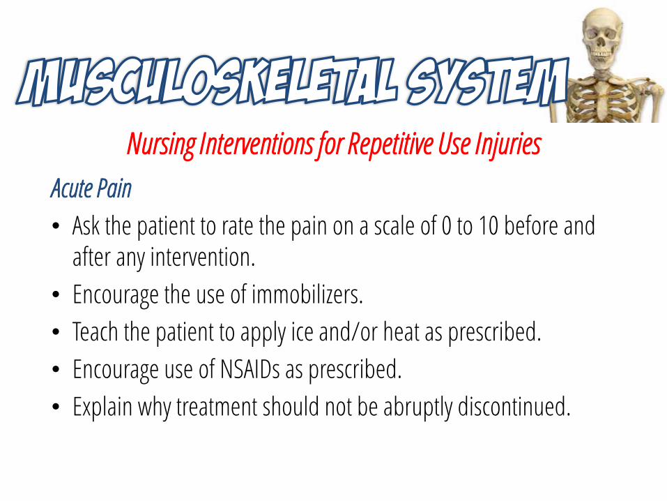
Nursing Interventions for Repetitive Use Injuries
Acute Pain
• Ask the patient to rate the pain on a scale of 0 to 10 before and after any intervention.
• Encourage the use of immobilizers.
• Teach the patient to apply ice and/or heat as prescribed.
• Encourage use of NSAIDs as prescribed.
• Explain why treatment should not be abruptly discontinued.
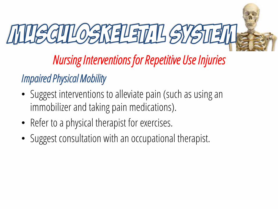
Nursing Interventions for Repetitive Use Injuries
Impaired Physical Mobility
• Suggest interventions to alleviate pain (such as using an immobilizer and taking pain medications).
• Refer to a physical therapist for exercises.
• Suggest consultation with an occupational therapist.
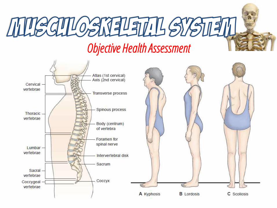
Objective Health Assessment
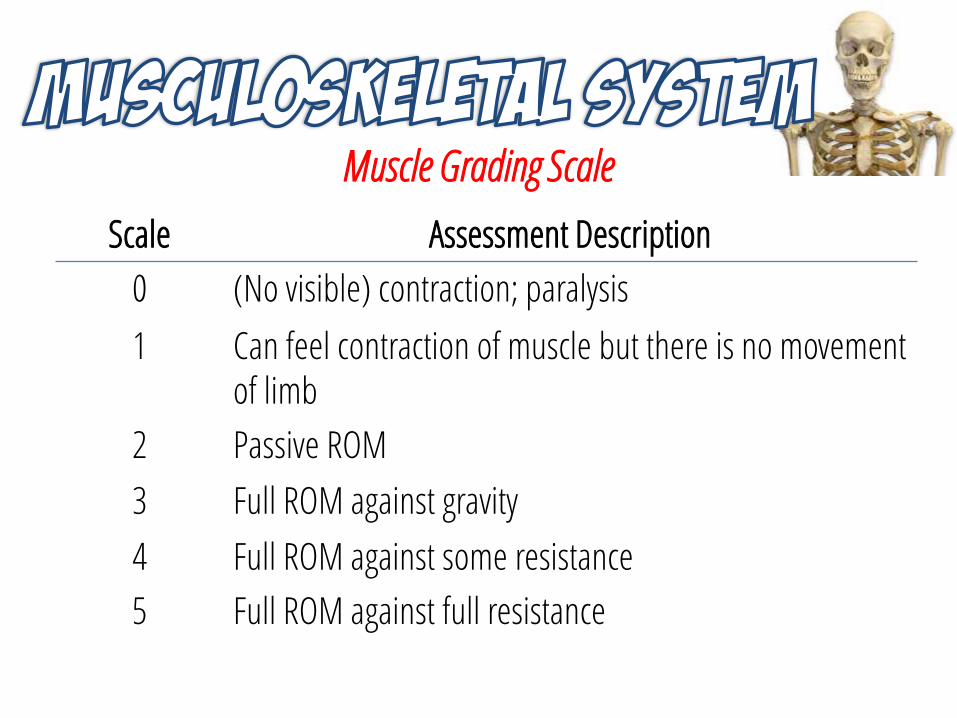
Muscle Grading Scale
Scale Assessment Description
0 (No visible) contraction; paralysis
1 Can feel contraction of muscle but there is no movement of limb
2 Passive ROM
3 Full ROM against gravity
4 Full ROM against some resistance
5 Full ROM against full resistance
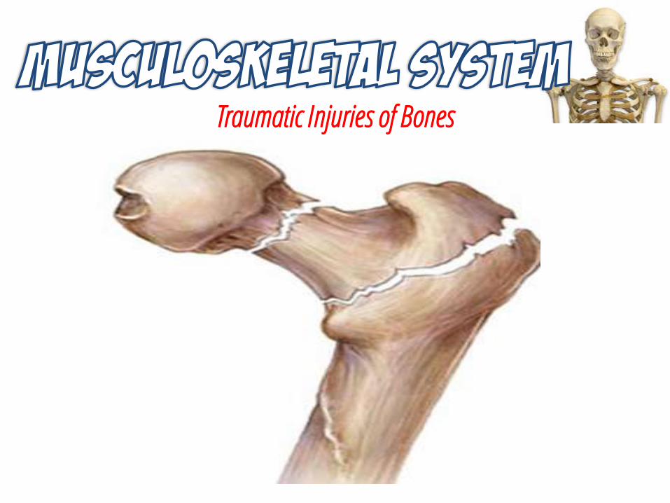
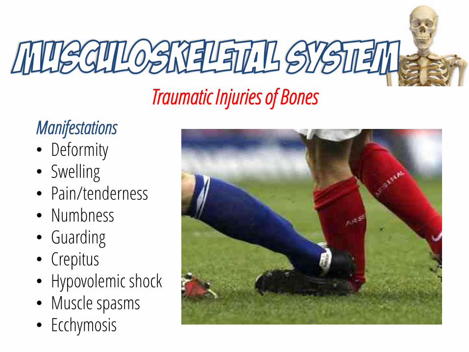
Traumatic Injuries of Bones
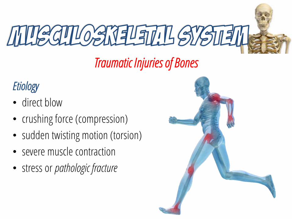
Traumatic Injuries of Bones
Etiology
• direct blow
• crushing force (compression)
• sudden twisting motion (torsion)
• severe muscle contraction
• stress or pathologic fracture
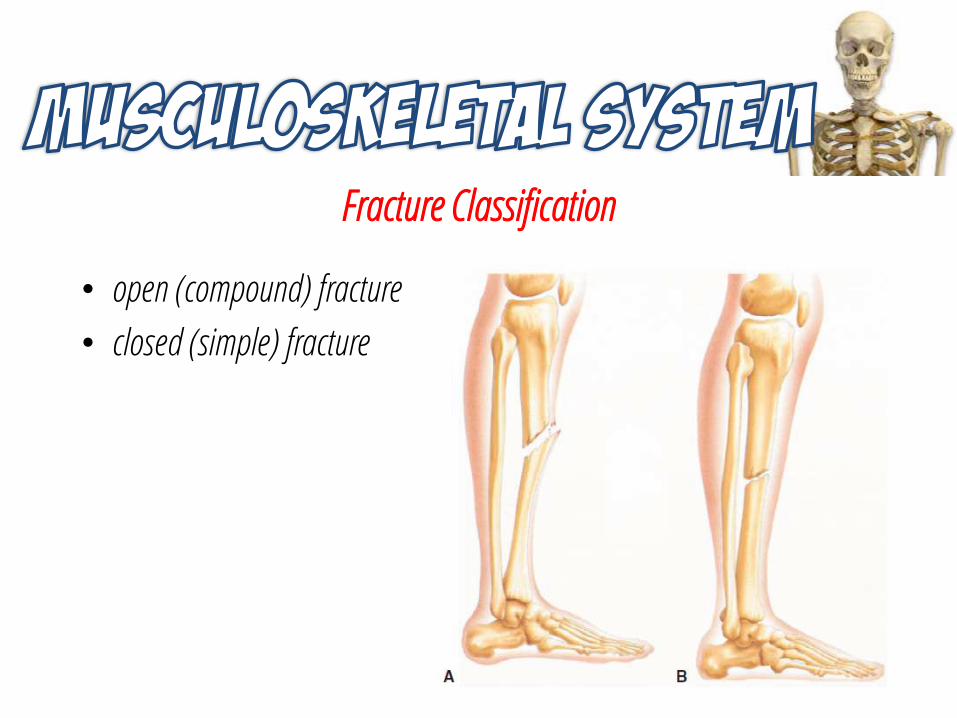
Fracture Classification
• open (compound) fracture
• closed (simple) fracture
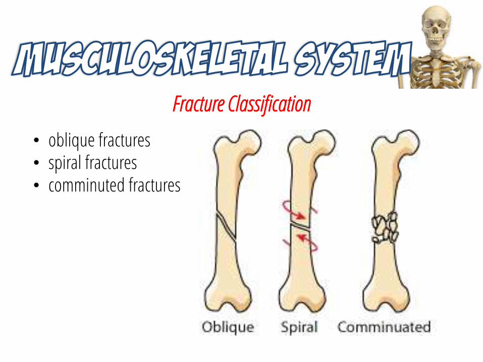
Fracture Classification
• oblique fractures• spiral fractures• comminuted fractures
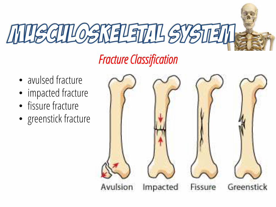
Fracture Classification
• avulsed fracture• impacted fracture• fissure fracture• greenstick fracture
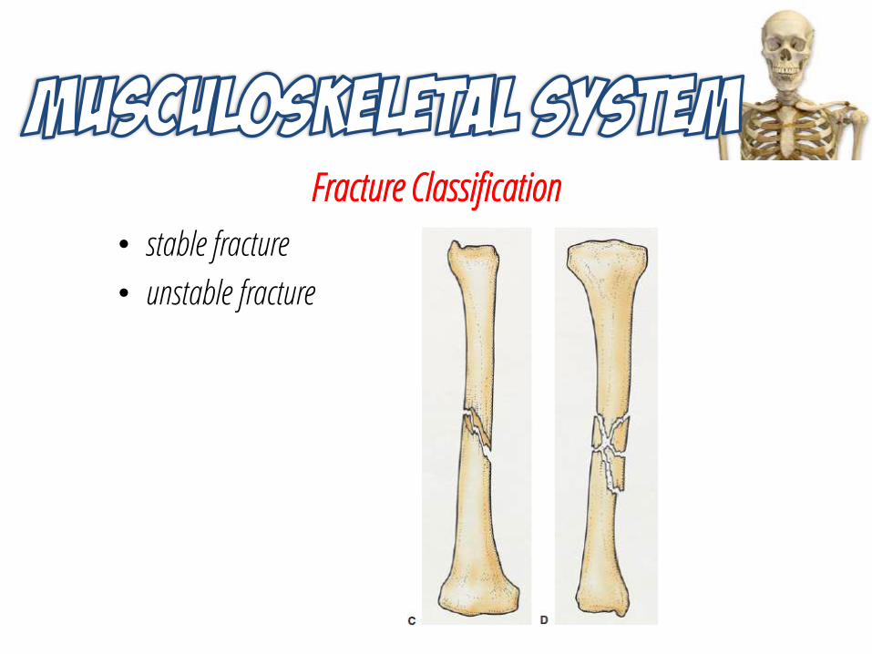
Fracture Classification
• stable fracture
• unstable fracture
Traumatic Injuries of Bones
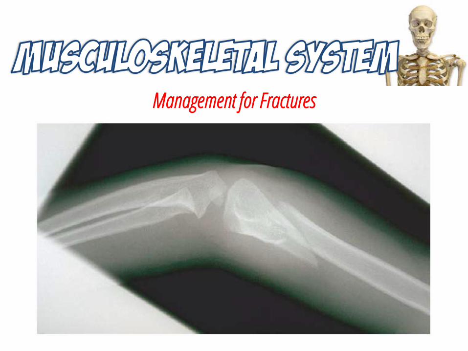
Manifestations • Deformity• Swelling• Pain/tenderness• Numbness• Guarding• Crepitus• Hypovolemic shock• Muscle spasms• Ecchymosis
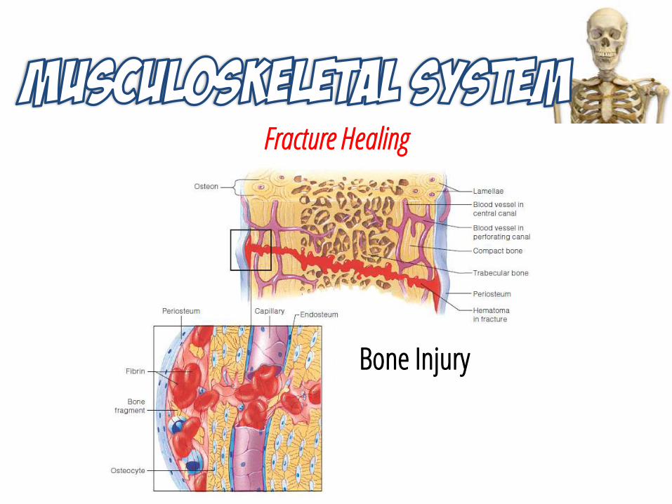
Fracture Healing
Bone Injury
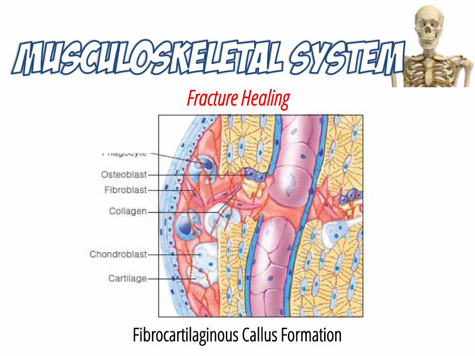
Fracture Healing
Fibrocartilaginous Callus Formation
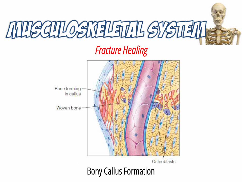
Fracture Healing
Bony Callus Formation
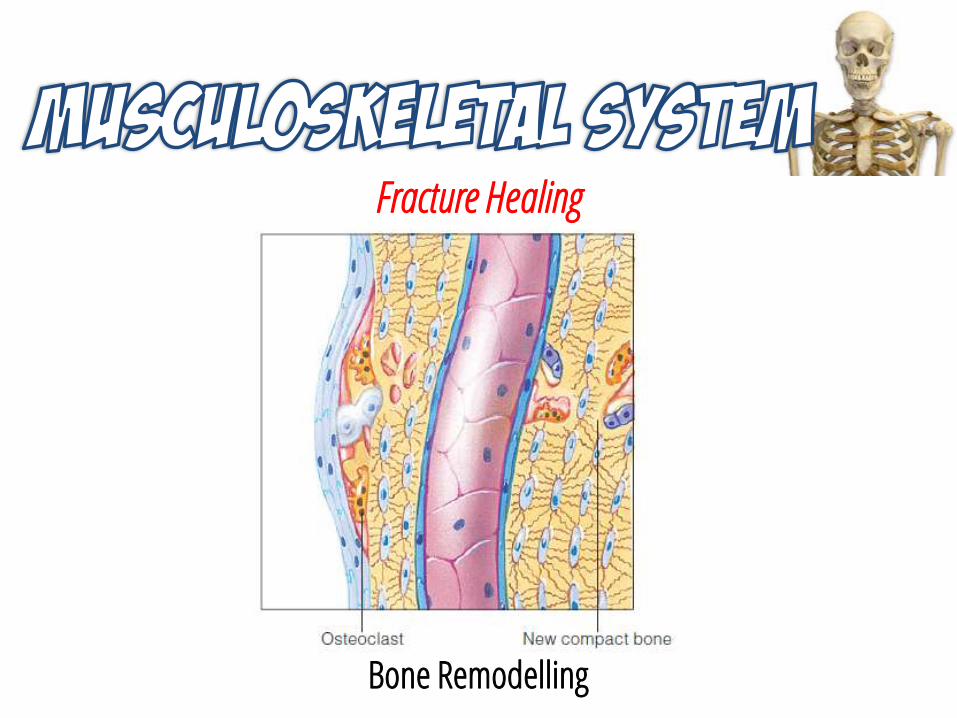
Fracture Healing
Bone Remodelling
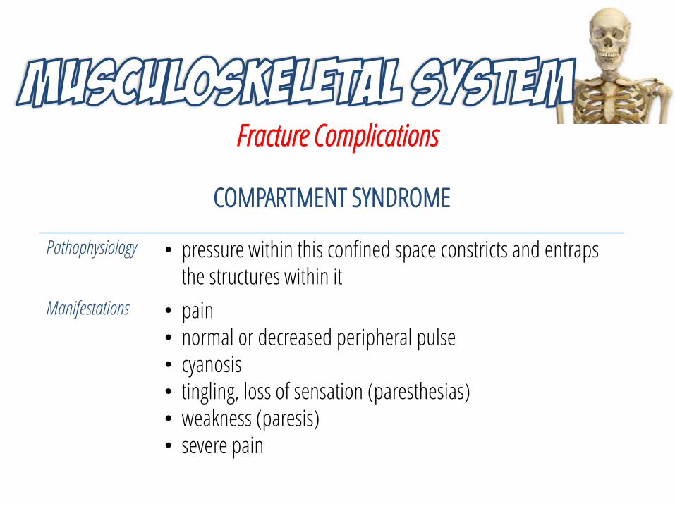
Fracture Complications
COMPARTMENT SYNDROME
Pathophysiology • pressure within this confined space constricts and entraps the structures within it
Manifestations • pain• normal or decreased peripheral pulse• cyanosis• tingling, loss of sensation (paresthesias)• weakness (paresis)• severe pain
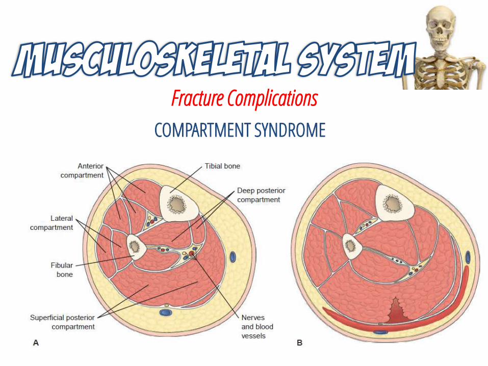
Fracture Complications
COMPARTMENT SYNDROME
Fracture Complications
COMPARTMENT SYNDROME
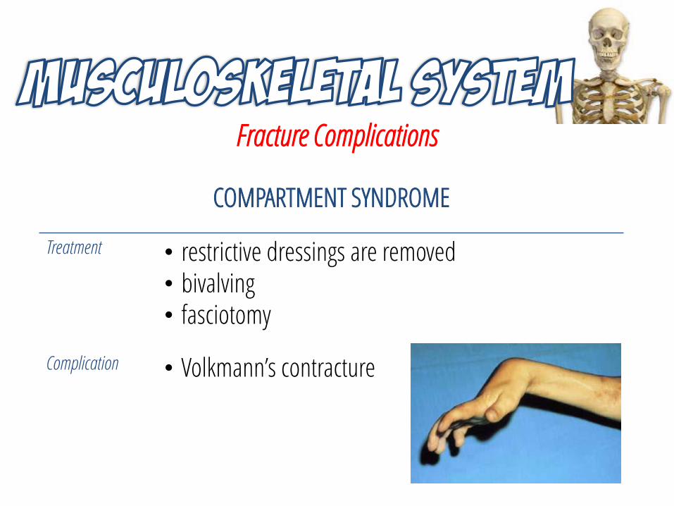
Treatment • restrictive dressings are removed • bivalving• fasciotomy
Complication • Volkmann’s contracture
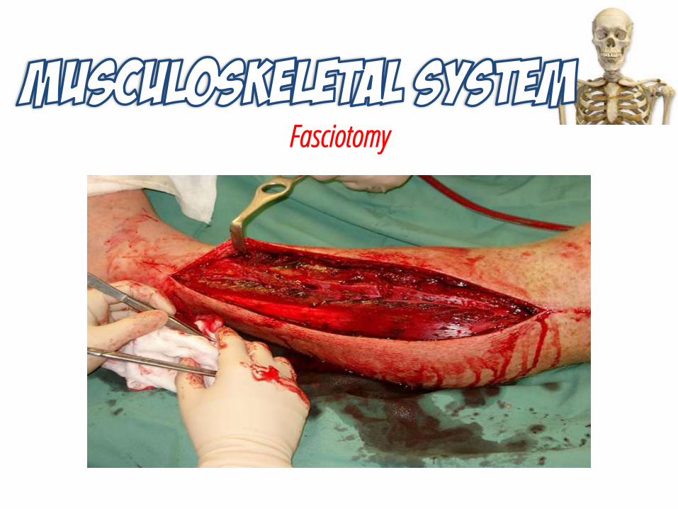
Fasciotomy
Fracture Complications
FAT EMBOLISM
Pathophysiology • fat globules lodge in the pulmonary vascular bed or peripheral circulation
Etiology • long bone fractures and other major trauma• hip replacement surgery
Manifestations • neurologic dysfunction• pulmonary insufficiency• petechial rash on the chest, axilla, and upper arms
Fracture Complications
FAT EMBOLISM
Treatment • early stabilization of long bone fractures• intubation and mechanical ventilation• fluid balance is closely monitored• corticosteroids
Fracture Complications
DEEP VEIN THROMBOSIS
Pathophysiology • blood clot forms along the intimal lining of a large vein, accompanied by inflammation of the vein wall
Etiology • venous stasis, or decreased blood flow• injury to blood vessel walls• altered blood coagulation
Manifestations • swelling, pain, tenderness, or cramping of the affected extremity
Fracture Complications
DEEP VEIN THROMBOSIS
Diagnosis • doppler ultrasonography• magnetic resonance imaging• venogram
Treatment • early immobilization of the fracture• early ambulation of the patient• prophylactic anticoagulation• antiembolism stockings and compression boots
Fracture Complications
INFECTION
Pathophysiology • Pseudomonas, Staphylococcus, or Clostridium organisms may invade the wound or bone
Complication • osteomyelitis
Fracture Complications
DELAYED UNION AND NONUNION
Pathophysiology • prolonged healing of bones beyond the usual time period• delayed union may lead to nonunion
Etiology • Injury-related: the type and location of facture and accompanying soft tissue injury
• System related: age, general health, immune status, chronic diseases, and smoking
Diagnosis • serial x-ray studies
Fracture Complications
DELAYED UNION AND NONUNION
Treatment • internal fixation and bone grafting• bone debridement• electrical or ultrasonic stimulation of the fracture site• growth hormone or parathyroid hormone stimulation
Fracture Complications
COMPLEX REGIONAL PAIN SYNDROME
Pathophysiology • pain receptors become sensitized to catecholamines, neurotransmitters associated with sympathetic nervous system activity
Etiology • female• older age
Diagnosis • history and physical examination• x-ray
Fracture Complications
COMPLEX REGIONAL PAIN SYNDROME
Manifestations • severe, diffuse, and burning pain • affected extremity is inflamed and edematous, later
becoming cool and pale• muscle wasting, skin and nail changes, and bone
abnormalities
Treatment • sympathetic nervous system blocking agent
Management for Fractures
Emergency Care
• Immobilizing the fracture
• Maintaining tissue perfusion
• Preventing infection
Diagnosis
• X-rays and bone scans
• Blood chemistry studies, complete blood count (CBC), and coagulation studies
Management for Fractures
Management for Fractures
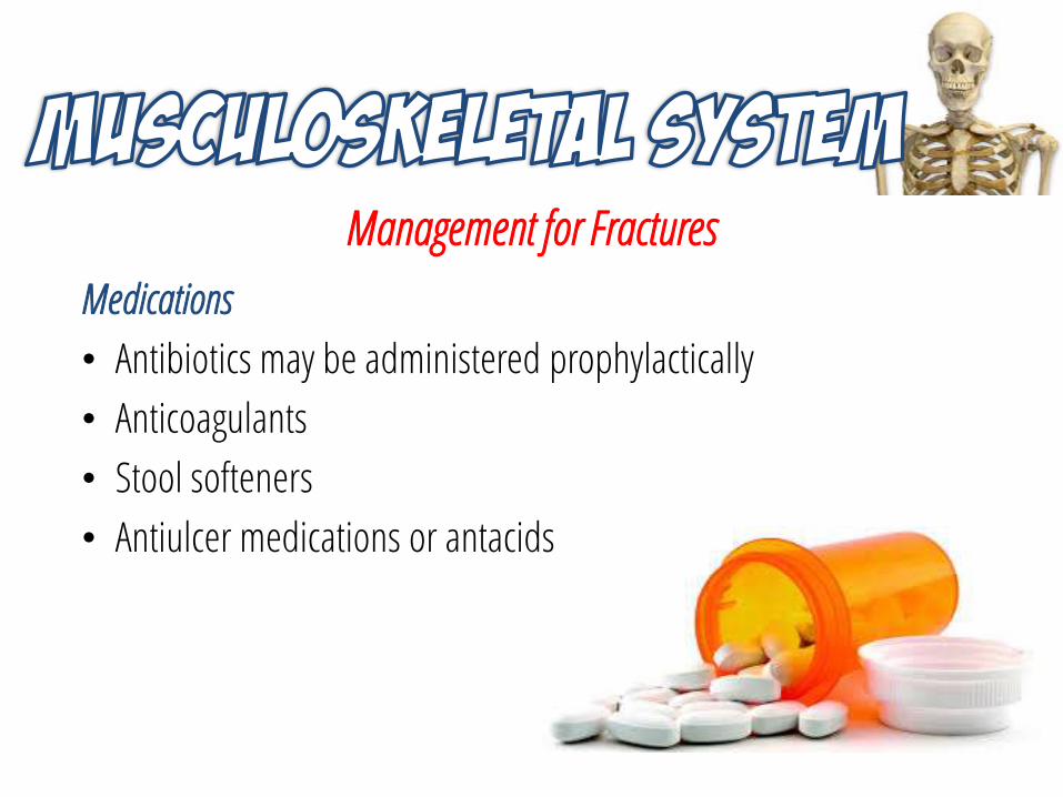
Medications
• Antibiotics may be administered prophylactically
• Anticoagulants
• Stool softeners
• Antiulcer medications or antacids
Management for Fractures
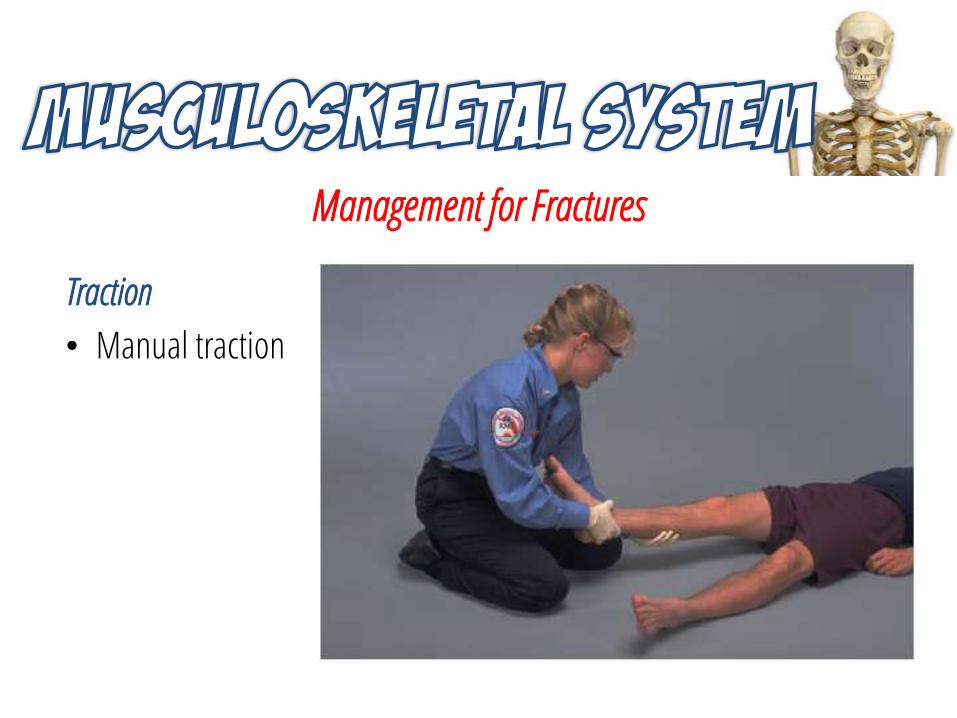
Traction
• Manual traction
Management for Fractures
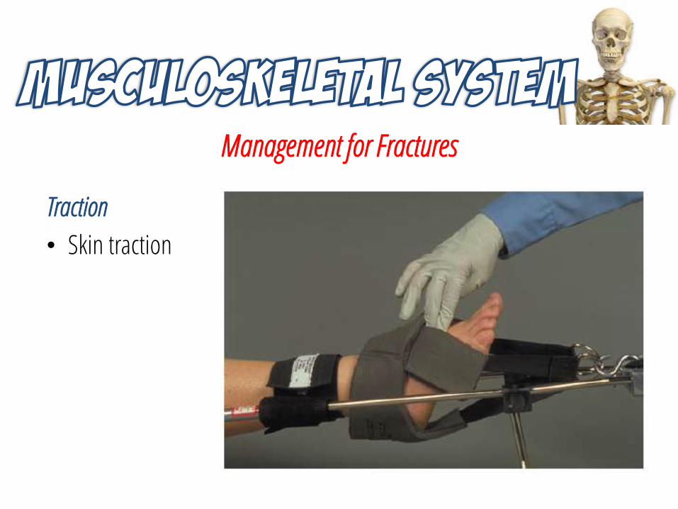
Traction
• Skin traction
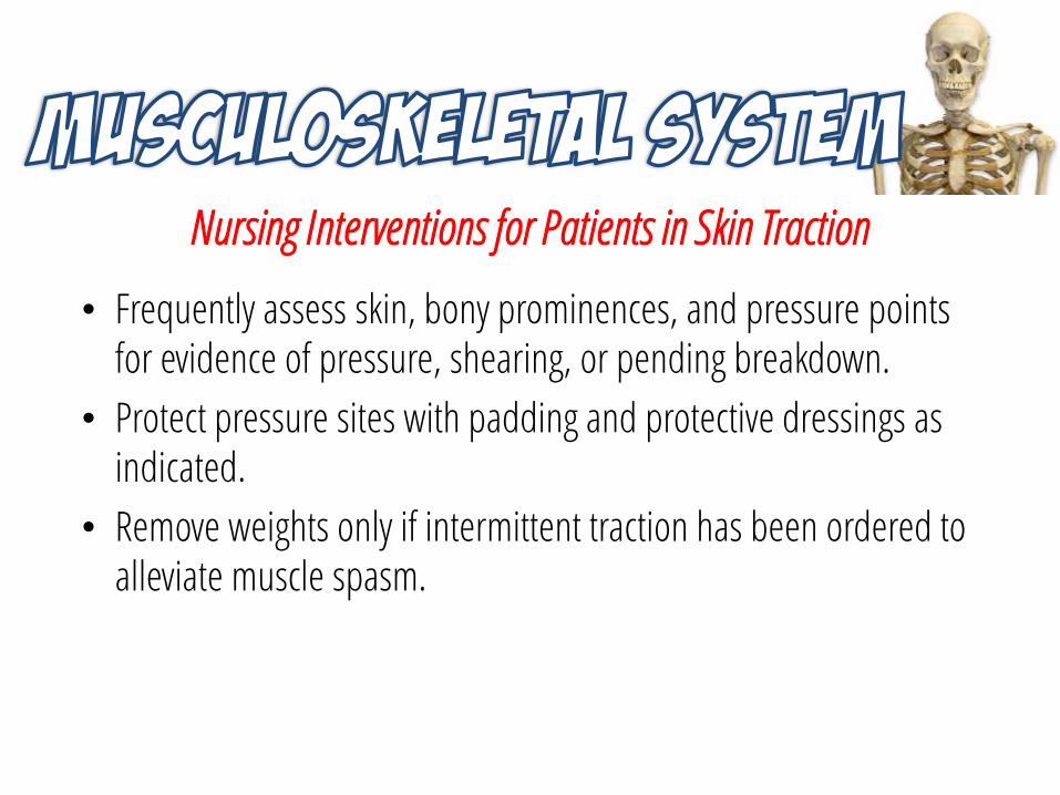
Nursing Interventions for Patients in Skin Traction
• Frequently assess skin, bony prominences, and pressure points for evidence of pressure, shearing, or pending breakdown.
• Protect pressure sites with padding and protective dressings as indicated.
• Remove weights only if intermittent traction has been ordered to alleviate muscle spasm.
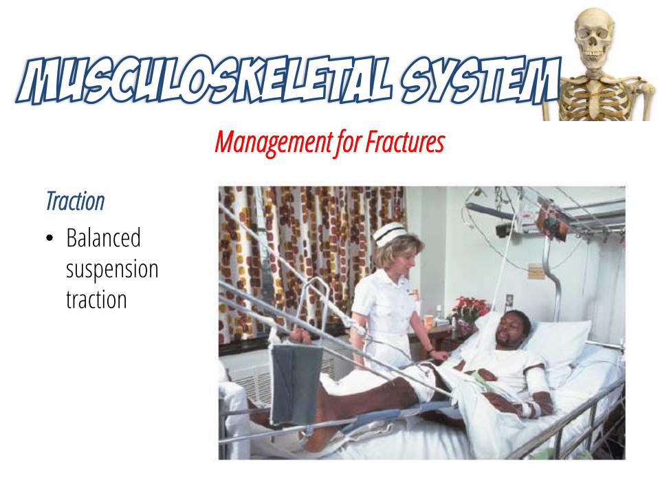
Management for Fractures
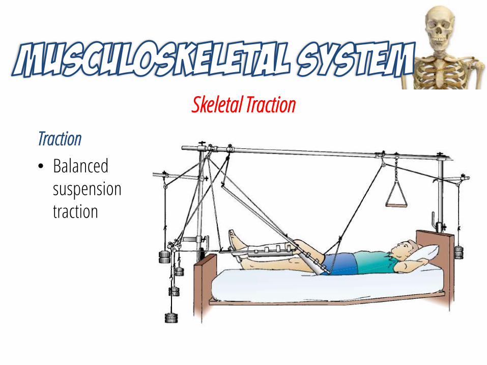
Traction
• Balanced suspension traction
Management for Fractures
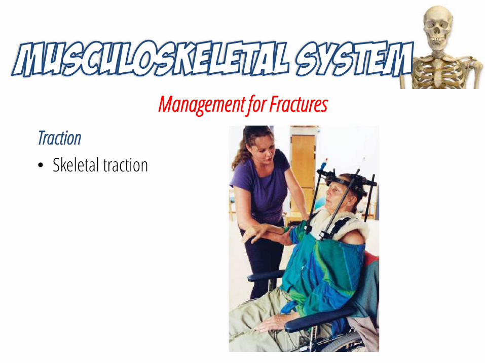
Traction
• Skeletal traction
Skeletal Traction
Traction
• Balanced suspension traction
Skeletal Traction
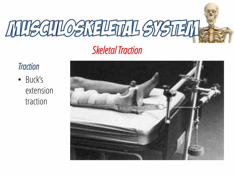
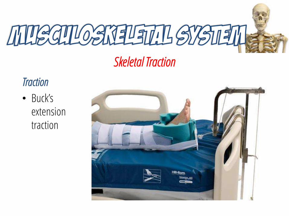
Traction
• Buck’s extension traction
Skeletal Traction
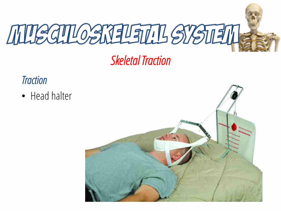
Traction
• Head halter
Skeletal Traction
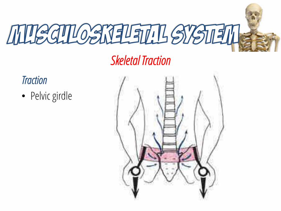
Traction
• Pelvic girdle
Skeletal Traction
Traction
• Buck’s extension traction
Skeletal Traction
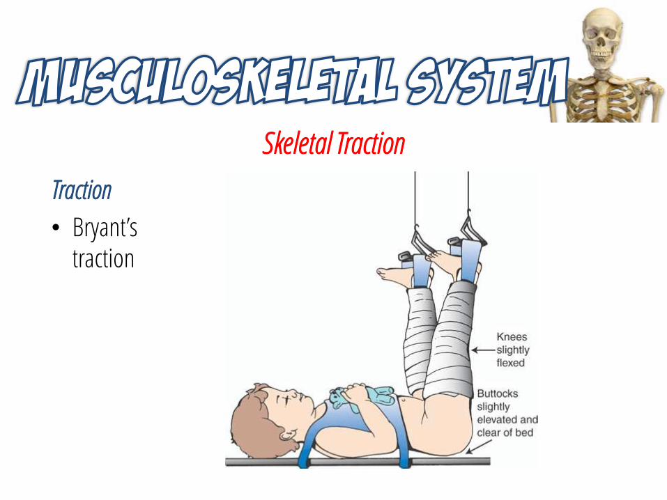
Traction
• Bryant’s traction
Skeletal Traction
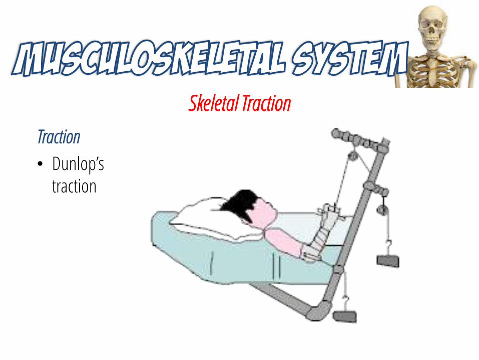
Traction
• Dunlop’s traction
Skeletal Traction
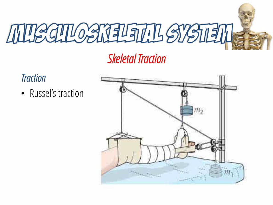
Traction
• Russel’s traction
Skeletal Traction
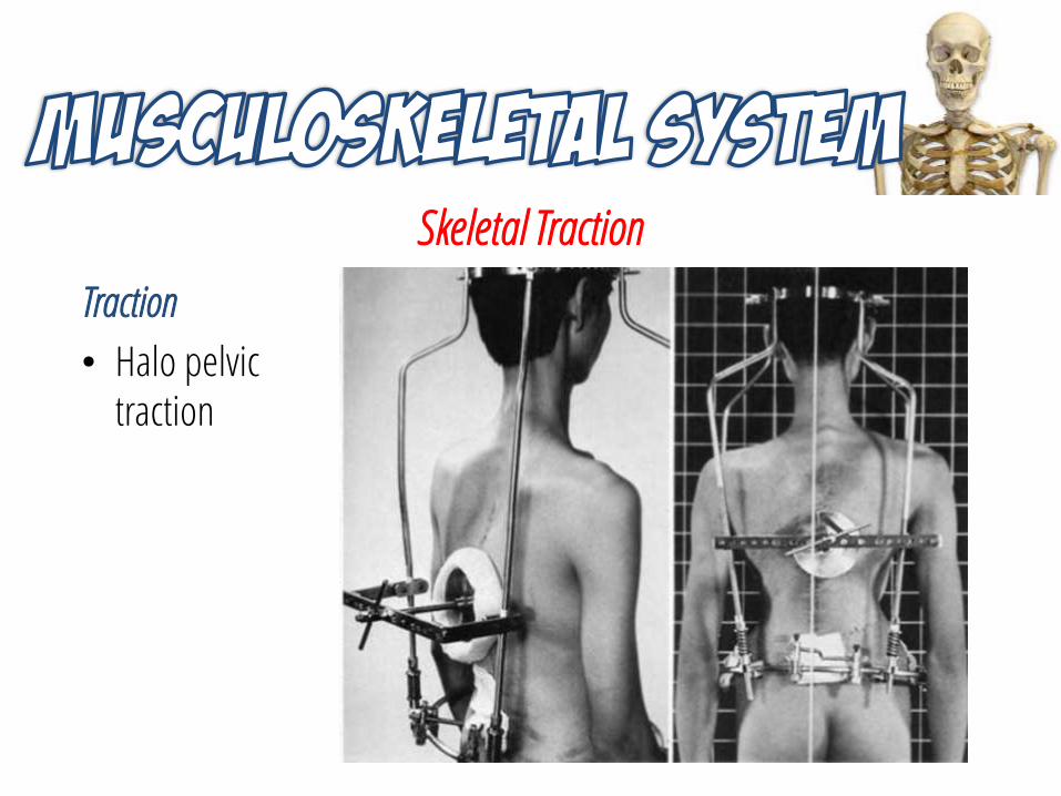
Traction
• Halo pelvic traction
Nursing Interventions for Patients in Skeletal Traction
• Never remove the weights.
• Frequently assess pin insertion sites and provide pin site care per policy.
• Report signs of infection at the pin sites, such as redness, drainage, and increased tenderness.
Nursing Interventions for Patients in Traction
Maintain the pulling force and direction of the traction:
• The patient’s weight provides counter traction.
• Center the patient on the bed; maintain body alignment with the direction of pull.
• Ensure that weights hang freely and do not touch the floor.
• Ensure that nothing is lying on or obstructing the ropes.
• Do not allow the knots at the end of the rope to come into contact with the pulley.
Nursing Interventions for Patients in Traction
• Perform neurovascular assessments frequently.
• Assess for common complications of immobility, including pressure ulcer formation, renal calculi, deep venous thrombosis, pneumonia, paralytic ileus, and loss of appetite.
• If a problem is detected, assist in repositioning. Stabilize the fracture site during repositioning.
• Teach the patient and family about the type and purpose of the traction.
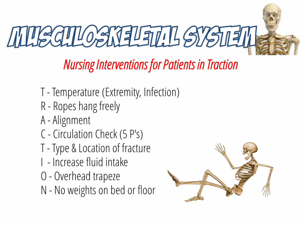
Nursing Interventions for Patients in Traction
T - Temperature (Extremity, Infection)R - Ropes hang freelyA - AlignmentC - Circulation Check (5 P's)T - Type & Location of fractureI - Increase fluid intakeO - Overhead trapezeN - No weights on bed or floor
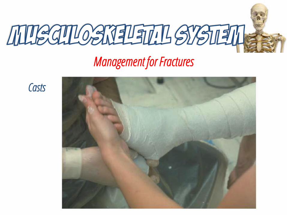
Management for Fractures
Casts
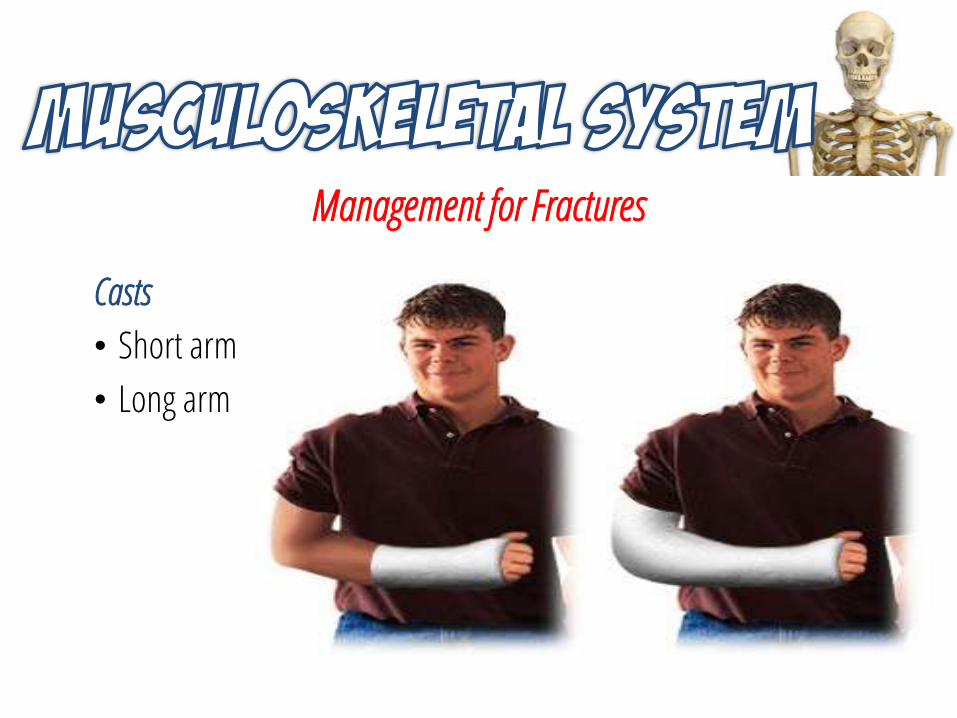
Management for Fractures
Casts
• Short arm
• Long arm
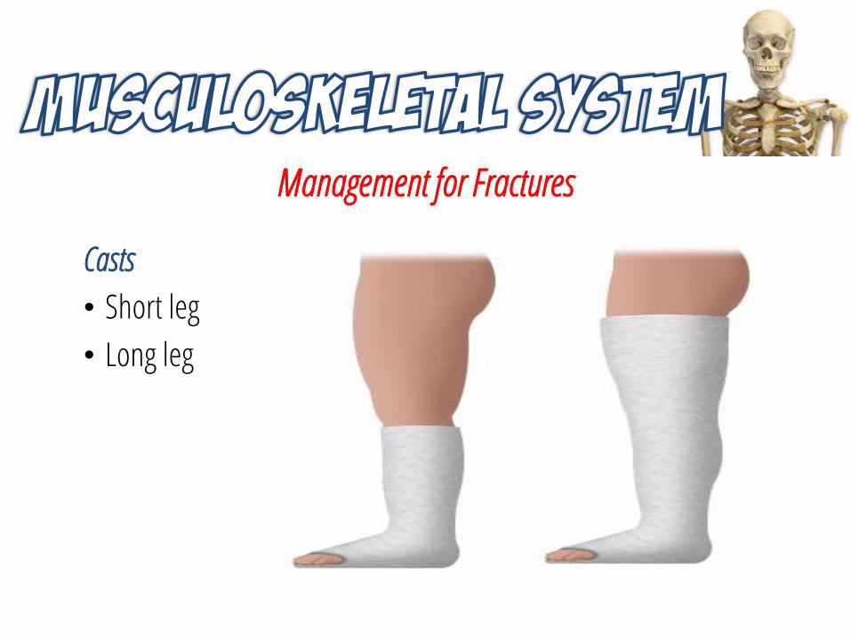
Management for Fractures
Casts
• Short leg
• Long leg
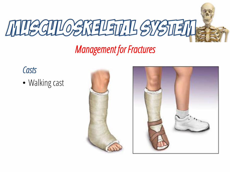
Management for Fractures
Casts
• Walking cast
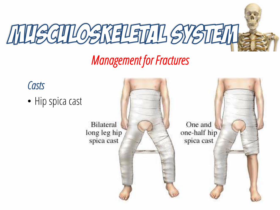
Management for Fractures
Casts
• Hip spica cast
Management for Fractures
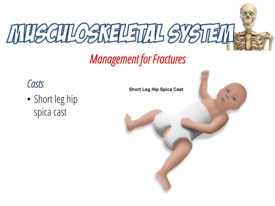
Casts
• Short leg hip spica cast
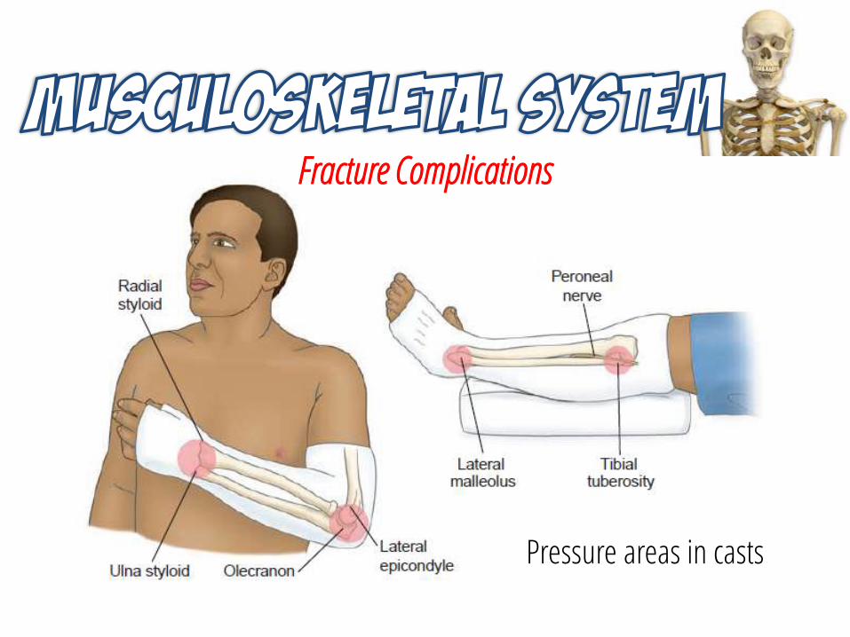
Fracture Complications
Pressure areas in casts
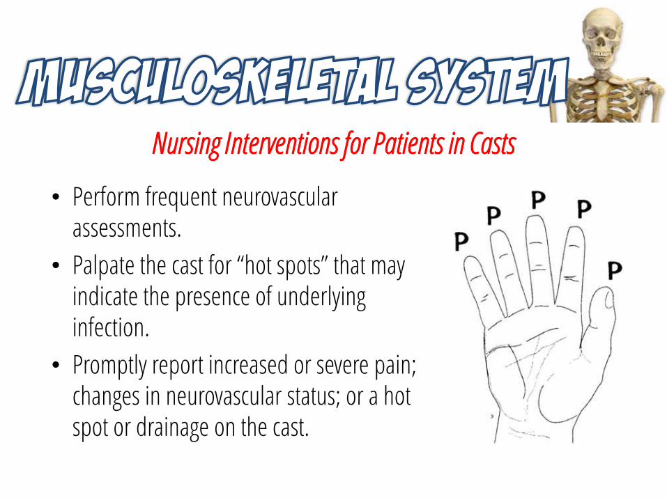
Nursing Interventions for Patients in Casts
• Perform frequent neurovascular assessments.
• Palpate the cast for “hot spots” that may indicate the presence of underlying infection.
• Promptly report increased or severe pain; changes in neurovascular status; or a hot spot or drainage on the cast.
Health Education for the Patient and Family
• Do not use a blow dryer to speed drying; do not cover the cast while it is drying.
• A sensation of warmth during drying is normal.
• Do not put anything into the cast.
• Keep the cast clean and dry; use plastic wrap as needed to protect it.
• If the cast is made of fiberglass, dry it with a blow dryer on the cool setting if it becomes wet.
Health Education for the Patient and Family
• Notify your doctor immediately if you develop increased pain, coolness, changes in color, increased swelling, and/or loss of sensation.
• A sling may be used to distribute the weight of the cast evenly around the neck.
• If crutches are used, arrange for physical therapist to teach correct crutch walking.
• When the cast is removed, an oscillating cast saw will be used.
Management for Fractures
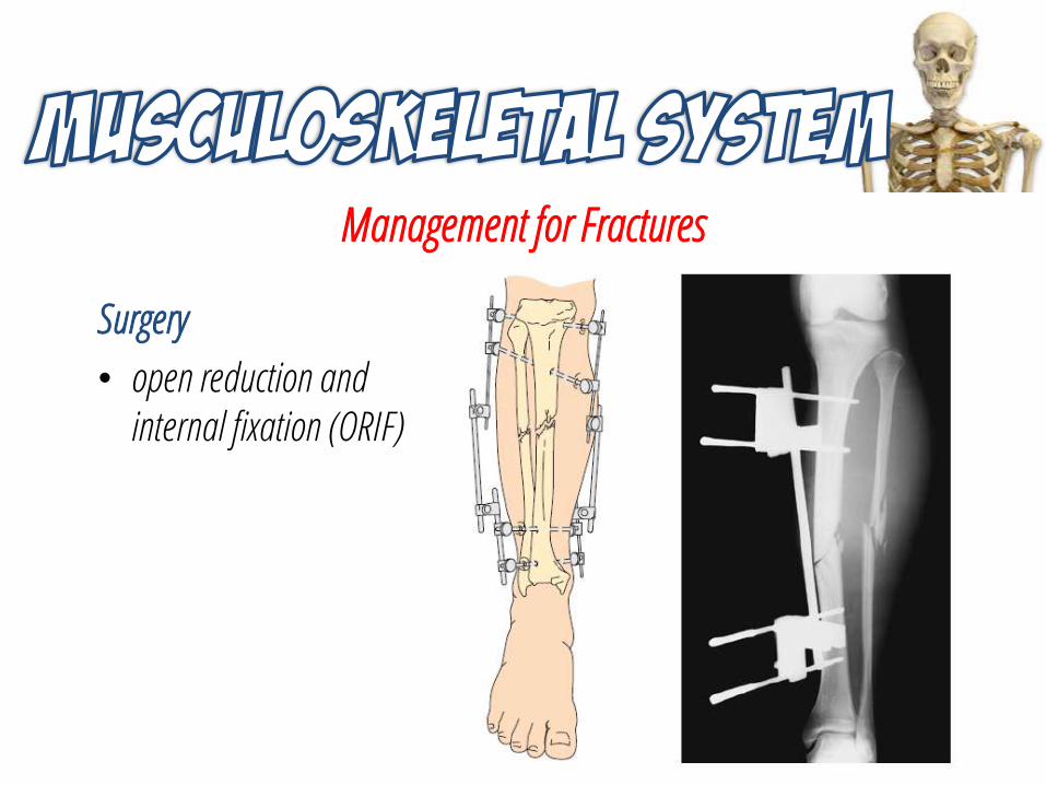
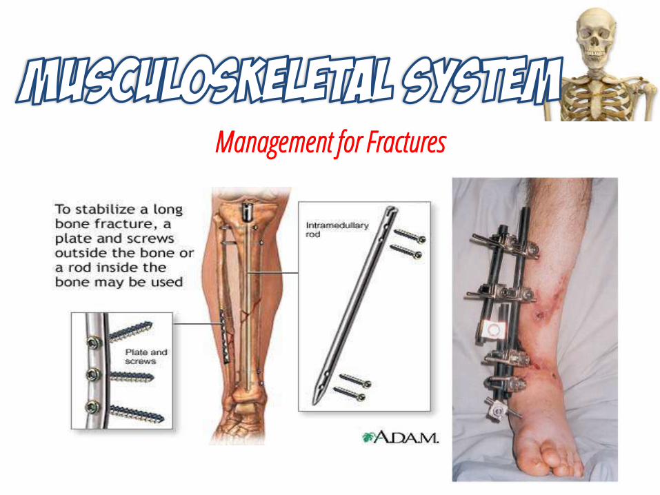
Surgery
• open reduction and internal fixation (ORIF)
Management for Fractures
Management for Fractures
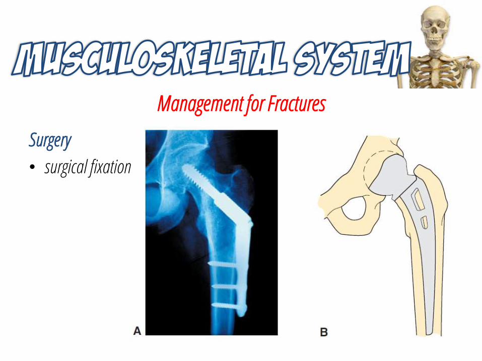
Surgery
• surgical fixation
Management for Fractures
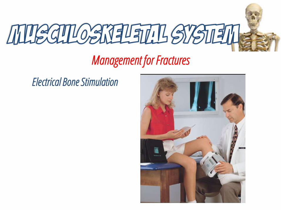
Electrical Bone Stimulation
Nursing Care for Fractures
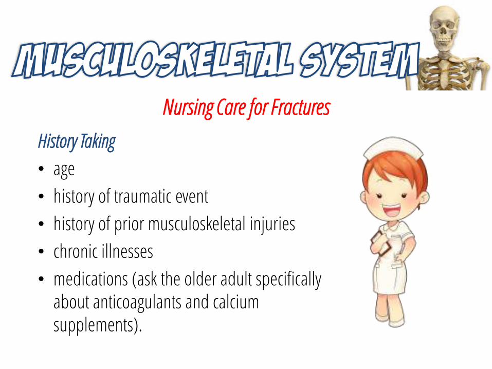
History Taking
• age
• history of traumatic event
• history of prior musculoskeletal injuries
• chronic illnesses
• medications (ask the older adult specifically about anticoagulants and calcium supplements).
Nursing Care for Fractures
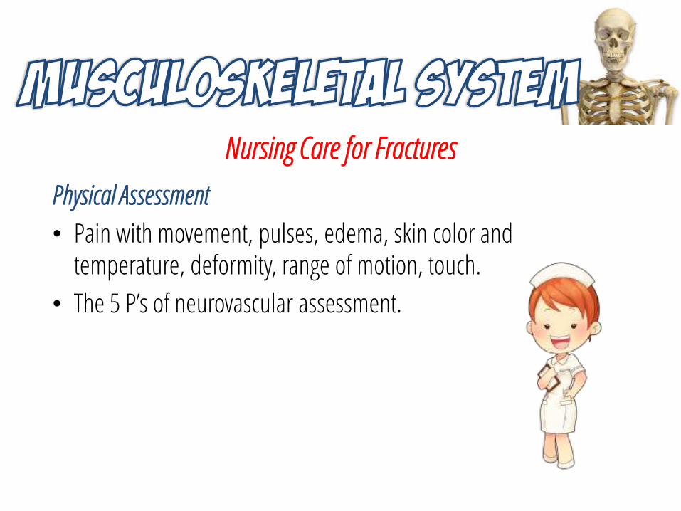
Physical Assessment
• Pain with movement, pulses, edema, skin color and temperature, deformity, range of motion, touch.
• The 5 P’s of neurovascular assessment.
Nursing Diagnosis and Interventions
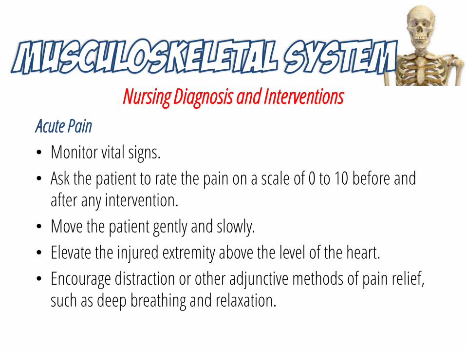
Acute Pain
• Monitor vital signs.
• Ask the patient to rate the pain on a scale of 0 to 10 before and after any intervention.
• Move the patient gently and slowly.
• Elevate the injured extremity above the level of the heart.
• Encourage distraction or other adjunctive methods of pain relief, such as deep breathing and relaxation.
Nursing Diagnosis and Interventions
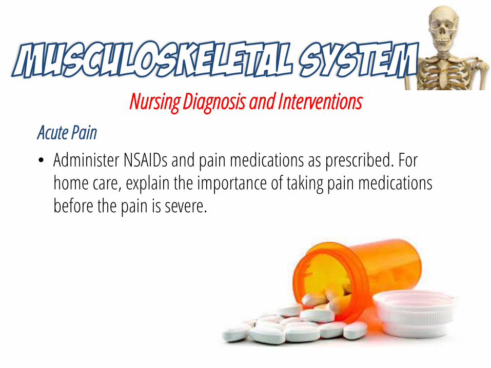
Acute Pain
• Administer NSAIDs and pain medications as prescribed. For home care, explain the importance of taking pain medications before the pain is severe.
Nursing Diagnosis and Interventions
Risk for Peripheral Neurovascular Dysfunction
• Support the injured extremity above and below the fracture site when moving the patient.
• Assess the 5 P’s every 1 to 2 hours. Report abnormal findings immediately.
• Assess nail beds for capillary refill. If nails are too thick or discolored, assess the skin around the nail.
• Monitor the extremity for edema and swelling.
• Assess for deep, throbbing, unrelenting pain.
Nursing Diagnosis and Interventions
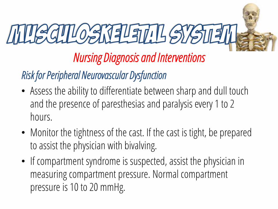
Risk for Peripheral Neurovascular Dysfunction
• Assess the ability to differentiate between sharp and dull touch and the presence of paresthesias and paralysis every 1 to 2 hours.
• Monitor the tightness of the cast. If the cast is tight, be prepared to assist the physician with bivalving.
• If compartment syndrome is suspected, assist the physician in measuring compartment pressure. Normal compartment pressure is 10 to 20 mmHg.
Nursing Diagnosis and Interventions
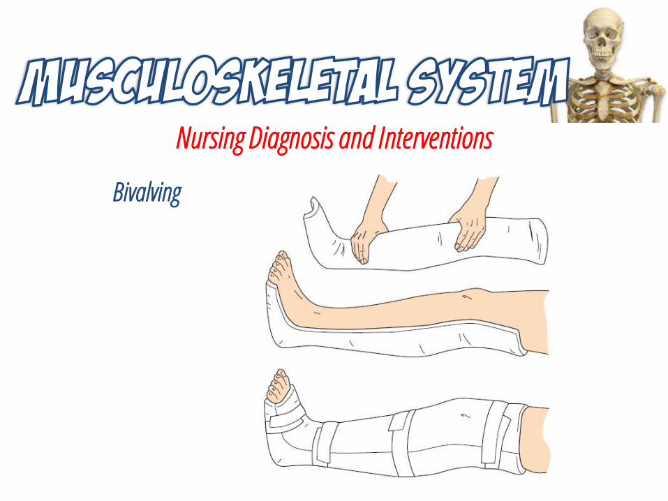
Bivalving
Nursing Diagnosis and Interventions
Risk for Peripheral Neurovascular Dysfunction
• Unless contraindicated, elevate the injured extremity above the level of the heart.
• Administer anticoagulant per physician’s order.
Nursing Diagnosis and Interventions
Risk for Infection
• For patients with skeletal pins, follow established guidelines for skeletal pin site care.
• Monitor vital signs and lab reports of WBCs.
• Use sterile technique for dressing changes.
• Assess the wound for size, color, and the presence of any drainage.
• Administer antibiotics per physician’s orders.
Nursing Diagnosis and Interventions
Impaired Physical Mobility
• Teach or assist patient with ROM exercises of the unaffected limbs.
• Teach isometric exercises, and encourage the patient to perform them every 4 hours.
• Encourage ambulation when able; provide assistance as necessary.
• Turn the patient on bed rest every 2 hours. If the patient is in traction, teach the patient to shift his or her weight every hour.
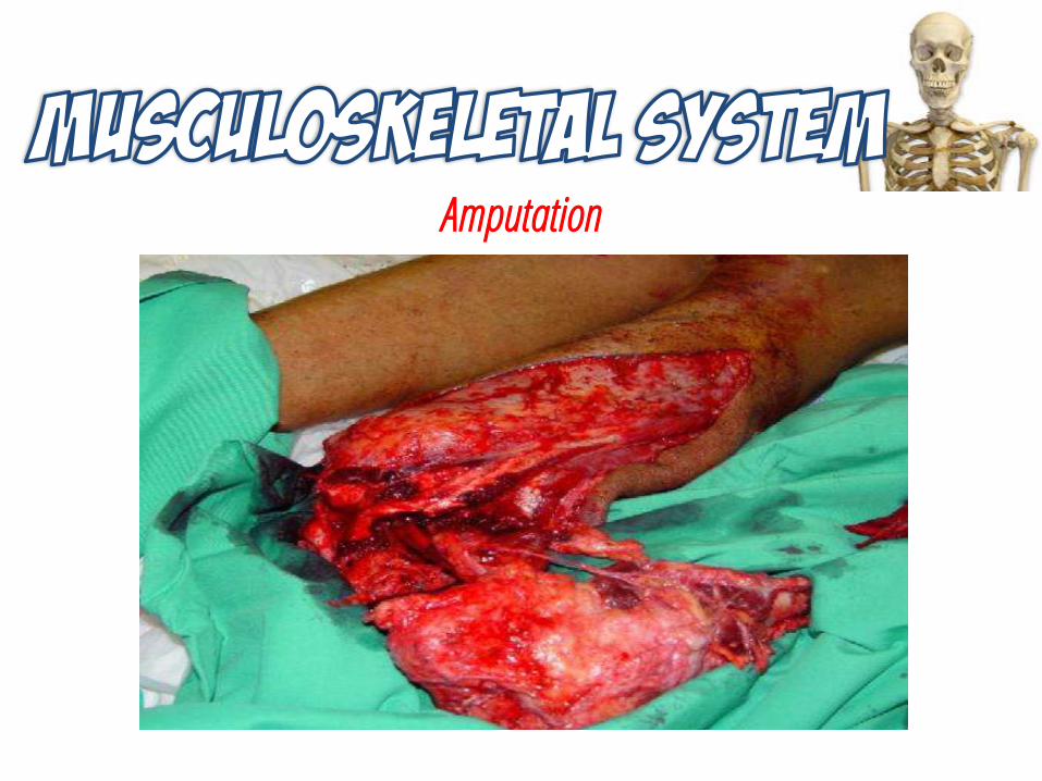
Amputation
Amputation
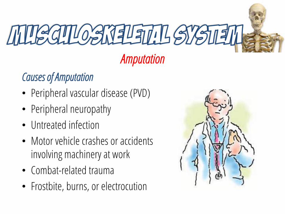
Causes of Amputation
• Peripheral vascular disease (PVD)
• Peripheral neuropathy
• Untreated infection
• Motor vehicle crashes or accidents involving machinery at work
• Combat-related trauma
• Frostbite, burns, or electrocution
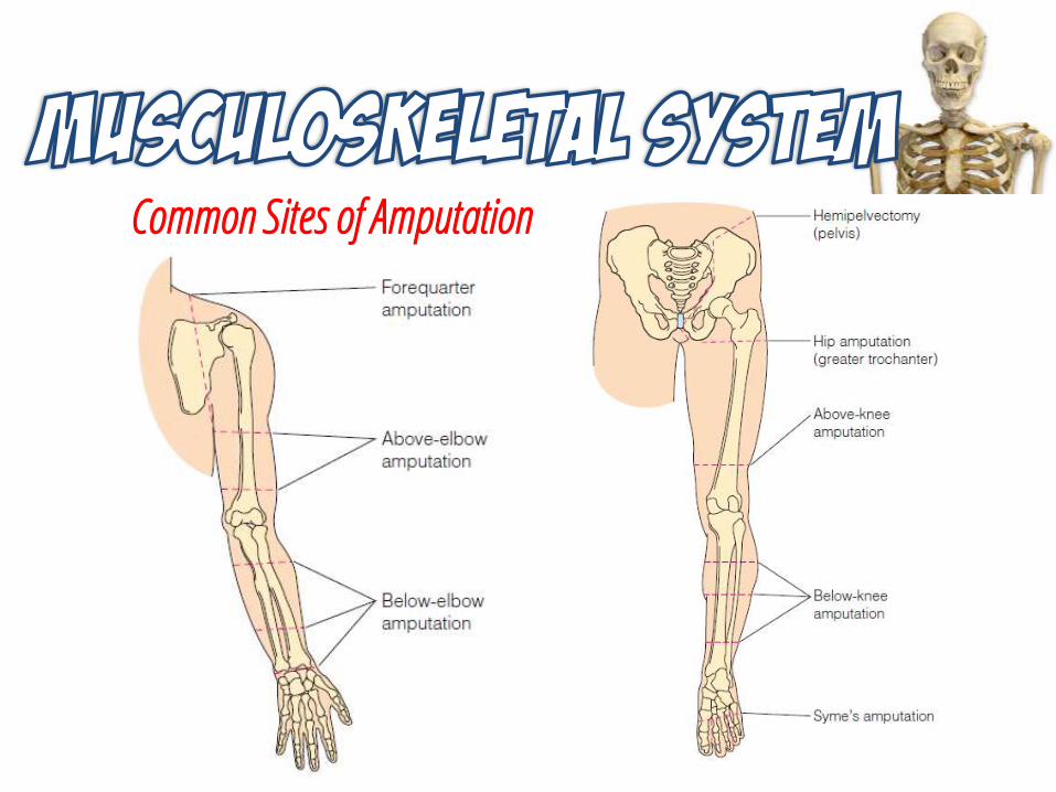
Common Sites of Amputation
Amputation Complications
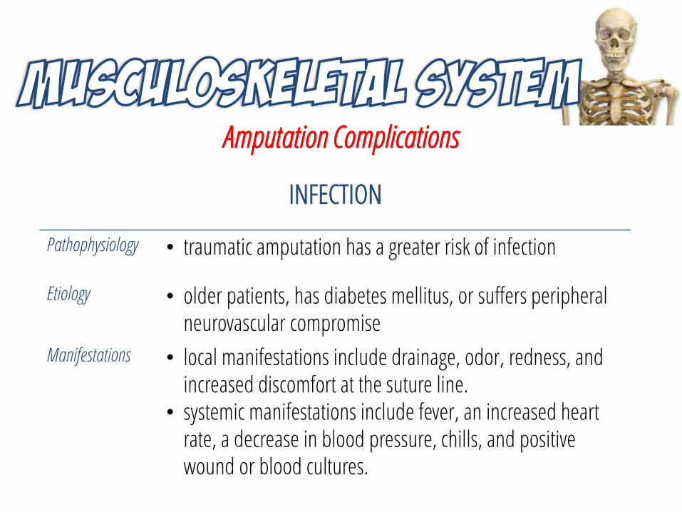
INFECTION
Pathophysiology • traumatic amputation has a greater risk of infection
Etiology • older patients, has diabetes mellitus, or suffers peripheral neurovascular compromise
Manifestations • local manifestations include drainage, odor, redness, and increased discomfort at the suture line.
• systemic manifestations include fever, an increased heart rate, a decrease in blood pressure, chills, and positive wound or blood cultures.
Amputation Complications
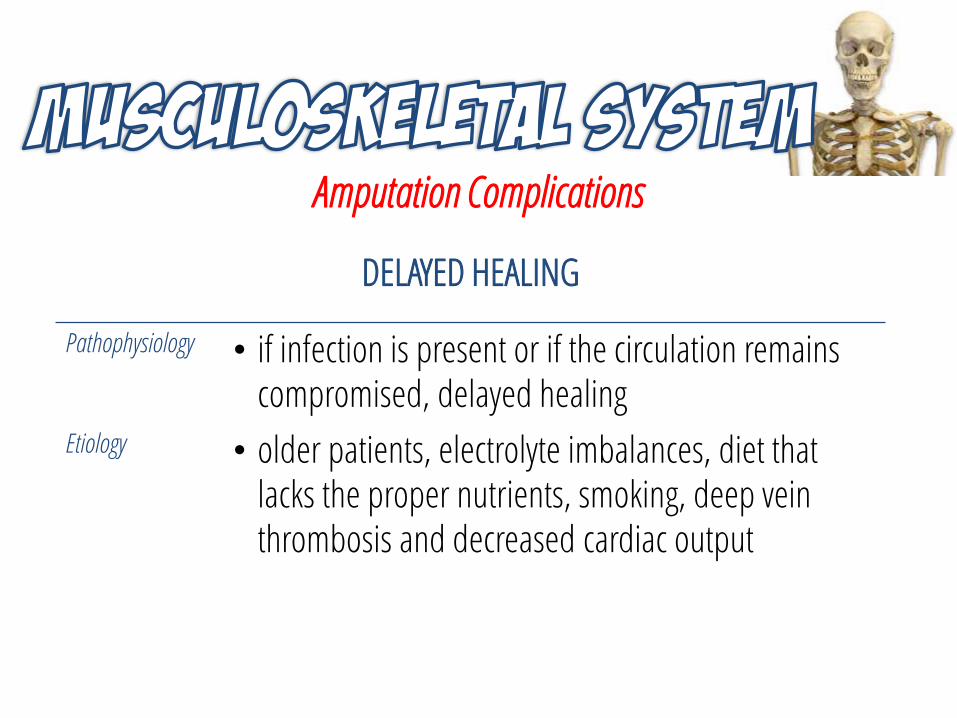
DELAYED HEALING
Pathophysiology • if infection is present or if the circulation remains compromised, delayed healing
Etiology • older patients, electrolyte imbalances, diet that lacks the proper nutrients, smoking, deep vein thrombosis and decreased cardiac output
Amputation Complications
CHRONIC STUMP PAIN
Pathophysiology • neuroma formation
Manifestations • severe burning pain
Treatment • medications• nerve blocks• transcutaneous electrical nerve stimulation (TENS)• surgical stump reconstruction

Amputation Complications
PHANTOM LIMB PAIN
Pathophysiology • pain in the amputated limb prior to its removal
Manifestations • tingling, numbness, cramping, or itching in the phantom foot or hand
Treatment • pain management• TENS• surgery
Amputation Complications
CONTRACTURES
Pathophysiology • abnormal flexion and fixation of a joint caused by muscle atrophy and shortening
Treatment • active or passive ROM exercises every 2 to 4 hours• postural exercises
Management for Amputation
Diagnosis
• Preoperative - Doppler flowmetry, segmental blood pressure determination, transcutaneous partial pressure oxygen readings, and angiography
• Postoperative - CBC, WBC, blood chemistries, and a vascular Doppler ultrasonography.
Management for Amputation
Medications
• analgesics
• antibiotics
• steroids
• H2 antagonists
Management for Amputation
Emergency Care
• Administer CPR as necessary, and control bleeding with direct pressure.
• Keep the person in a supine position with the legs elevated.
• Apply firm pressure to the bleeding area, using a towel or article of clothing.
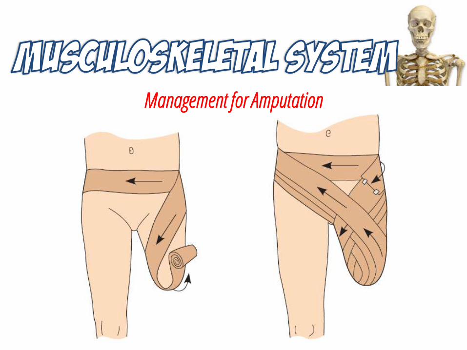
• Wrap the amputated part in a clean cloth. If possible, soak the cloth in saline.
Management for Amputation
Management for Amputation
Emergency Care
• Put the amputated part in a plastic bag and put the bag on ice.
• Send the amputated part to the emergency department with the injured person, and be sure the emergency personnel know what it is.
Management for Amputation
Assessment
• Health history: Mechanism of injury, current and past health problems, pain, occupation, ADLs, changes in sensation in the feet, cultural and/or religious guidelines for handling the amputated part.
• Physical examination: Bilateral neurovascular status of the extremities, bilateral capillary refill time, skin over the lower extremities (discoloration, edema, ulcerations, hair, gangrene).
Nursing Diagnosis and Interventions
Acute Pain
• Ask the patient to rate the pain on a scale of 0 to 10 before and after any intervention.
• Splint and support the injured area.
• Unless contraindicated, elevate the stump on a pillow for the first 24 hours after surgery.
• Move and turn the patient gently and slowly.
• Administer pain medications as prescribed. A PCA pump may be ordered by the physician.
Nursing Diagnosis and Interventions
Acute Pain
• Encourage deep breathing and relaxation exercises.
• Reposition patient every 2 hours; turn from side to side and onto abdomen.
Nursing Diagnosis and Interventions
Risk for Infection
• Assess the wound for redness, drainage, temperature, edema, and suture line approximation.
• Take the patient’s temperature every 4 hours.
• Monitor WBC count.
• Use aseptic technique to change the wound dressing.
• Administer antibiotics as ordered.
• Teach the patient stump-wrapping techniques.

Nursing Diagnosis and Interventions
Risk for Impaired Skin Integrity
• Wash the stump with soap and warm water and dry thoroughly.
• Inspect the stump for redness, irritation, or abrasions.
• Massage the end of the stump, beginning 3 weeks after surgery.
• Expose any open areas of skin on the remaining part of the limb for 1 hour four times a day.
• Change stump socks and elastic wraps each day. Wash these in mild soap and water, and allow to completely dry before using.
Nursing Diagnosis and Interventions
Risk for Complicated Grieving
• Encourage verbalization of feelings, using open-ended questions.
• Actively listen and maintain eye contact.
• Reflect on the patient’s feelings.
• Allow the patient to have unlimited visiting hours, if possible.
• If desired by the patient, provide spiritual support by encouraging activities such as visits from a spiritual leader, prayer, and meditation.
Nursing Diagnosis and Interventions
Disturbed Body Image
• Encourage verbalization of feelings.
• Allow the patient to wear clothing from home.
• Encourage the patient to look at the stump.
• Encourage the patient to care for the stump.
• Offer to have a fellow amputee visit the patient.
• Encourage active participation in rehabilitation.
Nursing Diagnosis and Interventions
Impaired Physical Mobility
• Perform ROM exercises on all joints.
• Maintain postoperative stump shrinkage devices. (elastic bandages, shrinker socks, an elastic stockinette, or a rigid plaster cast).
• Turn and reposition the patient every 2 hours.
• Reinforce teaching by the physical therapist in crutch walking or the use of assistive devices.
• Encourage active participation in physical therapy.
References1. LeMone, P. et al. (2011). Medical-Surgical Nursing: Critical
Thinking in Client Care. 5th Edition. New Jersey: Pearson Education, Inc.
2. Smeltzer, S. C. et al. (2010). Brunner & Suddarth’s Textbook of Medical-Surgical Nursing. 12th Edition. Philadelphia: Lippincott Williams and Wilkins.
3. Williams. L. S. & Hopper, P. D. (2011). Understanding Medical-Surgical Nursing. 5th Edition. Philadelphia: F. A. Davis Company