multifocal lenses in coral reef fishes · multifocal lenses in coral reef fishes 2925 camera (sony...
TRANSCRIPT
2923
IntroductionThe refractive power of a typical fish eye resides exclusively
in the powerful spherical lens, since the cornea is thin and hasfluid of similar refractive index on both sides (Matthiessen,1886). All lenses suffer from a number of optical aberrations.In spherical lenses, asymmetric aberrations such as astigmatismand coma are absent or at least of minor importance. There are,however, important symmetric aberrations. A homogeneousspherical lens focuses light passing through its periphery closerto the lens than light rays nearer to the optical axis (longitudinalspherical aberration, LSA). Fish lenses, however, areinhomogeneous gradient index lenses. Protein concentrationand thus refractive index is highest in the center and decreasesgradually towards the periphery. The refractive index profile isroughly parabolic and corrects a typical fish lens rather well,although not completely, for LSA (Maxwell, 1854; Matthiessen,1882; Matthiessen, 1886; Kröger et al., 1994).
Chromatic defocus originates from the prismatic effects ofsingle lenses. Short wavelengths (ultraviolet to blue) arerefracted more strongly and consequently focused closer to thelens than long wavelengths (red). The phenomenon is known aslongitudinal chromatic aberration (LCA) (Kröger andCampbell, 1996; Born and Wolf, 1999). This can cause a seriousproblem in fish eyes since depth of focus is short in opticalsystems with small f-numbers (f-number=focal length/diameterof aperture), as in most fishes. In addition, most teleosts do notconstrict their pupils even in bright light, since light flux isregulated by mechanisms located in the retina and retinalpigment epithelium (Walls, 1942; Douglas, 1982; Burnside and
Nagle, 1983). LCA thus usually exceeds depth of focus, whichmeans that fish lenses should only be able to focus a narrowspectral range on the retina, even in bright light when colorvision is possible and advantageous.
It has been shown in the African cichlid fish Astatotilapia(formerly Haplochromis) burtoni that the lens has residual LSAof complex shape. This LSA leads to several discrete focallengths for monochromatic light and such lenses are thereforecalled ‘multifocal’ lenses. If polychromatic light impinges onan A. burtoni lens, the lens focuses the wavelengths maximallyabsorbed (�max) by the cone photoreceptors at the same depth(Kröger et al., 1999). LCA is thus corrected for by accuratelytuned LSA, which in turn is dependent on the refractive indexprofile within the lens. Multifocal optical systems have beendemonstrated in some other freshwater fishes (Kröger et al.,1999; Malkki and Kröger, 2005) and a variety of terrestrialvertebrates (Kröger et al., 1999; Malmström and Kröger, 2006).We present here the first evidence for multifocal lenses inmarine teleosts. In addition we extend the study of multifocallenses to correlations between the optical properties of thelenses and the ecologies of different species.
Tropical and sub-tropical coral reefs are among the mostcolorful habitats on Earth (Chiao et al., 2000) and support largevarieties of animal species with different lifestyles (Dubinsky,1990). Clear water and proximity to the surface allow the useof the full spectrum of light, including the ultraviolet (UV)range. Color vision can provide a wealth of information in suchan environment and it is known that at least some coral reeffishes have well-developed color vision systems at the retinal
The optical properties of crystalline lenses were studiedin eleven species of coral reef fish from the Red Sea in Eilat,Israel. Three species each of diurnal planktivores,nocturnal planktivores and diurnal herbivores constitutedthree groups of animals with little within-group variability.In addition we studied two predators, which differed withrespect to body size, prey preference, hunting method anddiel activity period. All species studied have multifocallenses. There were statistically significant differences in theoptical properties of the lenses between the first three
groups and between the predatory species. The propertiesof the lenses correlate well with known complements ofvisual pigments and feeding habits. Lenticular zonesfocusing ultraviolet light were found in two diurnalplanktivores. The optical properties of the lens seem to bespecifically adapted to the visual needs of each species.
Key words: physiological optics, color vision, chromatic aberration,spherical aberration, visual pigments, Red Sea.
Summary
The Journal of Experimental Biology 210, 2923-2931Published by The Company of Biologists 2007doi:10.1242/jeb.002956
Multifocal lenses in coral reef fishes
Björn Karpestam1, Jonas Gustafsson2, Nadav Shashar3, Gadi Katzir4 and Ronald H. H. Kröger2,*1Eberhard Karls University Tübingen, Institute of Anatomy, Österbergstrasse 3, 72074 Tübingen, Germany,
2Lund University, Department of Cell and Organism Biology, Zoology Building, Helgonavägen 3, 22362 Lund, Sweden,3Ben Gurion University, Eilat Campus, Life Sciences Department, P.O.B. 653, Beer Sheva, 84105, Israel and
4University of Haifa, Department of Biology, Oranim, Tivon 36006, Israel*Author for correspondence (e-mail: [email protected])
Accepted 6 June 2007
THE JOURNAL OF EXPERIMENTAL BIOLOGY
2924
level (Losey et al., 2003; Marshall et al., 2003). Differentlifestyles and diel activity periods are expected to have led todifferent retinal and optical adaptations. In the present work weused recently described optical methods (Malkki and Kröger,2005) to investigate how the optical systems are matched to thecapabilities of the retinas. In addition, we studied whether thereare lifestyle-specific characteristics in the optical properties offish lenses.
Materials and methodsAnimals
Fish from 11 species were investigated, representing fourdifferent lifestyles: diurnal herbivores, diurnal planktivores,nocturnal planktivores and predators. The first three groupsconsisted of three species each and there was little within-groupvariability in body sizes and shapes, foraging strategies and dielactivity periods. In the latter group (predators) there were twospecies different in these respects. All animals were caught bySCUBA divers, using hand nets, in shallow waters not deeperthan 10·m on or near a coral reef in Eilat (the Gulf of Aqaba)under permit no. 18267 from the Israeli Natural Parks Authority.Characteristics of the selected species are summarized in theAppendix.
The animals were kept at the Inter University Institute ofEilat, Israel, in outdoor tanks with a continuous supply ofunfiltered seawater. Most fish were studied on the same day theywere caught; a few animals were kept for up to 1 week.
Optical investigationsThe methods we used have been described in detail elsewhere
(Malkki and Kröger, 2005). Here we briefly present theiressential features. All investigations were performed duringdaytime, i.e. on light-adapted fishes.
Lens examination in vivoPhotoretinoscopy. In the form used here, photoretinoscopy
(Fig.·1A) was developed by Schaeffel and co-workers(Schaeffel et al., 1987; Schaeffel et al., 1993). It can be usedwith live animals and gives an indication at what distance,relative to the camera, the eye is focused. If the eye has amultifocal optical system, ring-like structures are visible inphotoretinoscopic images (Kröger et al., 1999). With thismethod we could detect animals with eyes that were opticallyaberrant from the general pattern present in each particularspecies. Such deviations may be caused, for example, byintraocular parasites (Malkki and Kröger, 2005).
All caught fish were screened using photoretinoscopy andaberrant individuals discarded. One fish at a time was kept in asmall glass tank with unfiltered seawater. After allowing the fishto acclimatize for about 5·min, each eye was videotaped forapproximately 1–1.5·min and typical frames were later exportedusing Adobe Premiere 6.0 software.
Lens examination in vitroThe fish was sacrificed by rapid cerebral section and pithing.
Its total length (TL, tip of snout to end of tailfin) and standardlength (SL, tip of snout to base of tailfin) were measured to thenearest mm. One eye was excised, while the other one remainedin place and was kept moist with seawater until the first eye had
been completely processed. During extraction of the lens theexcised eye was immersed in phosphate-buffered saline (PBS;Na+=7.58·g·l–1, Cl–=4.88·g·l–1, HPO4
2–=0.757·g·l–1, H2PO4–=
0.259·g·l–1, pH·7.2, osmolality 290·mOsm) in order to preventdehydration of the lens.
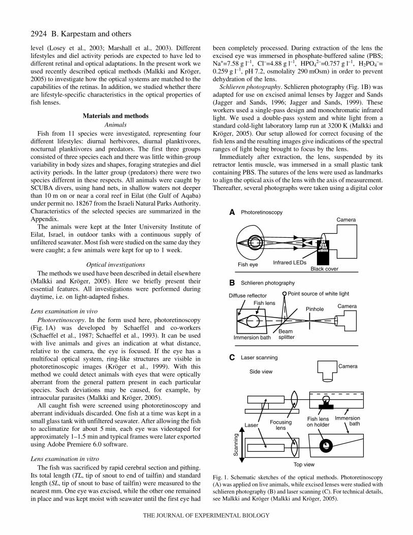
Schlieren photography. Schlieren photography (Fig.·1B) wasadapted for use on excised animal lenses by Jagger and Sands(Jagger and Sands, 1996; Jagger and Sands, 1999). Theseworkers used a single-pass design and monochromatic infraredlight. We used a double-pass system and white light from astandard cold-light laboratory lamp run at 3200·K (Malkki andKröger, 2005). Our setup allowed for correct focusing of thefish lens and the resulting images give indications of the spectralranges of light being brought to focus by the lens.
Immediately after extraction, the lens, suspended by itsretractor lentis muscle, was immersed in a small plastic tankcontaining PBS. The sutures of the lens were used as landmarksto align the optical axis of the lens with the axis of measurement.Thereafter, several photographs were taken using a digital color
B. Karpestam and others
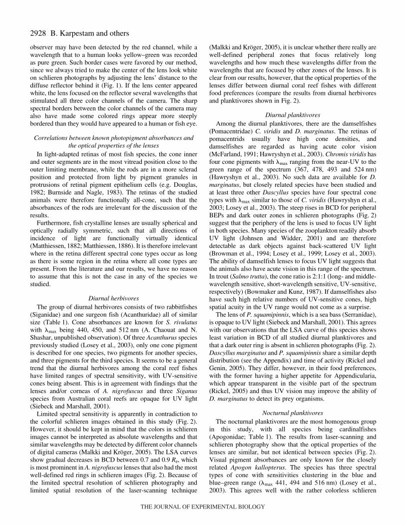
Fig.·1. Schematic sketches of the optical methods. Photoretinoscopy(A) was applied on live animals, while excised lenses were studied withschlieren photography (B) and laser scanning (C). For technical details,see Malkki and Kröger (Malkki and Kröger, 2005).
Camera
Fish lenson holderFocusing
lensLaser
Side view
Sca
nnin
g
Immersionbath
Point source of white light
Beamsplitter
Pinhole
Immersion bath
Fish lensDiffuse reflector
Camera
Laser scanning
Schlieren photography
Fish eyeBlack cover
Camera
Infrared LEDs
Photoretinoscopy
C
B
A
Top view
THE JOURNAL OF EXPERIMENTAL BIOLOGY
2925Multifocal lenses in coral reef fishes
camera (Sony DSC-F 707) with varying distances between thefish lens and the diffuse reflector (Fig. 1B).
Laser-scanning. The LSA of a lens was quantitativelydetermined using laser-scanning (Fig.·1C). Our setup was amodification of the system used previously (Kröger et al.,1994). Refinements increased speed and resolution ofmeasurement, and included semi-automated analysis of the rawdata (Malkki and Kröger, 2005). Data were obtained byscanning a thin laser beam of a wavelength of 547·nm (diode-pumped solid state laser) through a meridional plane of the lens.The results are equivalent to a transverse section through thesymmetrical wavefront aberrations of the lens and can bedirectly compared with schlieren images, in which variation infocal length is indicated by variation in color.
The lens was carefully placed on a plastic holder in the laserscanning unit (Fig. 1C). To correctly align the lens with the laserbeam, the direction of small grooves in the lens capsule wasdetermined using a light microscope. A small amount ofpolysterene microbeads (diameter 100·nm) was added to thePBS used for immersion. The microbeads scattered some light,which made the laser beam visible. Each lens was scanned twiceand an average LSA recorded.
From the video sequence of each scan, 200 frames wereexported to TIFF (tagged image file format, a non-compressedformat) images using Adobe Premiere 6.0. From these frames,the LSA was determined by using custom-written software(Malkki and Kröger, 2005). LSA curves were generated byplotting back center distance (BCD), i.e. the distance betweenthe center of the lens and the intercept of the exit beam and theoptical axis, as a function of beam entrance position (BEP), i.e.the lateral distance between the optical axis and the entrancebeam. All data were normalized to equatorial lens radius (Re),such that the results from lenses of different sizes could bepooled or compared.
We averaged the LSA curves across the optical axis over bothhalves of each lens because we were only interested in spherical
aberration, which is a symmetrical aberration. Each lens wastreated as an independent measurement because intra-animalvariance is higher than inter-animal variance in this kind ofmeasurement (Kröger et al., 2001). LSA curves were plottedwith 90% confidence intervals, because if two such intervals donot overlap, the probability of the average curves being identicalis less than 5%. Relative focal lengths were determined fromaveraged LSAs. The BCDs were weighted for their BEPs sinceperipheral regions of the lens contribute more to the image thancentral regions (Kröger and Campbell, 1996).
After all optical experiments on a lens were complete, itsdiameter was measured with calipers to the nearest 0.1·mm. Theentire procedure from sacrifice of the fish to completion of allmeasurements on both lenses lasted between 45 and 60·min perfish.
ResultsIn each species, 9–13 lenses were studied, each examined
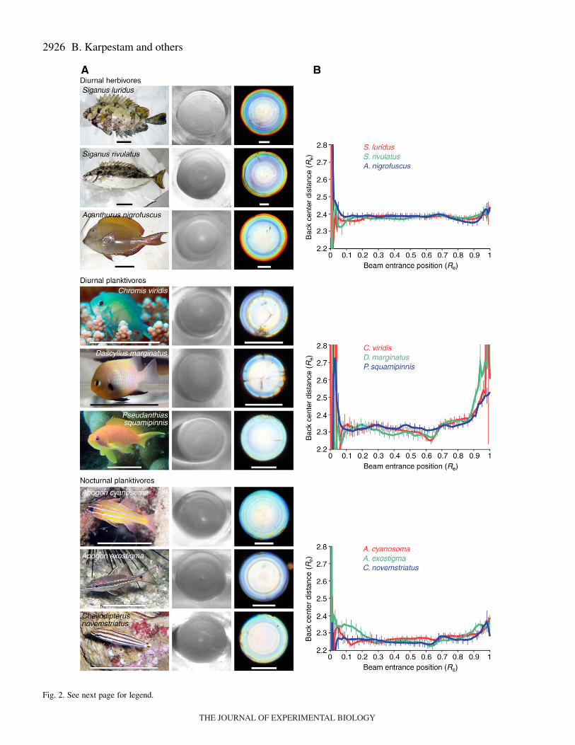
with all available methods (Table·1). All studied species hadmultifocal lenses (Figs·2 and 3). However, the optical designsof the lenses differed between species (Figs·2–4).
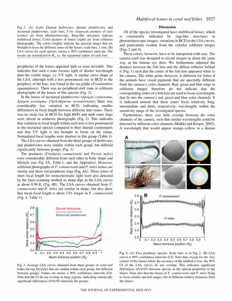
The peripheries of the lenses of diurnal herbivores (Siganusluridus, Siganus rivulatus, Acanthurus nigrofuscus) are red onschlieren photographs, which indicates that long wavelengthswere focused by these zones (Fig.·2A). Many differentlycolored rings are visible, especially in the large-eyed S. luridus.The LSA curves show less detail because of the method’s lowerspatial resolution (Malkki and Kröger, 2005). There is relativelylittle variation in BCD in the LSA curves, which indicates thatUV light is not focused on the retina.
The LSA curves of the diurnal planktivorous damselfishes(Chromis viridis and Dascyllus marginatus) showed steep risesin BCD for beams of high entrance positions (Fig.·2B). HighBCDs in a lens zone mean that such a zone focuses shortwavelengths on the retina, because focal length decreases withdecreasing wavelength. On schlieren photographs, the
Table·1. Basic data on the animals studied
Lens diameter NormalizedSpecies Order/Family No. animals No. lenses SL (mm) (mm) focal length (Re)
Diurnal planktivoresChromis viridis Perciformes/Pomacentridae 5 10 35±1 1.3±0.1 2.38Dascyllus marginatus Perciformes/Pomacentridae 8 13 39±7 1.5±0.2 2.40Pseudanthias squamipinnis Perciformes/Serranidae 7 12 67±17 1.9±0.2 2.36
Nocturnal planktivoresApogon cyanosoma Perciformes/Apogonidae 7 12 49±2 2.8±0.2 2.28Apogon exostigma Perciformes/Apogonidae 5 10 40±3 2.1±0.2 2.28Cheilodipterus novemstriatus Perciformes/Apogonidae 5 9 37±5 1.8±0.2 2.27
Diurnal herbivoresSiganus luridus Perciformes/Siganidae 6 12 155±20 4.6±0.8 2.39Siganus rivulatus Perciformes/Siganidae 6 10 156±39 3.9±0.9 2.39Acanthurus nigrofuscus Perciformes/Acanthuridae 6 12 120±9 3.3±0.2 2.38
PredatorsFistularia commersonii Syngnathiformes/Fistularidae 6 12 653±52 6.8±0.8 2.69Pterois miles Scorpaeniformes/Scorpaenida 6 12 127±43 3.9±1.1 2.38
Among the predators, F. commersonii is diurnal to crepuscular and P. miles is crepuscular to nocturnal. Standard length (SL) and lens diameter values are means ± s.d. Normalized focal lengths were calculated from the average LSA of each
species. Re is the equatorial radius of the lens.
THE JOURNAL OF EXPERIMENTAL BIOLOGY
2926 B. Karpestam and others
Fig. 2. See next page for legend.
THE JOURNAL OF EXPERIMENTAL BIOLOGY
2927Multifocal lenses in coral reef fishes
peripheries of the lenses appeared dark or were invisible. Thisindicates that such a zone focuses light of shorter wavelengththan the visible range, i.e. UV light. A similar curve shape ofthe LSA, although with a less pronounced rise in BCD in theperiphery of the lens, was found in the sea goldie (Pseudanthiassquamipinnis). There was no peripheral dark zone in schlierenphotographs of the lenses of this species (Fig.·2).
In the lenses of nocturnal planktivores (Apogon cyanosoma,Apogon exostigma, Cheilodipterus novemstriatus) there wasconsiderably less variation in BCD, indicating smallerdifferences in focal length between different lens zones. Therewas no steep rise in BCD for high BEPs and dark outer ringswere absent in schlieren photographs (Fig.·2). This indicatesthat variation in focal length within each lens is less pronouncedin the nocturnal species compared to their diurnal counterpartsand that UV light is not brought to focus on the retina.Normalized focal lengths were shortest in this group (Table·1).
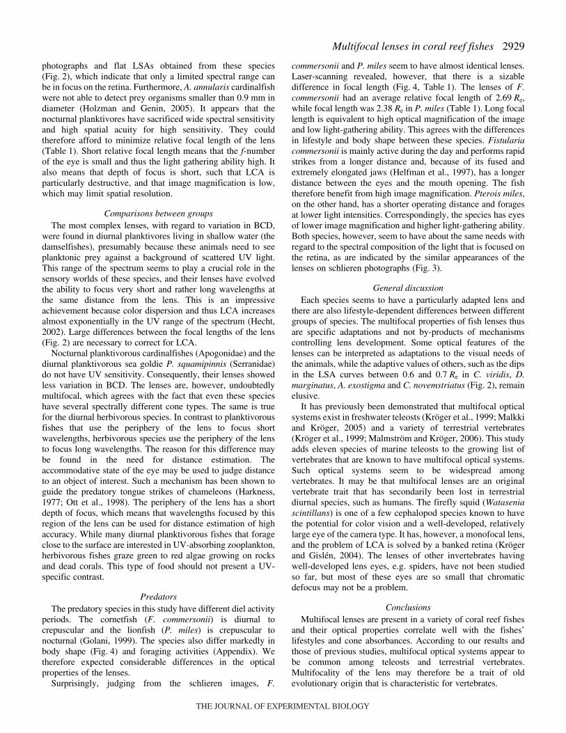
The LSA curves obtained from the three groups of herbivoresand planktivores were similar within each group, but differedsignificantly between groups (Fig.·3).
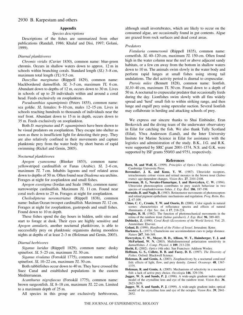
The predators (Fistularia commersonii and Pterois miles)were considerably different from each other in body shape andlifestyle (see Fig.·4A, Table·1, and the Appendix). However,schlieren photographs of F. commersonii and P. miles lenses aresimilar and show red peripheral rings (Fig.·4A). These zones ofshort focal length for monochromatic light were also detectedby the laser-scanning method as sharp dips in the LSA curvesat about 0.98·Re (Fig.·4B). The LSA curves obtained from F.commersonii and P. miles are similar in shape, but also showthat mean focal length is about 13% longer in F. commersonii(Fig.·4, Table·1).
DiscussionAll of the species investigated have multifocal lenses, which
is consistently indicated by ring-like structures inphotoretinoscopic images, variations in BCD in the LSA curves,and particularly evident from the colorful schlieren images(Figs·2 and 4).
These colors, however, have to be interpreted with care. Thecamera used was designed to record images in about the sameway as the human eye does. We furthermore adjusted thedistance between the fish lens and the diffuse reflector behindit (Fig.·1) such that the center of the fish lens appeared white tothe camera. The white point, however, is different for fishes ifthe animals have visual pigments that are spectrally differentfrom the camera’s color channels. Red, green and blue rings inschlieren images therefore do not indicate that thecorresponding zones of a fish lens are used to focus wavelengthsthat fit into the camera’s red, green and blue color channels. Itis indicated instead that these zones focus relatively long,intermediate and short, respectively, wavelengths within thesensitivity range of the investigated species.
Furthermore, there was little overlap between the colorchannels of the camera, such that similar wavelengths could bedetected by different color channels (Malkki and Kröger, 2005).A wavelength that would appear orange–yellow to a human
Fig.·2. (A) (Left) Diurnal herbivores, diurnal planktivores andnocturnal planktivores; scale bars, 5·cm. Grayscale pictures of eyes(centre) are from photoretinoscopy. Ring-like structures indicatemultifocal lenses. Color pictures of lenses (right) are from schlierenphotography. The colors roughly indicate the spectral ranges that arebrought to focus by different zones of the lenses; scale bars, 1·mm. (B)LSA curves for each species, means ± 90% confidence intervals. Theresults are normalized to Re, i.e. the equatorial radius of each lens.
Fig.·3. Average LSA curves obtained from three groups of coral reeffishes having lifestyles that are similar within each group, but differentbetween groups. Values are means ± 90% confidence intervals (CI).Note that the CI do not overlap in large regions, indicating statisticallysignificant differences (P<0.05) between the groups.
2.2
2.3
2.4
2.5
2.6
2.7
2.8
0 0.1 0.2 0.3 0.4 0.5 0.6 0.7 0.8 0.9 1
Bac
k ce
nter
dis
tanc
e (R
e)
Beam entrance position (Re)
Diurnal herbivoresDiurnal planktivoresNocturnal planktivores
Fig.·4. (A) Two predatory species. Scale bars as in Fig.·2. (B) LSAcurves ± 90% confidence intervals (CI). Note that, except for the verycenters of the lenses where the accuracy of the method is low, the 90%CI of the LSA curves do not overlap. This indicates significantdifferences (P<0.05) between species in the optical properties of thelenses. Note also that the lenses of F. commersonii and P. miles bringto focus similar spectral ranges, but at different relative distances fromthe lenses.
THE JOURNAL OF EXPERIMENTAL BIOLOGY
2928
observer may have been detected by the red channel, while awavelength that to a human looks yellow–green was recordedas pure green. Such border cases were favored by our method,since we always tried to make the center of the lens look whiteon schlieren photographs by adjusting the lens’ distance to thediffuse reflector behind it (Fig.·1). If the lens center appearedwhite, the lens focused on the reflector several wavelengths thatstimulated all three color channels of the camera. The sharpspectral borders between the color channels of the camera mayalso have made some colored rings appear more steeplybordered than they would have appeared to a human or fish eye.
Correlations between known photopigment absorbances andthe optical properties of the lenses
In light-adapted retinas of most fish species, the cone innerand outer segments are in the most vitread position close to theouter limiting membrane, while the rods are in a more scleradposition and protected from light by pigment granules inprotrusions of retinal pigment epithelium cells (e.g. Douglas,1982; Burnside and Nagle, 1983). The retinas of the studiedanimals were therefore functionally all-cone, such that theabsorbances of the rods are irrelevant for the discussion of theresults.
Furthermore, fish crystalline lenses are usually spherical andoptically radially symmetric, such that all directions ofincidence of light are functionally virtually identical(Matthiessen, 1882; Matthiessen, 1886). It is therefore irrelevantwhere in the retina different spectral cone types occur as longas there is some region in the retina where all cone types arepresent. From the literature and our results, we have no reasonto assume that this is not the case in any of the species westudied.
Diurnal herbivoresThe group of diurnal herbivores consists of two rabbitfishes
(Siganidae) and one surgeon fish (Acanthuridae) all of similarsize (Table·1). Cone absorbances are known for S. rivulatuswith �max being 440, 450, and 512·nm (A. Chaouat and N.Shashar, unpublished observation). Of three Acanthurus speciespreviously studied (Losey et al., 2003), only one cone pigmentis described for one species, two pigments for another species,and three pigments for the third species. It seems to be a generaltrend that the diurnal herbivores among the coral reef fisheshave limited ranges of spectral sensitivity, with UV-sensitivecones being absent. This is in agreement with findings that thelenses and/or corneas of A. nigrofuscus and three Siganusspecies from Australian coral reefs are opaque for UV light(Siebeck and Marshall, 2001).
Limited spectral sensitivity is apparently in contradiction tothe colorful schlieren images obtained in this study (Fig.·2).However, it should be kept in mind that the colors in schlierenimages cannot be interpreted as absolute wavelengths and thatsimilar wavelengths may be detected by different color channelsof digital cameras (Malkki and Kröger, 2005). The LSA curvesshow gradual decreases in BCD between 0.7 and 0.9·Re, whichis most prominent in A. nigrofuscus lenses that also had the mostwell-defined red rings in schlieren images (Fig.·2). Because ofthe limited spectral resolution of schlieren photography andlimited spatial resolution of the laser-scanning technique
(Malkki and Kröger, 2005), it is unclear whether there really arewell-defined peripheral zones that focus relatively longwavelengths and how much these wavelengths differ from thewavelengths that are focused by other zones of the lenses. It isclear from our results, however, that the optical properties of thelenses differ between diurnal coral reef fishes with differentfood preferences (compare the results from diurnal herbivoresand planktivores shown in Fig.·2).
Diurnal planktivoresAmong the diurnal planktivores, there are the damselfishes
(Pomacentridae) C. viridis and D. marginatus. The retinas ofpomacentrids usually have high cone densities, anddamselfishes are regarded as having acute color vision(McFarland, 1991; Hawryshyn et al., 2003). Chromis viridis hasfour cone pigments with �max ranging from the near-UV to thegreen range of the spectrum (367, 478, 493 and 524·nm)(Hawryshyn et al., 2003). No such data are available for D.marginatus, but closely related species have been studied andat least three other Dascyllus species have four spectral conetypes with �max similar to those of C. viridis (Hawryshyn et al.,2003; Losey et al., 2003). The steep rises in BCD for peripheralBEPs and dark outer zones in schlieren photographs (Fig.·2)suggest that the periphery of the lens is used to focus UV lightin both species. Many species of the zooplankton readily absorbUV light (Johnsen and Widder, 2001) and are thereforedetectable as dark objects against back-scattered UV light(Browman et al., 1994; Losey et al., 1999; Losey et al., 2003).The ability of damselfish lenses to focus UV light suggests thatthe animals also have acute vision in this range of the spectrum.In trout (Salmo trutta), the cone ratio is 2:1:1 (long- and middle-wavelength sensitive, short-wavelength sensitive, UV-sensitive,respectively) (Bowmaker and Kunz, 1987). If damselfishes alsohave such high relative numbers of UV-sensitive cones, highspatial acuity in the UV range would not come as a surprise.
The lens of P. squamipinnis, which is a sea bass (Serranidae),is opaque to UV light (Siebeck and Marshall, 2001). This agreeswith our observations that the LSA curve of this species showsleast variation in BCD of all studied diurnal planktivores andthat a dark outer ring is absent in schlieren photographs (Fig.·2).Dascyllus marginatus and P. squamipinnis share a similar depthdistribution (see the Appendix) and time of activity (Rickel andGenin, 2005). They differ, however, in their food preferences,with the former having a higher appetite for Appendicularia,which appear transparent in the visible part of the spectrum(Rickel, 2005) and thus UV vision may improve the ability ofD. marginatus to detect its prey organisms.
Nocturnal planktivoresThe nocturnal planktivores are the most homogenous group
in this study, with all species being cardinalfishes(Apogonidae; Table·1). The results from laser-scanning andschlieren photography show that the optical properties of thelenses are similar, but not identical between species (Fig.·2).Visual pigment absorbances are only known for the closelyrelated Apogon kallopterus. The species has three spectraltypes of cone with sensitivities clustering in the blue andblue–green range (�max 441, 494 and 516·nm) (Losey et al.,2003). This agrees well with the rather colorless schlieren
B. Karpestam and others
THE JOURNAL OF EXPERIMENTAL BIOLOGY
2929Multifocal lenses in coral reef fishes
photographs and flat LSAs obtained from these species(Fig.·2), which indicate that only a limited spectral range canbe in focus on the retina. Furthermore, A. annularis cardinalfishwere not able to detect prey organisms smaller than 0.9·mm indiameter (Holzman and Genin, 2005). It appears that thenocturnal planktivores have sacrificed wide spectral sensitivityand high spatial acuity for high sensitivity. They couldtherefore afford to minimize relative focal length of the lens(Table·1). Short relative focal length means that the f-numberof the eye is small and thus the light gathering ability high. Italso means that depth of focus is short, such that LCA isparticularly destructive, and that image magnification is low,which may limit spatial resolution.
Comparisons between groupsThe most complex lenses, with regard to variation in BCD,
were found in diurnal planktivores living in shallow water (thedamselfishes), presumably because these animals need to seeplanktonic prey against a background of scattered UV light.This range of the spectrum seems to play a crucial role in thesensory worlds of these species, and their lenses have evolvedthe ability to focus very short and rather long wavelengths atthe same distance from the lens. This is an impressiveachievement because color dispersion and thus LCA increasesalmost exponentially in the UV range of the spectrum (Hecht,2002). Large differences between the focal lengths of the lens(Fig. 2) are necessary to correct for LCA.
Nocturnal planktivorous cardinalfishes (Apogonidae) and thediurnal planktivorous sea goldie P. squamipinnis (Serranidae)do not have UV sensitivity. Consequently, their lenses showedless variation in BCD. The lenses are, however, undoubtedlymultifocal, which agrees with the fact that even these specieshave several spectrally different cone types. The same is truefor the diurnal herbivorous species. In contrast to planktivorousfishes that use the periphery of the lens to focus shortwavelengths, herbivorous species use the periphery of the lensto focus long wavelengths. The reason for this difference maybe found in the need for distance estimation. Theaccommodative state of the eye may be used to judge distanceto an object of interest. Such a mechanism has been shown toguide the predatory tongue strikes of chameleons (Harkness,1977; Ott et al., 1998). The periphery of the lens has a shortdepth of focus, which means that wavelengths focused by thisregion of the lens can be used for distance estimation of highaccuracy. While many diurnal planktivorous fishes that forageclose to the surface are interested in UV-absorbing zooplankton,herbivorous fishes graze green to red algae growing on rocksand dead corals. This type of food should not present a UV-specific contrast.
PredatorsThe predatory species in this study have different diel activity
periods. The cornetfish (F. commersonii) is diurnal tocrepuscular and the lionfish (P. miles) is crepuscular tonocturnal (Golani, 1999). The species also differ markedly inbody shape (Fig.·4) and foraging activities (Appendix). Wetherefore expected considerable differences in the opticalproperties of the lenses.
Surprisingly, judging from the schlieren images, F.
commersonii and P. miles seem to have almost identical lenses.Laser-scanning revealed, however, that there is a sizabledifference in focal length (Fig.·4, Table·1). The lenses of F.commersonii had an average relative focal length of 2.69·Re,while focal length was 2.38·Re in P. miles (Table·1). Long focallength is equivalent to high optical magnification of the imageand low light-gathering ability. This agrees with the differencesin lifestyle and body shape between these species. Fistulariacommersonii is mainly active during the day and performs rapidstrikes from a longer distance and, because of its fused andextremely elongated jaws (Helfman et al., 1997), has a longerdistance between the eyes and the mouth opening. The fishtherefore benefit from high image magnification. Pterois miles,on the other hand, has a shorter operating distance and foragesat lower light intensities. Correspondingly, the species has eyesof lower image magnification and higher light-gathering ability.Both species, however, seem to have about the same needs withregard to the spectral composition of the light that is focused onthe retina, as are indicated by the similar appearances of thelenses on schlieren photographs (Fig.·3).
General discussionEach species seems to have a particularly adapted lens and
there are also lifestyle-dependent differences between differentgroups of species. The multifocal properties of fish lenses thusare specific adaptations and not by-products of mechanismscontrolling lens development. Some optical features of thelenses can be interpreted as adaptations to the visual needs ofthe animals, while the adaptive values of others, such as the dipsin the LSA curves between 0.6 and 0.7·Re in C. viridis, D.marginatus, A. exostigma and C. novemstriatus (Fig.·2), remainelusive.
It has previously been demonstrated that multifocal opticalsystems exist in freshwater teleosts (Kröger et al., 1999; Malkkiand Kröger, 2005) and a variety of terrestrial vertebrates(Kröger et al., 1999; Malmström and Kröger, 2006). This studyadds eleven species of marine teleosts to the growing list ofvertebrates that are known to have multifocal optical systems.Such optical systems seem to be widespread amongvertebrates. It may be that multifocal lenses are an originalvertebrate trait that has secondarily been lost in terrestrialdiurnal species, such as humans. The firefly squid (Wataseniascintillans) is one of a few cephalopod species known to havethe potential for color vision and a well-developed, relativelylarge eye of the camera type. It has, however, a monofocal lens,and the problem of LCA is solved by a banked retina (Krögerand Gislén, 2004). The lenses of other invertebrates havingwell-developed lens eyes, e.g. spiders, have not been studiedso far, but most of these eyes are so small that chromaticdefocus may not be a problem.
ConclusionsMultifocal lenses are present in a variety of coral reef fishes
and their optical properties correlate well with the fishes’lifestyles and cone absorbances. According to our results andthose of previous studies, multifocal optical systems appear tobe common among teleosts and terrestrial vertebrates.Multifocality of the lens may therefore be a trait of oldevolutionary origin that is characteristic for vertebrates.
THE JOURNAL OF EXPERIMENTAL BIOLOGY
2930
AppendixSpecies descriptions
Descriptions of the fishes are summarized from otherpublications (Randall, 1986; Khalaf and Disi, 1997; Golani,1999).
Diurnal planktivoresChromis viridis (Cuvier 1830), common name: blue-green
chromis. Occurs in shallow waters down to approx. 12·m inschools within branching corals. Standard length (SL) 3–8·cm,maximum total length (TL) 9.5·cm.
Dascyllus marginatus (Rüppell 1829), common name:blackbordered damselfish. SL 3–5·cm, maximum TL 6·cm.Abundant down to depths of 12·m, occurs down to 30·m. Livesin schools of up to 20 individuals within and around a coralhead. Feeds exclusively on zooplankton.
Pseudoanthias squamipinnis (Peters 1855), common name:sea goldie. SL females: 8–10·cm, males 12–15·cm. Lives inschools reaching hundreds to thousands of individuals near thereef front. Abundant down to 15·m in depth, occurs down to35·m. Feeds exclusively on zooplankton.
Both D. marginatus and P. squamipinnis have been shown tobe visual predators on zooplankton. They escape into shelter assoon as there is insufficient light for detecting their prey. Theyare also relatively confined in their movements and captureplanktonic prey from the water body by short bursts of rapidswimming (Rickel and Genin, 2005).
Nocturnal planktivoresApogon cyanosoma (Bleeker 1853), common name:
yellowstriped cardinalfish or Fanas (Arabic). SL 2–6·cm,maximum TL 7·cm. Inhabits lagoons and reef related areasdown to depths of 50·m. Often found near Diadema sea urchins.Forages at night for zooplankton and small fishes.
Apogon exostigma (Jordan and Seale 1906), common name:narrowstripe cardinalfish. Maximum TL 11·cm. Found nearcoral reefs down to 25·m. Forages at night for zooplankton.
Cheilodipterus novemstriatus (Rüppell 1838), commonname: Indian Ocean twospot cardinalfish. Maximum TL 12·cm.Forages at night for zooplankton, gastropods and small fishes.Found down to 10·m depth.
These fishes spend the day hours in hidden, unlit sites andstart to forage at dusk. Their eyes are highly sensitive andApogon annularis, another nocturnal planktivore, is able tosuccessfully prey on planktonic organisms during moonlessnights at depths of at least 2–3·m (Holzman and Genin, 2003).
Diurnal herbivoresSiganus luridus (Rüppell 1829), common name: dusky
spinefoot. SL 5–25·cm, maximum TL 30·cm.Siganus rivulatus (Forsskål 1775), common name: marbled
spinefoot. SL 10–22·cm, maximum TL 30·cm.Both rabbitfishes occur down to 40·m. They have crossed the
Suez Canal and established populations in the easternMediterranean.
Acanthurus nigrofuscus (Forsskål 1775), common name:brown surgeonfish. SL 8–18·cm, maximum TL 22·cm. Limitedto a maximum depth of 25·m.
All species in this group are exclusively herbivorous,
although small invertebrates, which are likely to occur on theconsumed algae, are occasionally found in gut contents. Algaeare grazed from rock surfaces and dead coral areas.
PredatorsFistularia commersonii (Rüppell 1835), common name:
cornetfish. SL 40–120·cm, maximum TL 150·cm. Often foundhigh in the water column near the reef or above adjacent sandyhabitats, or a few·cm away from the bottom in shallow watersdown to 10·m. The animals swim slowly in the water body andperform rapid lunges at small fishes using strong tailundulations. The diel activity period is diurnal to crepuscular.
Pterois miles (Bennett 1828), common name: lionfish.SL10–40·cm, maximum TL 50·cm. Found down to a depth of50·m. A nocturnal to crepuscular predator that occasionally feedsduring the day. Lionfishes swim slowly with all fins widelyspread and ‘herd’ small fish to within striking range, and thenlunge and engulf prey using opercular suction. Several lionfishmay collaborate in herding and attacking schools of prey.
We express our sincere thanks to Shai Einbinder, EranBrokovich and the diving team of the underwater observatoryin Eilat for catching the fish. We also thank Tally Scotland(Eilat), Ylwa Andersson (Lund), and the Inter UniversityInsitute for Marine Science in Eilat for assistance with thelogistics and administration of the study. B.K., J.G. and R.K.were supported by SRC grant 2001-1574, N.S. and G.K. weresupported by ISF grants 550/03 and 9751, respectively.
ReferencesBorn, M. and Wolf, E. (1999). Principles of Optics (7th edn). Cambridge:
Cambridge University Press.Bowmaker, J. K. and Kunz, Y. W. (1987). Ultraviolet receptors,
tetrachromatic colour vision and retinal mosaics in the brown trout (Salmotrutta): age-dependent changes. Vision Res. 27, 2101-2108.
Browman, H. I., Novales-Flamarique, I. and Hawryshyn, C. W. (1994).Ultraviolet photoreception contributes to prey search behaviour in twospecies of zooplanktivorous fishes. J. Exp. Biol. 186, 187-198.
Burnside, B. and Nagle, B. (1983). Retinomotor movements of photoreceptorsand retinal pigment epithelium: mechanisms and regulation. Prog. Retin. Res.2, 67-109.
Chiao, C. C., Cronin, T. W. and Osorio, D. (2000). Color signals in naturalscenes: characteristics of reflectance spectra and effects of naturalilluminants. J. Opt. Soc. Am. A 17, 218-224.
Douglas, R. H. (1982). The function of photomechanical movements in theretina of the rainbow trout (Salmo gairdneri). J. Exp. Biol. 96, 389-403.
Dubinsky, Z. (1990). Coral Reefs (Ecosystems of the World Series). Vol. 25.Amsterdam: Elsevier.
Golani, D. (1999). Handbook of the Fishes of Israel. Jerusalem: Keter.Harkness, L. (1977). Chameleons use accommodation cues to judge distance.
Nature 267, 346-349.Hawryshyn, C. W., Moyer, H. D., Allison, W. T., Haimberger, T. J. and
McFarland, W. N. (2003). Multidimensional polarization sensitivity indamselfishes. J. Comp. Physiol. A 189, 213-220.
Hecht, E. (2002). Optics (4th edn). San Francisco: Addison Wesley.Helfman, G. S., Collete, B. B. and Facey, D. E. (1997). The Diversity of
Fishes. Oxford: Blackwell Science.Holzman, R. and Genin, A. (2003). Zooplanctivory by a nocturnal coral-reef
fish: effects of light, flow, and prey density. Limnol. Oceanogr. 48, 1367-1375.
Holzman, R. and Genin, A. (2005). Mechanisms of selectivity in a nocturnalfish: a lack of active prey choice. Oecologia 146, 329-336.
Jagger, W. S. and Sands, P. J. (1996). A wide-angle gradient index opticalmodel of the crystalline lens and eye of the rainbow trout. Vision Res. 36,2623-2639.
Jagger, W. S. and Sands, P. J. (1999). A wide-angle gradient index opticalmodel of the crystalline lens and eye of the octopus. Vision Res. 39, 2841-2852.
B. Karpestam and others
THE JOURNAL OF EXPERIMENTAL BIOLOGY
2931Multifocal lenses in coral reef fishes
Johnsen, S. and Widder, E. A. (2001). Ultraviolet absorption in transparentzooplankton and its implications for depth distribution and visual predation.Mar. Biol. 138, 717-730.
Khalaf, M. A. and Disi, A. M. (1997). Fishes of the Gulf of Aqaba. Aqaba:Marine Science Station.
Kröger, R. H. H. and Campbell, M. C. W. (1996). Dispersion and longitudinalchromatic aberration of the crystalline lens of the African cichlid fishHaplochromis burtoni. J. Opt. Soc. Am. A 13, 2341-2347.
Kröger, R. H. H. and Gislén, A. (2004). Compensation for longitudinalchromatic aberration in the eye of the firefly squid, Watasenia scintillans.Vision Res. 44, 2129-2134.
Kröger, R. H. H., Campbell, M. C. W., Munger, R. and Fernald, R. D.(1994). Refractive index distribution and spherical aberration in thecrystalline lens of the African cichlid fish Haplochromis burtoni. Vision Res.34, 1815-1822.
Kröger, R. H. H., Campbell, M. C. W., Fernald, R. D. and Wagner, H.-J.(1999). Multifocal lenses compensate for chromatic defocus in vertebrateeyes. J. Comp. Physiol. A 184, 361-369.
Kröger, R. H. H., Campbell, M. C. W. and Fernald, R. D. (2001). Thedevelopment of the crystalline lens is sensitive to visual input in the Africancichlid fish, Haplochromis burtoni. Vision Res. 41, 549-559.
Losey, G. S., Cronin, T. W., Goldsmith, T. H., Hyde, D., Marshall, N. J.and McFarland, W. N. (1999). The UV visual world of fishes: a review. J.Fish Biol. 54, 921-943.
Losey, G. S., McFarland, W. N., Loew, E. R., Zamzow, J. P., Nelson, P. A.and Marshall, N. J. (2003). Visual biology of Hawaiian coral reef fishes: I.Ocular transmission and visual pigments. Copeia 2003, 433-454.
Malkki, P. E. and Kröger, R. H. H. (2005). Visualization of chromaticcorrection of fish lenses by multiple focal lengths. J. Opt. A 7, 691-700.
Malmström, T. and Kröger, R. H. H. (2006). Pupil shapes and lens optics inthe eyes of terrestrial vertebrates. J. Exp. Biol. 209, 18-25.
Marshall, N. J., Jennings, K., Loew, E. R., McFarland, W. N. and Losey,
G. S. (2003). Visual biology of Hawaiian coral reef fishes. III. Environmentallight and an integrated approach to the ecology of reef fish vision. Copeia2003, 467-480.
Matthiessen, L. (1882). Ueber die Beziehungen, welche zwischen demBrechungsindex des Kerncentrums der Krystalllinse und den Dimensionendes Auges bestehen. Pfluegers Arch. 27, 510-523.
Matthiessen, L. (1886). Ueber den physikalisch-optischen Bau des Auges derCetaceen und der Fische. Pfluegers Arch. 38, 521-528.
Maxwell, J. C. (1854). Some solutions of problems 2. Cambridge Dublin Math.J. 8, 188-195.
McFarland, W. N. (1991). The visual world of coral reef fishes. In The Ecologyof Fishes on Coral Reefs (ed. P. Sale), pp. 16-38. New York: Academic Press.
Ott, M., Schaeffel, F. and Kirmse, W. (1998). Binocular vision andaccommodation in prey-catching chameleons. J. Comp. Physiol. A 182, 319-330.
Randall, J. E. (1986). Red Sea Reef Fishes. London: Immel Publishing.Rickel, S. (2005). Foraging in the flow: adaptations and limitations of
planktivory in coral reef fishes. PhD thesis, The Hebrew University ofJerusalem, Israel.
Rickel, S. and Genin, A. (2005). Twilight transitions in coral reef fishes: theinput of light-induced changes in foraging behaviour. Anim. Behav. 70, 133-144.
Schaeffel, F., Farkas, L. and Howland, H. C. (1987). Infraredphotoretinoscope. Appl. Opt. 26, 1505-1509.
Schaeffel, F., Wilhelm, H. and Zrenner, E. (1993). Inter-individual variabilityin the dynamics of natural accommodation in humans: relation to age andrefractive errors. J. Physiol. 461, 301-320.
Siebeck, U. E. and Marshall, N. J. (2001). Ocular media transmission ofcoral reef fish – can coral reef fish see ultraviolet light? Vision Res. 41,133-149.
Walls, G. L. (1942). The Vertebrate Eye and its Adaptive Radiation. New York:McGraw-Hill.
THE JOURNAL OF EXPERIMENTAL BIOLOGY