motion compensated reconstruction for myocardial perfusion mri
TRANSCRIPT
MODERATED POSTER PRESENTATION Open Access
Motion compensated reconstruction formyocardial perfusion MRISajan Goud Lingala1*, Edward DiBella2, Christophe Chefd’hotel3, Mariappan Nadar3, Mathews Jacob4
From 15th Annual SCMR Scientific SessionsOrlando, FL, USA. 2-5 February 2012
BackgroundClassical acceleration schemes for myocardial perfusionMRI like view sharing, k-t BLAST [Tsao et al. ‘03] aresensitive to motion artifacts which could arise in datawith inconsistent gating and/or breathing motion. Anatural approach to be robust to this is to estimate themotion and compensate for it during the recovery; tothis end we proposed a novel joint motion estimationand reconstruction scheme in [Lingala et al. ‘11]. Onegoal of this work is to further validate it by recoveringsuch data at considerable accelerations (R). In a 2ndgoal, we apply it to recover images acquired with anungated protocol [DiBella et al. ‘11]. This obtains dataat a rapid rate without any gating to provide robustnessto beat-beat variability. It also offers a high temporalresolution (~50ms), which ensures maximal informationis obtained during the brief first pass. This resolutionalso means the cardiac motion is captured, akin to cineimaging. Here, we aim to benefit the reconstructions bycompensating for this motion. We show its utility bycomparisons with standard gridding reconstruction (GR)and sliding window (SW) algorithm.
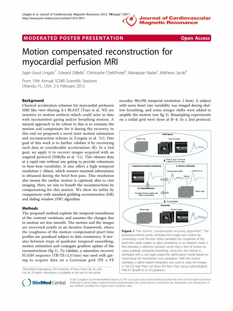
MethodsThe proposed method exploits the temporal smoothnessof the contrast variations, and assumes the changes dueto motion are less smooth. The motion and the imagesare recovered jointly in an iterative framework, wherethe roughness of the motion compensated pixel timeprofiles are penalized subject to data consistency. It iter-ates between steps of quadratic temporal smoothing,motion estimation and conjugate gradient update of thereconstructions (fig 1). To validate, a saturation recoveryFLASH sequence (TR/TE=2.5/1ms) was used with gat-ing to acquire data on a Cartesian grid (PE x FE
encodes: 90x190, temporal resolution: 1 beat). A subjectwith some heart rate variability was imaged during shal-low breathing, and some integer shifts were added toamplify the motion (see fig 2). Resampling experimentson a radial grid were done at R~4. In a 2nd protocol,
1Biomedical Engineering, The University of Iowa, Iowa City, IA, USAFull list of author information is available at the end of the article
Figure 1 The motion compensated recovery algorithm*: Theproposed scheme jointly estimates the images and motion byminimizing a cost function which penalizes the roughness of thepixel time series subject to data consistency. In an iterative mode, itfirst estimates a reference dynamic scene that is free of motion byusing quadratic temporal smoothing. Using this, the motion isestimated with a non-rigid coarse-fine deformation model based onmaximizing the normalized cross correlation. With the motionestimates, a spline based interpolant was used to warp the imagesin the CG step.*Here, we show the flow chart using a physiologicalPINCAT [Shariff et al. 07] phantom.
Lingala et al. Journal of Cardiovascular Magnetic Resonance 2012, 14(Suppl 1):M11http://www.jcmr-online.com/content/14/S1/M11
© 2012 Lingala et al; licensee BioMed Central Ltd. This is an open access article distributed under the terms of the Creative CommonsAttribution License (http://creativecommons.org/licenses/by/2.0), which permits unrestricted use, distribution, and reproduction inany medium, provided the original work is properly cited.

radial k space data with no gating was acquired (TR/TE=2.2/1.2 ms, 2.3x2.3mm pixel, 20 interleaved rays/frame with rotations across frames). 5 slices wereacquired after each saturation pulse, during a breathhold. This gave a total of 280 images/slice, where eachimage was acquired in 44 ms, and repeated every ~270ms (fig 2).
ResultsFrom fig 2, the images recovered with the proposedscheme were robust to motion blur and streaking arti-facts seen respectively with the SW and the GR meth-ods. Apart from its use in the recovery, the motionestimates here could be used for subsequent post-pro-cessing. For instance, to improve the temporal resolu-tion and/or the SNR of the ungated images or toimplement self-gating.
ConclusionsA novel motion compensation scheme for recoveringperfusion images with significant motion content wasdemonstrated. The experiments with both inconsistentgated, breathing and ungated data show promise; furtherstudies on multiple sets are required to thoroughly eval-uate the method.
FundingNSF AWARD CCF-0844812 and in part by NIHR01EB006155.
Author details1Biomedical Engineering, The University of Iowa, Iowa City, IA, USA.2Radiology, University of Utah, Salt Lake City, UT, USA. 3Siemens CorporateResearch, Princeton, NJ, USA. 4Electrical and Computer Engineering, TheUniversity of Iowa, Iowa City, IA, USA.
Published: 1 February 2012
doi:10.1186/1532-429X-14-S1-M11Cite this article as: Lingala et al.: Motion compensated reconstructionfor myocardial perfusion MRI. Journal of Cardiovascular MagneticResonance 2012 14(Suppl 1):M11.
Figure 2 Recovery of perfusion data with (I) inconsistent gating and shallow breathing and (II) ungated data. Few spatial frames and the imagetime profiles are shown. In (I), the first row shows the fully sampled data. The motion due to breathing and improper gating are shown by thegreen arrows. In (II), we consider applying the reconstructions to a single slice; the motion here is due to the cardiac dynamics. Here, we notethat the proposed scheme robustly recovers the data in the presence of motion, while sliding window and the gridding reconstructionsrespectively show temporal blur and streak artifacts. Specifically, for the ungated data, we note that if the motion is not compensated, it couldlead to misinterpreting the cardiac phase at a particular time frame. For instance in (II a), it is clear that the systolic phase is being imaged, whilesliding window in (II b) depicts this as a phase which looks like diastolic. The proposed scheme recovers the appropriate phase in (II c).
Lingala et al. Journal of Cardiovascular Magnetic Resonance 2012, 14(Suppl 1):M11http://www.jcmr-online.com/content/14/S1/M11
Page 2 of 2