morphoanatomic characterization of the stem and the leaf ... · employed in folk medicine as an...
TRANSCRIPT
667
Rev. Bras. Pl. Med., Campinas, v.17, n.4, supl. I, p.667-678, 2015.
Recebido para publicação em 16/07/2014Aceito para publicação em 20/02/2015
Morphoanatomic characterization of the stem and the leaf of Tabernaemontana catharinensis A.DC (Apocynaceae) and antimutagenic activity of its leaves
GUIDOTI, D.G.G.1*; GUIDOTI, D.T.1; ROCHA, C.L.M.S.C.1; MOURÃO, K.S.M.1
1Programa de Pós-graduação em Biologia Comparada da Universidade Estadual de Maringá, Avenida Colombo nº 5790, Jardim Universitário, CEP: 87020-900, Maringá/PR, Brasil. *Autor para correspondência: [email protected]
ABSTRACT: Tabernaemontana catharinensis A. DC (Apocynaceae) is used as a medicinal plant by the population. In order to contribute to the safe use of the plant as herbal medicine, this study aimed to morphoanatomically characterize the aereal vegetative organs of T. catharinensis and to evaluate the leaves’ mutagenic and antimutagenic activities. Histological blades of leaves and stem of T. catharinensis were performed; the methionine system (methG1) and Aspergillusnidulans conidia germination analysis were employed for mutagenic and antimutagenic evaluation. The morphoanatomic analysis did not show trichomes in the stem, petiole and leaf. Besides, it was observed both the presence of bi-collateral bundles - except in the foliar apex where the bundles were from the collateral type - as well as anamphistomatic leaf with paracyte stomata and sub-epidermal layer in the region of the leaf edges. The mutagenicity/antimutagenicity trial indicated a significant decrease of mutation frequency in comparison with the control group and showed that theT. catharinensis had antimutagenic activity within the type, time and form of treatment. Since the germination test showed that the conidia germination was accelerated from the bud phase, activities at the cell cycle level and polarized growth proved to be possible. The morphoanatomic analysis of the leaf and stem associated with the mutagenic and antimutagenic analyses contributes to the safe use of the plant by humans and also for the quality control of a possible phytotherapeutic drug.
Keywords: Aspergillus nidulans, genotoxicity, medicinal herbs, methionine system, morpho-anatomy.
RESUMO: Caracterização morfoanatômica do caule e da folha de Tabernaemontana catharinensis A.DC (Apocynaceae) e atividade antimutagênica de suas folhas Tabernaemontana catharinensis A. DC (Apocynaceae) é utilizada como planta medicinal pela população. A fim de contribuir para o uso seguro da planta como medicinal, este trabalho teve como objetivo caracterizar morfoanatomicamente os órgãos vegetativos aéreos de T. catharinensis e avaliar a atividade mutagênica e antimutagênica de suas folhas. Foram realizados cortes histológicos da folha e do caule de T. catharinensis e, para a avaliação mutagênica e antimutagênica, foi utilizado o sistema metionina (methG1) e análise da germinação de conídios em Aspergillus nidulans. A análise morfoanatômica evidenciou a ausência de tricomas no caule, pecíolo e folha; presença de feixes bicolaterais, com exceção no ápice foliar cujos feixes são do tipo colateral; folha anfiestomática com estômatos paracíticos e camada subepidérmica na região do bordo foliar. O ensaio de mutagenicidade/antimutagenicidade mostrou uma diminuição significativa da frequência de mutação em relação ao controle, indicando que nesse tipo, tempo e forma de tratamento, T. catharinensis apresentou atividade antimutagênica. O ensaio de germinação evidenciou que houve aceleração da germinação dos conídios, a partir da fase de botão, indicando uma possível atuação em nível de ativação de ciclo celular e crescimento polarizado. A análise morfoanatômica da folha e do caule associados à análise mutagênica e antimutagênica, contribuem para o uso seguro da planta pela população e para o controle de qualidade de um possível fitoterápico.
Palavras-chave: Aspergillus nidulans, genotoxicidade, plantas medicinais, sistema metionina, morfoanatomia.
10.1590/1983-084X/14_077
668
Rev. Bras. Pl. Med., Campinas, v.17, n.4, supl. I, p.667-678, 2015.
INTRODUCTIONSeveral plants have been used as folk
medicine for the treatment of several diseases (Melo-Reis et al., 2011), some of which, such as Solanum validinervium (Suarez et al., 2006), Morinda citrifolia (Brito et al., 2009) and Annona squamosa (Nandhakumar & Indumathi, 2012), showed highly interesting characteristics, while others, such as Bauhinia monandra (Macêdo et al., 2008), Alchornea grandulosa, A. castaneaefolia (Santos et al., 2010) and Cochlospermum regium (Castro et al., 2004; Andrade et al., 2008), proved to be genotoxic.
The study of medicinal plants is important since it analyzes compounds with a chemical and therapeutic potential and it is a safety strategy in the use of popular medicines (Arora et al., 2005). Plants precise identification and morphoanatomical characterization integrate the quality control of prime matter used in the preparation of phytotherapic compounds and warrant their reliability (Ming, 1994).
Tabernaemontana catharinensis A.DC (Apocynaceae) is a latex-producing tree found in Argentina, Uruguay, Paraguay, Brazil and Bolivia (Pereira et al., 2008). T. catharinensis infusion is employed in folk medicine as an antidote against snake bite, for the relief of toothache and as an antihelminthic potion.
Several assays have been performed with different sections of T. catharinensis and they revealed the following activities: antioxidant: the crude extract and hexane, water and alkaloid fractions from leaves and branches; the antioxidant effect varied between 53% and 95% determined by the coupled reaction of β-carotene and limonene acid (Pereira et al., 2005); anticancer activity: the crude extract of leaves against seven strains of human tumors (lung, breast, melanoma, colon, prostate, kidney) resistant to several drugs, with cytostatic and cytolithic effects, without any selectivity among the strains employed (Pereira et al., 2006); antiinflammatory: the stem´s bark with regard to edema induced in rats, with reduction by about 56% (Gomes et al., 2009).
The main chemical compounds of the secondary metabolites of Tabernaemontana correspond to indolic alkaloids (Fumagali et al., 2008), a class of compounds with a wide spectrum of pharmacologic activities (Federici et al., 2000).
Although several phytochemical studies on T. catharinensis reveal the species’ therapeutic activity, it use by people and the lack of pre-clinical and clinical research make the performance of mutagenicity and antimutagenicity assays mandatory with regard to different ways of its preparation. Further, several studies are required that describe its morphology and anatomy for its correct identification and consequently its quality control for the safe use
of the plant as a medicine. Current research characterizes the
morphoanatomy of the aerial segments of T. catharinensis and evaluates the genotoxic effect of medicinal preparations from its leaves, such as types of tea and water extracts used in folk medicine, with methionine (methG1) in Aspergillus nidulans as a test system.
MATERIAL AND METHODPlant material
Leaves and stem from Tabernaemontana catharinensis A. DC (Apocynaceae) were harvested in Maringá PR Brazil, 23º24’14.6”S; 51º56’23.3”W, in October 2011. Branches without flowers, leaves as from the third knot and the stem of the meristem region till the fourth knot were used. Voucher specimen of the plant was deposited in the Herbarium of the Universidade Estadual de Maringá (HUEM), registered under 21910, and identified by Dr. André Olmos Simões.
Anatomical studies were performed in fresh and fixed material employing the apical, median, lateral and basal regions. Leaf diaphanization was done following technique by Fuchs (1963) and FAA 50 (Johansen, 1940) and alcohol 70% (Jensen, 1962) were used respectively to fix and conserve the material.
Stem and leaf cuttings at several planes were undertaken under hand conditions and with a rotary microtome. They were stained with safranin and astra-blue (Gerlach, 1969).
Permanent laminas were manufactured from material included in Historesin - Leica, stained with toluidine blue (O’Brien et al., 1964) and mounted on Permount.
Histological and chemical tests were applied to the fresh material with Sudan IV solution for lipophilic compounds (Rawlins & Takahashi, 1952); water solution of ferric chloride 10% to localize phenolic compounds; fluoroglucinol acid (Johansen, 1940) to identify lignin; lugol reagent to localize starch (Jensen, 1962). The terminology employed to define morphological traits and leaf venation pattern followed Manual of Leaf Architecture (1999).
Methionine (methG1) system and conidia germination analysis in Aspergillus nidulans
Nutritional mutant strain of the fungus Aspergillus nidulans - biA1methG1, originary hailing from Glasgow, Scotland, was used for methionine (methG1) system and for the analysis of conidia germination. The strain is defective for biotin (chromosome I) and methionine production (chromosome IV).
669
Rev. Bras. Pl. Med., Campinas, v.17, n.4, supl. I, p.667-678, 2015.
Culture media were prepared according to Pontecorvo et al. (1959) and Clutterbuck (1974). Before treatment, strain biA1methG1 was cultured in a complete solid medium (CSM). Selective medium (SM) for the analysis of revertents consisted of minimum medium (MM), supplemented with biotin (0.02 µg.ml-1). Survival test medium (STM) was prepared with the same SM composition plus methionine (50 µg.ml-1).
Complete liquid medium (CLM), prepared with the same composition of complete solid medium, without the addition of sugar, was used to analyzed germination.
Preparation of extracts and treatmentLeaves were harvested in October 2011 on
the campus of the State University of Maringá (UEM), Maringá PR Brazil, and kept in a buffer at 37°C for approximately two weeks for drying.
Tea and water extract with boiling water and water at room temperature were prepared respectively, with dry and sliced leaves. Concentrations followed Pereira et al. (2006): 2.5 µg.ml-1 e 25 µg.ml-1, since results in the experiment showed that extract was efficient as from concentration 0.25 µg.ml-1. Solutions were filtered in double filter paper and later in a Milipore filter.
Methionine system (methG1) was employed for the mutagenesis/antimutagenesis analysis, following Lilly (1965). Conidia of strain biA1methG1 were harvested from 5-day-growth colonies, in CM, at 37º C and transferred to test tubes with 0.01% Tween 80, and stirred mechanically.
Conidia suspension was filtered in glass wool and divided into the test tubes: four were selected for treatments and one was the control with sterilized distilled water; test preparations were then added to the tubes with treatment.
After 4 h treatment time, tubes were centrifuged at 3000 rpm for 10 minutes and the precipitate was re-suspended in distilled water. Procedure was done in triplicate. Samples from each tube were counted in a hematimeter. Adequate dilutions were undertaken to estimate survival rates. Inoculate 0.1 ml of the suspension was diluted in 10 MSM plates for each test tube (treatment and control) and incubated for 3 days at 37ºC.
Analysis of revertents was performed by inoculating 0.1 ml of the suspension without any dilution in 10 SM plates for each tube (treatment and control) and incubated during 5 days at 37°C. Procedures were done in triplicate.
Statistical analysisStatistical analysis was undertaken following
Munson & Goodhead (1977), adapted for methionine system by Scott et al. (1982).
Analysis of conidia germinationGermination was analyzed by harvesting
conidia of strain biA1methG1 from colonies with 5 days of growth, in CM, at 37º C, transferred to test tubes with 0.01% Tween 80 and stirred mechanically.
Conidia suspension was filtered with glass wool and inoculated under five conditions: control (C) - containing sterile distilled water and treatments with tea and water extract from T. catharinensis leaves at concentrations 2.5 µg.ml-1 and 25 µg.ml-1. Further, 100 µl samples were transferred to microscope laminas and incubated at 37ºC in a wet chamber for 8 hours.
Laminas of each condition were analyzed every 2 hours and magnified 40 times under an optic microscope. Two hundred conidia were analyzed at each reading and conidia percentages were calculated at each germination phase (dormant, imbibed, germination bud and germination tube). The experiment was conducted in duplicate.
RESULT AND DISCUSSIONMorphology
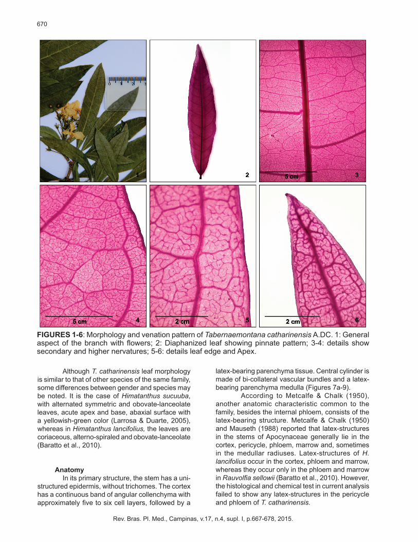
Tabernaemontana catharinensis is a shrub approximately 3.5 m long, with simpodial ramification. Its leaves are simples, glabras, opposite, marginally petiolated, with a milky sap, without stipulas and an elliptic laminar shape and acute asymmetric base. The apex is acumen-shaped (Figure 1). The border is slightly waved, without any lobules, but featuring a light green abaxial surface and dark green adaxial one.
Apezzato-da-Glória (1993) reports that when a species spreads over a wide environmental gradient, the species with meso-habitats have glabra or glabrascent leaves. This may be the case of the species under analysis, due to its wide geographic distribution in South American countries such as Brazil, Argentina, Paraguay and Uruguay (Pereira et al., 2005).
Leaves venation pattern is pine-shaped and primary nervation tapers towards on the apex; secondary nervation are slightly brochidodromous since they start from the primary nervation in a set of alternate arcs linked to the nervation immediately above (Figures 2-4).
Tertiary nervations are reticulated and randomized which anastomose with other tertiary or secondary nervations in an obtuse angle. Quaternary nervations are regular reticulated polygons and they anastomose with other nervations forming similar sized and shaped polygons. Quintenary nervations, dichotomized with two or more ramifications, form the venules (Figure 5). Venation on the edge region is anastomosed to venules that end adjacent to the border, apex included (Figures 5-6).
670
Rev. Bras. Pl. Med., Campinas, v.17, n.4, supl. I, p.667-678, 2015.
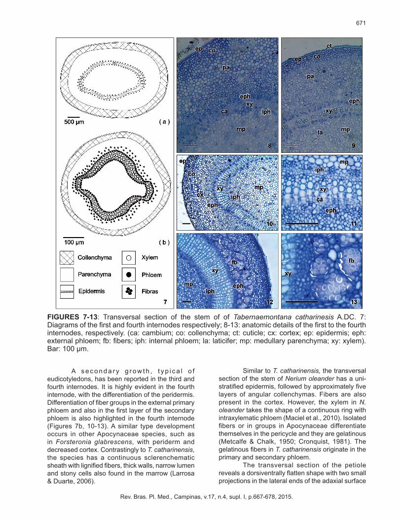
FIGURES 1-6: Morphology and venation pattern of Tabernaemontana catharinensis A.DC. 1: General aspect of the branch with flowers; 2: Diaphanized leaf showing pinnate pattern; 3-4: details show secondary and higher nervatures; 5-6: details leaf edge and Apex.
Although T. catharinensis leaf morphology is similar to that of other species of the same family, some differences between gender and species may be noted. It is the case of Himatanthus sucuuba, with alternated symmetric and obovate-lanceolate leaves, acute apex and base, abaxial surface with a yellowish-green color (Larrosa & Duarte, 2005), whereas in Himatanthus lancifolius, the leaves are coriaceous, alterno-spiraled and obovate-lanceolate (Baratto et al., 2010).
AnatomyIn its primary structure, the stem has a uni-
structured epidermis, without trichomes. The cortex has a continuous band of angular collenchyma with approximately five to six cell layers, followed by a
latex-bearing parenchyma tissue. Central cylinder is made of bi-collateral vascular bundles and a latex-bearing parenchyma medulla (Figures 7a-9).
According to Metcalfe & Chalk (1950),
another anatomic characteristic common to the family, besides the internal phloem, consists of the latex-bearing structure. Metcalfe & Chalk (1950) and Mauseth (1988) reported that latex-structures in the stems of Apocynaceae generally lie in the cortex, pericycle, phloem, marrow and, sometimes in the medullar radiuses. Latex-structures of H. lancifolius occur in the cortex, phloem and marrow, whereas they occur only in the phloem and marrow in Rauvolfia sellowii (Baratto et al., 2010). However, the histological and chemical test in current analysis failed to show any latex-structures in the pericycle and phloem of T. catharinensis.
671
Rev. Bras. Pl. Med., Campinas, v.17, n.4, supl. I, p.667-678, 2015.
FIGURES 7-13: Transversal section of the stem of of Tabernaemontana catharinesis A.DC. 7: Diagrams of the first and fourth internodes respectively; 8-13: anatomic details of the first to the fourth internodes, respectively. (ca: cambium; co: collenchyma; ct: cuticle; cx: cortex; ep: epidermis; eph: external phloem; fb: fibers; iph: internal phloem; la: laticifer; mp: medullary parenchyma; xy: xylem). Bar: 100 µm.
A s e c o n d a r y g r o w t h , t y p i c a l o f eudicotyledons, has been reported in the third and fourth internodes. It is highly evident in the fourth internode, with the differentiation of the peridermis. Differentiation of fiber groups in the external primary phloem and also in the first layer of the secondary phloem is also highlighted in the fourth internode (Figures 7b, 10-13). A similar type development occurs in other Apocynaceae species, such as in Forsteronia glabrescens, with periderm and decreased cortex. Contrastingly to T. catharinensis, the species has a continuous sclerenchematic sheath with lignified fibers, thick walls, narrow lumen and stony cells also found in the marrow (Larrosa & Duarte, 2006).
Similar to T. catharinensis, the transversal section of the stem of Nerium oleander has a uni-stratified epidermis, followed by approximately five layers of angular collenchymas. Fibers are also present in the cortex. However, the xylem in N. oleander takes the shape of a continuous ring with intraxylematic phloem (Maciel et al., 2010). Isolated fibers or in groups in Apocynaceae differentiate themselves in the pericycle and they are gelatinous (Metcalfe & Chalk, 1950; Cronquist, 1981). The gelatinous fibers in T. catharinensis originate in the primary and secondary phloem.
The transversal section of the petiole reveals a dorsiventrally flatten shape with two small projections in the lateral ends of the adaxial surface
672
Rev. Bras. Pl. Med., Campinas, v.17, n.4, supl. I, p.667-678, 2015.
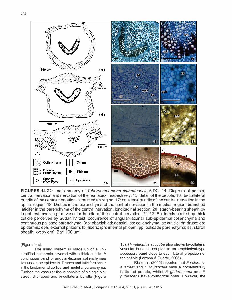
FIGURES 14-22: Leaf anatomy of Tabernaemontana catharinensis A.DC. 14: Diagram of petiole, central nervation and nervation of the leaf apex, respectively; 15: detail of the petiole; 16: bi-collateral bundle of the central nervation in the median region; 17: collateral bundle of the central nervation in the apical region; 18: Druses in the parenchyma of the central nervation in the median region; branched laticifer in the parenchyma of the central nervation, longitudinal section; 20: starch-bearing sheath by Lugol test involving the vascular bundle of the central nervation; 21-22: Epidermis coated by thick cuticle perceived by Sudan IV test, occurrence of angular-lacunar sub-epidermal collenchyma and continuous palisade parenchyma. (ab: abaxial; ad: adaxial; co: collenchyma; ct: cuticle; dr: druse; ep: epidermis; eph: external phloem; fb: fibers; iph: internal phloem; pp: palisade parenchyma; ss: starch sheath; xy: xylem). Bar: 100 µm.
(Figure 14c). The lining system is made up of a uni-
stratified epidermis covered with a thick cuticle. A continuous band of angular-lacunar collenchymas lies under the epidermis. Druses and laticifers occur in the fundamental cortical and medullar parenchyma. Further, the vascular tissue consists of a single big-sized, U-shaped and bi-collateral bundle (Figure
15). Himatanthus sucuuba also shows bi-collateral vascular bundles, coupled to an amphicrival-type accessory band close to each lateral projection of the petiole (Larrosa & Duarte, 2005).
Rio et al. (2005) reported that Forsteronia australis and F. thyrsoidea have a dorsiventrally flattened petiole, whilst F. glabrescens and F. pubescens have cylindrical ones. However, the
673
Rev. Bras. Pl. Med., Campinas, v.17, n.4, supl. I, p.667-678, 2015.
vascular bundle is bi-collateral, similar to all the above-mentioned species of T. catharinensis. Contrary to that in T. catharinensis, the petiole of F. glabrescens is concave-convex shaped, with simple tector trichomes coupled to non lignified fibers close to the internal phloem (Larrosa & Duarte, 2006).
Although the petiole structure continues within the central nervation the vessel becomes collateral in the leaf apex. The modification in the sheaves´ pattern occur separately (Figures 14c and 17). Druses and ramified laticifers occur in the fundamental parenchyma and a starch sheath
involves the vascular system of the central nerve as its continuation in the stem and the petiole (Figures 18-20).
Different ly to T. catharinensis, the Himatanthus sucuuba, described by Larrosa & Duarte (2005), has a V-shaped bi-collateral band in the petiole and central nervation. Several bi-small-size collateral vascular bundles occur towards the adaxial surface, which form a triangular arrangement with the bigger bundle. In the case of this species, the same authors registered ramified laticifers with thick walls and lipophilic contents in the fundamental
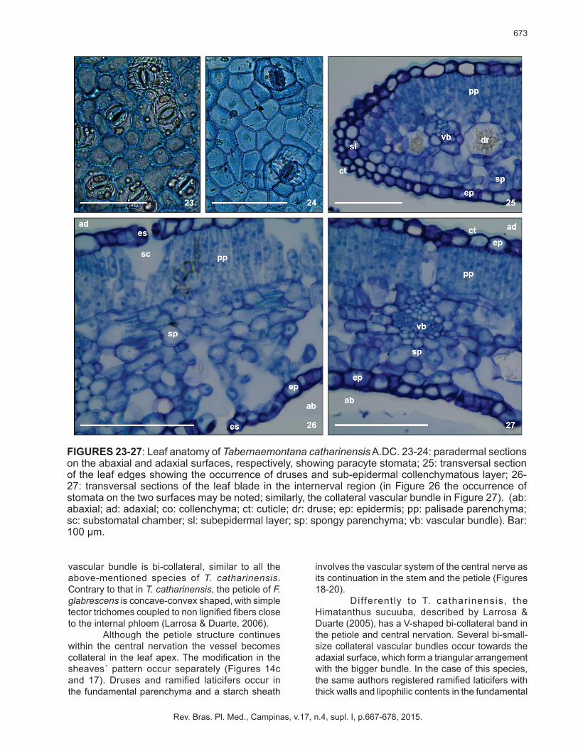
FIGURES 23-27: Leaf anatomy of Tabernaemontana catharinensis A.DC. 23-24: paradermal sections on the abaxial and adaxial surfaces, respectively, showing paracyte stomata; 25: transversal section of the leaf edges showing the occurrence of druses and sub-epidermal collenchymatous layer; 26-27: transversal sections of the leaf blade in the internerval region (in Figure 26 the occurrence of stomata on the two surfaces may be noted; similarly, the collateral vascular bundle in Figure 27). (ab: abaxial; ad: adaxial; co: collenchyma; ct: cuticle; dr: druse; ep: epidermis; pp: palisade parenchyma; sc: substomatal chamber; sl: subepidermal layer; sp: spongy parenchyma; vb: vascular bundle). Bar: 100 µm.
674
Rev. Bras. Pl. Med., Campinas, v.17, n.4, supl. I, p.667-678, 2015.
parenchyma of the central nervation and petiole, similar to the structure of T. catharinensis. The leaf of the T. catharinensis has a uni-stratified epidermis covered with a thick b cuticle (Figures 21-22). The leaf is amphistomatic with paracyte stomata predominantly on the abaxial surface (Figures 23-24).
According to Mott et al. (1982), amphistomatic leaves have as a rule a greater number of stomata and, consequently, a higher capacity to absorb carbonic acid to reach high photosynthesis levels. According to Metcalfe & Chalk (1950), stomata of the Apocynaceae are mainly anomocytic or paracytic and generally occur only on the abaxial surface.
Leaf coating system of T. catharinensis is organized similarly to that of other species of the family, with some differences: Himatanthus sucuuba has anomocytic stomata and the epidermis, coated with a striated cuticle, featured elongated cells in the periclinal direction (Larrosa & Duarte, 2005). Stomata of Forsteronia australis, F. glabrescens, F. pubescens and F. thyrsoidea are paracytic but restricted to the epidermis of the abaxial surface. Only the epidermis of F. australis is glabrous (Rio et al., 2005). Himathanthus lancifolius has anisocytic-type stomata only on the abaxial surface on which the cuticle is less thick (Baratto et al., 2010). Nerium oleander has a abaxial surface with great depressions, coupled to an epidermis on the surface coated with a thicker cuticle (Maciel et al., 2010).
The morphoanatomical analysis did not reveal trichomes in T. catharinensis. This factor was also reported by Rio et al. (2005) with regard to Forsteronia australis, F. glabrescens, F. pubescens and F. thyrsoidea, whose structures cannot be called trichome but merely emergencial since their origin is not exclusively protedermic (Rio et al., 2005). However, trichomes are a characteristic in other members of the family, such as the species Catharanthus roseus (Santos et al., 2009) and Nerium oleander (Maciel et al., 2010).
The mesophyll is dorsiventral, with two to three layers of cells which form the parenchyma palisade. The spongy parenchyma comprises approximately four layers of iso-diametric and slightly brachyform cells, with intercell spaces or gaps, approximately 50-60% of the mesophyll´s height. Druses are extant and collateral-type vascular sheaves are found along the mesophyll (Figures 26-27). A sub-epidermal layer of the thick wall may be observed at the edges (Figure 25).
The mesophyll is dorsiventral in F. glabrescens, with two or three layers of parenchyma palisade, of which the former is longer than the others. The spongy parenchyma is composed of three to five cell layers with lobules and small vascular collateral sheaves are surrounded by a
sheath of parenchymal bundles in the direction of the epidermis. The sub-epidermal layer is made up of parenchymal cells (Larrosa & Duarte, 2006).
Nerium oleander comprises an iso-bilateral mesophyll made of two to three layers of parenchyma palisade on the adaxial surface and of one to two layers on the abaxial surface. Spongy parenchyma comprises approximately six cell layers. A hypoderm follows the epidermis on the adaxial surface and comprises approximately two to three transparent cell layers (Maciel et al., 2010).
The spongy parenchyma is interrupted within the direction of the central nervation in the basal, median and apical regions of the leaf, whereas the palisade parenchyma lies throughout the angular-lacunar collenchyma. The later is made up of approximately five cell layers in the leaf´s basal and median regions and with approximately three to four layers in the apical region. However, the above does not occur in Forsteronia glabrescens (Larrosa & Duarte, 2006) and in Himatanthus sucuuba since the palisade parenchyma is gradually interrupted and is replaced by ring collenchyma (Larrosa & Duarte, 2005). According to Metcalfe & Chalk (1950), the mesophyll in Apocynaceae is usually dorsiventral and the vascular bundles of the central nervation are typically bi-collateral. According to Haberlandt (1928), the duplication of the phloem in the bi-collateral bundles may be probably due to an increase in a physiological demand to augment the conductor system area.
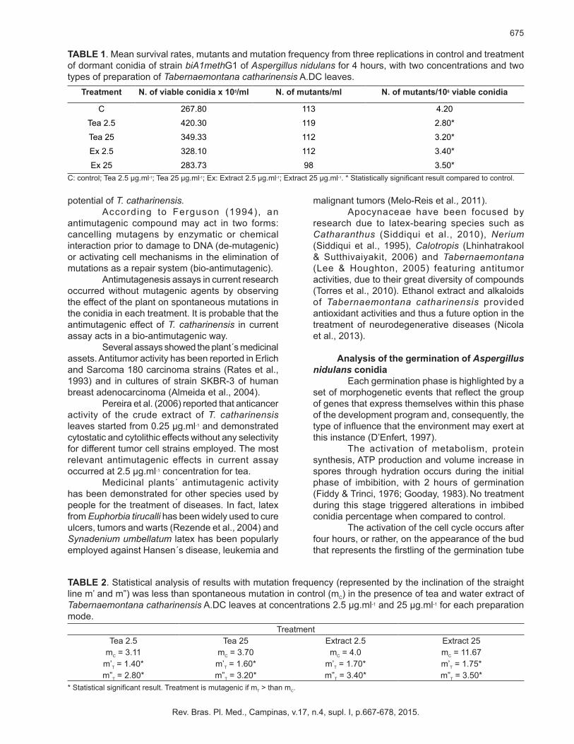
Mutagenesis and antimutagenesisTable 1 shows survival results, mutants and
mutation frequency of conidia treatment with tea and extract from the leaves of T. catharinensis. Results from T. catharinensis tea and extract revealed that no treatment was mutagenic since there was an increase in survival and a decrease in mutation frequency when compared to those of control. Statistical analysis showed that mutation frequency of the two teas and water extract concentrations is significantly lower that spontaneous mutation frequency (control). In other words, no mutagenic potential exists in T. catharinensis (Table 2).
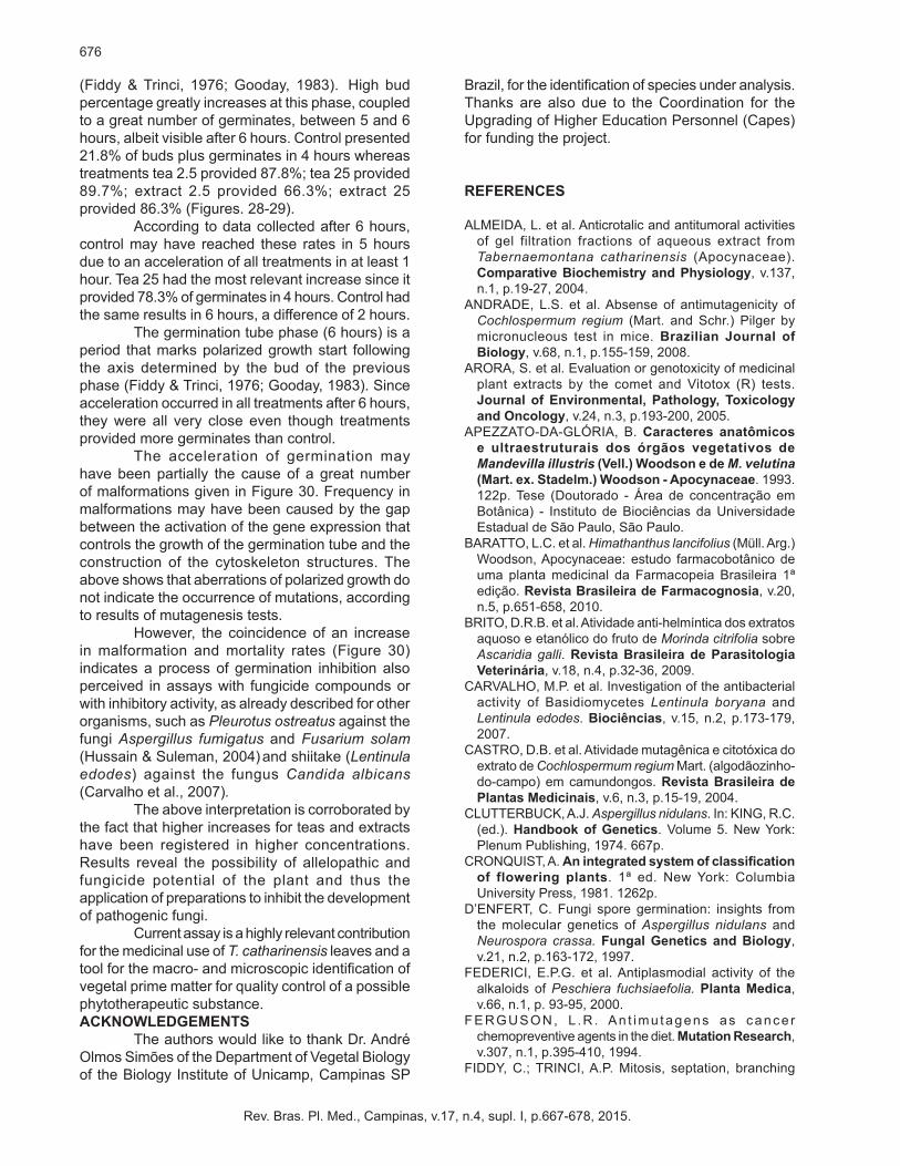
Foregrounded on the statistical method by Munson & Goodhead (1977), Rodrigues et al. (2003) suggested adapting the anti-mutagenic potential analysis. The tested compound is supposed to be anti-mutagenic if the inclination of the straight line calculated by spontaneous mutation rates (control) is higher than the inclination of the straight line calculated by induced mutation rates. As shown in Table 2, the frequency of mutation of the two concentrations of tea and water extract is significantly less than the frequency of spontaneous mutation (control) and indicates the antimutagenic
675
Rev. Bras. Pl. Med., Campinas, v.17, n.4, supl. I, p.667-678, 2015.
potential of T. catharinensis.According to Ferguson (1994), an
antimutagenic compound may act in two forms: cancelling mutagens by enzymatic or chemical interaction prior to damage to DNA (de-mutagenic) or activating cell mechanisms in the elimination of mutations as a repair system (bio-antimutagenic).
Antimutagenesis assays in current research occurred without mutagenic agents by observing the effect of the plant on spontaneous mutations in the conidia in each treatment. It is probable that the antimutagenic effect of T. catharinensis in current assay acts in a bio-antimutagenic way.
Several assays showed the plant´s medicinal assets. Antitumor activity has been reported in Erlich and Sarcoma 180 carcinoma strains (Rates et al., 1993) and in cultures of strain SKBR-3 of human breast adenocarcinoma (Almeida et al., 2004).
Pereira et al. (2006) reported that anticancer activity of the crude extract of T. catharinensis leaves started from 0.25 µg.ml-1 and demonstrated cytostatic and cytolithic effects without any selectivity for different tumor cell strains employed. The most relevant antimutagenic effects in current assay occurred at 2.5 µg.ml-1 concentration for tea.
Medicinal plants´ antimutagenic activity has been demonstrated for other species used by people for the treatment of diseases. In fact, latex from Euphorbia tirucalli has been widely used to cure ulcers, tumors and warts (Rezende et al., 2004) and Synadenium umbellatum latex has been popularly employed against Hansen´s disease, leukemia and
malignant tumors (Melo-Reis et al., 2011).Apocynaceae have been focused by
research due to latex-bearing species such as Catharanthus (Siddiqui et al., 2010), Nerium (Siddiqui et al., 1995), Calotropis (Lhinhatrakool & Sutthivaiyakit, 2006) and Tabernaemontana (Lee & Houghton, 2005) featuring antitumor activities, due to their great diversity of compounds (Torres et al., 2010). Ethanol extract and alkaloids of Tabernaemontana catharinensis provided antioxidant activities and thus a future option in the treatment of neurodegenerative diseases (Nicola et al., 2013).
Analysis of the germination of Aspergillus nidulans conidia
Each germination phase is highlighted by a set of morphogenetic events that reflect the group of genes that express themselves within this phase of the development program and, consequently, the type of influence that the environment may exert at this instance (D’Enfert, 1997).
The activation of metabolism, protein synthesis, ATP production and volume increase in spores through hydration occurs during the initial phase of imbibition, with 2 hours of germination (Fiddy & Trinci, 1976; Gooday, 1983). No treatment during this stage triggered alterations in imbibed conidia percentage when compared to control.
The activation of the cell cycle occurs after four hours, or rather, on the appearance of the bud that represents the firstling of the germination tube
TABLE 1. Mean survival rates, mutants and mutation frequency from three replications in control and treatment of dormant conidia of strain biA1methG1 of Aspergillus nidulans for 4 hours, with two concentrations and two types of preparation of Tabernaemontana catharinensis A.DC leaves.
C: control; Tea 2.5 µg.ml-1; Tea 25 µg.ml-1; Ex: Extract 2.5 µg.ml-1; Extract 25 µg.ml-1. * Statistically significant result compared to control.
Treatment N. of viable conidia x 105/ml N. of mutants/ml N. of mutants/106 viable conidia
C 267.80 113 4.20
Tea 2.5 420.30 119 2.80*
Tea 25 349.33 112 3.20*
Ex 2.5 328.10 112 3.40*
Ex 25 283.73 98 3.50*
TABLE 2. Statistical analysis of results with mutation frequency (represented by the inclination of the straight line m’ and m”) was less than spontaneous mutation in control (mC) in the presence of tea and water extract of Tabernaemontana catharinensis A.DC leaves at concentrations 2.5 µg.ml-1 and 25 µg.ml-1 for each preparation mode.
* Statistical significant result. Treatment is mutagenic if mT > than mC.
TreatmentTea 2.5 Tea 25 Extract 2.5 Extract 25
mC = 3.11 mC = 3.70 mC = 4.0 mC = 11.67m’T = 1.40* m’T = 1.60* m’T = 1.70* m’T = 1.75*m”T = 2.80* m”T = 3.20* m”T = 3.40* m”T = 3.50*
676
Rev. Bras. Pl. Med., Campinas, v.17, n.4, supl. I, p.667-678, 2015.
(Fiddy & Trinci, 1976; Gooday, 1983). High bud percentage greatly increases at this phase, coupled to a great number of germinates, between 5 and 6 hours, albeit visible after 6 hours. Control presented 21.8% of buds plus germinates in 4 hours whereas treatments tea 2.5 provided 87.8%; tea 25 provided 89.7%; extract 2.5 provided 66.3%; extract 25 provided 86.3% (Figures. 28-29).
According to data collected after 6 hours, control may have reached these rates in 5 hours due to an acceleration of all treatments in at least 1 hour. Tea 25 had the most relevant increase since it provided 78.3% of germinates in 4 hours. Control had the same results in 6 hours, a difference of 2 hours.
The germination tube phase (6 hours) is a period that marks polarized growth start following the axis determined by the bud of the previous phase (Fiddy & Trinci, 1976; Gooday, 1983). Since acceleration occurred in all treatments after 6 hours, they were all very close even though treatments provided more germinates than control.
The acceleration of germination may have been partially the cause of a great number of malformations given in Figure 30. Frequency in malformations may have been caused by the gap between the activation of the gene expression that controls the growth of the germination tube and the construction of the cytoskeleton structures. The above shows that aberrations of polarized growth do not indicate the occurrence of mutations, according to results of mutagenesis tests.
However, the coincidence of an increase in malformation and mortality rates (Figure 30) indicates a process of germination inhibition also perceived in assays with fungicide compounds or with inhibitory activity, as already described for other organisms, such as Pleurotus ostreatus against the fungi Aspergillus fumigatus and Fusarium solam (Hussain & Suleman, 2004) and shiitake (Lentinula edodes) against the fungus Candida albicans (Carvalho et al., 2007).
The above interpretation is corroborated by the fact that higher increases for teas and extracts have been registered in higher concentrations. Results reveal the possibility of allelopathic and fungicide potential of the plant and thus the application of preparations to inhibit the development of pathogenic fungi.
Current assay is a highly relevant contribution for the medicinal use of T. catharinensis leaves and a tool for the macro- and microscopic identification of vegetal prime matter for quality control of a possible phytotherapeutic substance.ACKNOWLEDGEMENTS
The authors would like to thank Dr. André Olmos Simões of the Department of Vegetal Biology of the Biology Institute of Unicamp, Campinas SP
Brazil, for the identification of species under analysis. Thanks are also due to the Coordination for the Upgrading of Higher Education Personnel (Capes) for funding the project.
REFERENCES
ALMEIDA, L. et al. Anticrotalic and antitumoral activities of gel filtration fractions of aqueous extract from Tabernaemontana catharinensis (Apocynaceae). Comparative Biochemistry and Physiology, v.137, n.1, p.19-27, 2004.
ANDRADE, L.S. et al. Absense of antimutagenicity of Cochlospermum regium (Mart. and Schr.) Pilger by micronucleous test in mice. Brazilian Journal of Biology, v.68, n.1, p.155-159, 2008.
ARORA, S. et al. Evaluation or genotoxicity of medicinal plant extracts by the comet and Vitotox (R) tests. Journal of Environmental, Pathology, Toxicology and Oncology, v.24, n.3, p.193-200, 2005.
APEZZATO-DA-GLÓRIA, B. Caracteres anatômicos e ultraestruturais dos órgãos vegetativos de Mandevilla illustris (Vell.) Woodson e de M. velutina (Mart. ex. Stadelm.) Woodson - Apocynaceae. 1993. 122p. Tese (Doutorado - Área de concentração em Botânica) - Instituto de Biociências da Universidade Estadual de São Paulo, São Paulo.
BARATTO, L.C. et al. Himathanthus lancifolius (Müll. Arg.) Woodson, Apocynaceae: estudo farmacobotânico de uma planta medicinal da Farmacopeia Brasileira 1ª edição. Revista Brasileira de Farmacognosia, v.20, n.5, p.651-658, 2010.
BRITO, D.R.B. et al. Atividade anti-helmíntica dos extratos aquoso e etanólico do fruto de Morinda citrifolia sobre Ascaridia galli. Revista Brasileira de Parasitologia Veterinária, v.18, n.4, p.32-36, 2009.
CARVALHO, M.P. et al. Investigation of the antibacterial activity of Basidiomycetes Lentinula boryana and Lentinula edodes. Biociências, v.15, n.2, p.173-179, 2007.
CASTRO, D.B. et al. Atividade mutagênica e citotóxica do extrato de Cochlospermum regium Mart. (algodãozinho-do-campo) em camundongos. Revista Brasileira de Plantas Medicinais, v.6, n.3, p.15-19, 2004.
CLUTTERBUCK, A.J. Aspergillus nidulans. In: KING, R.C. (ed.). Handbook of Genetics. Volume 5. New York: Plenum Publishing, 1974. 667p.
CRONQUIST, A. An integrated system of classification of flowering plants. 1ª ed. New York: Columbia University Press, 1981. 1262p.
D’ENFERT, C. Fungi spore germination: insights from the molecular genetics of Aspergillus nidulans and Neurospora crassa. Fungal Genetics and Biology, v.21, n.2, p.163-172, 1997.
FEDERICI, E.P.G. et al. Antiplasmodial activity of the alkaloids of Peschiera fuchsiaefolia. Planta Medica, v.66, n.1, p. 93-95, 2000.
FERGUSON, L .R . An t imu tagens as cance r chemopreventive agents in the diet. Mutation Research, v.307, n.1, p.395-410, 1994.
FIDDY, C.; TRINCI, A.P. Mitosis, septation, branching
677
Rev. Bras. Pl. Med., Campinas, v.17, n.4, supl. I, p.667-678, 2015.
and duplication cycle in Aspergillus nidulans. Journal of General Microbiology, v.97, n.2, p. 169-184, 1976.
FUCHS, C. Fuchsin staining with NaOH clearing for lignified elements of whole plants or plant organs. Stain Technology, v.38, n.3, p.141-144, 1963.
FUMAGALI, E. et al. Produção de metabólitos secundários em cultura de células e tecidos de plantas: o exemplo dos gêneros Tabernaemontana e Apidosperma. Brazilian Journal of Pharmacognosy, v.18, n.4, p.627-641, 2008.
GERLACH, G. Botanische microtechnik eine einführung. Volume 1. Stuttgard: Georg Thieme Verlag, 1969. 112p.
GOMES, R.C. et al. Antinociceptive and anti-inflamatory activit ies of Tabernaemontana catharinensis . Pharmaceutical Biology, v.47, n.4, p.372-376, 2009.
GOODAY, G.W. The hyphal tip. In: SMITH, J.E. (Ed.). Fungal Differentiation. A Contemporary Synthesis. Volume 4. New York: Dekker, 1983. 624p.
HABERLANDT, G. Physiological plant anatomy. Volume 1. London: McMillan and Co, 1928. 777p.
HUSSAIN, A.; SULEMAN, M.S. Evaluation of Pleurotus ostreatus Jaco. Ex. Fr., for its growth at different stages and its antifungical activity. Mycopathology, v.2, n.2, p.91-94, 2004.
JENSEN, W.A. Botanical histochemistry: principles and practice. 1ª ed. San Francisco: Freeman and Co, 1962. 408p.
JOHANSEN, W.A. Plant microtechnique. 1ª ed. New York: McGraw-Hill, 1940. 523p.
LARROSA, C.R.R.; DUARTE, M.R. Morfoanatomia de folhas de Himatanthus sucuuba (Spruce) Woodson, Apocynaceae. Acta Farmacêutica Bonaerense, v.24, n.2, p.165-171, 2005.
LARROSA, C.R.R.; DUARTE, M.R. Anatomia foliar e caulinar de Forsteronia glabrescens, Apocynaceae. Acta Farmacêutica Bonaerense, v.25, n.1, p.28-34, 2006.
LEE, C.C.; HOUGHTON, P. Cytotoxicity of plants from Malaysia and Thailand used traditionally to treat cancer. Journal of Ethnopharmacology, v.100, n.3, p.237-243, 2005.
LHINHATRAKOOL, T.; SUTTHIVAIYAKIT, S. 19-Nor- and 1820-epoxy-cardenolides from the leaves of Calotropis gigantea. Journal of Natural Products, v.69, n.8, p.1249-1251, 2006.
LILLY, L.J. An Investigation of the suitability of the supressors of meth1 in Aspergillus nidulans for the study of induced and spontaneous mutations. Mutation Research, v.2, n.2, p.192-195, 1965.
MACÊDO, M.F.S. et al. Determining the genotoxicity of an aqueous infusion of Bauhinia monandra leaves. Brazilian Journal of Pharmacognosy, v.18, n.4, p. 509-516, 2008.
MACIEL, V.E.O.; CORRÊA, P.G.; SILVA, M.D.; CHAGAS, M.G.S.; PIMENTEL, R.M.M. Anatomia e histoquímica do caule e folha de Nerium oleander L. In: X JORNADA DE ENSINO, PESQUISA E EXTENSÃO - JEPEX, 2010, Recife. Apresentação de trabalho. Resumo Expandido. Recife, 2010.
MANUAL OF LEAF ARCHITECTURE - Morphological description and categorization of dicotyledonous and net-veined monocotyledonous angiosperms
by Leaf Architecture Working Grouping, 1999. 65p.MAUSETH, J.D. Plant Anatomy. 1ª Ed. California: Menlo
Park, Benjamin Cummings, 1988. 560p.MELO-REIS, P.R. et al. Assessment of the mutagenic and
antimutagenic activity of Synadenium umbellatum Pax latex by micronucleous test in mice. Brazilian Journal of Biology, v.71, n.1, p.169-174, 2011.
METCALFE, C.R.; CHALK, L. Anatomy of the dicotyledons: leaves stem and wood in relation to taxonomy with notes on economic uses. Volume 2. Oxford: Clarendon Press, 1950. 1500p.
MING, L.C. Estudos e pesquisas de plantas medicinais na agronomia. Horticultura Brasileira, v.12, n.1, p.3-9, 1994.
MOTT, K.A. et al. The adaptive significance of amphistomatic leaves. Plant Cell and Environment, v.5, n.6, p.455-460, 1982.
MUNSON, R.J.; GOODHEAD, D.T. The relation between induced mutation frequency and cell survival - a theoretical approach and examination of experimental data for eukaryotes. Mutation Research, v.42, p.145-159, 1977.
NANDHAKUMAR, E.; INDUMATHI, P. In vitro antioxidante activities of methanol and aqueous extract of Annona squamosa (L.) fruit pulp. Journal of Acupunture and Meridian Studies, v.6, n.3, p.142-148, 2012.
NICOLA, C. et al. Chemical constituents antioxidant and anticholinesterasic activity of Tabernaemontana catharinensis. The Scientific World Journal, v.2013, p.1-10, 2013.
O’BRIEN, T.P. et al. Polychromatic staining of plant cell walls by toluidine blue O. Protoplasma, v.59, n.2, p.368-373, 1964.
PEREIRA, C.G. et al. Antioxidant and Antimycobacterial Activities of Tabernaemontana catharinensis extracts obtained by supercritical CO2 + cosolvent. Journal of Medicinal Food, v.8, n.4, p.533-538, 2005.
PEREIRA, C.G. e t a l . Ant icancer act iv i ty o f Tabernaemontana catharinensis extract obtained by supercritical fluid extraction. Revista Brasileira de Plantas Medicinais, v.8, n.4, p.144-149, 2006.
PEREIRA, P.S. et al. Chemical constituents from Tabernaemontana catharinensis root bark: a brief NMR review of indole alkaloids and in vitro cytotoxicity. Química Nova, v.31, n.1, p.20-24, 2008.
PONTECORVO, G. et al. The genetics of Aspergillus nidulans. Advances in Genetics, v.5, p.141-238, 1959.
RATES, S.M.K. et al. Chemical constituents and pharmacological activities of Peschiera australis. International Journal of Pharmaceutics, v.31, n.4, 1993.
RAWLINS, T.E.; TAKAHASHI, W.N. Techniques of plant histochemistry and virology. 1ª ed. California: The National Press, Millbrae, 1952. 125p.
REZENDE, J.R. et al. Efeito antimutagênico do latex de Euphorbia tirucalli no sistema metionina em Aspergillus nidulans. Acta Scientiarum, v. 26, n.4, p.481-484, 2004.
RIO, M.C.S. et al. Anatomia foliar como subsídio para a taxonomia de espécies de Forsteronia G. Mey. (Apocynaceae) dos cerrados paulistas. Revista Brasileira de Botânica, v.28, n.4, p. 713-726, 2005.
RODRIGUES, S.B. et al. Avaliação do potencial antimutagênica do Cogumelo do Sol (Agaricus blazei) no

678
Rev. Bras. Pl. Med., Campinas, v.17, n.4, supl. I, p.667-678, 2015.
sistema methG1 em Aspergillus (=Emericella) nidulans. Acta Scientiarum, v. 25, n.2, p. 513-517, 2003.
SANTOS, M.C.A. et al. Anatomia e histoquímica de folhas e raízes de vinca (Catharanthus roseus (L.) G. Don.). Revista de Biologia e Ciências da Terra, v. 9, n.1, p.24-30, 2009.
SANTOS, F.V. et al. Mutageniticy of two species of the genus Alchornea mensured by Salmonella microsome assay and micronucleous test. Brazilian Journal of Pharmacognosy, v. 20, n.3, p.382-389, 2010.
SCOTT, B.R. et al. Aspergillus nidulans: Systems and results of tests for induction of mitotic segregation and mutation. Haploid assay systems and overall response of all systems. Mutation Research, v.98, n.1, p.49-94, 1982.
SIDDIQUI, B.S. et al. Two cytotoxic pentacyclic triterpenoids
from Nerium oleander. Phytochemistry, v.39, n.1, p.171-4, 1995.
SIDDIQUI, M.J. et al. Cytotoxic activity of Catharanthus roseus (Apocynaceae) crude extracts and pure compound against human carcinoma cell l ine. International Journal of Pharmacology, v.6, n.1, p.43-47, 2010.
SUAREZ, L.E.C. et al. Compuestos fenólicos aislados de la espécie Solanum validinervium (Solanaceae) Seccion Geminata. Revista Colombiana de Química (online), v.35, n.1, p.59-65, 2006.
TORRES, M.J. et al. Purification and characterization of a cysteine endopeptidase from Vasconcellea quercifolia A. St.-Hil. latex displaying high substrate specificity. Journal of Agricultural Food Chemistry, v.58, n.34, p.11027-11035, 2010.