monitoring helicase activity with molecular beacons · monitoring helicase activity with molecular...
TRANSCRIPT

Vol. 45 ı No. 4 ı 2008 www.biotechniques.com ı BioTechniques ı 433
Research Reports
INTRODUCTION
Helicases are motor proteins that unwind double-stranded nucleic acids by utilizing free energy from ATP hydro-lysis. They are ubiquitous enzymes in the cellular milieu, functioning indiverse processes including DNA repli-cation, DNA repair, RNA transcription, and translation (1). The genomes of allcellular pathogens and many virusescode for helicases, and there has been great interest in exploiting helicasesas potential drug targets. However, with the exception of several potent compounds that inhibit a helicaseencoded by herpes simplex virus, few helicase inhibitors have entered clinical trials (2). One explanation for slowhelicase inhibitor development maybe that helicase activity is complex to monitor, making inhibitor identifi-cation and analysis difficult. Therefore, the goal of this study is to develop an improved helicase assay that could beused both for high-throughput screening and mechanistic analyses. Here, wedescribe how molecular beaconscommonly used for quantitative PCRcan serve this purpose.
Typically, helicase activity is measured by incubating a helicase withATP and a radiolabeled DNA duplex, terminating the reaction, and analyzing the products using non-denaturing
PAGE (3). Although such assays can be used to extract reaction details, they are cumbersome, time-consuming, and only yield a single time point for each reaction performed. This basic helicase assaycan be performed in a high-throughput format by filtering the products after the reaction is stopped (4), or by using radiolabeled biotinylated oligonucle-otides in a scintillation proximity assay(5). Electrochemiluminescence usedwith ruthenium-labeled biotinylated oligonucleotides (6) can circumvent the need for the radioactive biohazard. Nevertheless, all such assays only yielda single time point per reaction, limitingtheir potential usefulness.
Fluorescence has been the main toolused to date to increase the number of time points recorded in a single helicase reaction. To this end, fluorescent nucle-otides can be incorporated into DNA(7), two fluorescent moieties tethered toopposing strands (8–10), or fluorescent intercalating agents added to DNA(11). Each of these assays can be usedto monitor DNA (or RNA) unwinding in real time, but each has its own setof drawbacks. For example, most fluorescent nucleotide analogs [with theexception of 2-aminopurine (7)] disruptWatson-Crick base pairing, fluorescentmoieties or DNA ligands could affect unwinding rates, and ssDNA traps need
to be added to such assays to preventthe substrate from re-annealing.
To better monitor helicase action, we have designed a fluorescence-basedassay using molecular beacons: single-stranded nucleic acid molecules thatform stem loop structures. One end of the molecular beacon is attached toa fluorescent molecule and the other to a quencher such that, upon strandseparation and subsequent formation of the stem-loop structure, fluorescence is quenched (12). Intramolecular hairpinformation prevents strand re-annealing,and eliminates the need for the addition of ssDNA trap molecules to the reaction mixture. Furthermore, since most helicases typically contact one strand of a duplex more than its complement (13,14), the design of these new helicasesubstrates helps minimize the possibleimpact of the modifications on observed reaction rates.
As a model helicase to test this assay, we used a recombinant enzyme derived from hepatitis C virus (HCV)(15). HCV causes a liver disease that affects more than two percent of theworld population. The virus itself is apositive-sense ssRNA virus coding for a single polyprotein that is cleaved into structural and non-structural proteins. One of the non-structural proteins is a multifunctional enzyme possessingan N-terminal protease domain and a
Monitoring helicase activity with molecular beaconsCraig A. Belon and David N. Frick
BioTechniques 45:433-442 (October 2008)doi 10.2144/000112834
A high-throughput, fluorescence-based helicase assay using molecular beacons is described. The assay is tested using the NS3 heli-case encoded by the hepatitis C virus (HCV) and is shown to accurately monitor helicase action on both DNA and RNA. In the assay, a ssDNA oligonucleotide molecular beacon, featuring a fluorescent moiety attached to one end and a quencher attached to the other, is annealed to a second longer DNA or RNA oligonucleotide. Upon strand separation by a helicase and ATP, the beacon strand forms anintramolecular hairpin that brings the tethered fluorescent and quencher molecules into juxtaposition, quenching fluorescence. Unlikecurrently available real-time helicase assays, the molecular beacon!based helicase assay is irreversible. As such, it does not requirethe addition of extra DNA strands to prevent products from re-annealing. Several variants of the new assay are described and experi-mentally verified using both Cy3 and Cy5 beacons, including one based on a sequence from the HCV genome. The HCV genome!based molecular beacon helicase assay is used to demonstrate how such an assay can be used in high-throughput screens and to analyze HCV helicase inhibitors.
Department of Biochemistry and Molecular Biology, New York Medical College, Valhalla, NY, USA

434 ı BioTechniques ı www.biotechniques.com Vol. 44 ı No. 4 ı 2008
Research Reports
C-terminal ATPase/helicase domain. The helicase portion of non-structuralprotein 3 (NS3) is needed for HCV RNA replication (16,17), but while many potent small molecule inhibitors for this helicase target have been discovered(18–21), none have entered the clinic.Many assays specific for HCV helicaseactivity have been developeddd such as those based on ELISA (22), scintil-lation proximity assays (23), flashplatemethods (24), and FRET (25,26))) but they all suffer from the same drawbacksas the aforementioned general helicase assays.
Here we develop a simple assayfor evaluating helicase activity basedon molecular beacon technology and demonstrate its usefulness using HCV helicase as a model. Our new assay is advantageous because it is continuous,does not require modification of thestrand on which the helicase translo-cates, is essentially irreversible, and
eliminates the need for the addition of extra DNA to capture displaced strands.The assay should be useful for mecha-nistic analyses, is amenable to high-throughput screening, and could be used to rapidly evaluate enzyme inhibitors.
MATERIALS AND METHODS
Materials
DNA and RNA oligonucleotides were purchased from Integrated DNATechnologies (Coralville, IA, USA)in purified form as lyophilized solids. They were then dissolved in DNase/RNase-free water (FisherBiotech, FairLawn, NJ, USA), and concentrationswere determined from the extinction coefficients provided. Oligonucleotideswere modified with Cyanine 3 (Cy3),Cyanine 5 (Cy5), Iowa Black RQ
(IBRQ), and Black Hole Quencher(BHQ) (Integrated DNA Technologies). The sequences of the oligonucleotides used in the study are noted in Table 1.
Helicase substrates were prepared by combining single strands at a 1:1 molar ratio to a final concentration of 20 Min 10 mM Tris-HCl pH 8.5, placing in 95 C water, and allowing to cool to room temperature for 1 h to anneal.
The HCV NS3 helicase enzyme usedfor this study was a recombinant protein derived from the HCV 1b genotype, lacking the N-terminal NS3 proteasedomain. Cloning, expression, and purifi-cation of this truncated NS3 protein hasbeen described before (27). It containsa C-terminal His-tag consisting of the sequence “PNSSS VDKLA AALEH HHHHH.” Protein concentrations were determined by absorbance at 280 nmusing extinction coefficients calculatedusing the Sequence Analysis program (http://informagen.com/SA).
Molecular beacon!based unwindingassay
Unless otherwise noted, eachreaction contained 25 mM MOPS pH6.5, 2 mM MgCl2, 25 nM enzyme, 5 nMnucleic acid substrate, and was initiatedwith 0.5 mM ATP. Reactions werecarried out in 100 L, in triplicate, in white half-volume 96-well polystyrene plates at 22 C. Fluorescence wasmeasured as arbitrary units (a.u.) in each well every 40 s when using a 96-well plate. Data were collected using a Caryeclipse fluorescence spectrophotometer equipped with the microplate reader accessory (Varian, Inc., Palo Alto, CA, USA). Cy3-labeled substrates weremeasured for excitation/emissionat 552/570 nm (5/10 nm slit width).Cy5-labeled substrates were similarly measured at 643/667 nm and FRET measured at 570/667 nm. Stopped-flowdata was collected using an RX.2000 rapid kinetics spectrophotometer accessory (Applied Photophysics, Leatherhead, UK) with 1.0 mM ATP in syringe A and 2 reaction mixture insyringe B (50 mM MOPS pH 6.5, 4 mM MgCl2, 50 nM enzyme, 10 nM nucleic acid substrate). Reactions were started by mixing syringe A and B in a 1:1 ratiosuch that final concentrations of all components were identical to those forthe plate-based assays.
Table 1. Sequences of Oligonucleotides Used
Description Figure Sequence
Cy3 top strand 1A, 2A, 3A 5 -/IBRQ/AGTGC// GCTGTATCGTCAAGGCACT/Cy3/-3TT
Cy5 top strand 1B, 3A 5 -/Cy5/CCTACGCCACCAGCTCCGTAGG/BHQ/-3
Cy3 bottom strand 1A, 2A 5 -AGTGCCTTGACGATACAGC(T)20-3
Cy3 bottom strand (RNA) 2A 5 -AGUGCCUUGACGAUACAGC(U)20-3
Cy5 bottom strand 1B 5 -GGAGCTGGTGGCGTAGG(T)20-3
Hairpin Cy3 bottom strand 1A 5 -AGTGCCTTGACGATACAGCGCACT(T)19-3
Hairpin Cy5 bottom strand 1B 5 -CCTACGGAGCTGGTGGCGTAGG(T)20-3
FRET bottom strand 3A 5 -GGAGCTGGTGGCGTAGGCAAGAGTGCCTTGACGATACAGC(T)20-3
HCV genome top strand 4A 5 -/Cy5/GCTCCCCAATCGATGAACGGGGAGC/IBQ/-3
HCV genome bottom strand 4A 5 -GCTCCCCGTTCATCGATTGGGGAGC((T)20
Underlined regions correspond to hairpin-forming residues.
B
) was subtracted from each reaction. a.u., arbitrary units.
A
Data were analyzed using Graphpad Prism 4.0 (San Diego, CA, USA), and afirst-order exponential decay model was used to determine the pseudo-first order rate constant, kobskk . Data are shown with error bars of 1 standard deviation.
Radioactive unwinding assay
5 -32P labeled HCV genome template DNA was prepared by incubating 2 L of 10 M oligonucle-otide, 4 L of [ 32P]ATP (0.5 mCi, MP Biomedical, Solon, OH, USA), 2
L 10 reaction buffer (New EnglandBiolabs, Ipswich, MA, USA), 1 Lpolynucleotide kinase (New England Biolabs) and 11 L of water for 1 h at37 C. The resulting labeled oligonucle-otide was purified using a QIAquick Nucleotide Removal Kit (Qiagen, Valencia, CA, USA). Concentration wasdetermined by measuring A260 with
389400 M-1cm-1. The helicase substrate was then made as noted above.
Reaction conditions were identical to those used for continuous fluorescent assays. All reactants except ATP were added to the well, and 10 L placed into 2.5 L of stopping buffer for a t0point. The ATP was added, and 10 Laliquots removed and placed into 2.5 Lof 5 stopping buffer after 20 s, 40 s, 1min, 2 min, 3 min, 5 min, 7.5 min, 10 min, and 20 min. 5 stopping buffer contained 250 mM TRIS-Cl pH 7.5, 20mM EDTA, 0.5 SDS, 0.1 Nonidet P-40, 0.1 bromophenol blue, 0.1xylene cyanol FF, and 50 glycerol. Four microliters of each quenched aliquots (4 nM DNA) were run on a 12 non-denaturing polyacrylamidegel at a constant 200 V for 30 min.Radiolabeled ssDNA (2 L of 37.5 nM ssDNA in stopping buffer) andsubstrate DNA (2 L of 25 nM DNAin stopping buffer) controls were
) , y
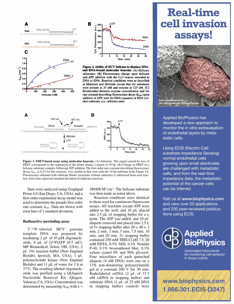
Figure 3. FRET-based assay using molecular beacons. (A) Substrate. The signal caused by loss of FRET corresponds to the separation of the donor strand, a region of 19 bp. (B) Change in FRET in a helicase substrate complex following ATP addition. The first order rate constant describing fluorescence decay, kobs, is 0.23 for this reaction, very similar to that seen with the 19 bp substrate from Figure 1A.Fluorescence obtained with substrate blanks (reactions without substrate) is subtracted from each reac-tion. Error bars represent standard deviation of triplicate reactions.
A B
Real-time cell invasion
assays!
Automated instruments for monitoring cell behavior in tissue culture.
www.biophysics.com 1.866.301.ECIS (3247)
www.biophysics.com
Irene Newsham et al., The MD Anderson Cancer Center

Research Reports
included. The gels were dried, exposedto a PhosphorImager screen for 14 hand imaged using a Storm 860 scanner(Molecular Dynamics, Sunnyvale, CA,USA), and quantified with ImageJ software (http://rsb.info.nih.gov/ij). Substrate fraction was calculated andcorrected for the presence of ssDNA att0 using Equation 1:
Substrate fractionDS(DS0 SS0)DS0 (DS SS)
,
[Eq.1]
where DS and SS are radioactive intensities for dsDNA and ssDNA at a given time and DS0 and SS0 are radio-active intensities at t0, respectively.
High-throughput screening
Two separate reactions, a positivecontrol (no inhibitor) and negativecontrol (dT20), were used. In both
instances, the HCV-derived DNA substrate shown in Figure 4A wasused. These reactions were staggered in a checkerboard pattern on a 384-well plate. The no-inhibitor reactions were identical to those described above for the real-time assay. For a negative control dT20 inhibited reaction, themixture contained an additional 1 M of dT20, a short ssDNA that competes with the dsDNA substrate. For each well,only two data points were collected:one before the addition of ATP (F0) and one at 30 min (F30). F0 was simply usedas a quality control to ensure that theinhibitor did not itself cause quenchingof fluorescence; only F30 was evaluated to determine reaction progress. Zfactors (28) were determined with Equation 2:
,
[Eq. 2]
where p and n are the standarddeviations of the positive and negative controls, respectively, and p and n
are similarly the means of the positive and negative controls. Pilot uninhibited and inhibited reactions (n = 16) were monitored continuously for fluores-cence. Experiments with n = 103 or 104 only used the F30 data point to computequantitative parameters. The coefficient of variance (CV) was determined as shown in Equation 3:
CV 100,
[Eq. 3]
where is the standard deviationand is average of all points read at 30 min.
To determine repeatability andday-to-day variability of the assay,inhibited and uninhibited controls in
BioTechniquess
www.BioTechniques.com/subscribe
Free Subscription
Vol. 45 ı No. 4 ı 2008 www.biotechniques.com ı BioTechniques ı 437
Research Reports
384-well white plates were conducted on different days using the Eclipse Fluorescence Spectrophotometer(Varian, Inc.). To optimize assaysensitivity, the same reactions were performed in 384-well black plates and read with a Tecan Infinite M200Fluorescence Microplate Reader (Männendorf, Switzerland).
RESULTS
Molecular beacons, hairpin oligo-nucleotides modified at either end with a fluorophore and quencher, arecommonly used to measure DNA concentrations during real-time polymerase chain reactions. In real-timePCR, an increase in fluorescence is observed when the molecular beacon anneals to amplified DNA. We hypoth-esized that if a corresponding recip-rocal decrease in fluorescence occurswhen the beacon is separated from itstarget DNA, then such a signal couldbe used to monitor the action of a DNAor RNA helicase. To test this idea, we used an enzyme capable of unwinding duplex DNA or RNA, the HCV NS3protein, and molecular beacons previ-ously shown to anneal to either DNA or RNA (29). Although most helicases prefer to unwind either DNA or RNA, HCV helicase has a uniquely relaxed specificity; even though HCV is an RNA virus and the helicase likely neverencounters DNA, the recombinantprotein actually unwinds DNA better than RNA (30).
Cy3- and Cy5-based molecularbeacons in helicase assays
To examine the consequence of HCV helicase displacing DNA-bound molecular beacons, four different duplexes were generated with eitherCy3- (Figure 1A) or Cy5- (Figure 1B) labeled probes. Each probe was annealed either to an oligonucleotide that could form an intermolecularhairpin (longer duplex) or one that couldnot (shorter duplex). HCV NS3 helicase must first bind to a ssDNA region asit moves from 3 to 5 unwinding a duplex, so long strands were designed with a 20 nucleotide long 3 overhang consisting of 20 dT residues for DNA
templates, or a 3 -U20 overhang for the RNA template strand.
In reactions incubated with the DNAsubstrate and HCV helicase alone, the fluorescence did not change. However, when ATP was added to the reaction, a rapid decrease in fluorescence wasobserved. Reactions were reproducible,with triplicate independent reactions yielding fluorescence traces thatoverlaid almost perfectly.
Hairpin formation by either strand of the substrate did not prevent the
helicase from unwinding the duplex.Fluorescence decay fit well to a first order equation and the observed rateconstants describing the reaction are proportional to the duplex length (Figure 1C). Results were similar with both Cy3 and Cy5 beacons.However, the same amount of Cy5probes produced stronger signalsthan Cy3-based beacons, with lower signal-to-noise ratios (Figure 1, A andB). It is important to note that most reactions proceeded to completion in
0 5 10 15 200.0
0.2
0.4
0.6
0.8
1.0
L P N R 3'UTRCy5-GCTCCCCAATCGATGAACGGGGAGC-IBQ
3'-TTTTTTTTTTTTTTTTTTTTCGAGGGGTTAGCTACTTGCCCCTCG[32P]-5'
Cy5-GCTCCCCAATCG
AIBQ-CGAGGGG CAAG
3'-T20CGAGGGGTTAGCT
A5'-GCTCCCC GTTC
T
ATP
Helicase
HCV: NS5B
Time (min)
Subs
trate
(fra
ctio
n)
1 2 3 4 5 6 7 8 9 10 11 12
0 10 20 30 40 500
200
400
600
100806040201050
ATP Helicase (nM)
Time (min)
Cy5
Flu
ores
cenc
e (a
.u.)
0 1 2 3 4 50
100
200
300
23 °C
27 °C30 °C33 °C
37 °C
Time (min)
Cy5
Fluo
resc
ence
(a.u
.)
02550751000.00.20.40.60.81.0
50 nM5 nM
500 nM
Temperature (°C)
Subs
trat
e
(frac
tion)
0 2 4 6 8 10 120
50
100
150
200
250
Time (hr)
Cy5
Flu
ores
cenc
e (a
.u.)
E
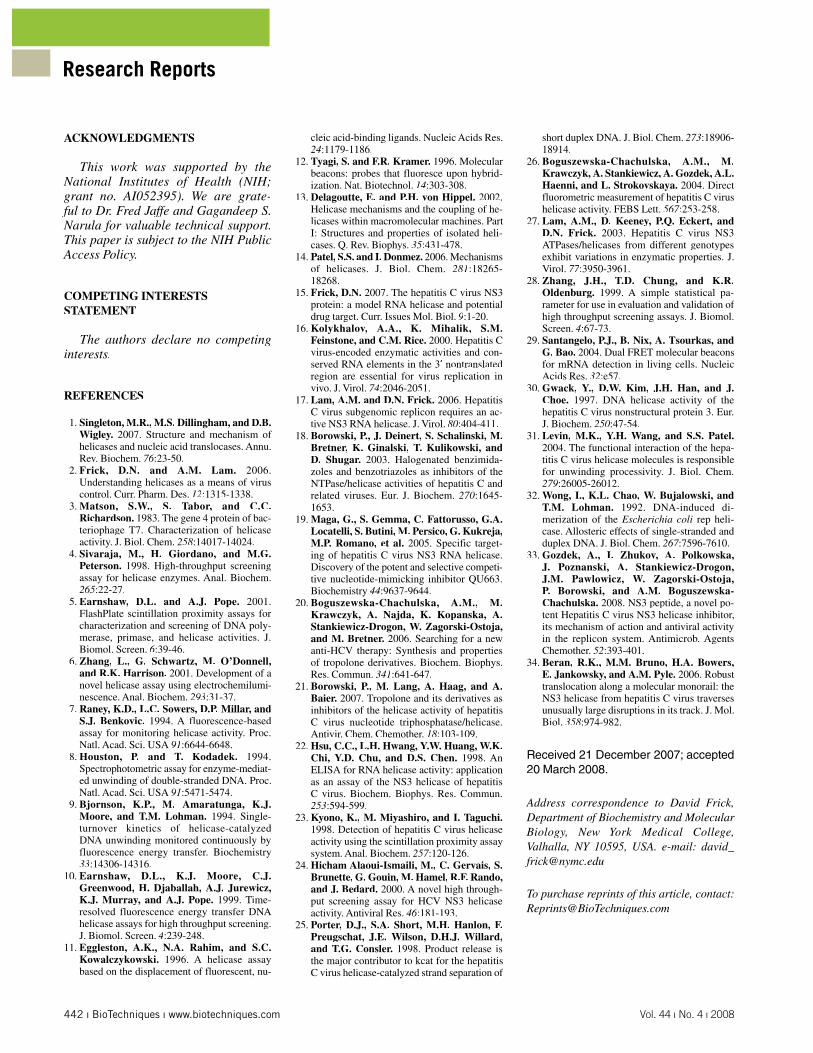
Figure 4. A HCV genome based molecular beacon assay. (A) Comparison of a molecular beacon based assay to a conven-tional electrophoretic mobilityshift (EMSA) gel-based helicase assay. Two simultaneous reactions were run with the substrate shown.One was monitored continuouslyfor fluorescence (gray line), andaliquots were removed from theother and analyzed on three sepa-rate gels (squares). Fluorescenceobtained from substrate blanks is subtracted and fractional fluores-cence (F/Fo) is plotted. The error bars denote the standard devia-tion among the EMSAs and the dashed line represents the 95confidence band for data derived from the EMSAs. (B) Sample gel from one of the EMSAs. Lane 1 represents an aliquot removed before adding ATP to start the re-action (t0); lanes 2–10 representaliquots removed after 20 s, 40 s,1 min, 2 min, 3 min, 5 min, 7.5 min, 10 min, and 20 min. Lane 11is the [32P] oligonucleotide alone and lane 12 is substrate alone.(C) The unwinding reaction as afunction of helicase concentra-tion. Error bars represent standarddeviation of triplicate reactions. (D) The unwinding reaction(25 nM helicase) as a functionof temperature. Because of the rapid progress of the reaction at elevated temperatures, these data were collected using a rapid-mixing stopped-flow device. (E) Substrate stability. Very little fluo-rescence change is observed for 12h when the substrate is incubated in reaction buffer without enzyme or ATP at 22 C (black line). Theinset shows annealing curves for the same substrate at 5 nM (black),50 nM (red), and 500 nM (blue). Fluorescence was monitored while decreasing temperature from 95 C. a.u., arbitrary units.
A
B
C
D

438 ı BioTechniques ı www.biotechniques.com Vol. 44 ı No. 4 ı 2008
Research Reports
the timescale measured, even in theabsence of trap DNA used to capturethe dissociated DNA. This result stands in stark contrast to other fluorescent HCV helicase assays, which absolutely require the presence of a capture strand (25,26).
With the 17-mer substrate shown in Figure 1B, it is conceivable that the helicase could simultaneously load and unwind along the 3 -ssDNA regions of both strands. To test this, the Cy5 beacon was annealed to an oligonucleotide lacking the 3 -dT20 leader sequence. When this blunt-end substrate was used in the same helicase assay, no change influorescence occurred, indicating the enzyme was not unwinding from the 3 -BHQ-GGATG end (data not shown).
The same beacons were then used to analyze the action of HCV helicaseon RNA (Figure 2A). With RNA, the beacons fluoresced strongly only when annealed to complementary strands that do not form intermolecular hairpins(i.e., those that form the 17- and 19-mer duplexes). This may be due to the fact that RNA secondary structures are more stable than comparable DNA struc-
tures or that duplex RNA structures are less stable than a DNA double helix. Regardless, the shorter RNA-DNA heteroduplexes fluoresced at levels similar to that seen with the DNA duplex (Figure 2B). When incubatedwith HCV helicase and ATP, the fluorescence of the RNA-DNA duplex likewise rapidly decreased. Both the initial rates and final amplitude of thefluorescence decrease were lower when the beacons were annealed to RNA (Figure 2B). With both DNA and RNA, observed rate constants (and thus initial reaction rates) were linear with enzyme concentration. However at each enzyme concentration, RNA was unwoundmore slowly than DNA (Figure 2C), as is typically seen with this enzyme (15).
Dual FRET molecular beacons can be used to determine DNA unwinding
To further evaluate molecular beacon based helicase assays using another technique, we combined the two molecular beacon substrates
shown in Figure 1 to produce a dualFRET molecular beacon based onone developed by Santangelo et al.(29) (Figure 3A). In this setup, two beacons were annealed to the same oligonucleotide such that Cy3 emittedlight that could be absorbed by Cy5. The displacement of either probe would therefore lead to a subsequent decreasein FRET between Cy3 and Cy5. To testthis, the dual FRET molecular beacon was used under our standard helicaseassay conditions, and FRET decreased only after addition of the helicase and ATP (Figure 3B). Again, the signal was highly reproducible and rates of decrease were proportional to the amount of enzyme in solution.
An HCV genome based molecularbeacon helicase assay
Since our goal is to eventually usethis assay to screen for HCV inhibitors, we also designed another helicasesubstrate based on a hairpin-forming region of the HCV genome located at the end of the open reading frame encoding the HCV polyprotein nearthe 3 untranslated region (Figure 4A).Using the HCV substrate, we performedsimultaneous experiments with thebeacon annealed to a radiolabeled oligo-nucleotide. We measured fluorescence continuously in one well and used a conventional gel-based electrophoretic mobility shift assay (EMSA) to analyzefractions removed from another well(Figure 4B).
Upon incubation with HCV helicase and ATP, a fluorescence decrease was again observed and when raw fluores-cence was converted to fractionalfluorescence remaining, the data wereoverlaid with that obtained from a standard assay (Figure 4A). Wheneach data set is fit to a first-order rate equation, the kobskk obtained with fluores-cence (0.22 min-1) was slightly slower than that obtained with an EMSA (0.25 min-1). To examine if this difference was due to a slower change in fluorescence(possibly due to hairpin formation, for example) or experimental error,the same samples were analyzed on two additional gels (error bars, Figure 4A). The dotted lines on Figure 4A show the 95 prediction band for thebest-fit curve of the three EMSA datasets. The fluorescence data all fit within
Figure 5. High-throughput screening. (A) Representative data from two continuously monitored in-hibited and non-inhibited reactions. (B and C) 104 inhibited and 103 non-inhibited reactions, carried out simultaneously in a 384-well plate with a final reaction volume of 50 L. Identical sets of the same reactions were repeated on two different days. (B) Data obtained with a Cary Eclipse microplate reader with excitation/emission measured at 643/667 nm (slit widths of 5/10 nm, respectively) (C) Data obtainedwith black plates in a Tecan microplate reader with excitation/emission measured at 643/670 nm (9/20 nm slit widths). Substrate blanks (reactions without substrates) were not subtracted from reactions. (D) Data using the fluorescent assay to evaluate four inhibitors. dT18 competes with the DNA substrate, while , -methylene-ATP and , -imido-ATP are non-hydrolyzable ATP analogs that compete with ATP. NS3pep is a potent, newly described peptide inhibitor of NS3 helicase with the sequence RRGRTGRGRRGIYR(33). a.u., arbitrary units. Error bars represent standard deviation of triplicate reactions.
A B

Research Reports
this range, demonstrating that the slight differences in rates are likely due to error introduced during the elaborate EMSA. Helicase activity was also monitored at varying concentrations of enzyme (Figure 4C) in well plates, as well as at different temperatures usinga stopped-flow device due to the shorter reaction times at elevated temperature (Figure 4D).
The gel in Figure 4B reveals that therewas a considerable amount of ssDNA present before ATP addition (lane 1), which is equivalent to the amount of freessDNA in the substrate after annealing (lane 12) where the material is about 65 dsDNA. Because the ssDNA inthe starting material could be due eitherto substrate instability or incompleteannealing, we performed experiments to judge the stability and annealingof the molecular beacon substrates.The two Cy5-labeled substrates from Figure 1B and the one from Figure 4A (17, 22, and 25 bp, respectively) were monitored for fluorescence at 22 C in the absence of enzyme and ATP. No fluorescence decrease was observedafter 12 h, indicating the substrates were very stable and did not spontane-ously separate (Figure 4E). To examinesubstrate annealing, the constituentstrands of each DNA substrate were combined and fluorescence monitoredto judge annealing at 22 C. At 5 nM,the half-times of annealing for the 17-and 22-bp substrates were 1.5 and 4.6 h respectively, while the 25-bp substrateonly annealed to 10 of its maximum fluorescence after 12 h (data not shown). Even upon heating to 95 C, the 25-bp substrate showed little annealing at 5 nM. The fraction annealed after heating to 95 C increased significantlyat 50 and 500 nM (Figure 4E, inset).We conclude that incomplete annealingleads to the ssDNA contamination of the substrate (Figure 4B, lane 12).
The presence of ssDNA in helicasesubstrates is common and is dealt with by either using an accounting equation (e.g., Equation 3 of Reference 31) or by purifying the substrates with the use of native polyacrylamide gels (32). Herewe used Equation 1 (see Radioactiveunwinding assay section) to correct for ssDNA contaminates, but we also purified the substrates on gels (data notshown). The fluorescent labels usedhere permit direct DNA visualizationwithout dyes or UV light, greatly facili-
tating purification. Thus, we recommend such purification if these substrates are used in mechanistic studies sincehelicases bind ssDNA and slower rateswould be observed with contaminatedsubstrates. Additionally, mechanistic studies would require a comparison of data obtained with fluorescent beaconsto data obtained with the same DNAsubstrate lacking fluorescent moieties.
High-throughput screening
To determine if a molecular beacon!based helicase assay is suitablefor high-throughput screening (HTS), a series of pilot screens were conducted using the HCV-based substrate (Figure 5). All reactions were the same as thosedescribed in Figure 4A except that they were performed in 384-well plates at a volume of 50 L. 16 pilot reactionsss 8uninhibited and 8 inhibited with anoligonucleotide decoy substrate (1 M
dT20))) were monitored continuouslyto follow reaction progress. After deter-mining that the positive control reactionwas near complete after 30 min (Figure 5A), the rest of the reactions were only read twice: before ATP addition and 30 min after ATP addition. Next, pilotscreens were each carried out with104 inhibited and 103 uninhibited control reactions per plate. Positive and negative control reactions were staggered in a checkerboard patternon the 384-well plate. 30 min after thestart of the reaction, fluorescence from inhibited and uninhibited reactions was statistically different (Figure 5B).To examine day-to-day repeatability of the assay, additional assays wereperformed under identical conditions on another day with nearly identical results. Although the assay-to-assayprecision was high in a Cary Eclipse plate reader (Varian, Inc.) (CVs 3 ), we were somewhat disappointed by

440 ı BioTechniques ı www.biotechniques.com Vol. 44 ı No. 4 ı 2008
Research Reports
the relative low Z factors (0.5). We reasoned that the low score might be due to low instrument sensitivity. The Varian instrument is not compatible with standard black microplates and as a result a relatively high background is seen from the necessary white plates. We therefore repeated the high-throughput screen in black 384-well plates using a Tecan microplate reader, again with two separate sets of reactions on 2 d. Although results with the Tecan instrument were somewhatless precise (CVs 5 –10 ) the Zfactor was significantly higher at 0.7, indicating an excellent high-throughput assay (Figure 5C).
To demonstrate how this assay can be used to characterize various classesof NS3 inhibitors, we compared various known inhibitors with the oligonucle-otide dT18, a representative compoundthat binds in place of the RNA or DNA substrate. Two non-hydrolyzableATP analogs ( , methylene-ATP and , imido-ATP) that competewith ATP binding, and a newly reported peptide inhibitor, NS3pep (RRGRTGRGRRGIYR) (33), were analyzed. All reactions were performedin triplicate and IC50 values were calculated using a sigmoidal dose-response curve. The oligonucleotidedT18 was the most potent inhibitor with an IC50 of 30 nM, NS3pep inhibited with an IC50 of 100 nM, and the ATP analogs bound much more weakly:
, methylene-ATP had an IC50 of 140 M, which was more than 10 times lower than , imido-ATP, with an IC50 of 1.8 mM (Figure 5D).
DISCUSSION
Here we describe a new technique to monitor the activity of an importantclass of enzymes. Using a molecular beacon annealed to a DNA or RNAoligonucleotide, the progression of a helicase along its substrate can bemonitored in real time. Unlike conven-tional gel-based helicase assays, fluores-cence-based assaysss and in particular,the new molecular beacon!basedhelicase assayyy are easily amenable to high-throughput screening. Moreover,these new assays differ from previ-ously described continuous helicase assays (7–10,25,26) in three critical ways. First, all modifications are made
on only one strand of the helicase substrate. Second, these assays monitor a decrease in fluorescence. Third, the products form hairpins, making the reaction essentially irreversible duringthe timescale of the experiment.
The fact that both the fluorophoreand quencher are part of the same strand is important because it is increasinglyrecognized that helicases contact one strand of a duplex more than the other,displacing the complementary strandbut making fewer specific contacts with it (13,14). In previously reported helicase assays, both strands are normally modified with a fluorophoreon one strand and a quencher on thecomplement. As a result, the modifi-cation on either strand could influencehelicase progression. To avoid thispotential complication, both modifica-tions are made on the strand believed tointeract least with the helicase.
If this assay is to be used with ahelicase other than the HCV helicase,appropriate substrates will need to bedesigned. The substrates used here were designed with a 3 single-strandedtail sequence because the HCV helicaserequires a 3 single-stranded tail for optimal activity. Substrate require-ments for other helicases vary widely. Other helicases likewise require3 single-stranded tails for optimalactivity, whereas some require 5 tails, some are bidirectional, some work at blunt ends or nicks, and some only act on forked substrates. It is also possible that some RNA helicases might notdisplace a DNA-based beacon from RNA. If that is the case, dual-labeled RNA probes could be used, but these are more difficult to synthesize and, atpresent, are expensive and not widely commercially available.
While many of the beacons usedhere were based on ones used in previous studies (29), the design of the HCV genome based molecular beaconsubstrate was aided by the use of the publicly available software mFold (http://frontend.bioinfo.rpi.edu/appli-cations/mfold). Commercial software is also available for designing molecular beacons, such as Beacon Designer(Premier Biosoft International, Palo Alto, CA, USA). Considering thatmolecular beacons are commonly usedin quantitative PCR, it should be noted that molecular beacon!based helicaseassays could be monitored using almost
any commercially available real-timequantitative PCR apparatus.
The fact that the molecular beacon!based helicase assay monitors fluorescence decrease is notablebecause previous continuous helicaseassays typically monitor the increase of fluorescence after a fluorophore-bearing strand is separated from a quencher-bearing strand. In such asetup where fluorescence increase ismonitored, it is somewhat challengingto convert fluorescence change into a fraction of unwound substrate, especially if an end point is not immedi-ately obvious. With these molecular beacon assays, typically 95 of the signal is lost when the beacon isdisplaced. As a result, data from a molecular beacon!based helicase assay can be easily converted to a percentageof duplex remaining, simply by calcu-lating fractional fluorescence remaining(F/Fo) after subtracting backgroundfluorescence. The resulting rates arealmost identical to those obtainedby conventional gel-based helicaseassays but far more time points can beobtained per reaction, enabling a more rigorous kinetic analysis.
Probably the most important feature of a molecular beacon!based helicaseassay is that it is essentially irreversible. To prevent substrates from re-annealingafter strand separation, many assays add, at some point, a high concentration of a complementary ssDNA/ssRNA.Prior studies have reported diverse, and conflicting, impacts of thesecapture or trapping single-stranded oligonucleotides, ranging from greatlyinhibiting the reaction (8) to actually increasing the observed reaction rate (34). A molecular beacon solves this problem, especially when it is annealedto a fully complementary strand thatis also capable of forming a hairpin.As a result, initial rates observed in molecular beacon!based helicase assays are linear with both time and enzyme concentration.
In conclusion, molecular beaconsenable the study of steady-state rates of helicase-catalyzed reactions in atrue high-throughput manner. Such atool should be valuable for mechanistic analyses, helicase inhibitor identifi-cation, and structure activity relation-ships.
442 ı BioTechniques ı www.biotechniques.com Vol. 44 ı No. 4 ı 2008
Research Reports
ACKNOWLEDGMENTS
This work was supported by the National Institutes of Health (NIH;grant no. AI052395). We are grate-ful to Dr. Fred Jaffe and Gagandeep S. Narula for valuable technical support. This paper is subject to the NIH PublicAccess Policy.
COMPETING INTERESTSSTATEMENT
The authors declare no competinginterests.
REFERENCES
1. Singleton, M.R., M.S. Dillingham, and D.B.Wigley. 2007. Structure and mechanism of helicases and nucleic acid translocases. Annu.Rev. Biochem. 76:23-50.
2. Frick, D.N. and A.M. Lam. 2006. Understanding helicases as a means of virus control. Curr. Pharm. Des. 12:1315-1338.
3. Matson, S.W., S. Tabor, and C.C.Richardson. 1983. The gene 4 protein of bac-teriophage T7. Characterization of helicase activity. J. Biol. Chem. 258:14017-14024.
4. Sivaraja, M., H. Giordano, and M.G.Peterson. 1998. High-throughput screening assay for helicase enzymes. Anal. Biochem.265:22-27.
5. Earnshaw, D.L. and A.J. Pope. 2001. FlashPlate scintillation proximity assays for characterization and screening of DNA poly-merase, primase, and helicase activities. J. Biomol. Screen. 6:39-46.
6. Zhang, L., G. Schwartz, M. O’Donnell,and R.K. Harrison. 2001. Development of a novel helicase assay using electrochemilumi-nescence. Anal. Biochem. 293:31-37.
7. Raney, K.D., L.C. Sowers, D.P. Millar, andS.J. Benkovic. 1994. A fluorescence-basedassay for monitoring helicase activity. Proc. Natl. Acad. Sci. USA 91:6644-6648.
8. Houston, P. and T. Kodadek. 1994.Spectrophotometric assay for enzyme-mediat-ed unwinding of double-stranded DNA. Proc.Natl. Acad. Sci. USA 91:5471-5474.
9. Bjornson, K.P., M. Amaratunga, K.J.Moore, and T.M. Lohman. 1994. Single-turnover kinetics of helicase-catalyzedDNA unwinding monitored continuously by fluorescence energy transfer. Biochemistry33:14306-14316.
10. Earnshaw, D.L., K.J. Moore, C.J.Greenwood, H. Djaballah, A.J. Jurewicz,K.J. Murray, and A.J. Pope. 1999. Time-resolved fluorescence energy transfer DNAhelicase assays for high throughput screening.J. Biomol. Screen. 4:239-248.
11. Eggleston, A.K., N.A. Rahim, and S.C.Kowalczykowski. 1996. A helicase assay based on the displacement of fluorescent, nu-
cleic acid-binding ligands. Nucleic Acids Res.24:1179-1186.
12. Tyagi, S. and F.R. Kramer. 1996. Molecular beacons: probes that fluoresce upon hybrid-ization. Nat. Biotechnol. 14:303-308.
13. Delagoutte, E. and P.H. von Hippel. 2002.Helicase mechanisms and the coupling of he-licases within macromolecular machines. Part I: Structures and properties of isolated heli-cases. Q. Rev. Biophys. 35:431-478.
14. Patel, S.S. and I. Donmez. 2006. Mechanisms of helicases. J. Biol. Chem. 281:18265-18268.
15. Frick, D.N. 2007. The hepatitis C virus NS3protein: a model RNA helicase and potentialdrug target. Curr. Issues Mol. Biol. 9:1-20.
16. Kolykhalov, A.A., K. Mihalik, S.M.Feinstone, and C.M. Rice. 2000. Hepatitis Cvirus-encoded enzymatic activities and con-served RNA elements in the 3 nontranslated region are essential for virus replication in vivo. J. Virol. 74:2046-2051.
17. Lam, A.M. and D.N. Frick. 2006. Hepatitis C virus subgenomic replicon requires an ac-tive NS3 RNA helicase. J. Virol. 80:404-411.
18. Borowski, P., J. Deinert, S. Schalinski, M.Bretner, K. Ginalski, T. Kulikowski, andD. Shugar. 2003. Halogenated benzimida-zoles and benzotriazoles as inhibitors of theNTPase/helicase activities of hepatitis C andrelated viruses. Eur. J. Biochem. 270:1645-1653.
19. Maga, G., S. Gemma, C. Fattorusso, G.A.Locatelli, S. Butini, M. Persico, G. Kukreja,M.P. Romano, et al. 2005. Specific target-ing of hepatitis C virus NS3 RNA helicase. Discovery of the potent and selective competi-tive nucleotide-mimicking inhibitor QU663. Biochemistry 44:9637-9644.
20. Boguszewska-Chachulska, A.M., M.Krawczyk, A. Najda, K. Kopanska, A.Stankiewicz-Drogon, W. Zagorski-Ostoja, and M. Bretner. 2006. Searching for a new anti-HCV therapy: Synthesis and properties of tropolone derivatives. Biochem. Biophys.Res. Commun. 341:641-647.
21. Borowski, P., M. Lang, A. Haag, and A.Baier. 2007. Tropolone and its derivatives as inhibitors of the helicase activity of hepatitisC virus nucleotide triphosphatase/helicase.Antivir. Chem. Chemother. 18:103-109.
22. Hsu, C.C., L.H. Hwang, Y.W. Huang, W.K.Chi, Y.D. Chu, and D.S. Chen. 1998. An ELISA for RNA helicase activity: application as an assay of the NS3 helicase of hepatitis C virus. Biochem. Biophys. Res. Commun. 253:594-599.
23. Kyono, K., M. Miyashiro, and I. Taguchi.1998. Detection of hepatitis C virus helicase activity using the scintillation proximity assay system. Anal. Biochem. 257:120-126.
24. Hicham Alaoui-Ismaili, M., C. Gervais, S.Brunette, G. Gouin, M. Hamel, R.F. Rando,and J. Bedard. 2000. A novel high through-put screening assay for HCV NS3 helicase activity. Antiviral Res. 46:181-193.
25. Porter, D.J., S.A. Short, M.H. Hanlon, F.Preugschat, J.E. Wilson, D.H.J. Willard,and T.G. Consler. 1998. Product release is the major contributor to kcat for the hepatitisC virus helicase-catalyzed strand separation of
short duplex DNA. J. Biol. Chem. 273:18906-18914.
26. Boguszewska-Chachulska, A.M., M.Krawczyk, A. Stankiewicz, A. Gozdek, A.L.Haenni, and L. Strokovskaya. 2004. Direct fluorometric measurement of hepatitis C virushelicase activity. FEBS Lett. 567:253-258.
27. Lam, A.M., D. Keeney, P.Q. Eckert, andD.N. Frick. 2003. Hepatitis C virus NS3 ATPases/helicases from different genotypes exhibit variations in enzymatic properties. J.Virol. 77:3950-3961.
28. Zhang, J.H., T.D. Chung, and K.R.Oldenburg. 1999. A simple statistical pa-rameter for use in evaluation and validation of high throughput screening assays. J. Biomol. Screen. 4:67-73.
29. Santangelo, P.J., B. Nix, A. Tsourkas, andG. Bao. 2004. Dual FRET molecular beacons for mRNA detection in living cells. NucleicAcids Res. 32:e57.
30. Gwack, Y., D.W. Kim, J.H. Han, and J.Choe. 1997. DNA helicase activity of the hepatitis C virus nonstructural protein 3. Eur. J. Biochem. 250:47-54.
31. Levin, M.K., Y.H. Wang, and S.S. Patel.2004. The functional interaction of the hepa-titis C virus helicase molecules is responsible for unwinding processivity. J. Biol. Chem. 279:26005-26012.
32. Wong, I., K.L. Chao, W. Bujalowski, andT.M. Lohman. 1992. DNA-induced di-merization of the Escherichia coli rep heli-case. Allosteric effects of single-stranded and duplex DNA. J. Biol. Chem. 267:7596-7610.
33. Gozdek, A., I. Zhukov, A. Polkowska,J. Poznanski, A. Stankiewicz-Drogon,J.M. Pawlowicz, W. Zagorski-Ostoja,P. Borowski, and A.M. Boguszewska-Chachulska. 2008. NS3 peptide, a novel po-tent Hepatitis C virus NS3 helicase inhibitor,its mechanism of action and antiviral activity in the replicon system. Antimicrob. Agents Chemother. 52:393-401.
34. Beran, R.K., M.M. Bruno, H.A. Bowers,E. Jankowsky, and A.M. Pyle. 2006. Robust translocation along a molecular monorail: theNS3 helicase from hepatitis C virus traverses unusually large disruptions in its track. J. Mol. Biol. 358:974-982.
Received 21 December 2007; accepted20 March 2008.
Address correspondence to David Frick,Department of Biochemistry and Molecular Biology, New York Medical College,Valhalla, NY 10595, USA. e-mail: [email protected]
To purchase reprints of this article, contact:[email protected]