molecular structures of acetylcholinesterase …of purified acetylcholinesterase (dudai and silman,...
TRANSCRIPT
Proc. Nat. Acad. Sci. USAVol. 70, No. 9, pp. 2473-2476, September 1973
Molecular Structures of Acetylcholinesterase from Electric OrganTissue of the Electric Eel
(electron microscopy/analytical ultracentrifugation/maleylation/protein subunits/membrane enzymes)
YADIN DUDAI*, MAX HERZBERGt, AND ISRAEL SILMAN** Department of Biophysics, The Weizmann Institute of Science, Rehovot, Israel; and t Department of Life Sciences,Bar Ilan University, Ramat Gan, Israel
Communicated by David Nachmansohn, May 23, 1973
ABSTRACT Three purified molecular forms of acetyl-cholinesterase (EC 3.1.1.7) with sedimentation coefficientsof 18 S, 14 S, and 11 S were studied by analytical ultra-centrifugation and electron microscopy. The three specieshave molecular weights of (1.1 ± 0.1) X 106, (7.5 4 1.5) X106, and (3.35 4- 0.25) X 106, respectively. Electron micro-graphs reveal that the 18S and 14S forms are asymmetric,composed of a head, containing a large number of sub-units, and an elongated tail. The 11 S form of acetylcholin-esterase is apparently a tetrameric structure devoid of thetail. Maleylation of 18S and 14S acetylcholinesterasesabolishes their tendency to aggregate at low ionic strength.
Acetylcholinesterase (acetylcholine hydrolase, EC 3.1.1.7) inextracts of fresh electric organ tissue of electric eels consistsof several components distinguished by their sedimentationcoefficients (1, 2). The major component is an 18S form, whichaggregates at low ionic strength (2, 3). A 14S component,which also aggregates at low ionic strength, is present insmaller amounts. Treatment of electric organ tissue withproteolytic enzymes or autolysis converts all the acetyl-cholinesterase to an 11S form, which does not aggregate atlow ionic strength and is not present in fresh tissue (2, 3).We have purified, by affinity chromatography, the dif-
ferent molecular forms of acetylcholinesterase present infresh tissue or obtained after proteolysis (3, 4). Purified 18Sand 14S acetylcholinesterases retain their tendency to ag-gregate at low ionic strength, and the 18S, 14S, and 11Sacetylcholinesterases all display similar patterns on poly-acrylamide gel electrophoresis in the presence of sodiumdodecyl sulfate and 2-mercaptoethanol (4). Massouli6 et al.(5) have suggested, on the basis of gel-filtration studies, that18S and 14S acetylcholinesterases have an asymmetric struc-ture, in contrast with 11S acetylcholinesterase, which isglobular.
In the following we will show, by electron microscopy andultracentrifugation, that 18S and 14S acetylcholinesterasesdiffer markedly in their quaternary structure from the 11Sform. Similar electron microscopic observations have beenrecently reported by Rieger et al. (6). We will also show thatthe tendency of 18S and 14S acetylcholinesterases to aggregateat low ionic strength can be prevented by chemical modifica-tion. The relevance of these observations to the relationshipof acetylcholinesterase with the electroplax membrane will bediscussed.
2473
METHODS
18S, 14S, and llS forms of acetylcholinesterase were purifiedby affinity chromatography (3, 4). The 18S and -14S enzymeswere separated from each other by sucrose gradient centrifu-gation as follows: aliquots of 1.8 ml of a mixture of purified18S and 14S enzymes (containing about 1 mg/ml of enzyme)were layered on a 5-20% linear sucrose gradient in 1.0 MNaCl-0.01 M phosphate (pH 7.0) of total volume of 28 ml,with a 5-ml cushion of 60% sucrose in the same buffer at thebottom. Centrifugation was performed in an SW27 rotor inan L2 65-B Beckman preparative ultracentrifuge at 27,000rpm at 40 for 20 hr. The 18S and 14S samples thus obtainedwere dialyzed against 1.0 M NaCl-0.01 M phosphate (pH7.0).
Analytical ultracentrifugation was performed by the high-speed meniscus depletion method of Yphantis (7), with aBeckman model E ultracentrifuge equipped with an electronicspeed-control unit. Aluminum-filled epon double-sector cells(12 mm) were used in an An-F aluminum rotor with four cellholes (for speeds higher than 10,000 rpm) or an An-J alum-inum rotor with four cell holes (for speeds less than 10,000rpm). Filling of the cells, the criteria for equilibrium, runningof blanks, and analysis of the Rayleigh patterns were as de-scribed by Godfrey and Harrington (8).
Electron microscopy was performed with a Jeol 100 Belectron microscope, with an accelerating voltage of 80 kV,magnifications of 25,000-60,000, and an objective aperture ofof 40 ,um. Negatively stained samples of acetylcholinesterasewere prepared by the technique of Huxley and Zubay (9).The best results were obtained when the enzyme was stainedwith a 1% solution of uranyl acetate (pH 3.5) over holes.
Purified 18S + 14S acetylcholinesterase was treated withmaleic anhydride according to Butler et al. (10). A sample of300 ug of purified 18S + 14S enzyme in 0.6 ml of 0.5 M NaCl-0.11 M phosphate (pH 7.5) was treated with 20 Ml of a 7.5%(w/v) solution of maleic anhydride in redistilled dioxane,and the reaction mixture was kept for 3 hr at room tempera-ture (250). Control aliquots of acetylcholinesterase were treatedas above, but the maleic anhydride solution was replaced by20 Ad of redistilled dioxane or 20 ul of H20. The samples werethen dialyzed overnight at 4° against 1 M NaCl-0.01 M phos-phate (pH 7.0). Acetylcholinesterase activity of the samples
Dow
nloa
ded
by g
uest
on
Mar
ch 2
0, 2
020

Proc. Nat. Acad. Sci. USA 70 (1973)
14
X 12 (A),,., .. . _
3j 68
D ~~~(B)W 40
0 100 200 300 400 500 600
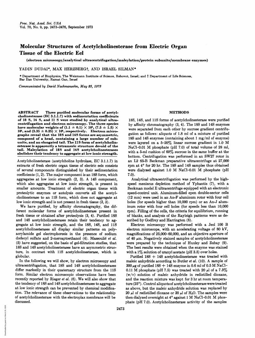
FRINGE DISPLACEMENT (,Mm)FIG. 1. The weight-average molecular weight of 18S and of
11S acetyicholinesterase plotted against fringe displacement as
computed by the high-speed equilibrium ultracentrifugationprogram of Roark and Yphantis (11). (A) 18S acetylcholin-esterase, centrifuged at 6000 rpm; (B) 118 acetylcholinesterase,centrifuged at 10,000 rpm.
was assayed before and after maleylation by the l)H-statmethod (3). Sucrose gradient centrifugation and enzymic as-
say of the fractions from the gradient were Iperformed as de-scribed (3).
RESULTSEquilibrium sedimentation measurements on purified 1 iS and18S acetylcholinesterases yield the results shown in Fig. 1.The molecular weight of the 18S form is (1.1 i 0.1) X 106,assuming a v of 0.72, as computed from amino-acid analysisof purified acetylcholinesterase (Dudai and Silman, un-published results). The molecular weight for 11S acetyl-cholinesterase according to Fig. 1 is 350,000 i 10,000, againfrom a v of 0.72. This preparation was obtained by trypticdigestion of fresh tissue (acetylcholinesterase A in ref. 3).Another preparation of 11S acetylcholinesterase, obtainedfrom toluene-treated tissue (acetylcholinesterase C in ref. 3),displayed similar homogeneity on equilibrium sedimentation,and had a molecular weight of 320,000 + 10,000.A preparation of 14S acetylcholinesterase, prepared as
described in Methods, appeared to be rather heterogeneous onequilibrium sedimentation, although on acrylamide gel elec-trophoresis in the presence of sodium dodecyl sulfate it dis-played a pattern similar to those observed for 18S and 11Sacetylcholinesterase (4). From the ultracentrifugation mea-surements, a molecular weight of 750,000 + 150,000 could beestimated.
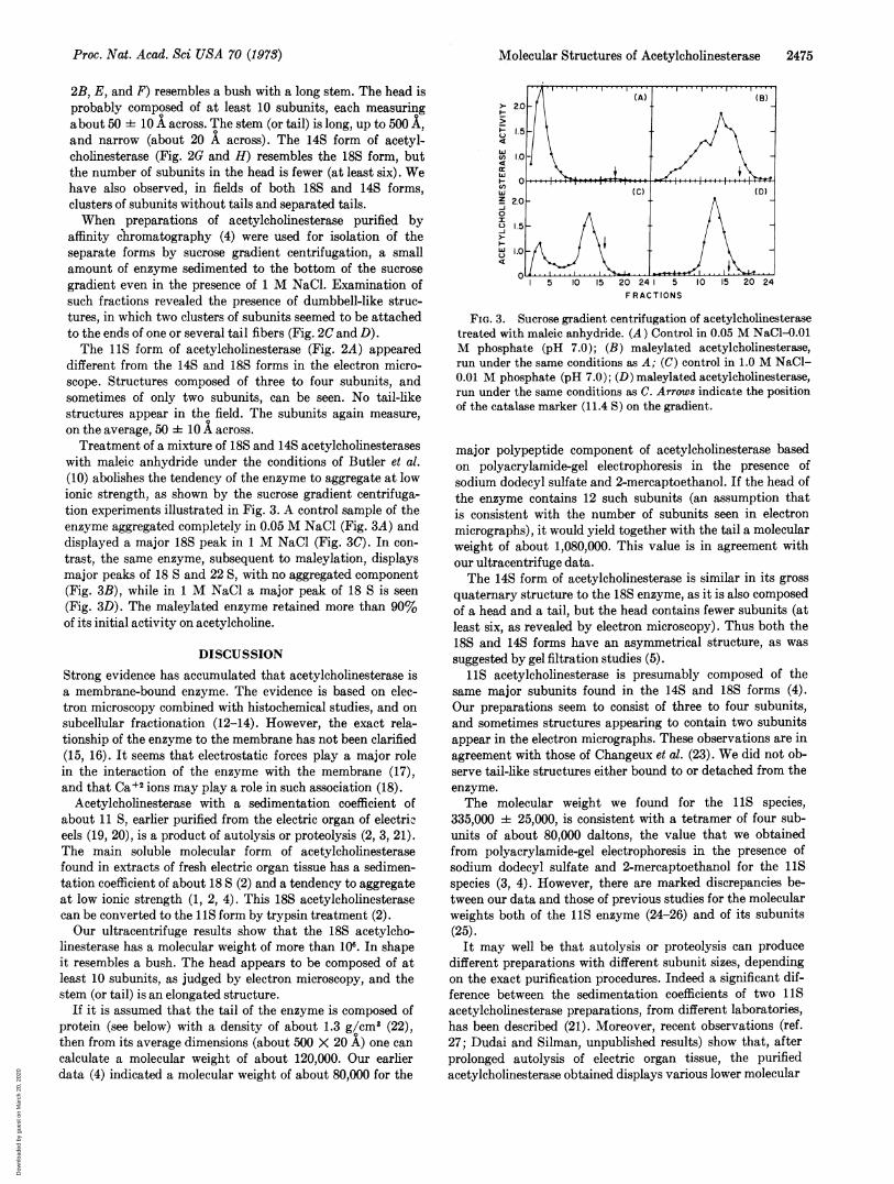
Electron micrographs of the different molecular forms ofacetylcholinesterase are shown in Fig. 2. The 18S form (Fig.
-~ %-4 ,;
C D E
.. "I H1, -4 14ZMA" , .2-k., 16.
Z#I
i1
1
i 41,MA
a
F G HFIG. 2. Electron micrographs of various molecular forms of acetylcholinesterase, stained with 1% uranyl acetate. (A) 11S ace-
tylcholinesterase, X - 225,000; insert shows a single molecule, X - 650,000. (B) 18S enzyme, X - 225,000; arrows indicate tails. (C)])umbbell form of acetylcholinesterase, X - 225,000. (D) Dumbbell form of acetylcholinesterase, X - 340,000. (E and F) 18S acetylcho-linesterase, X - 340,000. (G and H) 14S acetylcholinesterase, X - 340,000.
2474 Biochemistry: Dudai et al.
Dow
nloa
ded
by g
uest
on
Mar
ch 2
0, 2
020
Molecular Structures of Acetyicholinesterase 2475
2B, E, and F) resembles a bush with a long stem. The head isprobably composed of at least 10 subunits, each measuringabout 50 i 10 A across. The stem (or tail) is long, up to 500 ,and narrow (about 20 A across). The 14S form of acetyl-cholinesterase (Fig. 2G and H) resembles the 18S form, butthe number of subunits in the head is fewer (at least six). Wehave also observed, in fields of both 18S and 14S forms,clusters of subunits without tails and separated tails.When preparations of acetylcholinesterase purified by
affinity chromatography (4) were used for isolation of theseparate forms by sucrose gradient centrifugation, a smallamount of enzyme sedimented to the bottom of the sucrosegradient even in the presence of 1 M NaCl. Examination ofsuch fractions revealed the presence of dumbbell-like struc-tures, in which two clusters of subunits seemed to be attachedto the ends of one or several tail fibers (Fig. 2C and D).The 11S form of acetylcholinesterase (Fig. 2A) appeared
different from the 14S and 18S forms in the electron micro-scope. Structures composed of three to four subunits, andsometimes of only two subunits, can be seen. No tail-likestructures appear in the field. The subunits again measure,on the average, 50 i 10 A across.Treatment of a mixture of 18S and 14S acetylcholinesterases
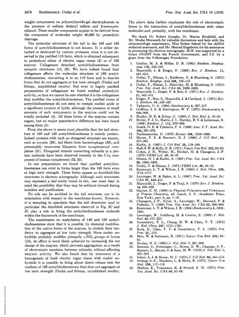
with maleic anhydride under the conditions of Butler et al.(10) abolishes the tendency of the enzyme to aggregate at lowionic strength, as shown by the sucrose gradient centrifuga-tion experiments illustrated in Fig. 3. A control sample of theenzyme aggregated completely in 0.05 M NaCl (Fig. 3A) anddisplayed a major 18S peak in 1 M NaCl (Fig. 3C). In con-trast, the same enzyme, subsequent to maleylation, displaysmajor peaks of 18 S and 22 S, with no aggregated component(Fig. 3B), while in 1 M NaCl a major peak of 18 S is seen(Fig. 3D). The maleylated enzyme retained more than 90%of its initial activity on acetylcholine.
DISCUSSION
Strong evidence has accumulated that acetylcholinesterase isa membrane-bound enzyme. The evidence is based on elec-tron microscopy combined with histochemical studies, and onsubcellular fractionation (12-14). However, the exact rela-tionship of the enzyme to the membrane has not been clarified(15, 16). It seems that electrostatic forces play a major rolein the interaction of the enzyme with the membrane (17),and that Ca+2 ions may play a role in such association (18).
Acetylcholinesterase with a sedimentation coefficient ofabout 11 S, earlier purified from the electric organ of electriceels (19, 20), is a product of autolysis or proteolysis (2, 3, 21).The main soluble molecular form of acetylcholinesterasefound in extracts of fresh electric organ tissue has a sedimen-tation coefficient of about 18 S (2) and a tendency to aggregateat low ionic strength (1, 2, 4). This 18S acetylcholinesterasecan be converted to the llS form by trypsin treatment (2).Our ultracentrifuge results show that the 18S acetylcho-
linesterase has a molecular weight of more than 106. In shapeit resembles a bush. The head appears to be composed of atleast 10 subunits, as judged by electron microscopy, and thestem (or tail) is an elongated structure.
If it is assumed that the tail of the enzyme is composed ofprotein (see below) with a density of about 1.3 g/cm3 (22),then from its average dimensions (about 500 X 20 A) one cancalculate a molecular weight of about 120,000. Our earlierdata (4) indicated a molecular weight of about 80,000 for the
> 2I-
u
4
w
i I
w
C,,
w
z 2.-i0
I
-I
-JW)
(A) (B)
1.5
1.0
C(C) (D)
!.0
.5
.0
0 ....5 10 15 20 24 5 10 15 20 24
F RACTIONS
FIG. 3. Sucrose gradient centrifugation of acetylcholinesterasetreated with maleic anhydride. (A) Control in 0.05 M NaCl-0.01M phosphate (pH 7.0); (B) maleylated acetylcholinesterase,run under the same conditions as A; (C) control in 1.0 M NaCl-0.01 M phosphate (pH 7.0); (D) maleylated acetylcholinesterase,run under the same conditions as C. Arrows indicate the positionof the catalase marker (11.4 S) on the gradient.
major polypeptide component of acetylcholinesterase basedon polyacrylamide-gel electrophoresis in the presence ofsodium dodecyl sulfate and 2-mercaptoethanol. If the head ofthe enzyme contains 12 such subunits (an assumption thatis consistent with the number of subunits seen in electronmicrographs), it would yield together with the tail a molecularweight of about 1,080,000. This value is in agreement withour ultracentrifuge data.The 14S form of acetylcholinesterase is similar in its gross
quaternary structure to the 18S enzyme, as it is also composedof a head and a tail, but the head contains fewer subunits (atleast six, as revealed by electron microscopy). Thus both the18S and 14S forms have an asymmetrical structure, as wassuggested by gel filtration studies (5).
11S acetylcholinesterase is presumably composed of thesame major subunits found in the 14S and 18S forms (4).Our preparations seem to consist of three to four subunits,and sometimes structures appearing to contain two subunitsappear in the electron micrographs. These observations are inagreement with those of Changeux et al. (23). We did not ob-serve tail-like structures either bound to or detached from theenzyme.The molecular weight we found for the 11S species,
335,000 i 25,000, is consistent with a tetramer of four sub-units of about 80,000 daltons, the value that we obtainedfrom polyacrylamide-gel electrophoresis in the presence ofsodium dodecyl sulfate and 2-mercaptoethanol for the 11Sspecies (3, 4). However, there are marked discrepancies be-tween our data and those of previous studies for the molecularweights both of the 11S enzyme (24-26) and of its subunits(25).
It may well be that autolysis or proteolysis can producedifferent preparations with different subunit sizes, dependingon the exact purification procedures. Indeed a significant dif-ference between the sedimentation coefficients of two 11Sacetylcholinesterase preparations, from different laboratories,has been described (21). Moreover, recent observations (ref.27; Dudai and Silman, unpublished results) show that, afterprolonged autolysis of electric organ tissue, the purifiedacetylcholinesterase obtained displays various lower molecular
Proc. Nat. Acad. Sci USA 70 (1978)
Dow
nloa
ded
by g
uest
on
Mar
ch 2
0, 2
020
Proc. Nat. Acad. Sci. USA 70 (1973)
weight components on polyacrylamide-gel electrophoresis inthe presence of sodium dodecyl sulfate and 2-mercapto-ethanol. These smaller components appear to be derived fromthe component of molecular weight 80,000 by proteolyticcleavage.The molecular structure of the tail in the 18S and 14S
forms of acetylcholinesterase is not known. It is either de-tached or destroyed by various proteases, since it is not ob-served in the purified 11S form, which is obtained subsequentto proteolysis either of electric organ tissue (3) or of 18Senzyme. Collagenase detached acetylcholinesterase fromsynaptic structures (15, 28). We have indeed found thatcollagenase affects the molecular structure of 18S acetyl-cholinesterase, converting it to an 11S form and to heavierforms that do not aggregate at low ionic strength (Dudai andSilman, unpublished results). But even in highly purifiedpreparations of collagenase we found residual proteolyticactivity, so that we cannot be sure whether it is the collagenasethat modifies the acetylcholinesterase. The purified forms ofacetylcholinesterase do not seem to contain nucleic acids ora significant amount of lipids, although the presence of smallamounts of such components in the enzyme could not betotally excluded (4). All three forms of the enzyme containsugars, but no major quantitative difference has been foundamong them (4).From the above it seems most plausible that the tail struc-
ture of 18S and 14S acetylcholinesterase is mainly l)rotein.Indeed proteins with such an elongated structure are known,such as myosin (29), tail fibers from bacteriophage (30), andpresumably transverse filaments from synaptonemal com-plexes (31). Elongated strands connecting subunits in a pro-tein molecule have been observed recently in the Clq com-ponent of human complement (32, 33).
In our preparations we found that purified acetylcho-linesterase can exist in forms larger than the 18S form evenat high ionic strength. These forms appear as dumbbell-likestructures in electron micrographs. Although such structuresmay represent a real entity found in situ, one must bear inmind the possibility that they may be artifacts formed duringisolation and purification.No role can be ascribed to the tail structure, nor is its
orientation with respect to the membrane known. However,it is tempting to speculate that the tail structures (and inparticular the dumbbell structures observed in Fig. 2C andD) play a role in fixing the acetylcholinesterase moleculewithin the framework of the membrane.The experiments on maleylation of 14S and 18S acetyl-
cholinesterase show that it is possible, by chemical modifica-tion of the native forms of the enzyme, to abolish their ten-dency to aggregate at low ionic strength. Since maleic an-hydride probably modifies primarily e-NH2 groups of lysine(10), its effect is most likely achieved by increasing the netcharge of the enzyme, which prevents aggregation, as a resultof electrostatic repulsion between subunits, without affectingenzymic activity. We also found that by treatment of ahomogenate of fresh electric organ tissue with maleic an-hydride it is possible to bring about direct release into themedium of 18S acetylcholinesterase that does not aggregate atlow ionic strength (Dudai and Silman, unpublished results).
The above data further emphasize the role of electrostaticforces in the interaction of acetylcholinesterase with othermolecules and, probably, with the membrane.
We thank Dr. Robert Josephs, Dr. Martin Hirshfeld, andMr. Moshe Mevarech for valuable discussions and help with theultracentrifuge experiments, Miss Esther Saksel for her skillfultechnical assistance, and Mr. Shmuel Engelstein for his assistancein processing the electron micrographs. M.H. was supported by aGrant DGRST from the French Government, and I.S. by agrant from the Volkswagen Foundation.
1. Grafius, M. A. & Millar, D. B. (1965) Biochim. Biophys.Acta 110, 540-547.
2. Massouli6, J. & Rieger, F. (1969) Eur. J. Biochem. 11,441-455.
3. Dudai, Y., Silman, I., Kalderon, N. & Blumberg, S. (1972)Biochim. Biophys. Acta 268, 138-157.
4. Dudai, Y., Silman, I., Shinitzky, M. & Blumberg, S. (1972)Proc. Nat. Acad. Sci. USA 69, 2400-2403.
5. Massouli6, J., Rieger, F. & Bon, S. (1971) Eur. J. Biochem.21, 542-551.
6. Rieger, F., Bon, S., Massouli6, J. & Cartlaud, J. (1973) Eur.J. Biochem. 34,539-547.
7. Yphantis, D. A. (1964) Biochemistry 3, 297-317.8. Godfrey, J. E. & Harrington, W. F. (1970) Biochemistry 9,
894-908.9. Huxley, H. E. & Zubay, G. (1960) J. Mol. Biol. 2, 10-18.
10. Butler, P. J. G., Harris, J. I., Hartley, B. S. & Leberman, R.(1969) Biochem. J. 112, 679-689.
11. Roark, D. E. & Yphantis, P. A. (1969) Ann. N.Y. Acad. Sci.164, 265-276.
12. Nachmansohn, D. (1970) Science 168, 1059-1066.13. Bloom, F. E. & Barrnett, R. J. (1966) J. Cell Biol. 29,
475-495.14. Karlin, A. (1965) J. Cell Biol. 25, 159-169.15. Hall, Z. W. & Kelly, R. B. (1971) Nature New Biol. 232,62-63.16. Cohen, J. B., Weber, M., Huchet, M. & Changeux, J.-P.
(1972) FEBS Lett. 26, 43-47.17. Silman, H. I. & Karlin, A. (1967) Proc. Nat. Acad. Sci. USA
58, 1664-1668.18. Dudai, Y. & Silman, I. (1973) FEBS Lett. 30, 49-52.19. Kremzner, L. T. & Wilson, I. B. (1963) J. Biol. Chem. 238,
1714-1717.20. Leuzinger, W. & Baker, A. L. (1967) Proc. Nat. Acad. Sci.
USA 57, 446-451.21. Massoulie, J., Rieger, F. & Tsuji, S. (1970) Eur. J. Biochem.
14, 430-439.22. Slayter, E. M. (1969) in Physical Principles and Techniques
of Protein Chemistry, ed. Leach, J. S. (Academic Press,New York), part A, pp. 1-58.
23. Changeux, J.-P., Ryter, A., Leuzinger, W., Barrand, P. &Podleski, T. (1969) Proc. Nat. Acad. Sci. USA 62, 986-993.
24. Kremzner, L. T. & Wilson, I. B. (1964) Biochemistry 3, 1902-1905.
25. Leuzinger, W., Goldberg, M. & Cauvin, E. (1969) J. M1ol.Biol. 40, 217-225.
26. Rosenberry, T. L., Chang, H. W. & Chen, Y. T. (1972)J. Biol. Chem. 247, 1555-1565.
27. Bock, E., Chen, Y. T. & Rosenberry, T. L. (1973) Fed.Proc. 32, 510.
28. Betz, W. & Sakmann, B. (1971) Nature New Biol. 232, 94-95.
29. Huxley, H. E. (1963) J. Mol. Biol. 7, 281-308.30. Brenner, S., Streisinger, G., Horne, 1I. W., Champe, S. P.,
Barnett, L., Benzer, S. & Rees, M. W. (1959) J. Mol. Biol. 1,281-292.
31. Solari, A. J. & Moses, M. J. (1973) J. Cell Biol. 56, 145-152.32. Svehag, S.-E., Manhem, L. & Bloth, B. (1972) Nature Neow
Biol. 238, 117-118.33. Shelton, E., Yonemasu, K. & Stroud, RI. M. (1972) Proc.
Nat. Acad. Sci. USA 69, 65-68.
9A76 Biochemistry: Dudai et al.
Dow
nloa
ded
by g
uest
on
Mar
ch 2
0, 2
020