molecular staging of colorectal cancer: new paradigm or waste of time?
TRANSCRIPT
Review
10.1517/17530059.1.1.31 © 2007 Informa UK Ltd ISSN 1753-0059 31
1. Introduction
2. Molecular heterogeneity
3. Approaches to molecular
staging
4. Biomarkers
5. Molecular technology
6. Genome analysis
7. Transcriptome analysis
8. The future
9. Conclusion
10. Expert opinion
Molecular staging of colorectal cancer: new paradigm or waste of time? Jamie Murphy , Sina Dorudi & Stephen A Bustin † †Centre for Academic Surgery, Barts and the London, Queen Mary ’ s School of Medicine and Dentistry, Royal London Hospital, E1 1BB, London
Colorectal cancer staging describes disease spread by assessing tumour penetration of the bowel wall and the presence of lymph node involvement and distant metastases. Although useful for stratifying patients into risk groups that indicate prognosis as well as treatment, conventional staging frequently fails to predict the biological behaviour of individual tumours within the same stage. Accordingly, up to a third of patients undergoing apparently curative surgery develop systemic disease and die from cancer-related causes. This heterogeneity of clinical outcome is mirrored by similarly extensive molecular diversity, where progression to systemic disease is associated with a range of genetic and epigenetic alterations. This has led to the idea that tumour stratification based on molecular taxonomy might increase the prognostic accuracy for the individual patient. However, despite the description of many putative individual and multiple molecular biomarkers, the data generated by molecular diagnostic assays have not proved to be either robust or consistently reproducible. Experimental bias exists throughout the entire process, starting with sample collection and ending with data interpretation. There is an urgent need to integrate clinical, biochemical, genetic and molecular data, and findings must be validated in prospective, well-controlled clinical studies of large numbers of diverse patients across multiple institutions. Only then can the implementation of molecular staging assays into clinical practice be considered.
Keywords: colorectal cancer , histopathology , microarray , molecular staging, PCR
Expert Opin. Med. Diagn. (2007) 1(1):31-45
1. Introduction
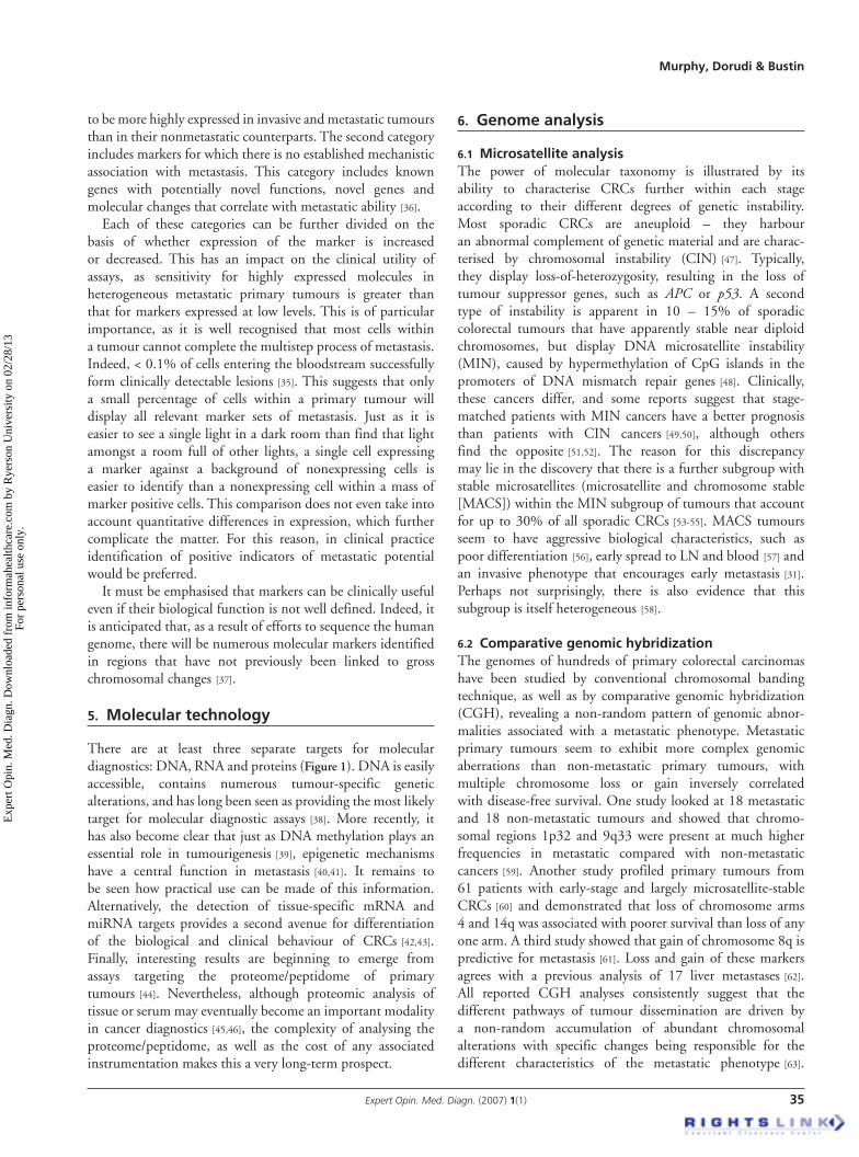
Prognosis for colorectal cancer (CRC) patients is predicted by pathological assessment of the clinical stage of the tumour following surgery. The most widely used staging system, TNM (tumour, node, metastasis), describes the anatomical extent of tumour invasion into the bowel wall, and any associated spread of tumour cells to lymph nodes (LN) and distant organs ( Figure 1 ). The predictive power of TNM staging is augmented by the residual tumour (R) classification, which describes the absence (R0), or microscopic (R1) or macroscopic (R2) presence of residual tumour following surgical resection. Other important pathological findings that convey poor prognosis include lymphovascular and perineural tumour invasion and stratification of TNM groups based on the presence or absence of elevated serum levels of carcinoembryonic antigen (CEA) on preoperative clinical examination [1] . At present, when performed accurately, TNM staging provides the best prognostic data available for patients that have undergone putatively curative CRC resection [2] , although there is some debate concerning its ongoing revision [3] . However, prognostic data associated with particular
Exp
ert O
pin.
Med
. Dia
gn. D
ownl
oade
d fr
om in
form
ahea
lthca
re.c
om b
y R
yers
on U
nive
rsity
on
02/2
8/13
For
pers
onal
use
onl
y.

Molecular staging of colorectal cancer: new paradigm or waste of time?
32 Expert Opin. Med. Diagn. (2007) 1(1)
TNM stages are based upon group data and are not tailored to the individual patient. As a consequence, there is substantial prognostic heterogeneity within each TNM stage [4] , resulting in the understaging of tumours and treatment failure for approximately a third of patients with N0/R0 tumours [5] . The existence of a subgroup of individuals with early CRC that harbour clinically significant occult disease has major clinical relevance, as both adjuvant (postoperative) and, to a lesser extent, neoadjuvant (preoperative) treatments have been shown to prolong disease-free and overall survival in subgroups of patients with colonic and rectal cancers, respectively [6,7] . As a consequence, stratification of individuals prior to surgery into more accurate prognostic subgroups within a given TNM stage is crucial, as it has a direct impact on clinical manage-ment of individual patients [8] . Accordingly it is becoming increasingly clear that additional factors, whether morpho-logical or mole cular, will be needed for future clinical man-agement [9] . Although analysis of various molecular factors such as microsatellite instability (MSI), expression levels of thymidylate synthase and of numerous other genes, TP53 sta-tus, K-ras mutations and absence of deleted in colon cancer ( DCC ) has been advocated for either determining prognosis or predicting response to therapy in patients with CRC, for now there is insufficient evidence to recommend their routine clinical use [10] .
Nevertheless, there can be no doubt that, in time, sets of molecular markers will be identified that will become useful
aids to clinical decision making [11,12] . For example, a study of 393 patients showed that retention of heterozygocity at three loci on chromosomes 17p and 18q predicts sensitivity of that tumour to adjuvant chemotherapy in 80% of patients, whereas chemotherapy had no effect in patients with no retained heterozygosity [13] . Reduced expression of another marker, Raf kinase inhibitor protein (RKIP), predicts meta-static recurrence and reduced disease-free survival independent of sex, age, mitotic index, lymphatic and vascular invasion, depth of invasion and tumour site [14] . Several other meta-stasis suppressors have been identified, most of which inhibit metastasis not by tumour cell killing, but by modulation of cell signalling. Not only do these markers hold promise if validated in appropriate, randomised clinical trials, but understanding their role will also help with the rational development of antimetastatic drug therapy.
2. Molecular heterogeneity
There is increasing recognition that the commonly illustrated, linear model of colorectal tumorigenesis [15] is inaccurate. Instead it is becoming accepted that CRC refers to a range of diseases characterised by multiple molecular pathways [16] . This is also dramatically changing our understanding of the molecular biology of metastasis formation, as it predicts that different molecular pathways will be associated with different clinical outcomes, despite the tumours being histologically
Figure 1. Conventional and molecular staging modalities. A. Conventional staging involves histological analysis of resected tumour, surrounding tissue and LN. B. Molecular staging might involve additional analysis of blood or bone marrow and detection of metastasis-associated protein or peptides, genetic or epigenetic alterations and/or mRNA or miRNA signatures.LN: Lymph node.
Tissue, LN sample
Tissue, LN, blood sample
TranscriptomemiRNA
Genomemethylome
Histopathology
Proteomepeptidome
A.
B.
Exp
ert O
pin.
Med
. Dia
gn. D
ownl
oade
d fr
om in
form
ahea
lthca
re.c
om b
y R
yers
on U
nive
rsity
on
02/2
8/13
For
pers
onal
use
onl
y.

Murphy, Dorudi & Bustin
Expert Opin. Med. Diagn. (2007) 1(1) 33
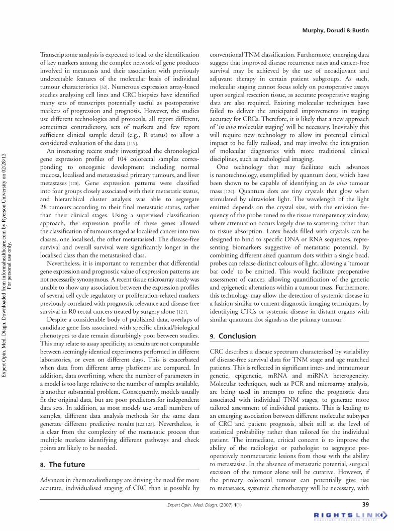
identical ( Figure 2 ). Unfortunately, it is also becoming clear that each molecular subgroup can be further stratified into additional, distinct subgroups with the ultimate implication that each individual tumour may turn out to be unique. Nevertheless, the likelihood remains that successful metastases must have passed through common key stages that, once identified, can yield universal sets of prognostic markers.
Systemic dissemination of tumour cells is a prerequisite of malignant tumour progression and is correlated with the epithelial to mesenchymal transition, a loss of epithelial differentiation and the acquisition of a migratory phenotype [17] . Individual tumours shows distinct subareas of proliferation and cell-cycle arrest, epithelial differentiation and epithelial to mesenchymal transition, cell adhesion and dissemination. This morphological and functional heterogeneity is a function of intratumoural genetic heterogeneity, with most of the advanced CRCs composed of one predominant clone and other minor ones [18] . In addition, LN and liver metastases from the same patient do not always show similar genetic aberrations, further suggesting that there are multiple pathways through which colorectal metastases arise [19] , and that the
LN metastases could be derived from different areas of the primary tumour. Furthermore, the important steps that enable metastasis cannot be explained solely by irreversible genetic alterations [20] and it is becoming clear that reversible, dynamic changes, for example in cellular adhesion and catenin localisation, occur during invasion, metastasis and expansion of CRCs [21] . Moreover, it is clear that the tumour environ-ment plays a regulatory role [22] and that host genetics influences tumour metastasis [23] .
On the other hand, if polymorphism are the driving force behind the metastasis-associated expression profiles, then it may be possible to sample any tissue and determine host-specific predictive genetic profiles. This could pose a real problem, as it suggests that a tumour-derived metastatic signature may not be sufficient for accurate prognosis; there may be an additional requirement for determining host polymorphism associated with metastasis. On the other hand, if polymorphism are the driving force behind the metastasis Taken together, this information leads to the redefinition of CRC as a spectrum of complex diseases that have developed via multiple, often-alternative molecular
Figure 2. Colorectal tumourigenesis as a multi-pathway disease. The signifi cant heterogeneity in colorectal cancers is refl ected in the wide variation of location, histology, the degree of microsatellite instability, levels of methylation, the extent of chromosomal instability and the presence of germline or somatic mutations (indicated by *) as well as epigenetic alterations (indicated by me) of numerous genes implicated in apoptosis and DNA repair. The involvement of JC virus remains an intriguing, albeit unproven hypothesis.CIMP: CpG island methylator phenotype; CIN: Chromosome instability; FAP: Familial adenomatous polyposis; HNPCC: Herteditary non-polyposis colorectal cancer; MACS: Microsatellite and chromosome instability; MSI: Microsatellite instability.
TGF-BIIR*IGF-2R*
BAX*PTEN
B-catLOH 5q
KRAS*
LOH 18q
p53*p53*
18qLOH
APC*
KRAS*
MGMTme
SFRP1,2me
WIF-1me
PTEN*
Kras*BRAF*
hMLH1me
TGF-BIIR*IGF-2R*
BAX*
JC virus ?
Precursor lesions
Epigenetic events APC*JC virus ?
CIMP
hMLH1*
Clinically heterogeneous
MSIMSS CINMACSMSI
FAP HNPCC
?
Exp
ert O
pin.
Med
. Dia
gn. D
ownl
oade
d fr
om in
form
ahea
lthca
re.c
om b
y R
yers
on U
nive
rsity
on
02/2
8/13
For
pers
onal
use
onl
y.

Molecular staging of colorectal cancer: new paradigm or waste of time?
34 Expert Opin. Med. Diagn. (2007) 1(1)
pathways driven by a combination of host influence and selective pressure-associated expression profiles, then it may be possible to sample any tissue and determine host-specific predictive genetic profiles. This would have the additional advantage of permitting population screening and identifying high-risk patients well before they develop cancers.
3. Approaches to molecular staging
Although molecular diagnostic tools that can accurately predict an individual patient ’ s clinical outcome on the basis of tumour biology and host response alone remain a long-term prospect, the classification of CRCs based on molecular heterogeneity has raised expectations for the use of this technology to refine prognostic data from TNM staging. There are two different, albeit not exclusive, conceptual approaches to generating more accurate disease outcome data.
The first approach is based on the hypothesis that treatment failure following surgery is due to the presence of occult disease that cannot be detected preoperatively by radiological assessment, or predicted by standard histopathological techniques following tumour resection. This occult disease takes the form of circulating tumour cells (CTCs) entering the lymphatic and vascular circulation and travelling through the body until they arrest in a distant organ, extravasate and form metastases. These cells should be detectable in blood or LN samples; indeed, cytological examination of blood smears can detect CTCs in between 25% [24] and 67% [25] of samples. However, this method of detection under-estimates the true number of circulating cells as there is a considerable loss in the preparation of blood cell concentrates, and other large mononuclear cells can mimic the appearance of cancer cells [26] . Consequently, PCR-based methods have been developed for the detection in blood and LN of tumour-specific genetic alteration (PCR) or colon-specific mRNAs (RT-PCR) [27] .
A second approach is based on the notion that the metastatic potential of a cancer is encoded in the bulk of the primary tumour [28] and can be predicted from its genotypic or mRNA expression profile [29] . Consequently, there have been numerous attempts to analyse primary tumours for genomic features, such as microsatellite [30] and ploidy [31] status, as well as transcriptome parameters, such as mRNA [32] and micro RNA [33] expression profiles. In theory, this approach permits an ‘ assumption-free ’ means of identifying alterations, genes or regulatory RNAs not previously known to play a role in metastasis. These factors could be potential biomarkers, allowing the subtyping of tumours into more accurate prognostic groups than the TNM classification.
4. Biomarkers
The ultimate power of molecular staging lies in its potential to discriminate preoperatively between colorectal tumours that are likely to have metastasised at the time of surgery, and
those that have not. Choosing useful markers of metastasis requires a better understanding of the metastatic process, and specifically how it differs from the process of tumourigenesis, which refers to the ability of cells to proliferate continuously in the absence of the persistent stimulation by a triggering agent. Successful metastasis reflects the influence of several distinct parameters, including genetic determinants that are largely distinct from those that mediate malignant transformation [34] . Attributes of malignant cells include but are not limited to, undifferentiated morphology/cytology, vascular density, necrosis, high mitotic index, aneuploidy and high nuclear:cytoplasmic ratio [35] . The utility of these characteristics as markers is limited, as their assessment is largely subjective.
When searching for markers of metastasis it is worth bearing in mind that only those mutations that give some kind of selective survival advantage will be selected, making the cell ‘ fitter ’ . Selection pressures change as the tumour evolves, hence different stages of tumour growth will result in the selection of different mutations. Importantly, as selection cannot foresee what mutations or gene expression patterns might be advanta-geous for metastasis, dissemination of tumour cells to, and successful growth at, a distant target organ is likely to drive evolution of metastatic deposits in a different direction to the primary tumour. The exception is the rare occasion when neutral mutations arise that confer neither a selective advantage nor disadvantage at the time, but by chance give a selective advantage to the metastasis. However, such bystander mutations will not form part of the genetic pathway leading to metastasis and are unlikely to be useful biomarkers. It is useful to distinguish between different classes of metastasis gene [34] : ‘ metastasis initiation ’ genes provide a selective advan-tage in primary tumours, enabling cell mobilisation and intravasation. Mutations in or expression of such genes are selected for as invasiveness provides a local advantage by enabling tumour cells to seek vasculature and other favourable elements of the microenvironment. ‘ Metastasis progression ’ genes are those that contribute to primary tumourigenesis, but also fulfil an additional function that happens to be advantageous at the site of metastasis. This may explain why some primary tumour gene-expression signatures correlate with risk of organ-specific dissemination. The third class of metastasis genes provide a selective advantage at secondary sites, but not in the primary tumour, thus participating in metastatic colonisation, but not in primary tumour devel-opment. As they increase the fitness of metastatic tumour cells at the target, their altered expression becomes identifiable only in successfully metastasised tumours. Such genes would rarely be present in primary tumour ‘ poor-prognosis ’ gene expression signatures.
What characteristics define a suitable marker of metastasis? In general, markers fall into two categories: the first predicts metastatic propensity based on expression and/or activity of molecules with an established role in metastasis. For example, matrix metalloproteinases might be expected
Exp
ert O
pin.
Med
. Dia
gn. D
ownl
oade
d fr
om in
form
ahea
lthca
re.c
om b
y R
yers
on U
nive
rsity
on
02/2
8/13
For
pers
onal
use
onl
y.
Murphy, Dorudi & Bustin
Expert Opin. Med. Diagn. (2007) 1(1) 35
to be more highly expressed in invasive and metastatic tumours than in their nonmetastatic counterparts. The second category includes markers for which there is no established mechanistic association with metastasis. This category includes known genes with potentially novel functions, novel genes and molecular changes that correlate with metastatic ability [36] .
Each of these categories can be further divided on the basis of whether expression of the marker is increased or decreased. This has an impact on the clinical utility of assays, as sensitivity for highly expressed molecules in heterogeneous metastatic primary tumours is greater than that for markers expressed at low levels. This is of particular importance, as it is well recognised that most cells within a tumour cannot complete the multistep process of metastasis. Indeed, < 0.1% of cells entering the bloodstream successfully form clinically detectable lesions [35] . This suggests that only a small per centage of cells within a primary tumour will display all relevant marker sets of metastasis. Just as it is easier to see a single light in a dark room than find that light amongst a room full of other lights, a single cell expressing a marker against a background of nonexpressing cells is easier to identify than a nonexpressing cell within a mass of marker positive cells. This comparison does not even take into account quantitative differences in expression, which further complicate the matter. For this reason, in clinical practice identification of positive indicators of metastatic potential would be preferred.
It must be emphasised that markers can be clinically useful even if their biological function is not well defined. Indeed, it is anticipated that, as a result of efforts to sequence the human genome, there will be numerous molecular markers identified in regions that have not previously been linked to gross chromosomal changes [37] .
5. Molecular technology
There are at least three separate targets for molecular diagnostics: DNA, RNA and proteins ( Figure 1 ). DNA is easily accessible, contains numerous tumour-specific genetic alterations, and has long been seen as providing the most likely target for molecular diagnostic assays [38] . More recently, it has also become clear that just as DNA methylation plays an essential role in tumourigenesis [39] , epigenetic mechanisms have a central function in metastasis [40,41] . It remains to be seen how practical use can be made of this information. Alternatively, the detection of tissue-specific mRNA and miRNA targets provides a second avenue for differentiation of the biological and clinical behaviour of CRCs [42,43] . Finally, interesting results are beginning to emerge from assays targeting the proteome/peptidome of primary tumours [44] . Nevertheless, although proteomic analysis of tissue or serum may eventually become an important modality in cancer diagnostics [45,46] , the complexity of analysing the proteome/peptidome, as well as the cost of any associated instrumentation makes this a very long-term prospect.
6. Genome analysis
6.1 Microsatellite analysis The power of molecular taxonomy is illustrated by its ability to characterise CRCs further within each stage according to their different degrees of genetic instability. Most sporadic CRCs are aneuploid – they harbour an abnormal complement of genetic material and are charac-terised by chromosomal instability (CIN) [47] . Typically, they display loss-of-heterozygosity, resulting in the loss of tumour suppressor genes, such as APC or p53 . A second type of instability is apparent in 10 – 15% of sporadic colorectal tumours that have apparently stable near diploid chromosomes, but display DNA microsatellite instability (MIN), caused by hypermethylation of CpG islands in the promoters of DNA mismatch repair genes [48] . Clinically, these cancers differ, and some reports suggest that stage-matched patients with MIN cancers have a better prognosis than patients with CIN cancers [49,50] , although others find the opposite [51,52] . The reason for this discrepancy may lie in the discovery that there is a further subgroup with stable microsatellites (microsatellite and chromosome stable [MACS]) within the MIN subgroup of tumours that account for up to 30% of all sporadic CRCs [53-55] . MACS tumours seem to have aggressive biological characteristics, such as poor differentiation [56] , early spread to LN and blood [57] and an invasive phenotype that encourages early metastasis [31] . Perhaps not surprisingly, there is also evidence that this subgroup is itself heterogeneous [58] .
6.2 Comparative genomic hybridization The genomes of hundreds of primary colorectal carcinomas have been studied by conventional chromosomal banding technique, as well as by comparative genomic hybridization (CGH), revealing a non-random pattern of genomic abnor-malities associated with a metastatic phenotype. Metastatic primary tumours seem to exhibit more complex genomic aberrations than non-metastatic primary tumours, with multiple chromosome loss or gain inversely correlated with disease-free survival. One study looked at 18 metastatic and 18 non-metastatic tumours and showed that chromo-somal regions 1p32 and 9q33 were present at much higher frequencies in metastatic compared with non-metastatic cancers [59] . Another study profiled primary tumours from 61 patients with early-stage and largely microsatellite-stable CRCs [60] and demonstrated that loss of chromosome arms 4 and 14q was associated with poorer survival than loss of any one arm. A third study showed that gain of chromosome 8q is predictive for metastasis [61] . Loss and gain of these markers agrees with a previous analysis of 17 liver metastases [62] . All reported CGH analyses consistently suggest that the different pathways of tumour dissemination are driven by a non-random accumulation of abundant chromosomal alterations with specific changes being responsible for the different characteristics of the metastatic phenotype [63] .
Exp
ert O
pin.
Med
. Dia
gn. D
ownl
oade
d fr
om in
form
ahea
lthca
re.c
om b
y R
yers
on U
nive
rsity
on
02/2
8/13
For
pers
onal
use
onl
y.

Molecular staging of colorectal cancer: new paradigm or waste of time?
36 Expert Opin. Med. Diagn. (2007) 1(1)
These data imply that genetic profiling of primary tumours of patients with early-stage CRC may be useful in the mainstream management of patients, although the results still suggest a statistical, rather than an individual, risk of distant recurrence. Furthermore, a remaining problem is that MACS tumours, which have no gross chromosomal alterations, seem to have the worst prognosis. In practical terms, it is likely that although CGH can point towards regions of the genome that are important for prognosis, other, more rapier-like technologies, such as the polymerase chain reaction (PCR), are required to pinpoint the specific alterations that can predict individual outcome.
6.3 Polymerase chain reaction DNA-targeted PCR is a robust, sensitive and reliable method for detecting differences in gene structure, such as loss of heterozygosity, chromosome rearrangements or point mutations. However, the heterogeneity of primary tumours precludes a simple interpretation of results generated from single-target PCR assays carried out on limited biopsies. It is essential to analyse multiple biopsies, ideally microdissected from separate areas of the tumour, as CRCs are usually made up from several clones [18,64] . This is not a trivial matter, which adds to the technical complexity and cost of the assay, and hampers its translation into routine clinical practice.
A second option makes use of the fact that LN involvement is already an established prognostic indicator and aims to use the PCR to detect submicroscopic (occult) disease in LN harvested following surgery [65] . The major technical limitation here is that there are no known mutations that occur consistently in CRCs, making a universal assay unfeasible at this time [66] . Furthermore, even the presence of actual metastases in LN is not prognostic for individual patients; hence the presence of occult metastases may also only be useful for stratifying groups of patients, not providing prognostic information for individuals. This is born out by two studies that have looked at LN from cancers harbouring mutations in the K-ras and/or p53 genes and assessed as tumour-free by TNM staging. Using allele-specific PCR, both were able to identify occult disease and showed that its presence was predictive of poorer survival [67,68] . However, even this best-case scenario failed to predict relapse for individual patients, as between 27 and 69% of patients that were LN positive by PCR did not develop distant recurrences. Indeed, a patient in the negative control group also died [68] . Furthermore, K-ras mutations are present in only 40 – 50% of CRCs, and mutations in the p53 gene, although a more frequent finding in colorectal carcinoma, are not confined to a single site. These results have important, albeit unwelcome implications, as they suggest that a straightforward model that evaluates metastasis risk simply from the presence or absence of tumour cells is of little value.
Although blood is not analysed as part of conventional staging, PCR-based studies have been performed on blood
samples as blood is easily accessible pre and postoperatively. Interestingly, the detection of free genomic DNA in plasma/serum of cancer patients has been reported [69,70] , with one study showing a significantly reduced survival rate for patients with free-circulating tumour-associated DNA [71] . However, circulating DNA can be detected in some normal patients, and cannot in some cancer patients. Furthermore, these studies involved relatively few patients and short follow-up periods. Detection of tumour-specific mutations is no more accurate: in one study high levels of circulating tumour cells did not lead to distant disease [72] and in another one only 2/5 and 4/9 patients, respectively, from two groups with concordant K-ras mutations in their primary cancers and serum developed distant recurrences [73] . The reluctant but realistic conclusion must be that detection of universal free genomic or of tumour-derived (i.e., mutated) DNA is not prognostic for individual patients. The most obvious explanation for this is that the assays are not detecting CTCs, much less CTCs with metastatic potential. DNA is very stable and its detection may be a reflection of tumour burden, resulting in the persistence of tumour DNA released by cell necrosis or apoptosis [69] . Together with the LN data, these results merely provide further evidence for our contention that detection of a tumour cell per se or its DNA is of little relevance for individual patient prognosis.
7. Transcriptome analysis
7.1 Real-time polymerase chain reaction Although there are no tumour-specific genes for CRC, malignant cells continue to express markers that are character-istic of their tissue of origin. Therefore, a range of epithelial cell markers, which are normally absent from the blood, have been proposed as targets for circulating putative CRC cells. RNA is rapidly degraded and so its presence in the blood suggests active expression by CTCs, in contrast to fragments of DNA, which are stable and may represent tumour DNA released previously during CTC apoptosis [74] . Therefore, the use of RT-PCR has been evaluated extensively in the molecular assessment of tumour stage and disease recurrence [65,75] . However, again there are significant technical and conceptual limitations [76] that hold back its adaptation into clinical practice [77] . For example, there is a lack of standardised protocols [78] , it is unclear just how tissue-specific mRNAs are, a problem exacerbated by the use of quantitative methods such as real-time PCR that can easily detect illegitimate transcription [76] and the significant variability of the RT-step makes it problematic to delineate universal, clinically useful quantitative ‘ cutoff ’ points [79] . Therefore, at present it is best to think of RT-PCR as a ‘ proof of principle ’ rather than as a robust and reliable clinical assay [32] .
There have been numerous studies reporting the post operative detection of mRNA markers, such as carcinoembryonic antigen (CEA), cytokeratins (ck), mucins, CD44 and guanylyl cyclase C (GCC) in different tissue
Exp
ert O
pin.
Med
. Dia
gn. D
ownl
oade
d fr
om in
form
ahea
lthca
re.c
om b
y R
yers
on U
nive
rsity
on
02/2
8/13
For
pers
onal
use
onl
y.

Murphy, Dorudi & Bustin
Expert Opin. Med. Diagn. (2007) 1(1) 37
compartments, which have attempted to assess their prognostic significance [80] . One report suggests that it is possible to distinguish histologically positive from histologically negative LNs by counting the number of CEA-expressing cells [81] . However, there was significant overlap between the two groups and cell numbers were calculated relative to a CEA-expressing cell line, ignoring inter-sample or inter-patient variation of CEA mRNA levels. A second study attempted to overcome this by calculating CEA copy numbers in LN relative to 18S RNA levels, and used cutoff levels to suggest that high CEA mRNA levels might be predictive of distant recurrence [82] . In keeping with these data, a third study also concluded that quantification of CEA mRNA levels in the LN from patients with advanced CRC yielded prognostic information [83] . However, quantitating the amount of an mRNA does not allow the calculation of the number of CTCs as the expression of most genes varies by several orders of magnitude between tumours in different individuals, and often varies in the tumour of the same individual [84] . In addition, none of these authors discussed how to implement a relative quantification assay in clinical practice.
In complete contrast, studies using both conventional [85] and real-time [76] PCR (RT-PCR) have reported the detection of CEA mRNA in up to 85% of control LN, with significant overlap of CEA copy numbers between histologically involved and uninvolved LN. There was no correlation between CEA copy numbers and prognosis, suggesting that a CEA-based assay is unable to identify patients at risk of distant disease recurrence.
An interesting variation describes the use of immunobead RT-PCR to isolate epithelial-derived tumour cells and target CEA, laminin 2, ephrin B4, matrilysin and ck20 mRNA to detect perioperatively the presence of colon tumour cells within the peritoneal cavity of colon cancer patients [86] . Patients who were marker positive for dissemi-nated cells in postresection lavage samples showed a significantly poorer prognosis independent of other risk factors. However, no information was provided on the R-status or RNA quality, and the process of obtaining samples for analysis is not simple. Interestingly, and in line with the reasoning described above, the same study also looked at the same markers in blood, but did not report any association between the presence of these markers and prognosis.
Indeed, RT-PCR assays have been extensively used for the detection of supposedly tissue-specific mRNAs in blood, with conflicting results. Some aim to determine preoperatively the metastatic potential of primary tumours, others quantitate postoperatively systemic disease burden. Some reports suggest that CEA mRNA levels in the blood of CRC patients are associated with disease stage [87] and may be of prognostic value [88,89] . These studies contrast with others that question its specificity and suggest that peripheral blood is not a suitable compartment for detection of tumour cells [78,85,90,91] , or advocate analysis of yet another tissue compartment [92] . Similar, contradictory results have been reported for other
tissue-specific markers [32] . This discordance challenges the practical usefulness of RT-PCR assays. Most importantly, when individual results are analysed in detail, there is a significant percentage of patients that test positive for the marker in question, yet survive for 5 years or do not test positive for the marker yet die within 5 years [76,91] .
Many assays claim a specificity rate of 100%, indicating that none of the control samples are positive for the marker of interest. However, other assays, targeting the same markers, suggest that there is no absolute specificity for any of the markers [76,93,94] . Incidentally, one study also reported that the false-negative rate was ≥ 25% of patients known to have metastatic or unresectable CRC had a negative RT-PCR test [94] . Clearly, at best RT-PCR assays may provide some additional information for patient groups, at worst the data may be irrelevant.
Although the development of blood based assays that yield prognostic information may be clinically attractive, it must be remembered that early studies suggested < 0.1% of CTCs survive in the circulation, and only 0.01% form metastases [95] . This challenges the whole concept of deriving prognostic information by simply detecting, or even quantitating the number of CTCs. Crucially, it is becoming apparent that the capacity to form metastases depends on the presence of a very small subset of cells able to self-renew and differentiate into the bulk tumour population [96,97] . It was also thought that most CTCs are removed from the circulation within 24 h by elimination in the first capillary bed they encounter [98] . However, recent work using cyto-plasmically labelled tumour cells (as opposed to nuclear-labelled cells, which are more vulnerable to destruction) has found that most cells survive in the circulation for several days following injection, and the inefficient part of the metastatic process seems to be the variable growth of the cancer cell at the secondary site [99] .
The development of serum-based assays is further complicated by the fact the source of blood for analysis seems to be important. Theoretically, CTCs must pass through the liver, lungs and the microcirculation of the other tissues of the body before they pass into the systemic venous circulation. This suggests that detection of CTCs might be expected to be more likely in portal venous (PV) rather than in systemic venous (SV) blood. One study found 57% of patients were positive for CTC in PV blood during resection of a colorectal tumour, as opposed to 50% of patients who were positive in their SV samples [26] . Another found more PV samples were positive before, during and after surgery compared with SV samples [74] . However, 3 of the 17 who were positive in the peripheral blood were negative in all portal samples. It is interesting to note these three patients had late-stage disease and it may be that tumour cells were bypassing the portal circulation and entering the systemic circulation directly. Yet another study found concordance between SV and PV samples for CEA mRNA in only 65% of cases [100] . Another one found that detection of ck20 and CEA mRNA in
Exp
ert O
pin.
Med
. Dia
gn. D
ownl
oade
d fr
om in
form
ahea
lthca
re.c
om b
y R
yers
on U
nive
rsity
on
02/2
8/13
For
pers
onal
use
onl
y.

Molecular staging of colorectal cancer: new paradigm or waste of time?
38 Expert Opin. Med. Diagn. (2007) 1(1)
SV blood had no prognostic value. Consequently, it is not surprising that preoperative [101] peripheral blood sample positive for CEA or pre and postoperative samples positive for ck20 and CEA [102] have not been shown to have prognostic value. A comprehensive review concludes that the presence of CTC cannot be considered to be a reliable indicator of prognosis in any common solid malignancy [80] .
The importance of appropriate sampling must also be considered, as each assay represents but a single snapshot of a complex and dynamic process [103] . It is unclear whether the release of tumour cells is a continuous process or whether cancer cells are intermittently shed into the blood [104] . If the latter, this would result in sampling errors if single samples are taken. One study involving sequential intra-operative sampling found that of the 5 patients positive for CTC at any of the 4 sampling time points, 2 (40%) were positive at a single time point only [105] . Another report demonstrated that by increasing the sampling frequency (from once to twice), the detection of CTC is significantly increased [106] . A third investigation that sampled SV and PV blood twice found that of the 14 patients who were positive in PV blood, 5 (36%) were positive in the second sample only, and so would have been considered negative if a single sample was taken [74] . In the SV samples, 3 out of 17 (18%) patients were positive in the second sample only. In this context it is also important to note that multicell clumps are more likely to metastasise than single cells [104] and that detection or quantification of mRNA levels cannot distinguish between the two.
One less obvious reason for unreliable data generated by serum assays is that the detection of CTC may be affected by the degree of intra-operative blood loss and subsequent administration of intravenous fluids, thereby diluting the blood volume and reducing the likelihood of tumour cell detection during or immediately after surgery [107] . Environmental factors may also affect the likelihood of any tumour metastasising: the metastatic process might be enhanced by the surgical intervention. It is known the entrapment of tumour cells in the microcirculation of a target organ is facilitated by the presence of fibrin and platelets. Therefore, activation of coagulation that occurs during an operation may facilitate metastases [108] . Furthermore, surgical stress [109] and perioperative blood transfusion [110] have been shown to induce immune suppression, which, in turn, is believed to increase the efficiency of the metastatic process. Conversely, there are no convincing data to suggest variations in surgical technique influence tumour metastasis intraoperatively [111] .
Taken together, a disinterested analysis of most RT-PCR data points towards a conceptual flaw underlying the attempts to use such assays to allow prediction of successful distant metastasis. They are based on a simplistic view of the biology and kinetics of tumour cell traffic through the lymphatic and systemic circulation and subsequent metastasis development. Instead, RT-PCR may simply be detecting cells of no biological significance [112] and variability in survival within each staging
category probably reflects not only the inaccuracy of detecting occult residual disease, but also a lack of understanding of the sequestration, release and subsequent trafficking of the tumour cell in both the lymphatic and systemic circulation. None of the RT-PCR assays address the question of the biological rele-vance of detecting tumour cells in blood or LN and do not provide any information regarding their metastatic potential. Furthermore, the genotype of LN metastases differs from that of the main clone in the primary tumour in > 50% of patients, with a significant minority displaying a genotype not detected in the primary tumour at all [113] . In addition, these studies do not take into account the role of patient genotype, or that of the immune system in allowing or suppressing metastasis: humans are genetically polymorphic, and the outcome of metastasis depends on the interplay of tumour cells with vari-ous host factors including the organ microenvironment, which can influence the biology of cancer growth, angiogenesis and metastasis. Therefore, it is not surprising that the detection of supposedly tissue-specific mRNA, and extrapolating from that to the presence of occult disease, has not produced robust and reproducible data with a prognostic value for the individual patient. As a result, despite this vast effort, PCR-based tech-niques have failed to capture the enthusiasm of clinicians and have still not been validated clinically in prospective studies.
Finally, there is a lack of standardisation of RT-PCR techniques, resulting in widely different sensitivities and speci-ficities between laboratories. Illegitimate transcription, the transcription of tissue-specific genes in non-specific cells, is easily detected, and the presence of non-malignant epithelial cells in the blood following venepuncture [114] may reduce assay specificity. In addition, as RT-PCR detects the number of mRNA copies, not the number of CTCs, calculating mRNA copy numbers may not accurately relate to an increase in tumour cells [74] . False positives can frequently occur with RT-PCR, particularly with non-probe-based real-time RT-PCR assays currently being developed. Equally, there is a problem with false negatives: the marker of interest may not be expressed, or detectable due to tumour cell heterogeneity or because there is a poorly-differentiated subclone that has lost the ability to express the tissue-specific marker [115] . It must also be remembered that PCR inhibitors are present within all body fluids and tissues, and it is important to note the in vitro sensitivity reported by many studies will be higher than the true in vivo sensitivity, as the cell lines used to determine the sensitivity are known to strongly express the marker of interest in the absence of PCR inhibitors. The high variability in RT and PCR efficiencies, combined with subjective data analysis and interpretation, also contributes to a significant problem with the reliability of any RT-PCR-based assay [116-118] .
7.2 Microarrays Microarrays permit the investigation of the expression of thousands of genes within a single patient sample and remove the practical limit on the number of candidate genes for which expression can be studied conveniently and in parallel.
Exp
ert O
pin.
Med
. Dia
gn. D
ownl
oade
d fr
om in
form
ahea
lthca
re.c
om b
y R
yers
on U
nive
rsity
on
02/2
8/13
For
pers
onal
use
onl
y.
Murphy, Dorudi & Bustin
Expert Opin. Med. Diagn. (2007) 1(1) 39
Transcriptome analysis is expected to lead to the identification of key markers among the complex network of gene products involved in metastasis and their association with previously undetectable features of the molecular basis of individual tumour characteristics [32] . Numerous expression array-based studies analysing cell lines and CRC biopsies have identified many sets of transcripts potentially useful as postoperative markers of progression and prognosis. However, the studies use different technologies and protocols, all report different, sometimes contradictory, sets of markers and few report sufficient clinical sample detail (e.g., R status) to allow a considered evaluation of the data [119] .
An interesting recent study investigated the chronological gene expression profiles of 104 colorectal samples corres-ponding to oncogenic development including normal mucosa, localised and metastasised primary tumours, and liver metastases [120] . Gene expression patterns were classified into four groups closely associated with their metastatic status, and hierarchical cluster analysis was able to segregate 28 tumours according to their final metastatic status, rather than their clinical stages. Using a supervised classification approach, the expression profile of these genes allowed the classification of tumours staged as localised cancer into two classes, one localised, the other metastasised. The disease-free survival and overall survival were significantly longer in the localised class than the metastasised class.
Nevertheless, it is important to remember that differential gene expression and prognostic value of expression patterns are not necessarily synonymous. A recent tissue microarray study was unable to show any association between the expression profiles of several cell cycle regulatory or proliferation-related markers previously correlated with prognostic relevance and disease-free survival in R0 rectal cancers treated by surgery alone [121] .
Despite a considerable body of published data, overlaps of candidate gene lists associated with specific clinical/biological phenotypes to date remain disturbingly poor between studies. This may relate to assay specificity, as results are not comparable between seemingly identical experiments performed in different laboratories, or even on different days. This is exacerbated when data from different array platforms are compared. In addition, data overfitting, where the number of parameters in a model is too large relative to the number of samples available, is another substantial problem. Consequently, models usually fit the original data, but are poor predictors for independent data sets. In addition, as most models use small numbers of samples, different data analysis methods for the same data generate different predictive results [122,123] . Nevertheless, it is clear from the complexity of the metastatic process that multiple markers identifying different pathways and check points are likely to be needed.
8. The future
Advances in chemoradiotherapy are driving the need for more accurate, individualised staging of CRC than is possible by
conventional TNM classification. Furthermore, emerging data suggest that improved disease recurrence rates and cancer-free survival may be achieved by the use of neoadjuvant and adjuvant therapy in certain patient subgroups. As such, molecular staging cannot focus solely on postoperative assays upon surgical resection tissue, as accurate preoperative staging data are also required. Existing molecular techniques have failed to deliver the anticipated improvements in staging accuracy for CRCs. Therefore, it is likely that a new approach of ‘ in vivo molecular staging ’ will be necessary. Inevitably this will require new technology to allow its potential clinical impact to be fully realised, and may involve the integration of molecular diagnostics with more traditional clinical disciplines, such as radiological imaging.
One technology that may facilitate such advances is nanotechnology, exemplified by quantum dots, which have been shown to be capable of identifying an in vivo tumour mass [124] . Quantum dots are tiny crystals that glow when stimulated by ultraviolet light. The wavelength of the light emitted depends on the crystal size, with the emission fre-quency of the probe tuned to the tissue transparency window, where attenuation occurs largely due to scattering rather than to tissue absorption. Latex beads filled with crystals can be designed to bind to specific DNA or RNA sequences, repre-senting biomarkers suggestive of metastatic potential. By combining different sized quantum dots within a single bead, probes can release distinct colours of light, allowing a ‘ tumour bar code ’ to be emitted. This would facilitate preoperative assessment of cancer, allowing quantification of the genetic and epigenetic alterations within a tumour mass. Furthermore, this technology may allow the detection of systemic disease in a fashion similar to current diagnostic imaging techniques, by identifying CTCs or systemic disease in distant organs with similar quantum dot signals as the primary tumour.
9. Conclusion
CRC describes a disease spectrum characterised by variability of disease-free survival data for TNM stage and age matched patients. This is reflected in significant inter- and intratumour genetic, epigenetic, mRNA and miRNA heterogeneity. Molecular techniques, such as PCR and microarray analysis, are being used in attempts to refine the prognostic data associated with individual TNM stages, to generate more tailored assessment of individual patients. This is leading to an emerging association between different molecular subtypes of CRC and patient prognosis, albeit still at the level of statistical probability rather than tailored for the individual patient. The immediate, critical concern is to improve the ability of the radiologist or pathologist to segregate pre-operatively nonmetastatic lesions from those with the ability to metastasise. In the absence of metastatic potential, surgical excision of the tumour alone will be curative. However, if the primary colorectal tumour can potentially give rise to metastases, systemic chemotherapy will be necessary, with
Exp
ert O
pin.
Med
. Dia
gn. D
ownl
oade
d fr
om in
form
ahea
lthca
re.c
om b
y R
yers
on U
nive
rsity
on
02/2
8/13
For
pers
onal
use
onl
y.

Molecular staging of colorectal cancer: new paradigm or waste of time?
40 Expert Opin. Med. Diagn. (2007) 1(1)
preoperative local radiotherapy reserved for those patients with rectal tumours. Although numerous individual markers and several expression profiles have been reported as being independent predictors of clinical outcome, none have been universally validated. The acceptance of any markers into clinical practice must be supported by reproducible validation using a standardised assay condition or diagnostic criteria in multiple independent large sets of samples, as well as by different laboratories. In addition, in light of the additional expense and resources, which will be required to implement molecular assays, demonstrable benefit must be shown over the current gold standard of TNM staging. Therefore, at present molecular diagnosis is sadly a pipe-dream.
10. Expert opinion
Molecular biology techniques, such as PCR and microarrays, allow the detection and quantification of putative cancer cells circulating through the body, as well as the stratification of primary tumours according to their genetic and expression profiles. Although they provide outstanding research tools for analysing the alterations in molecular pathways underlying colorectal tumourigenesis and metastasis, their value in clinical diagnostics is less apparent. Undoubtedly, their use allows tumours that are grouped within the same histopathological stage to be further subdivided according to molecular criteria, such as genomic stability or expression signature. Although it may prove possible to assign patients to more accurate molecular staging categories, these will still provide an overall, rather than individual assessment of treatment failure risk. Biological and technical problems, for example, tumour and host heterogeneity, lack of standardisation of sample source, CTC detection methods and small sample sizes, suggests that for individual patients the clinical advantage of quantitating
CTCs, determining genetic profiles or establishing expression signatures remains, at best, unclear.
Without doubt the latest molecular tools, such as tissue-specific genetic knockouts, siRNA-mediated knockdown and the development of protein, tissue and interaction arrays are combining with PCR-based and microarray technology to unravel the molecular networks involved in regulating the metastatic cascade. Furthermore, they are assisting in identify-ing the critical role played by the host response in determining metastatic fitness and an overall scenario is being painted of extreme complexity at the molecular, cellular, tissue and host levels. However, the expectation that accurate detection of occult circulating malignant cells can help to refine TNM staging and associated prognosis for individual CRC patients, as well as monitor their response to therapy and highlight early the possibility of recurrence remains unfulfilled.
Although there is a clear, and urgent, necessity to improve preoperative staging of CRCs, it is unclear which, if any, of the present approaches to molecular staging will significantly improve conventional staging in the medium term. Indeed the only molecular diagnostic tests presently available clinically, which determine chromosomal or microsatellite instability, have limited prognostic use for individual CRC patients. Therefore, the most likely clinical application of molecular technology may prove to be in conjunction with traditional imaging modalities. This is not unprecedented, as imaging based upon increased tumour glucose meta bolism has already reached clinical practice. However, even then the lack of reli-able tumour specific genetic and/or expression signatures will remain a major stumbling block for ‘ molecular imaging ’ .
Declaration of interest
The authors have no conflict of interest to declare and no fee has been received for preparation of the manuscript.
Exp
ert O
pin.
Med
. Dia
gn. D
ownl
oade
d fr
om in
form
ahea
lthca
re.c
om b
y R
yers
on U
nive
rsity
on
02/2
8/13
For
pers
onal
use
onl
y.
Murphy, Dorudi & Bustin
Expert Opin. Med. Diagn. (2007) 1(1) 41
Bibliography Papers of special note have been highlighted as either of interest (•) or of considerable interest (••) to readers.
1 . QUIRKE P, MORRIS E: Reporting colorectal cancer. Histopathology ( 2007 ) 50 (1): 103 -112.
2 . NELSON H, PETRELLI N, CARLIN A et al. : Guidelines 2000 for colon and rectal cancer surgery. J. Natl. Cancer Inst. ( 2001 ) 93 (8): 583 -596.
3 . QUIRKE P, WILLIAMS GT, ECTORS N et al. : The future of the TNM staging system in colorectal cancer: time for a debate? Lancet Oncol. ( 2007 ) 8 (7): 651 -657.
4 . JEMAL A, MURRAY T, WARD E et al. : Cancer statistics, 2005 . CA Cancer J. Clin. ( 2005 ) 55 (1): 10 -30.
5 . RATTO C, SOFO L, IPPOLITI M et al. : Prognostic factors in colorectal cancer. Literature review for clinical application. Dis. Colon Rectum ( 1998 ) 41 (8): 1033 -1049.
6 . MIDGLEY R, KERR DJ: Adjuvant chemotherapy for stage II colorectal cancer: the time is right! Nat. Clin. Pract. Oncol. ( 2005 ) 2 (7): 364 -369.
7 . SEBAG -MONTEFIORE D: Developments in the use of chemoradiotherapy in rectal cancer. Colorec. Dis. ( 2006 ) 8 (Suppl. 3): 4 -17.
8 . STEIN U, SCHLAG PM: Clinical, biological, and molecular aspects of metastasis in colorectal cancer. Recent Results Cancer Res. ( 2007 ) 176 : 1 -80.
9 . JASS JR, O BRIEN MJ, RIDDELL RH et al. : Recommendations for the reporting of surgically resected specimens of colorectal carcinoma. Hum. Pathol. ( 2007 ) 38 (4): 537 -545.
• Clear critique of present staging procedure defi ciencies.
10 . DUFFY MJ, VAN DALEN A, HAGLUND C et al. : Tumour markers in colorectal cancer: European group on tumour markers (EGTM) guidelines for clinical use. Eur. J. Cancer ( 2007 ).
11 . ROUKOS DH, MURRAY S, BRIASOULIS E: Molecular genetic tools shape a roadmap towards a more accurate prognostic prediction and personalized management of cancer. Cancer Biol. Ther. ( 2007 ) 6 (3): 308 -312.
12 . MAURER GD, LEUPOLD JH, SCHEWE DM et al. : Analysis of specifi c transcriptional regulators as early predictors of independent prognostic relevance in
resected colorectal cancer. Clin. Cancer Res. ( 2007 ) 13 (4): 1123 -1132.
• Defi nes novel high-risk groups of colorectal cancer patients based on transcriptional regulators of the invasion-related gene u-PAR.
13 . BARRATT PL, SEYMOUR MT, STENNING SP et al. : DNA markers predicting benefi t from adjuvant fl uorouracil in patients with colon cancer: a molecular study. Lancet ( 2002 ) 360 (9343): 1381 -1391.
14 . AL -MULLA F, HAGAN S, BEHBEHANI AI et al. : Raf kinase inhibitor protein expression in a survival analysis of colorectal cancer patients. J. Clin. Oncol. ( 2006 ) 24 (36): 5672 -5679.
15 . VOGELSTEIN B, FEARON ER, HAMILTON SR et al. : Genetic alterations during colorectal-tumor development. N. Engl. J. Med. ( 1988 ) 319 (9): 525 -532.
• First correlation of histopathological and molecular changes in a cancer.
16 . TAKAYAMA T, MIYANISHI K, HAYASHI T et al. : Colorectal cancer: genetics of development and metastasis. J. Gastroenterol. ( 2006 ) 41 (3): 185 -192.
17 . THIERY JP: Epithelial–mesenchymal transitions in development and pathologies. Curr. Opin. Cell Biol. ( 2003 ) 15 (6): 740 -746.
18 . LOSI L, BAISSE B, BOUZOURENE H et al. : Evolution of intratumoral genetic heterogeneity during colorectal cancer progression. Carcinogenesis ( 2005 ) 26 (5): 916 -922.
• Elegant paper describing in detail the accumulation of genetic alterations giving rise to different clonal populations within tumours.
19 . AL -MULLA F, KEITH WN, PICKFORD IR et al. : Comparative genomic hybridization analysis of primary colorectal carcinomas and their synchronous metastases. Genes Chromosomes Cancer ( 1999 ) 24 (4): 306 -314.
20 . BRABLETZ T, JUNG A, REU S et al. : Variable β -catenin expression in colorectal cancers indicates tumor progression driven by the tumor environment. Proc. Natl. Acad. Sci. USA ( 2001 ) 98 (18): 10356 -10361.
21 . BARKER N, CLEVERS H: Tumor environment: a potent driving force in colorectal cancer? Trends Mol. Med. ( 2001 ) 7 (12): 535 -537.
22 . BISSELL MJ, RADISKY D: Putting tumours in context. Nat. Rev. Cancer ( 2001 ) 1 (1): 46 -54.
23 . HUNTER KW, CRAWFORD NP: Germ line polymorphism in metastatic progression. Cancer Res. ( 2006 ) 66 (3): 1251 -1254.
•• Highly insightful discussion of the importance of host responses to metastasis.
24 . FISHER ER, TURNBULL RB Jr: The cytologic demonstration and signifi cance of tumor cells in the mesenteric venous blood in patients with colorectal carcinoma. Surg. Gynecol. Obstet. ( 1955 ) 100 (1): 102 -108.
25 . ENGELL HC: Cancer cells in the circulating blood; a clinical study on the occurrence of cancer cells in the peripheral blood and in venous blood draining the tumour area at operation. Acta Chir. Scand. Suppl. ( 1955 ) 201 : 1 -70.
26 . GRIFFITHS JD, MCKINNA JA, ROWBOTHAM HD et al. : Carcinoma of the colon and rectum: circulating malignant cells and fi ve-year survival. Cancer ( 1973 ) 31 (1): 226 -236.
27 . BUSTIN SA: Nucleic acid quantifi cation and disease outcome prediction in colorectal cancer. Personalized Med. ( 2006 ) 3 (2): 207 -216.
28 . RAMASWAMY S, ROSS KN, LANDER ES et al. : A molecular signature of metastasis in primary solid tumors. Nat. Genet. ( 2003 ) 33 (1): 49 -54.
•• Powerful study identifying molecular signature associated with metastasis, albeit not in CRC.
29 . AHMED FE: Molecular markers that predict response to colon cancer therapy. Expert Rev. Mol. Diagn. ( 2005 ) 5 (3): 353 -375.
30 . RIBIC CM, SARGENT DJ, MOORE MJ et al. : Tumor microsatellite-instability status as a predictor of benefi t from fl uorouracil-based adjuvant chemotherapy for colon cancer. N. Engl. J. Med. ( 2003 ) 349 (3): 247 -257.
•• Early paper identifying a practical benefi t from molecular stratifi cation of CRC.
31 . HAWKINS NJ, TOMLINSON I, MEAGHER A et al. : Microsatellite-stable diploid carcinoma: a biologically distinct and aggressive subset of sporadic colorectal cancer. Br. J. Cancer ( 2001 ) 84 (2): 232 -236.
32 . BUSTIN SA, DORUDI S: Gene expression profi ling for molecular staging
Exp
ert O
pin.
Med
. Dia
gn. D
ownl
oade
d fr
om in
form
ahea
lthca
re.c
om b
y R
yers
on U
nive
rsity
on
02/2
8/13
For
pers
onal
use
onl
y.
Molecular staging of colorectal cancer: new paradigm or waste of time?
42 Expert Opin. Med. Diagn. (2007) 1(1)
and prognosis prediction in colorectal cancer. Expert Rev. Mol. Diagn. ( 2004 ) 4 (5): 599 -607.
33 . CUMMINS JM, VELCULESCU VE: Implications of micro-RNA profi ling for cancer diagnosis. Oncogene ( 2006 ) 25 (46): 6220 -6227.
• Description of a novel layer of dysregulation affecting cancer behaviour.
34 . NGUYEN DX, MASSAGUE J: Genetic determinants of cancer metastasis. Nat. Rev. Genet. ( 2007 ) 8 (5): 341 -352.
35 . FIDLER IJ: The biology of cancer metastasis or, ‘you cannot fi x it if you do not know how it works ’ . Bioessays ( 1991 ) 13 (10): 551 -554.
36 . HABERMANN JK, PAULSEN U, ROBLICK UJ et al. : Stage-specifi c alterations of the genome, transcriptome, and proteome during colorectal carcinogenesis. Genes Chromosomes Cancer ( 2007 ) 46 (1): 10 -26.
• Demonstration that recurrent patterns of chromosomal imbalances, as well as specifi c gene and protein expression changes, correlate with distinct stages of colorectal cancer progression
37 . CONSORTIUM TWTCC: Genome-wide association study of 14,000 cases of seven common diseases and 3,000 shared controls. Nature ( 2007 ) 447 (7145): 661 -678.
38 . BOOTH RA: Minimally invasive biomarkers for detection and staging of colorectal cancer. Cancer Lett. ( 2007 ) 249 (1): 87 -96.
39 . TING AH, MCGARVEY KM, BAYLIN SB: The cancer epigenome – components and functional correlates. Genes Dev. ( 2006 ) 20 (23): 3215 -3231.
40 . MIRANDA E, DESTRO A, MALESCI A et al. : Genetic and epigenetic changes in primary metastatic and nonmetastatic colorectal cancer. Br. J. Cancer ( 2006 ) 95 (8): 1101 -1107.
• Description of epigenetic changes associated with metastasis.
41 . WENDT MK, JOHANESEN PA, KANG-DECKER N et al. : Silencing of epithelial CXCL12 expression by DNA hypermethylation promotes colonic carcinoma metastasis. Oncogene ( 2006 ) 25 (36): 4986 -4997.
•• Shows the importance of directed migrationspecifi ed by a metastasis-associatedsuppressor gene.
42 . BANDRES E, CUBEDO E, AGIRRE X et al. : Identifi cation by real-time PCR of 13 mature microRNAs differentially expressed in colorectal cancer and non-tumoral tissues. Mol. Cancer ( 2006 ) 5: 9 .
43 . CUMMINS JM, HE Y, LEARY RJ et al. : The colorectal microRNAome. Proc. Natl. Acad. Sci. USA ( 2006 ) 103 (10): 3687 -3692.
44 . XU WH, CHEN YD, HU Y et al. : Preoperative molecular staging of colorectal cancers by CM10 ProteinChip and SELDI-TOF-MS analysis. Zhonghua Zhong Liu Za Zhi ( 2006 ) 28 (10): 753 -757.
45 . VILLANUEVA J, PHILIP J, ENTENBERG D et al. : Serum peptide profi ling by magnetic particle-assisted, automated sample processing and MALDI-TOF mass spectrometry. Anal. Chem. ( 2004 ) 76 (6): 1560 -1570.
46 . SMITH FM, GALLAGHER WM, FOX E et al. : Combination of SELDI-TOF-MS and data mining provides early-stage response prediction for rectal tumors undergoing multimodal neoadjuvant therapy. Ann. Surg. ( 2007 ) 245 (2): 259 -266.
47 . LENGAUER C, KINZLER KW, VOGELSTEIN B: Genetic instability in colorectal cancers. Nature ( 1997 ) 386 (6625): 623 -627.
48 . CUNNINGHAM JM, CHRISTENSEN ER, TESTER DJ et al. : Hypermethylation of the hMLH1 promoter in colon cancer with microsatellite instability. Cancer Res. ( 1998 ) 58 (15): 3455 -3460.
49 . SAMOWITZ WS, CURTIN K, MA KN et al. : Microsatellite instability in sporadic colon cancer is associated with an improved prognosis at the population level. Cancer Epidemiol. Biomarkers Prev. ( 2001 ) 10 (9): 917 -923.
50 . BENATTI P, GAFA R, BARANA D et al. : Microsatellite instability and colorectal cancer prognosis. Clin. Cancer Res. ( 2005 ) 11 (23): 8332 -8340.
51 . SALAHSHOR S, KRESSNER U, FISCHER H et al. : Microsatellite instability in sporadic colorectal cancer is not an independent prognostic factor. Br. J. Cancer ( 1999 ) 81 (2): 190 -193.
52 . FEELEY KM, FULLARD JF, HENEGHAN MA et al. : Microsatellite instability in sporadic colorectal carcinoma is not an indicator of prognosis. J. Pathol. ( 1999 ) 188 (1): 14 -17.
53 . CURTIS LJ, GEORGIADES IB, WHITE S et al. : Specifi c patterns of chromosomal abnormalities are associated with RER status in sporadic colorectal cancer. J. Pathol. ( 2000 ) 192 (4): 440 -445.
54 . GEORGIADES IB, CURTIS LJ, MORRIS RM et al. : Heterogeneity studies identify a subset of sporadic colorectal cancers without evidence for chromosomal or microsatellite instability. Oncogene ( 1999 ) 18 (56): 7933 -7940.
55 . YAO T, KAJIWARA M, KOUZUKI T et al. : Villous tumor of the colon and rectum with special reference to roles of p53 and bcl-2 in adenoma-carcinoma sequence. Pathol. Int. ( 1999 ) 49 (5): 374 -382.
56 . TANG R, CHANGCHIEN CR, WU MC et al. : Colorectal cancer without high microsatellite instability and chromosomal instability – an alternative genetic pathway to human colorectal cancer. Carcinogenesis ( 2004 ) 25 (5): 841 -846.
57 . CHAN TL, CURTIS LC, LEUNG SY et al. : Early-onset colorectal cancer with stable microsatellite DNA and near-diploid chromosomes. Oncogene ( 2001 ) 20 (35): 4871 -4876.
58 . JONES AM, DOUGLAS EJ, HALFORD SE et al. : Array-CGH analysis of microsatellite-stable, near-diploid bowel cancers and comparison with other types of colorectal carcinoma. Oncogene ( 2005 ) 24 (1): 118 -129.
59 . GHADIMI BM, GRADE M, MONKEMEYER C et al. : Distinct chromosomal profi les in metastasizing and non-metastasizing colorectal carcinomas. Cell Oncol. ( 2006 ) 28 (5-6): 273 -281.
60 . AL -MULLA F, BEHBEHANI AI, BITAR MS et al. : Genetic profi ling of stage I and II colorectal cancer may predict metastatic relapse. Mod. Pathol. ( 2006 ) 19 (5): 648 -658.
61 . GHADIMI BM, GRADE M, LIERSCH T et al. : Gain of chromosome 8q23-24 is a predictive marker for lymph node positivity in colorectal cancer. Clin. Cancer Res. ( 2003 ) 9 (5): 1808 -1814.
62 . DIEP CB, PARADA LA, TEIXEIRA MR et al. : Genetic profi ling of colorectal cancer liver metastases by combined comparative genomic hybridization and G-banding analysis. Genes Chromosomes Cancer ( 2003 ) 36 (2): 189 -197.
Exp
ert O
pin.
Med
. Dia
gn. D
ownl
oade
d fr
om in
form
ahea
lthca
re.c
om b
y R
yers
on U
nive
rsity
on
02/2
8/13
For
pers
onal
use
onl
y.
Murphy, Dorudi & Bustin
Expert Opin. Med. Diagn. (2007) 1(1) 43
63 . KNOSEL T, SCHLUNS K, STEIN U et al. : Chromosomal alterations during lymphatic and liver metastasis formation of colorectal cancer. Neoplasia ( 2004 ) 6 (1): 23 -28.
64 . BAISSE B, BOUZOURENE H, SARAGA EP et al. : Intratumor genetic heterogeneity in advanced human colorectal adenocarcinoma. Int. J. Cancer ( 2001 ) 93 (3): 346 -352.
• First demonstration that colorectal cancers are made up from several individual clones.
65 . BUSTIN SA, DORUDI S: Molecular assessment of tumour stage and disease recurrence using PCR- based assays. Mol. Med. Today ( 1998 ) 4 (9): 389 -396.
66 . VLEMS FA, WOBBES T, PUNT CJ et al. : Detection and clinical relevance of tumor cells in blood and bone marrow of patients with colorectal cancer. Anticancer Res. ( 2003 ) 23 (1B): 523 -530.
67 . HAYASHI N, ITO I, YANAGISAWA A et al. : Genetic diagnosis of lymph-node metastasis in colorectal cancer. Lancet ( 1995 ) 345 (8960): 1257 -1259.
68 . CLARKE GA, RYAN E, CROWE JP et al. : Tumour-derived mutated K-ras codon 12 expression in regional lymph nodes of stage II colorectal cancer patients is not associated with increased risk of cancer-related death. Int. J. Colorec. Dis. ( 2001 ) 16 (2): 108 -111.
69 . LEON SA, SHAPIRO B, SKLAROFF DM et al. : Free DNA in the serum of cancer patients and the effect of therapy. Cancer Res. ( 1977 ) 37 (3): 646 -650.
70 . ANKER P, LEFORT F, VASIOUKHIN V et al. : K-ras mutations are found in DNA extracted from the plasma of patients with colorectal cancer. Gastroenterology ( 1997 ) 112 (4): 1114 -1120.
71 . LECOMTE T, BERGER A, ZINZINDOHOUE F et al. : Detection of free-circulating tumor-associated DNA in plasma of colorectal cancer patients and its association with prognosis. Int. J. Cancer ( 2002 ) 100 (5): 542 -548.
72 . FUJITA S, SUGANO K, FUKAYAMA N et al. : Detection of K-ras point mutations in mesenteric venous blood from colorectal cancer patients by enriched polymerase chain reaction and single-strand conformation polymorphism analysis. Jpn. J. Clin. Oncol. ( 1996 ) 26 (6): 417 -421.
73 . RYAN BM, MCMANUS RO, DALY JS et al. : Serum mutant K-ras in the colorectal
adenoma-to-carcinoma sequence. Implications for diagnosis, postoperative follow-up, and early detection of recurrent disease. Ann. NY Acad. Sci. ( 2000 ) 906 : 9 -30.
74 . TIEN YW, LEE PH, WANG SM et al. : Simultaneous detection of colonic epithelial cells in portal venous and peripheral blood during colorectal cancer surgery. Dis. Colon Rectum ( 2002 ) 45 (1): 23 -29.
75 . MOCELLIN S, ROSSI CR, PILATI P et al. : Quantitative real-time PCR: a powerful ally in cancer research. Trends Mol. Med. ( 2003 ) 9 (5): 189 -195.
76 . BUSTIN SA, SIDDIQI S, AHMED S et al. : Quantifi cation of cytokeratin 20, carcinoembryonic antigen and guanylyl cyclase C mRNA levels in lymph nodes may not predict treatment failure in colorectal cancer patients. Int. J. Cancer ( 2004 ) 108 (3): 412 -417.
• Quantifi cation of cancer cells in lymph nodes is not prognostic.
77 . KEILHOLZ U, WILLHAUCK M, RIMOLDI D et al. : Reliability of reverse transcription-polymerase chain reaction (RT-PCR)-based assays for the detection of circulating tumour cells: a quality-assurance initiative of the EORTC melanoma cooperative group. Eur. J. Cancer ( 1998 ) 34 : 750 -753.
78 . VLEMS FA, LADANYI A, GERTLER R et al. : Reliability of quantitative reverse-transcriptase-PCR-based detection of tumour cells in the blood between different laboratories using a standardised protocol. Eur. J. Cancer ( 2003 ) 39 (3): 388 -396.
79 . BUSTIN SA: Quantifi cation of mRNA using real-time reverse transcription PCR (RT-PCR): trends and problems. J. Mol. Endocrinol. ( 2002 ) 29 (1): 23 -39.
80 . TSAVELLAS G, PATEL H, ALLEN-MERSH TG: Detection and clinical signifi cance of occult tumour cells in colorectal cancer. Br. J. Surg. ( 2001 ) 88 (10): 1307 -1320.
• Clear and realistic review of molecular methods for detection of occult disease.
81 . MIYAKE Y, FUJIWARA Y, OHUE M et al. : Quantifi cation of micrometastases in lymph nodes of colorectal cancer using real-time fl uorescence polymerase chain reaction. Int. J. Oncol. ( 2000 ) 16 (2): 289 -293.
82 . OBERG AN, LINDMARK GE, ISRAELSSON AC et al. : Detection of
occult tumour cells in lymph nodes of colorectal cancer patients using real-time quantitative RT-PCR for CEA and CK20 mRNAS. Int. J. Cancer ( 2004 ) 111 (1): 101 -110.
83 . HO SB, HYSLOP A, ALBRECHT R et al. : Quantifi cation of colorectal cancer micrometastases in lymph nodes by nested and real-time reverse transcriptase-PCR analysis for carcinoembryonic antigen. Clin. Cancer Res. ( 2004 ) 10 (17): 5777 -5784.
84 . BUSTIN SA, GYSELMAN VG, WILLIAMS NS et al. : Detection of cytokeratins 19/20 and guanylyl cyclase C in peripheral blood of colorectal cancer patients. Br. J. Cancer ( 1999 ) 79 (11-12): 1813 -1820.
• First application of real-time RT-PCR to detection of CTCs.
85 . BOSTICK PJ, CHATTERJEE S, CHI DD et al. : Limitations of specifi c reverse-transcriptase polymerase chain reaction markers in the detection of metastases in the lymph nodes and blood of breast cancer patients. J. Clin. Oncol. ( 1998 ) 16 (8): 2632 -2640.
• Early warning of potential pitfalls associated with molecular staging.
86 . LLOYD JM, MCIVER CM, STEPHENSON SA et al. : Identifi cation of early-stage colorectal cancer patients at risk of relapse post-resection by immunobead reverse transcription-PCR analysis of peritoneal lavage fl uid for malignant cells. Clin. Cancer Res. ( 2006 ) 12 (2): 417 -423.
87 . GUO J, XIAO B, ZHANG X et al. : Combined use of positive and negative immunomagnetic isolation followed by real-time RT-PCR for detection of the circulating tumor cells in patients with colorectal cancers. J. Mol. Med. ( 2004 ) 82 (11): 768 -774.
88 . ITO S, NAKANISHI H, HIRAI T et al. : Quantitative detection of CEA expressing free tumor cells in the peripheral blood of colorectal cancer patients during surgery with real-time RT-PCR on a LightCycler. Cancer Lett. ( 2002 ) 183 (2): 195 -203.
89 . MIURA M, ICHIKAWA Y, TANAKA K et al. : Real-time PCR (TaqMan PCR) quantifi cation of carcinoembryonic antigen (CEA) mRNA in the peripheral blood of colorectal cancer patients. Anticancer Res. ( 2003 ) 23 (2B): 1271 -1276.
Exp
ert O
pin.
Med
. Dia
gn. D
ownl
oade
d fr
om in
form
ahea
lthca
re.c
om b
y R
yers
on U
nive
rsity
on
02/2
8/13
For
pers
onal
use
onl
y.

Molecular staging of colorectal cancer: new paradigm or waste of time?
44 Expert Opin. Med. Diagn. (2007) 1(1)
90 . WONG IH, YEO W, CHAN AT et al. : Quantitative relationship of the circulating tumor burden assessed by reverse transcription-polymerase chain reaction for cytokeratin 19 mRNA in peripheral blood of colorectal cancer patients with Dukes ’ stage, serum carcinoembryonic antigen level and tumor progression. Cancer Lett. ( 2001 ) 162 (1): 65 -73.
91 . SCHUSTER R, MAX N, MANN B et al. : Quantitative real-time RT-PCR for detection of disseminated tumor cells in peripheral blood of patients with colorectal cancer using different mRNA markers. Int. J. Cancer ( 2004 ) 108 (2): 219 -227.
• Demonstration that blood is not a suitable tissue compartment for the detection of colorectal cancer cells.
92 . GULLER U, ZAJAC P, SCHNIDER A et al. : Disseminated single tumor cells as detected by real-time quantitative polymerase chain reaction represent a prognostic factor in patients undergoing surgery for colorectal cancer. Ann. Surg. ( 2002 ) 236 (6): 768 -775.
93 . WYLD DK, SELBY P, PERREN TJ et al. : Detection of colorectal cancer cells in peripheral blood by reverse-transcriptase polymerase chain reaction for cytokeratin 20. Int. J. Cancer ( 1998 ) 79 (3): 288 -293.
94 . PATEL H, LE MARER N, WHARTON RQ et al. : Clearance of circulating tumor cells after excision of primary colorectal cancer. Ann. Surg. ( 2002 ) 235 (2): 226 -231.
95 . FIDLER IJ: The relationship of embolic homogeneity, number, size and viability to the incidence of experimental metastasis. Eur. J. Cancer ( 1973 ) 9 (3): 223 -227.
96 . O ’ BRIEN CA, POLLETT A, GALLINGER S et al. : A human colon cancer cell capable of initiating tumour growth in immunodefi cient mice. Nature ( 2007 ) 445 (7123): 106 -110.
97 . RICCI -VITIANI L, LOMBARDI DG, PILOZZI E et al. : Identifi cation and expansion of human colon-cancer-initiating cells. Nature ( 2007 ) 445 (7123): 111 -115.
98 . WEISS L, DIMITROV DS: A fl uid mechanical analysis of the velocity, adhesion, and destruction of cancer cells in capillaries during metastasis. Cell Biophys. ( 1984 ) 6 (1): 9 -22.
99 . CHAMBERS AF, NAUMOV GN, VARGHESE HJ et al. : Critical steps in hematogenous metastasis: an overview.
Surg. Oncol. Clin. North Am. ( 2001 ) 10 (2): vii , 243-255.
100 . BESSA X, CASTELLS A, LACY AM et al. : Laparoscopic-assisted vs. open colectomy for colorectal cancer: infl uence on neoplastic cell mobilization. J. Gastrointest Surg. ( 2001 ) 5 (1): 66 -73.
101 . BESSA X, ELIZALDE JI, BOIX L et al. : Lack of prognostic infl uence of circulating tumor cells in peripheral blood of patients with colorectal cancer. Gastroenterology ( 2001 ) 120 (5): 1084 -1092.
102 . YAMAGUCHI K, TAKAGI Y, AOKI S et al. : Signifi cant detection of circulating cancer cells in the blood by reverse transcriptase-polymerase chain reaction during colorectal cancer resection. Ann. Surg. ( 2000 ) 232 (1): 58 -65.
103 . CHAMBERS AF, GROOM AC, MACDONALD IC: Dissemination and growth of cancer cells in metastatic sites. Nat. Rev. Cancer ( 2002 ) 2 (8): 563 -572.
104 . GLAVES D, HUBEN RP, WEISS L: Haematogenous dissemination of cells from human renal adenocarcinomas. Br. J. Cancer ( 1988 ) 57 (1): 32 -35.
105 . MORI M, MIMORI K, UEO H et al. : Molecular detection of circulating solid carcinoma cells in the peripheral blood: the concept of early systemic disease. Int. J. Cancer ( 1996 ) 68 (6): 739 -743.
106 . WHARTON RQ, JONAS SK, GLOVER C et al. : Increased detection of circulating tumor cells in the blood of colorectal carcinoma patients using two reverse transcription-PCR assays and multiple blood samples. Clin. Cancer Res. ( 1999 ) 5 (12): 4158 -4163.
107 . WEITZ J, KIENLE P, LACROIX J et al. : Dissemination of tumor cells in patients undergoing surgery for colorectal cancer. Clin. Cancer Res. ( 1998 ) 4 (2): 343 -348.
108 . BROWN DC, PURUSHOTHAM AD, BIRNIE GD et al. : Detection of intraoperative tumor cell dissemination in patients with breast cancer by use of reverse transcription and polymerase chain reaction. Surgery ( 1995 ) 117 (1): 95 -101.
109 . LUNDY J: Anesthesia and surgery: a double-edged sword for the cancer patient. J. Surg. Oncol. ( 1980 ) 14 (1): 61 -65.
110 . ZIV Y, SHOHAT B, GELERENTER I et al. : The infl uence of perioperative transfusions on the immunological profi le of patients with colorectal adenocarcinoma. Panminerva Med. ( 1992 ) 34 (1): 4 -12.
111 . ATKIN G, CHOPADA A, MITCHELL I: Colorectal cancer metastasis: in the surgeon ’ s hands? Int. Semin. Surg. Oncol. ( 2005 ) 2 (1): 5 .
112 . ZETTER BR: The cellular basis of site-specifi c tumor metastasis. N. Engl. J. Med. ( 1990 ) 322 (9): 605 -612.
113 . SOUTER RG, WELLS C, TARIN D et al. : Surgical and pathologic complications associated with peritoneovenous shunts in management of malignant ascites. Cancer ( 1985 ) 55 (9): 1973 -1978.
114 . WHARTON RQ, PATEL H, JONAS SK et al. : Venesection needle coring increases positive results with RT-PCR for detection of circulating cells expressing CEA mRNA. Clin. Exp. Metast. ( 2000 ) 18 (4): 291 -294.
115 . GHOSSEIN RA, BHATTACHARYA S: Molecular detection and characterisation of circulating tumour cells and micrometastases in solid tumours. Eur. J. Cancer ( 2000 ) 36 (13): 1681 -1694.
116 . BUSTIN SA, NOLAN T: Pitfalls of quantitative real-time reverse-transcription polymerase chain reaction. J. Biomol. Tech. ( 2004 ) 15 (3): 155 -166.
117 . HUGGETT J, DHEDA K, BUSTIN S et al. : Real-time RT-PCR normalisation; strategies and considerations. Genes Immun. ( 2005 ) 6 (4): 279 -284.
118 . NOLAN T, HANDS RE, BUSTIN SA: Quantifi cation of mRNA using real-time RT-PCR. Nat. Protocols ( 2006 ) 1 (3): 1559 -1582.
119 . BUSTIN SA, MUELLER R: Real-time reverse transcription PCR and the detection of occult disease in colorectal cancer. Mol. Aspects Med. ( 2006 ) 27 : 192 -223.
120 . YAMASAKI M, TAKEMASA I, KOMORI T et al. : The gene expression profi le represents the molecular nature of liver metastasis in colorectal cancer. Int. J. Oncol. ( 2007 ) 30 (1): 129 -138.
121 . HOOS A, NISSAN A, STOJADINOVIC A et al. : Tissue microarray molecular profi ling of early, node-negative adenocarcinoma of the rectum: a comprehensive analysis. Clin. Cancer Res. ( 2002 ) 8 (12): 3841 -3849.
122 . SHIH W, CHETTY R, TSAO MS: Expression profi ling by microarrays in colorectal cancer. Oncol. Rep. ( 2005 ) 13 (3): 517 -524.
123 . DRAGHICI S, KHATRI P, EKLUND AC et al. : Reliability and reproducibility issues
Exp
ert O
pin.
Med
. Dia
gn. D
ownl
oade
d fr
om in
form
ahea
lthca
re.c
om b
y R
yers
on U
nive
rsity
on
02/2
8/13
For
pers
onal
use
onl
y.

Murphy, Dorudi & Bustin
Expert Opin. Med. Diagn. (2007) 1(1) 45
in DNA microarray measurements. Trends Genet. ( 2006 ) 22 (2): 101 -109.
124 . LE MASNE DE CHERMONT Q, CHANEAC C, SEGUIN J et al. : Nanoprobes with near-infrared persistent luminescence for in vivo imaging. Proc. Natl. Acad. Sci. USA ( 2007 ) 104 (22): 9266 -9271.
•• Imaginative paper describing the in vivo imaging of tumours.
Affi liation Jamie Murphy B Chr (Cantab) MRCS (Eng), Clinical Research Fellow , Sina Dorudi PhD MD FRCS FRCS (Gen),Professor of Surgical Oncology & Stephen A Bustin † BA(mod) PhD, Professor of Molecular Science † Author for correspondence Centre for Academic Surgery Barts and the London Queen Mary’s School for Medicine and Dentistry, 3rd Floor Alexandra Wing, Royal London Hospital, London E1 1BB, UK Tel: + 44 20 7377 7000 (extn) 2431 ; E-mail: [email protected]
Exp
ert O
pin.
Med
. Dia
gn. D
ownl
oade
d fr
om in
form
ahea
lthca
re.c
om b
y R
yers
on U
nive
rsity
on
02/2
8/13
For
pers
onal
use
onl
y.

Exp
ert O
pin.
Med
. Dia
gn. D
ownl
oade
d fr
om in
form
ahea
lthca
re.c
om b
y R
yers
on U
nive
rsity
on
02/2
8/13
For
pers
onal
use
onl
y.