molecular pathogenesis of colorectal cancer || the association between inflammation and colorectal...
TRANSCRIPT

67K.M. Haigis (ed.), Molecular Pathogenesis of Colorectal Cancer, DOI 10.1007/978-1-4614-8412-7_3, © Springer Science+Business Media New York 2013
Abstract Infl ammation plays an important role in the development and progres-sion of many forms of cancer, including colorectal cancer (CRC). Several lines of evidence support a major role for an infl ammatory background in CRC: (1) infl am-matory bowel disease patients have a higher risk of developing CRC; (2) regular use of non-steroidal anti-infl ammatory drugs (e.g., aspirin) prevents CRC development in cancer-free patients and promotes regression of established cancers; and (3) even CRCs that do not develop under infl ammatory conditions are strongly infi ltrated with multiple types of pro-tumorigenic immune cells. In this chapter, the associa-tion between chronic infl ammation and CRC is reviewed and the major molecular mechanisms leading to tumor development are summarized. In addition, the contri-butions of distinct immune cell populations for cancer progression are discussed. Finally, the implications of these associations for cancer prevention and treatment are highlighted.
Keywords Colorectal cancer • Colitis-associated cancer • Infl ammatory bowel dis-ease • Infl ammation • Immune system • Immunotherapy • STAT3 • NFkB • IL-6 • TNF-α
Chapter 3 The Association Between Infl ammation and Colorectal Cancer
Maria José Oliveira and Sérgia Velho
M. J. Oliveira (*) INEB—Institute for Biomedical Engineering, University of Porto , Rua do Campo Alegre, 823 , Porto 4150-180 , Portugal
Department of Pathology and Oncology , Faculty of Medicine, University of Porto , Alameda Professor Hernâni Monteiro , Porto 4200-319 , Portugal e-mail: [email protected]
S. Velho Molecular Pathology Unit, Center for Cancer Research, and Center for Systems Biology, Massachusetts General Hospital, Harvard Medical School , 149 13th Street, 7.372 , Charlestown , MA 02129 , USA
Institute of Molecular Pathology and Immunology of the University of Porto , Porto , Portugal e-mail: [email protected]; [email protected]

68
3.1 The Link Between Infl ammation and Cancer
Infl ammation is a natural bodily defense raised in response to injury caused by external stimuli, such as pollutants, irritants, radiation, viruses, bacteria, pathogens, or cellular damage. An initial response, known as acute infl ammation, occurs when injured tissues transiently recruit, from the blood system, plasma enriched in solu-ble factors (vascular stage) and leukocytes, such as eosinophils, monocytes and neu-trophils (cellular stage). At the site of injury, phagocytic leukocytes engulf and digest the external aggressors, causing necrosis and pro-infl ammatory mediators. A cascade of signaling mediators (histamine, cytokines, chemokines, and proteases) propagates and consolidates the infl ammatory response, which, once resolved, leads to tissue healing and repair (Coussens and Werb 2002 ). Nevertheless, if injury is sustained, a prolonged unresolved condition with a distinct pattern of cellular and molecular mediators, known as chronic infl ammation, takes place. Several diseases, such as heart disease, autoimmunity, arthritis, Alzheimer’s and cancer have been associated with chronic infl ammation. The evidence that infl ammation and cancer are tightly linked comes from the nineteenth century, when Rudolf Virchow reported that infl ammatory cells were present in tumor biopsy specimens and that tumors often developed in the setting of chronic infl ammation (Balkwill and Mantovani 2001 ). Since then, several chronic infections have been associated with cancer development, as is the case for Helicobacter pylori and gastric carcinoma (Peek and Blaser 2002 ) the Human papillomavirus (HPV) and cervical cancer (Woodman et al. 2007 ; Moody and Laimins 2010 ), the Epstein–Barr virus and lymphoma (Henle and Henle 1973 ; Young and Murray 2003 ) and Schistosoma haemotobium and urinary bladder cancer (Botelho et al. 2011 ; Gelfand et al. 1967 ).
Over the past decades, efforts have been made to understand the connection between infl ammation and cancer. Currently, it is accepted that infl ammation plays dual and opposing roles in carcinogenesis (Rizzo et al. 2011 ): because it promotes the eradication of nascent tumor cells, it protects the organism from cancer, but since it establishes microenvironmental conditions sustaining tumor cell activities, it aids tumor development and selects malignant cells that escape immune system recognition (Schreiber et al. 2011 ). Concurrently, tumor cells release cytokines and chemokines and can sustain the infl ammatory response, modulate the activity of infl ammatory cells, and promote cellular phenotypes associated with transforma-tion (e.g., proliferation, migration, and invasion). Three major immune hallmarks have been defi ned for the successful progression of cancer: (1) the ability to survive in a chronically-infl amed microenvironment; (2) the capacity to evade immune sur-veillance; (3) the ability to induce immune suppression (Cavallo et al. 2011 ).
The infl ammatory microenvironment is known to create favorable conditions that foster the different steps of cancer development (initiation, promotion, and progres-sion) by inducing genotoxic stress and by enhancing cell proliferation, survival, migration, angiogenesis, invasion, and metastasis (Schreiber et al. 2011 ; Greten et al. 2004 ). The role that infl ammation plays in the different steps of cancer devel-opment results from a complex cross talk between infl ammatory cells and cancer cells. This cross talk is ensured through two pathways: the extrinsic pathway through
M.J. Oliveira and S. Velho

69
which infl ammation induces changes at the cancer cells and the intrinsic pathway by which cancer cells modulate the infl ammatory response (Mantovani et al. 2008 ). The extrinsic pathway involves the continuous release of reactive oxygen species (ROS) and reactive nitrogen intermediates (RNI) by infl ammatory cells, namely neutro-phils, macrophages and dendritic cells, reducing DNA repair or causing DNA dam-age and epigenetic changes (Grady and Carethers 2008 ), and predisposing to the acquisition of genomic instability and dysplasia. In addition, infl ammatory media-tors, such as cytokines and chemokines, secreted by infl ammatory cells may induce, in a paracrine manner, the activation of cancer-related signaling pathways (Mantovani et al. 2008 ; Grivennikov et al. 2010 ). In the intrinsic pathway, the activation of cer-tain oncogenes (e.g., RET, RAS, MYC, and B-RAF) in tumor cells can modulate the infl ammatory reaction by inducing the expression of an infl ammatory transcriptome (Mantovani et al. 2008 ; Borrello et al. 2008 ; Sumimoto et al. 2006 ; Guerra et al. 2007 ; Sparmann and Bar-Sagi 2004 ). Both the extrinsic and the intrinsic pathways converge upon the activation of several transcription factors, such as the nuclear factor-kb (NF-kB), signal transducer and activator of transcription 3 (STAT3), hypoxia-inducible factor 1α (HiF1α), and SMAD-family members in both tumor and infl ammatory cells. These transcription factors coordinate (1) the production of pro-infl ammatory chemokines and cytokines, which in turn increase the production of more infl ammatory mediators and contribute for the generation/maintenance of a cancer-related infl ammatory microenvironment; (2) the expression of several anti-apoptotic proteins (e.g., Bcl-2, Bcl-XL among others); (3) the expression of TP53; (4) the expression of cell cycle-related proteins; (5) the expression of c-MYC; and (6) the production of other molecules involved in the control of angiogenesis, tumor growth and invasion (Mantovani et al. 2008 ; Grivennikov et al. 2010 ).
Chronic infl ammation has been associated with distinct forms of cancer, includ-ing colorectal cancer (CRC). This association was supported by studies revealing higher CRC incidence in individuals with infl ammatory bowel disease (IBD) and reduced incidence upon treatment with common anti-infl ammatory drugs, such as aspirin (Munkholm 2003 ). In addition, the presence of chronic intestinal infl amma-tion is considered, together with hereditary CRC syndromes, a high risk factor for the development of CRC (Xie and Itzkowitz 2008 ). In this chapter the association between chronic infl ammation and CRC will be reviewed and the major molecular mechanisms summarized. The contribution of distinct immune cell populations for cancer progression will be discussed. Finally, the implications of this association for cancer treatment will be highlighted as well.
3.2 The Link Between Infl ammation and Colorectal Cancer: Insights from Infl ammatory Bowel Disease
IBD, in the form of ulcerative colitis (UC) or Crohn’s disease (CD), is characterized by an abnormal immune reaction developed in response to antigens of commensal intestinal bacteria, resulting in chronic infl ammation of the gastrointestinal tract.
3 The Association Between Infl ammation and Colorectal Cancer

70
There is now a general consensus that multiple factors contribute to the develop-ment of the disease, for example intestinal microenvironmental changes, alterations to the commensal fl ora (i.e., dysbiosis), disturbances in the innate adaptive immune responses, and genetic variations increasing susceptibility (Triantafi llidis et al. 2011 ; Schirbel and Fiocchi 2010 ). Polymorphisms found in several genes, including NOD2 / CARD15 , DLG5 , SLC22A4 , SLC22A5 , ABCB1 / MDR1 , ATG16L1 , and IL23R , have been associated with increased susceptibility to IBD (Wirtz and Neurath 2007 ; Cummings et al. 2007a , b ).
The association between the presence of IBD and an increased risk for CRC was fi rst described by Rosenberg and Crohn in 1925 . In fact, this infl ammatory condition represents a paradigm for the development of a type of infl ammation-driven CRC, known as colitis-associated cancer (CAC). Epidemiologic data shows that, although patients with IBD represent only a small fraction (1–2 %) of all CRC patients (Kraus and Arber 2009 ), the presence of this infl ammatory disorder is considered, together with hereditary syndromes such as familial adenomatous polyposis (FAP) and hereditary non-polyposis colorectal cancer (HNPCC), a high-risk factor for the development of CRC (Xie and Itzkowitz 2008 ). Progression to CAC is related to the duration of the infl ammatory disease and the extent and severity of infl ammation (Gupta et al. 2007 ; Rutter et al. 2004 ). In general, UC patients have a 2.75 higher overall incidence rate ratio of developing CRC than the general population (Rizzo et al. 2011 ; Bernstein et al. 2001 ). A large meta-analysis study estimated that the risk of CRC in UC patients is 2 % after 10 years, 8 % after 20 years and 18 % after 30 years of disease (Eaden 2004 ). Nevertheless, prospective data obtained from a sur-veillance program estimated that the likelihood of UC patients developing CRC is 2.5 % at 20 years, 7.6 % at 30 years, and 10.8 % at 40 years (Rutter et al. 2006 ). The risk of CRC in CD patients is less documented. A meta-analysis study estimated that 2.9 % of the CD patients will develop CRC after 10 years of disease, 5.6 % after 20 years, and 8.3 % after 30 years (Canavan et al. 2006 ). Nevertheless, in contrast to UC, which exclusively affects the mucosal lining of the colon and rectum, Crohn’s patients can develop infl ammation in any part of the gastrointestinal tract (although showing predominance in the terminal ileum and colon). When infl ammation occurs exclusively in the colon, CD patients have a 5.6-fold increased risk of developing CRC. The risk decreases to 3.2 if the disease locates at the ileocolonic region, whereas, the risk for patients with only ileum disease is not different from the gen-eral population (Rizzo et al. 2011 ; Ekbom et al. 1990 ). Some studies also showed an association between the age at onset of the IBD and CRC development. An early onset of IBD often correlates with the presence of widespread neoplasia, whereas localized tumors are frequently associated with late-onset (Brackmann et al. 2009 ; Delaunoit et al. 2006 ). Other studies, however, have failed to fi nd a correlation between the age at onset and the development of CRC in IBD patients (Rutter et al. 2006 ). Moreover, other factors such as the presence of sclerosing cholangitis, a chronic liver disease caused by progressive infl ammation (Kornfeld et al. 1997 ; Shetty et al. 1999 ; Torres et al. 2011 ; Soetikno et al. 2002 ), and family history of sporadic CRC (SCRC) (Askling et al. 2001 ; Nuako et al. 1998 ) further increase the risk associated with the development CRC in IBD patients (Rutter et al. 2004 ).
M.J. Oliveira and S. Velho
71
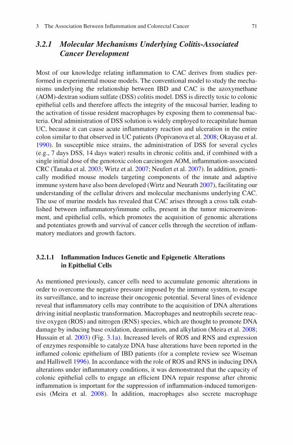
3.2.1 Molecular Mechanisms Underlying Colitis-Associated Cancer Development
Most of our knowledge relating infl ammation to CAC derives from studies per-formed in experimental mouse models. The conventional model to study the mecha-nisms underlying the relationship between IBD and CAC is the azoxymethane (AOM)-dextran sodium sulfate (DSS) colitis model. DSS is directly toxic to colonic epithelial cells and therefore affects the integrity of the mucosal barrier, leading to the activation of tissue resident macrophages by exposing them to commensal bac-teria. Oral administration of DSS solution is widely employed to recapitulate human UC, because it can cause acute infl ammatory reaction and ulceration in the entire colon similar to that observed in UC patients (Popivanova et al. 2008 ; Okayasu et al. 1990 ). In susceptible mice strains, the administration of DSS for several cycles (e.g., 7 days DSS, 14 days water) results in chronic colitis and, if combined with a single initial dose of the genotoxic colon carcinogen AOM, infl ammation- associated CRC (Tanaka et al. 2003 ; Wirtz et al. 2007 ; Neufert et al. 2007 ). In addition, geneti-cally modifi ed mouse models targeting components of the innate and adaptive immune system have also been developed (Wirtz and Neurath 2007 ), facilitating our understanding of the cellular drivers and molecular mechanisms underlying CAC. The use of murine models has revealed that CAC arises through a cross talk estab-lished between infl ammatory/immune cells, present in the tumor microenviron-ment, and epithelial cells, which promotes the acquisition of genomic alterations and potentiates growth and survival of cancer cells through the secretion of infl am-matory mediators and growth factors.
3.2.1.1 Infl ammation Induces Genetic and Epigenetic Alterations in Epithelial Cells
As mentioned previously, cancer cells need to accumulate genomic alterations in order to overcome the negative pressure imposed by the immune system, to escape its surveillance, and to increase their oncogenic potential. Several lines of evidence reveal that infl ammatory cells may contribute to the acquisition of DNA alterations driving initial neoplastic transformation. Macrophages and neutrophils secrete reac-tive oxygen (ROS) and nitrogen (RNS) species, which are thought to promote DNA damage by inducing base oxidation, deamination, and alkylation (Meira et al. 2008 ; Hussain et al. 2003 ) (Fig. 3.1a ). Increased levels of ROS and RNS and expression of enzymes responsible to catalyze DNA base alterations have been reported in the infl amed colonic epithelium of IBD patients (for a complete review see Wiseman and Halliwell 1996 ). In accordance with the role of ROS and RNS in inducing DNA alterations under infl ammatory conditions, it was demonstrated that the capacity of colonic epithelial cells to engage an effi cient DNA repair response after chronic infl ammation is important for the suppression of infl ammation-induced tumorigen-esis (Meira et al. 2008 ). In addition, macrophages also secrete macrophage
3 The Association Between Infl ammation and Colorectal Cancer

72
inhibitory factor (MIF), which was shown to suppress the transcription of the tumor suppressor TP53 gene, inhibiting the DNA-damage response and, perhaps, leading to an increase in genomic instability (Fingerle-Rowson et al. 2003 ).
In addition to genetic alterations, infl ammation is also associated with the pres-ence of a methylator phenotype, leading to epigenetic silencing of genes that are important for preventing tumor development (McCabe et al. 2009 ; Issa et al. 2001 ). In vivo modeling of CAC also suggests that infl ammation-induced DNA methyla-tion occurs in the early phases of the disease, affecting noncancerous mucosa (Hartnett and Egan 2012 ). In fact, 60 % of genes hypermethylated in CRCs are reported to exhibit aberrant methylation in infl amed noncancerous tissues, suggest-ing that infl ammation creates a signature of aberrant DNA methylation similar to what is found in cancers (Hahn et al. 2008 ). Some authors speculate that in early phases of tumor development, methylation is important to silence tumor specifi c antigens, therefore, keeping tumor cells hidden from the immune system in an equi-librium phase (Hosking 2012 ).
The mechanisms by which infl ammation promotes changes in DNA methylation are not fully characterized. Methylation has been reported to occur after DNA dam-age induced by ROS, as halogenated pyrimidines, one form of ROS-induced dam-age, mimic 5-methylcytosine and stimulate DNA methyltransferase 1 (DNMT1)-mediated CpG methylation (McCabe et al. 2009 ; O’Hagan et al. 2011 ;
PROGRESSION
STAT3
VEGF
TNF-a
IL-21IL-23
IL-6
IL-10IL-17
INITIATION
ROS
RNS
PROMOTION
STAT3NFKB
CCLs
TNF-a
IL-21IL-23
IL-6
IL-10IL-17
a b c
Fig. 3.1 Schematic representation of the molecular and cellular contributors in colitis-associated cancer development. ( a ) Infl ammatory cells secrete ROS and RNS that induce genetic and epigen-etic alterations in epithelial cells, initiating neoplastic transformation. ( b ) Tumor cell outgrowth is promoted by a complex interaction between immune cells that secrete pro- and anti-infl ammatory mediators that will promote proliferation and survival of tumor cells by activating STAT3- and NFkB-dependent signaling. Tumor cells secrete chemokines (CCLs) that will recruit more infl am-matory cells thus contributing to sustain an infl ammatory reaction ( c ) Secretion of VEGF may contribute to tumor progression by inducing angiogenesis and enabling migration of both tumor and infl ammatory cells
M.J. Oliveira and S. Velho

73
Katsurano et al. 2012 ). In addition, in vitro studies using CRC cell lines and the analyses of CAC from mouse models of colitis have shown that the expression of infl ammatory cytokines, such as IL-6, IL-1β, tumor necrosis factor alpha (TNF-α), and interferon-γ, is associated with increased expression and stability of enzymes involved in DNA methylation, such as DNMT1 and DNMT3b (Hartnett and Egan 2012 ; Katsurano et al. 2012 ; Foran et al. 2010 ; Kominsky et al. 2011 ). Increased expression of DNMT1 was also observed in human CAC (Foran et al. 2010 ). Furthermore, over-expression of enzymes central to all cellular methylation mecha-nisms, S-adenosylmethionine synthetase and S-adenosylhomocysteine hydrolase, was found to occur in a DSS mouse model (Kominsky et al. 2011 ). Recent data using in vitro cell culture and a mouse model of colitis also showed that oxidative damage and infl ammation increase the recruitment of silencing complexes contain-ing DNMTs to the promoter CpG islands of genes, some of them previously shown to undergo infl ammation and tumor-specifi c DNA methylation in models of intesti-nal infl ammation and human cancers (O’Hagan et al. 2011 ).
3.2.1.2 Infl ammation Contributes to CAC Promotion and Progression
The pro-tumorigenic properties of the tumor-associated infl ammatory reaction are also linked to the capacity of infl ammation-related factors to stimulate proliferation, survival, angiogenesis, and migration of epithelial cells, allowing the expansion of tumor initiating cells (Grivennikov et al. 2009 ). The effect exerted by some of these factors on the modulation of cancer cell-related activities will be discussed in detail in this section.
NFκB
In the DSS mouse model of chronic colitis, NFκB signaling is induced in activated macrophages, stimulating the production and secretion of pro-infl ammatory cyto-kines by these cells. Secreted cytokines then activate NFκB signaling in intestinal epithelial cells (IECs), promoting the expression of survival molecules (Karin and Greten 2005 ) (Fig. 3.1b ). In accordance with these observations, enterocyte-specifi c ablation of IKK-β, an activator of NFκB, was shown to decrease tumor incidence drastically in response to AOM-DSS treatment, without affecting the size and com-position of tumors or the induction of oncogenic mutations. This fi nding indicates that the IKK-β-dependent NFκB-activated pathway operates during early tumor promotion. In addition, deletion of IKK-β in enterocytes enhanced the loss of intes-tinal barrier function induced by DSS and caused more infl ammation, suggesting that the tumor-promoting function of NFκB in enterocytes is associated with its ability to suppress apoptosis of pre-neoplastic progenitors (Greten et al. 2004 ; Karin and Greten 2005 ). On the other hand, deletion of IKK-β in myeloid cells (dendritic cells and macrophages) resulted in a signifi cant decrease in tumor number and size, but without affecting apoptosis of epithelial cells. The difference between
3 The Association Between Infl ammation and Colorectal Cancer

74
enterocyte and myeloid-specifi c ablation of NFκB was mainly associated with a decrease in the expression of pro-infl ammatory cytokines by myeloid cells that may serve as tumor growth factors (Greten et al. 2004 ).
IL-6/STAT3
Several studies using mouse models of chronic colitis have highlighted the role of the pro-infl ammatory cytokine interleukin (IL)-6 in the development of CAC. It has been suggested that IL-6 contributes to increase tumor burden and multiplicity in the early stages of CAC development, as well to maintain tumor growth at late stages of the disease by stimulating proliferation and survival of neoplastic IECs. Additionally, IL-6 also helps to perpetuate infl ammation by infl uencing the continuous recruit-ment and activation of infl ammatory cells to sites of infl ammation (Grivennikov et al. 2009 ; Becker et al. 2004 ; Bollrath et al. 2009 ). Myeloid cells and T lympho-cytes present in the lamina propria or infi ltrating the tumor tissue were described as the main sources of IL-6 secretion to the microenvironment during CAC develop-ment (Grivennikov et al. 2009 ; Becker et al. 2004 , 2005 ; Matsumoto et al. 2010 ) (Fig. 3.1b ). Grivennikov and colleagues (Grivennikov et al. 2009 ) reported that IL-6 expression by myeloid cells is driven by an NFκB-dependent mechanism, whereas, expression of IL-6 by T-lymphocytes was reported to occur during CAC progression and seems to be controlled by TGF-β signaling (Becker et al. 2004 ). Upon secretion into the microenvironment, IL-6 stimulates proliferation and survival of IECs through the activation of STAT3 (Greten et al. 2004 ; Grivennikov et al. 2009 ; Becker et al. 2004 ; Bollrath et al. 2009 ) (Fig. 3.1b ). The activation of STAT3 was shown to occur downstream IL-6 binding to its receptors (gp130 and IL-6 receptor) expressed in the surface of epithelial cells or due to IL-6 trans-signaling induced by macro-phage-derived IL-6/soluble IL-6R (Matsumoto et al. 2010 ; Grivennikov and Karin 2011 ). Consistent with the role of IL-6 in CAC development, a reduction in tumor number and in tumor size was observed in IL-6 null mice treated with DSS, along with inhibition of progression from adenoma to carcinoma. These data show that IL-6 signaling plays an important role during early stages of CAC (Grivennikov et al. 2009 ). In accordance with the role of IL-6/STAT3 pathway activation, deletion of STAT3 in IECs reduces cell proliferation, increases apoptosis and colitis, and reduces the number and size of tumors. These data demonstrate a critical role for epithelial STAT3 activation in infl ammation-induced tumor formation and growth (Grivennikov et al. 2009 ; Becker et al. 2004 ; Bollrath et al. 2009 ). Curiously, the ablation of STAT3 in IECs had a stronger effect than ablation of IL-6, suggesting that other factors may contribute to induce STAT3 activation. In this vein, other cytokines, such as IL-11, IL-22 and IL-23, were also reported to induce STAT3 acti-vation in IECs in mouse models of colitis (Grivennikov et al. 2009 ; Bollrath et al. 2009 ; Sugimoto et al. 2008 ; Pickert et al. 2009 ; Grivennikov and Karin 2010 ).
In vivo studies also showed that activation of STAT3 in T cells plays a pathogenic role in chronic colitis by inducing prolonged survival of pro-infl ammatory T cells and disruption of immune tolerance (Sugimoto 2008 ; Atreya et al. 2000 ; Takeda
M.J. Oliveira and S. Velho

75
et al. 1998 ). In addition, IL-6 was shown to induce polarization of T lymphocytes towards more pro-tumorigenic subtypes, such as T helper 17 (Th17), while inhibit-ing the differentiation of the suppressor T regulatory cells (Tregs), in this way con-tributing to enhancement of the infl ammatory reaction (Grivennikov and Karin 2011 ; Dominitzki et al. 2007 ). Mice lacking STAT3 specifi cally in macrophages and neutrophils showed abnormal activation of these cells and impaired expression of IL-10 signaling. These mice developed chronic enterocolitis and showed enhanced T cell polarization towards Th1 cell activity, as is the case in IL-10-defi cient mice (Kuhn et al. 1993 ; Berg et al. 1996 ; Takeda et al. 1999 ). These fi ndings demonstrate that STAT3 activation in myeloid cells is essential for anti- infl ammatory reactions mediated by IL-10 (Takeda et al. 1999 ). Latter studies confi rmed that tumor-associ-ated macrophages (TAMs) released IL-6, which in turn induced STAT3-mediated IL10 production in tumor cells, favoring immunesuppression and tumor progression (Herbeuval et al. 2004 ). Altogether, these results point to a role for IL-6 in promot-ing a strong infl ammatory response, contributing to the continuous release of pro-tumorigenic factors through T-cells recruitment/activation, which can be coun-ter-balanced by the induction of an immune- suppressive phenotype in myeloid cells, sustaining a pro-oncogenic microenvironment.
Consistent with the in vitro and in vivo studies aforementioned, higher levels of IL-6, an increase in active STAT3, and lower levels of SOCS3 (a negative regulator of STAT3 activation), were reported in the blood and infl amed mucosa, as well as in dysplasias and cancers, of IBD patients in comparison to patients with inactive UC and controls (Li et al. 2010 ), demonstrating the importance of this signaling path-way in the human context. In addition, constitutive activation of STAT3 was reported in immune cells (mainly macrophages and T lymphocytes) present in actively infl amed areas of both CD and UC patients (Wick et al. 2012 ; Lovato et al. 2003 ; Musso et al. 2005 ).
TNF-α
TNF-α is another NFκB-regulated pro-infl ammatory cytokine playing a central role in the initiation and progression of CAC (Bollrath and Greten 2009 ). Similar to IL-6, TNF-α is also involved in the control of infl ammatory cells recruitment and known to induce survival of epithelial cells (Fig. 3.1b ). In addition, TNF-α and IL-6 were reported to cross-regulate each other, contributing to the enhancement of chronic infl ammation and intestinal tumorigenesis (Grivennikov et al. 2009 ).
TNF-α expression levels were shown to increase after AOM/DSS treatment and this was associated with an increase in the number of infi ltrating leukocytes express-ing its major receptor (TNFR1), in the lamina propria and submucosal regions of the colon. During CAC development TNF-α is mostly produced by macrophages and T lymphocytes (Fig. 3.1b ), although epithelial cells might also express this cytokine. The tumor-promoting properties of TNF-α are probably linked to its role as an inducer of NFκB signaling in epithelial cells, therefore stimulating survival (Grivennikov and Karin 2011 ). In accordance with this view, two studies have
3 The Association Between Infl ammation and Colorectal Cancer

76
addressed the role of TNF-α in activating NFκB in epithelial cells. In one study, an increase in the NFκB signaling and simultaneous up-regulation of TNFR2 were observed during the progression from normal mucosa to CAC in DSS-treated mice (Onizawa et al. 2009 ). In this case, up-regulation of TNFR2 in colon cancer cells was mediated primarily by STAT3 activity upon IL-6 and TNF-α stimulation (Hamilton et al. 2011 ). Over-expression of this receptor was also reported to occur in epithelial cells from IBD patients (Mizoguchi et al. 2002 ). In the other study, NFκB activation was shown to occur downstream of the TNFR1-RAF1 signaling cascade (Edelblum et al. 2008 ). In conclusion, TNF-α stimulates NFκB activation in epithelial cells through both of its receptors.
TNF-α levels are increased in the mucosa of IBD patients (Kollias 2004 ; Roberts- Thomson et al. 2011 ) and its contribution to CAC development makes it a valuable molecule for targeted therapies. Consistent with this notion, AOM/DSS treated mice lacking TNFR1 or treated with Etanercept, a specifi c antagonist of TNF-α, showed reduced mucosal damage, reduced infi ltration of macrophages and neutro-phils, and attenuated tumor formation (Popivanova et al. 2008 ). The same inhibitory effect on tumor growth was observed when TNF-α was inhibited using monoclonal antibodies during late stages of CAC development (Grivennikov et al. 2009 ; Onizawa et al. 2009 ). In addition, TNF-α inhibition in AOM/DSS-treated mice was also shown to reduce angiogenesis, possibly due to inhibition of leucocytes recruit-ment and consequent inhibition of ciclooxigenase 2 (COX-2) expression (Popivanova et al. 2008 ; Goel et al. 2011 ). The benefi cial effects of anti-TNF-α targeted therapies using monoclonal antibodies have been extensively shown in CD and UC patients that are refractory to conventional therapies, such as aminosalicylates, corticoste-roids, or immunosuppressors (Triantafi llidis et al. 2011 ). From the observations made in experimental mouse models of colitis, TNF-α inhibition seems to be anti- tumorigenic, but the effect of TNF-α inhibitors in CAC from human patients remains to be elucidated.
IL-21
Interleukin (IL)-21, a T-cell-derived cytokine, is over-produced in IBD (Fina et al. 2008 ) and its role in CAC development has been recently demonstrated. After AOM/DSS treatment, IL-21 knockout mice showed reduced mucosal damage, reduced infi ltration of T cells, and diminished production of IL-6 and IL-17A. Absence of IL-21 reduced STAT3 activation in epithelial and stromal cells and resulted in the development of fewer and smaller tumors compared with wild-type mice (Stolfi et al. 2011 ). In addition, IL-21 induces the polarization of T cells towards Th17-mediated chronic intestinal infl ammation, characterized by high levels of IL-17A, and reduces the production of interferon-γ (IFN-γ), which exerts anti-tumor activity by enhancing the capacity of cytotoxic CD8 T cells. Thus IL-21 supports chronic infl ammation and reduces tumor immune surveillance, promoting a tumor-support-ive microenvironment in the colon (Danese et al. 2011 ; Jauch et al. 2011 ).
M.J. Oliveira and S. Velho

77
Chemokine Expression
Chemokines are important components of cancer-related infl ammation where they play a key role in orchestrating the recruitment and positioning of leukocytes (Bonecchi et al. 2011 ). NFκB activation in IECs was shown to induce the expression of several chemokines involved in the recruitment of more myeloid cells to sites of infl ammation (Bollrath and Greten 2009 ; Eckmann et al. 2008 ). In the DSS model of colitis, mice lacking CCR5 do not develop colitis (Goel et al. 2011 ; Andres et al. 2000 ). In the AOM-DSS model of infl ammation-induced colon carcinogenesis, CCR2 knockout mice exhibit less macrophage infi ltration and lower tumor num-bers, indicating that CCL2, its ligand, is a crucial mediator of the initiation and promotion of CAC and that targeting CCR2 may be useful in treating CAC (Goel et al. 2011 ; Popivanova et al. 2009 ). The use of CCL2 antagonists inhibited COX-2 expression, reduced angiogenesis, and decreased the number and size of colon tumors in mice (Popivanova et al. 2009 ).
Angiogenesis
Angiogenesis is a fundamental process underlying tumor growth and progression, since it allows the diffusion of oxygen and nutrients to tumor cells and provides a conduit through which cancer cells can metastasize (Keith and Simon 2008 ). An increase in the microvessel density has also been shown to play an important role in the pathogenesis of IBD and CAC (Danese et al. 2006 ). Vascular endothelial growth factor (VEGF), a strong pro-angiogenic factor involved in the induction of endothe-lial cell proliferation, migration, survival and permeability, was shown to be up- regulated in the infl amed mucosa of IBD (Danese et al. 2006 ; Tsiolakidou et al. 2008 ; Alkim et al. 2012 ; Scaldaferri et al. 2009 ) and in CAC patients (Fig. 3.1c ) (Waldner et al. 2010 ). Similarly, the expression of VEGF receptor 2 was found to be up-regulated in the mucosa of IBD patients, mainly localized in endothelial cells, however others have also shown it in epithelial cells from infl amed mucosa and CAC (Scaldaferri et al. 2009 ; Waldner et al. 2010 ; Frysz-Naglak et al. 2011 ). Together, the abovementioned observations highlight the importance of the micro-vasculature and of the associated angiogenic factors in colitis and CAC.
3.3 Colitis-Associated Colorectal Cancer Versus Sporadic Colorectal Cancer
CAC is an example of the extrinsic pathway of infl ammation-induced cancer in which components of the immune system are the main drivers of carcinogenesis. In sporadic colorectal cancer (SCC), infl ammation also plays a role, but these tumors are unlikely to be initiated by infl ammation because most tumor immune cells are
3 The Association Between Infl ammation and Colorectal Cancer

78
presumably recruited after the tumor is formed (to be discussed in more detail in Sects. 3.4 and 3.5 of this chapter) (Terzic et al. 2010 ). Because the etiologic factors underlying cancer development in these two subsets of malignancies are different, it is reasonable to ask if both types of cancers share the same clinical, pathologic, and molecular features, or if they are two distinct entities. In the next subsections, we will highlight the similarities and differences between these two subtypes of CRC.
3.3.1 Clinical and Pathologic Features of CAC Versus SCC
Carcinomas arising in CD and UC patients have strikingly similar clinicopathologic features, namely age at onset, tumor location, and histology, suggestive of an infl am-mation-related signature of carcinogenesis (Choi and Zelig 1994 ; Svrcek et al. 2007 ). Some studies have, however, shown that CRC arising in CD patients appears at more advanced stages (Kiran et al. 2010 ) and are associated with worse overall survival (Ouaissi et al. 2011 ) when compared with CRC from UC patients. Nevertheless, compared with sporadic colorectal carcinoma (SCRC), CRC arising in patients with IBD has several distinguishing clinical features. In general, CAC affects individuals at a younger age (Itzkowitz and Yio 2004 ) and CAC presents more often with muci-nous or signet ring cell histology. Due to the presence of widespread infl ammation, CRC arising in IBD patients tend to be macroscopically heterogeneous and poorly delimited, irregular, and frequently multifocal, suggesting a broader fi eld effect of mucosal infl ammation (Brackmann et al. 2009 ; Delaunoit et al. 2006 ). CAC is fre-quently anaplastic, broadly infi ltrating, and rapidly growing, progressing to invasive adenocarcinoma from fl at and non-polypoid dysplasia more frequently than SCRC. In accordance, it was reported that CAC patients show a poorer survival rate when compared to SCRC in the background population (Brackmann et al. 2009 ). In con-trast, other studies failed to identify a difference in the survival rates for IBD-associated and SCRC (Delaunoit et al. 2006 ; Kiran et al. 2010 ).
3.3.2 Molecular Alterations of CAC Versus SCRC
The molecular alterations accompanying pathogenesis are well described for SCRC. Regarding the molecular landscape, SCRC can be primarily divided in two major groups according to the type of genetic instability they exhibit. The majority (85 %) of SCRC are characterized by the presence of chromosomal instability (CIN), which consists of great losses and gains of chromosomal material. The remaining 15 % of the tumors are characterized by the accumulation of numerous mutations through-out the genome due to inactivation of mismatch repair (MMR) genes, creating a phenotype known as microsatellite instability (MSI) (Ionov et al. 1993 ). It is accepted that CIN and MSI SCRCs develop through different pathways of neoplas-tic transformation, which are associated with specifi c molecular alterations. CIN
M.J. Oliveira and S. Velho

79
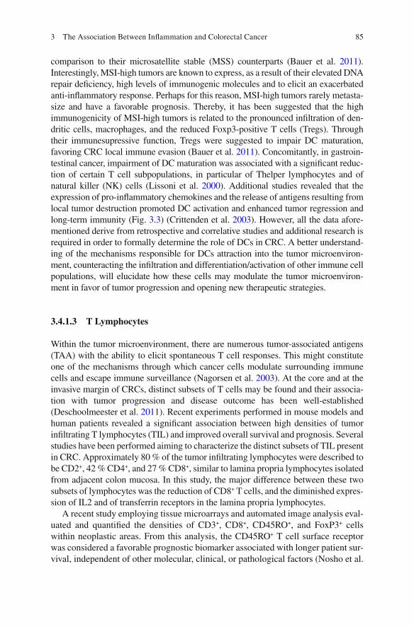
tumors follow the adenoma—carcinoma pathway described by Fearon and Vogelstein in ( 1990 ). In this pathway, inactivation of the adenomatous polyposis coli ( APC ) gene is considered to be the trigger to adenoma formation and it occurs in about 80 % of SCCs. Progression to more advanced stages and the eventual for-mation of adenocarcinoma proceeds with the acquisition of alterations in other fun-damental genes such as KRAS , deleted in CRC ( DCC ), SMAD2 , SMAD4 , and, lastly, TP53 (Fig. 3.2a ). On the other hand, a serrated neoplasia pathway, which is fre-quently associated with the early occurrence of BRAF mutations and the CpG island methylator phenotype, a surrogate marker of widespread methylation at the
Aberrantcrypt foci
Earlyadenoma
Intermediateadenoma
Lateadenoma
Carcinoma(MSS/CIN)
APC KRASDCC
SMAD2/SMAD4 p53
Adenoma to carcinoma sequence
Serrated pathway
Small hyperplastic
polyp
Large hyperplastic
polyp
Serratedadenoma
Serrated adenoma
(high grade dysplasia)
Carcinoma(MSI)
BRAFMLH-1
methylation
Inflammation-associated pathway
Colitis Low gradedysplasia
Intermediategrade
dysplasia
High gradedysplasia CAC
p53
CIMP
APCKRAS
MSI
B-RAF
Methylation
DCCSMAD2/SMAD4
a
b
c
Fig. 3.2 Pathways for colorectal cancer development. ( a ) In the adenoma-carcinoma sequence, inactivating mutations of APC gene are the onset of adenoma formation. Acquisition of further genetic alterations, namely KRAS oncogenic activation and TP53 inactivating mutations, will then function as driving forces towards progression from adenoma to carcinoma; ( b ) BRAF mutation is a marker for the serrated polyp pathway that has its origin in hyperplastic polyps and a potential end point as a MSI carcinoma. CIMP develops early in this sequence and MSI develops late due to MLH1 silencing by promoter methylation; ( c ) CAC development frequently occurs through the adenoma-carcinoma sequence although a serrated pathway has also been described. In both cases, the sequence of molecular events driving tumor development differs from the one described for non-infl ammation-associated CRCs. In CAC development, TP53 mutations are the initiating event in the adenoma-carcinoma sequence and loss of APC occurs prior to malignant transformation. In the serrated pathway of CAC development, BRAF mutations occur in a late stage of tumor devel-opment. In its turn, the role of CpG island methylation is still controversial. It seems to be impor-tant only for the progression of pre-malignant lesions and not so relevant in an established cancer
3 The Association Between Infl ammation and Colorectal Cancer

80
promoter regions of cancer-associated genes, is mainly associated with the develop-ment of sporadic MSI tumors. In this pathway, loss of function of MLH1 due to promoter hypermethylation occurs at late stages and results in the progression to MSI adenocarcinoma (Jass 2005 ; O’Brien et al. 2006 ; Velho et al. 2008 ) (Fig. 3.2b ).
CRC developing in the context of IBD shares many of the molecular alterations found in both CIN and MSI SCRCs, although the type of precursor lesion and the sequence of molecular events leading to neoplastic transformation differ from the one described for SCRC (Xie and Itzkowitz 2008 ). Unlike SCRC that arises from well-defi ned adenomas, CAC may also arise from fl at dysplasia areas through a sequence of chronic infl ammation, injury, dysplasia and carcinoma (Terzic et al. 2010 ) (Fig. 3.2c ). In contrast to SCRC, APC inactivation in CAC is not so frequent (14–33 %) and occurs in the late stages of the infl ammation-associated dysplasia—carcinoma pathway (Umetani et al. 1999 ; Sepulveda and Aisner 2010 ). Conversely, TP53 mutations, which are often found in late stages of the adenoma—carcinoma sequence, occur early in the development of CAC and are often found in non- dysplastic mucosa (Xie and Itzkowitz 2008 ; Kraus and Arber 2009 ). TP53 mutations were found in about 19 % of biopsies from IBD patients without dysplasia and the frequency increased with progression to higher grades of dysplasia (Sepulveda and Aisner 2010 ). In CAC, TP53 mutations were described to occur in frequencies above 50 % (Harpaz and Polydorides 2010 ; Aust et al. 2005 ; Sanchez et al. 2011 ). Other alterations known to play a role in the adenoma-carcinoma sequence, such as loss of DCC , SMAD2 and SMAD4 , were also found to occur at early phases of CAC devel-opment (Harpaz and Polydorides 2010 ). Another important gene in the development and progression of SCRC is KRAS . Mutations in this gene also play a role in the development of CAC and are found in high-grade dysplasia (Umetani et al. 1999 ). KRAS mutations, which are very frequent in SCRC (approximately 35 %) (Lau and Haigis 2009 ; Oliveira et al. 2004 ), occur in only about 20 % of IBD- related cancers (Umetani et al. 1999 ; Aust et al. 2005 ; Lyda et al. 2000 ; Holzmann et al. 1998 ).
Alterations similar to the mutator pathway were also described to occur during the pathogenesis of CAC, and MSI is also considered one of the mechanisms accounting for neoplastic progression in IBD patients (Umetani et al. 1999 ) (Fig. 3.2c ). MSI was early found in infl amed mucosa of IBD patients without signs of dysplasia (Tahara et al. 2005 ). In addition, a high incidence of MSI was described in UC patients with long-standing severe infl ammation, probably refl ecting genomic instability caused by repeated infl ammatory stress (Ishitsuka et al. 2001 ). The majority of the studies in which the frequency of MSI was analyzed reported that approximately 15 % of CAC display the mutator phenotype (Svrcek et al. 2007 ; Umetani et al. 1999 ; Schulmann et al. 2005 ), although others described higher fre-quencies (Tahara et al. 2005 ). Compared with sporadic MSI CRCs, MSI CAC patients presented with a younger age at diagnosis, and there was neither female nor right-sided predominance as it is characteristic of MSI SCC (Svrcek et al. 2007 ; Schulmann et al. 2005 ). Furthermore, there is some disagreement about the MMR defects underlying MSI in CAC. Methylation of MLH1 promoter region, which is the foremost mechanism causing MSI in SCRC, was described to occur in IBD- related neoplasias (Aust et al. 2005 ; Schulmann et al. 2005 ; Fleisher et al. 2000 ), although most of these studies addressed methylation in a region of the promoter
M.J. Oliveira and S. Velho

81
that is not related with silencing of the gene (Svrcek et al. 2007 ). Indeed, only a small proximal region of the MLH1 promoter (C region) has been demonstrated to harbor a methylation status that correlates invariably with the loss of gene expres-sion (Svrcek et al. 2007 ; Deng et al. 1999 ; Capel et al. 2007 ). When analyzing a large series of IBD-related neoplasias, Svrcek and colleagues (Kiran et al. 2010 ) found that, unlike sporadic MSI CRCs, methylation at the C-region of MLH1 pro-moter occurs in a low frequency and, instead, MSI IBD neoplasias presented hetero-geneous MMR defects involving MLH1 , MSH2 , MSH6 , or PMS2 genes.
Other molecular markers of the MSI pathway, such as BRAF mutations and CIMP, were also described to occur during CAC development. BRAF V600E hotspot mutations were described in 33.3 % of MSI IBD-associated neoplasias, a frequency comparable to the one found in MSI SCRC (Kiran et al. 2010 ), although, they are not considered an initiating event in CAC development (Aust et al. 2005 ) (Fig. 3.2c ). The presence of CIMP is also a common feature of SCRC, in particular in MSI tumors, but in CAC its role is still controversial. Due to the aforementioned effect of infl ammation in the induction of DNA methylation, it would be expected that CAC exhibited similar, if not higher levels, of DNA methylation than SCC. Instead, lower levels of CIMP and lower levels of methylation of age-associated genes have been reported to occur in CAC in comparison to SCRC (Sanchez et al. 2011 ; Konishi et al. 2007 ; Olaru et al. 2012 ). As described previously, the presence of methylation is a common feature of colitis-associated dysplastic lesions, how-ever, the methylator phenotype does not seem to play a major role during CAC progression. In order to explain this discrepancy, some authors proposed that the presence of colitis-associated methylation creates a fi eld defect resulting in prema-ture aging of epithelial cells, therefore, increasing the risk of malignancy. Because, in CAC, methylation seems to play a minor role, genetic alterations are thought to be the main drivers of immune escape, leading to a more aggressive clinical course than epigenetic changes (Issa et al. 2001 ; Konishi et al. 2007 ).
3.4 The Role of Infl ammation in Other Forms of Colorectal Cancer
CRC can be classifi ed as: (1) inherited, including non-polyposis colorectal cancer (HNPCC) and familial adenomatous polyposis coli (FAP); (2) SCRC; and (3) infl am-mation-driven CRC (CAC) (Xie and Itzkowitz 2008 ). Although most CRCs are unlikely to be initiated by infl ammation, they recruit and activate distinct immune cells, creating an infl ammatory microenvironment. The cytokines and chemokines released by immune cells may then promote a pro-infl ammatory response, counter-acting tumor growth and survival, or an anti-infl ammatory response, sustaining tumor cell activities (Terzic et al. 2010 ; Mantovani et al. 2004 ) (Fig. 3.3 ). Early studies profi ling immune population distribution within CRC indicated that, at peritumoral regions, the infl ammatory infi ltrate consisted of 47 % lymphocytes, 19 % plasma cells, 15 % macrophages/monocytes, 5 % granulated mast cells, and 15 % polymor-phonucleated (PMN) cells. Necrotic areas of the tumors were abundant in PMN and
3 The Association Between Infl ammation and Colorectal Cancer

82
macrophages (Svennevig et al. 1982 ). The role of innate immune cells, such as macrophages, natural killer (NK) and dendritic (DC) cells or of adaptive immune cells, such as T and B lymphocytes, in CRC cell activities will be overviewed.
3.4.1 Tumor Immune and Infl ammatory Cell Infi ltration
3.4.1.1 Macrophages
In the majority of the tumors, macrophages are a major component of the host leu-kocytic infi ltrate (Condeelis and Pollard 2006 ). In the 1970s, studies aiming to understand the role of monocytes/macrophages on tumor progression suggested
Motility
AngiogenesisMetastasis
Proliferation
Immune escapeInvasion
M2/TAM
Treg
MDSC
B cell NK
M1CD4+ T cell
CD8+
T cellDC
Cytotoxicity Apoptosis
Immune surveillance
B cell
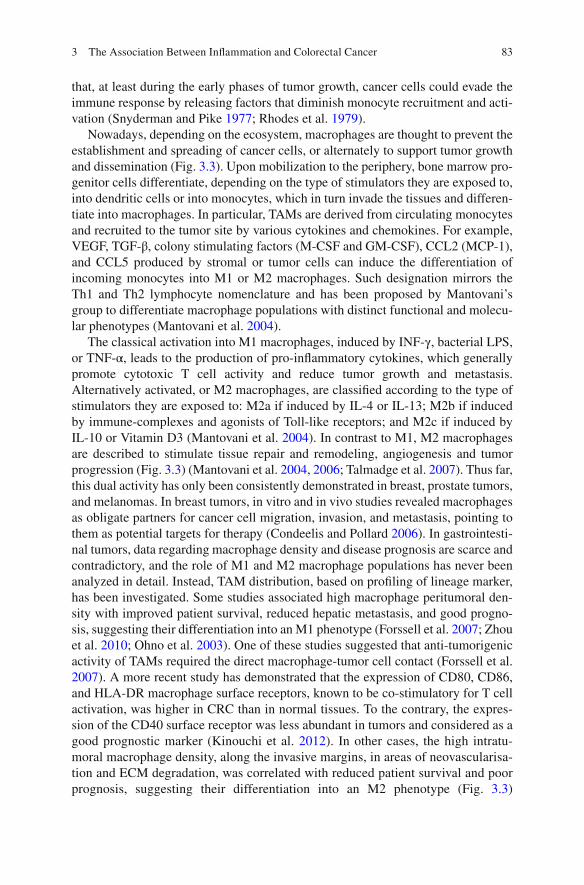
Fig. 3.3 Fine-tune regulation of the immune response in sporadic colorectal cancer. A strong infl ammatory component is often present in the stroma of CRCs that do not develop in response to a pre-existent chronic infl ammatory condition. The stroma of these cancers may be populated by pro-tumorigenic immune cells (M2/TAM, B cell, Treg, MDSC) that promote immune evasion, proliferation, motility, invasion, and metastization, or by anti-tumorigenic immune cells (Dc, CD4 + T cell, CD8 + T cell, B cell, NK e M1/TAM) that stimulate immune surveillance, cytotoxicity, and apoptosis. Infl ammatory modulators (antigens, cytokines, and chemokines) produced by tumors cells shift the balance towards a more pro-tumorigenic or a more anti-tumorigenic immune response
M.J. Oliveira and S. Velho
83
that, at least during the early phases of tumor growth, cancer cells could evade the immune response by releasing factors that diminish monocyte recruitment and acti-vation (Snyderman and Pike 1977 ; Rhodes et al. 1979 ).
Nowadays, depending on the ecosystem, macrophages are thought to prevent the establishment and spreading of cancer cells, or alternately to support tumor growth and dissemination (Fig. 3.3 ). Upon mobilization to the periphery, bone marrow pro-genitor cells differentiate, depending on the type of stimulators they are exposed to, into dendritic cells or into monocytes, which in turn invade the tissues and differen-tiate into macrophages. In particular, TAMs are derived from circulating monocytes and recruited to the tumor site by various cytokines and chemokines. For example, VEGF, TGF-β, colony stimulating factors (M-CSF and GM-CSF), CCL2 (MCP-1), and CCL5 produced by stromal or tumor cells can induce the differentiation of incoming monocytes into M1 or M2 macrophages. Such designation mirrors the Th1 and Th2 lymphocyte nomenclature and has been proposed by Mantovani’s group to differentiate macrophage populations with distinct functional and molecu-lar phenotypes (Mantovani et al. 2004 ).
The classical activation into M1 macrophages, induced by INF-γ, bacterial LPS, or TNF-α, leads to the production of pro-infl ammatory cytokines, which generally promote cytotoxic T cell activity and reduce tumor growth and metastasis. Alternatively activated, or M2 macrophages, are classifi ed according to the type of stimulators they are exposed to: M2a if induced by IL-4 or IL-13; M2b if induced by immune-complexes and agonists of Toll-like receptors; and M2c if induced by IL-10 or Vitamin D3 (Mantovani et al. 2004 ). In contrast to M1, M2 macrophages are described to stimulate tissue repair and remodeling, angiogenesis and tumor progression (Fig. 3.3 ) (Mantovani et al. 2004 , 2006 ; Talmadge et al. 2007 ). Thus far, this dual activity has only been consistently demonstrated in breast, prostate tumors, and melanomas. In breast tumors, in vitro and in vivo studies revealed macrophages as obligate partners for cancer cell migration, invasion, and metastasis, pointing to them as potential targets for therapy (Condeelis and Pollard 2006 ). In gastrointesti-nal tumors, data regarding macrophage density and disease prognosis are scarce and contradictory, and the role of M1 and M2 macrophage populations has never been analyzed in detail. Instead, TAM distribution, based on profi ling of lineage marker, has been investigated. Some studies associated high macrophage peritumoral den-sity with improved patient survival, reduced hepatic metastasis, and good progno-sis, suggesting their differentiation into an M1 phenotype (Forssell et al. 2007 ; Zhou et al. 2010 ; Ohno et al. 2003 ). One of these studies suggested that anti-tumorigenic activity of TAMs required the direct macrophage-tumor cell contact (Forssell et al. 2007 ). A more recent study has demonstrated that the expression of CD80, CD86, and HLA-DR macrophage surface receptors, known to be co-stimulatory for T cell activation, was higher in CRC than in normal tissues. To the contrary, the expres-sion of the CD40 surface receptor was less abundant in tumors and considered as a good prognostic marker (Kinouchi et al. 2012 ). In other cases, the high intratu-moral macrophage density, along the invasive margins, in areas of neovascularisa-tion and ECM degradation, was correlated with reduced patient survival and poor prognosis, suggesting their differentiation into an M2 phenotype (Fig. 3.3 )
3 The Association Between Infl ammation and Colorectal Cancer

84
(Oosterling et al. 2005 ; Ishigami et al. 2003 ; Bailey et al. 2007 ). Such contradiction may refl ect differences in number, grade, stage, and tumor size, but also in the meth-ods used to assess macrophage distribution and infi ltration. Another hypothesis, not confi rmed by any of these studies, is that the identifi ed macrophage populations are not necessarily the same. In fact, peritumoral macrophages might be less exposed to tumor-derived cytokines and tumor modulation and, thereby, differentiate into anti- tumor macrophages, producing cytotoxic molecules (such as ROS, NO and TNF-α) and exerting anti-infl ammatory activities. It is possible, however, that when tumors progress by escaping immune surveillance, the tumor microenvironment gets hypoxic, enriched in tumor-derived metabolic products or cytokines, leading to the differentiation of incoming monocytes into pro-tumor macrophages (Mantovani et al. 2004 ; Erreni et al. 2011 ).
In CRC, TAMs are described to exert anti-tumoral or pro-tumoral activities (Fig. 3.3 ). Anti-tumoral activities may occur directly, by inducing tumor cytotoxic-ity, or indirectly, by modulating the host immune response, and have been suggested to be related with the presence of M1-polarized macrophages (Mantovani et al. 2004 ; Erreni et al. 2011 ). TAMs are described to induce tumor cell apoptosis via FAS-ligand mediated pathways, reducing tumor size and metastasis (Sugita et al. 2002 ). Macrophage production of ROS and NOS, upon T lymphocytes or natural killer (NK) cells stimulation, may also lead to tumor cell death. Tumor cell release of macrophage migration inhibitory factor (MIF) or granulocyte/macrophage- colony stimulating factors (GM-CSF or M-CSF) affect macrophage differentiation, survival, proliferation, migration, and metabolism, promoting phagocytosis, tumor cell lysis and the release of pro-infl ammatory cytokines (Pozzi and Weiser 1992 ; Shinohara et al. 2000 ).
Upon modulation by the tumor microenvironment, TAMs may share many func-tional characteristics with M2 macrophages, suppressing the infl ammatory response and inducing angiogenesis, tissue remodeling, tumor invasion and metastasis (Fig. 3.3 ) (Condeelis and Pollard 2006 ; Mantovani et al. 2006 ; Ruffell et al. 2012 ; Sica et al. 2006 ). Despite these evidences, their role in cancer progression is still controversial.
3.4.1.2 Dendritic Cells
Dendritic cells play (DCs) an important role in the infl ammatory process. They are unique antigen-presenting cells (APCs), able to induce primary immune responses, but also capable of promoting immunological tolerance and regulation of T cell- mediated immune responses. Several studies have been performed to evaluate the degree and subsets of DC infi ltration in CRC. DCs are more common in normal colon mucosa than in the tumor microenvironment, and nearly absent at metastatic tumors (Schwaab et al. 2001 ). In another study, lower levels of DCs infi ltration in the tumor stroma and of CD83 + DCs at tumor invasive margins were associated with high fre-quency of distant metastasis and with reduced patient survival (Gulubova et al. 2012 ). In CRC, mature DC infi ltration seems to be enhanced in MSI-high tumors in
M.J. Oliveira and S. Velho
85
comparison to their microsatellite stable (MSS) counterparts (Bauer et al. 2011 ). Interestingly, MSI-high tumors are known to express, as a result of their elevated DNA repair defi ciency, high levels of immunogenic molecules and to elicit an exacerbated anti-infl ammatory response. Perhaps for this reason, MSI-high tumors rarely metasta-size and have a favorable prognosis. Thereby, it has been suggested that the high immunogenicity of MSI-high tumors is related to the pronounced infi ltration of den-dritic cells, macrophages, and the reduced Foxp3-positive T cells (Tregs). Through their immunesupressive function, Tregs were suggested to impair DC maturation, favoring CRC local immune evasion (Bauer et al. 2011 ). Concomitantly, in gastroin-testinal cancer, impairment of DC maturation was associated with a signifi cant reduc-tion of certain T cell subpopulations, in particular of Thelper lymphocytes and of natural killer (NK) cells (Lissoni et al. 2000 ). Additional studies revealed that the expression of pro-infl ammatory chemokines and the release of antigens resulting from local tumor destruction promoted DC activation and enhanced tumor regression and long-term immunity (Fig. 3.3 ) (Crittenden et al. 2003 ). However, all the data afore-mentioned derive from retrospective and correlative studies and additional research is required in order to formally determine the role of DCs in CRC. A better understand-ing of the mechanisms responsible for DCs attraction into the tumor microenviron-ment, counteracting the infi ltration and differentiation/activation of other immune cell populations, will elucidate how these cells may modulate the tumor microenviron-ment in favor of tumor progression and opening new therapeutic strategies.
3.4.1.3 T Lymphocytes
Within the tumor microenvironment, there are numerous tumor-associated antigens (TAA) with the ability to elicit spontaneous T cell responses. This might constitute one of the mechanisms through which cancer cells modulate surrounding immune cells and escape immune surveillance (Nagorsen et al. 2003 ). At the core and at the invasive margin of CRCs, distinct subsets of T cells may be found and their associa-tion with tumor progression and disease outcome has been well-established (Deschoolmeester et al. 2011 ). Recent experiments performed in mouse models and human patients revealed a signifi cant association between high densities of tumor infi ltrating T lymphocytes (TIL) and improved overall survival and prognosis. Several studies have been performed aiming to characterize the distinct subsets of TIL present in CRC. Approximately 80 % of the tumor infi ltrating lymphocytes were described to be CD2 + , 42 % CD4 + , and 27 % CD8 + , similar to lamina propria lymphocytes isolated from adjacent colon mucosa. In this study, the major difference between these two subsets of lymphocytes was the reduction of CD8 + T cells, and the diminished expres-sion of IL2 and of transferrin receptors in the lamina propria lymphocytes.
A recent study employing tissue microarrays and automated image analysis eval-uated and quantifi ed the densities of CD3 + , CD8 + , CD45RO + , and FoxP3 + cells within neoplastic areas. From this analysis, the CD45RO + T cell surface receptor was considered a favorable prognostic biomarker associated with longer patient sur-vival, independent of other molecular, clinical, or pathological factors (Nosho et al.
3 The Association Between Infl ammation and Colorectal Cancer

86
2010 ). Additional studies revealed that the combined analysis of CD8 + and CD45RO + cells in specifi c tumor areas could be a marker to predict tumor recur-rence and survival in patients with early stage CRC (Pages et al. 2009 ).
Interestingly, the absence of early signs of metastatic invasion, such as lympho-vascular emboli, was correlated with a signifi cant increase of the density of memory T cells in situ. Pioneer studies performed by Galon and collaborators demonstrated that an increase in intra-tumoral expression of markers for cytotoxic effector T cells was associated with absence of early metastasis and a decrease in tumor recurrence (Galon et al. 2006 ). In general, the proportion of tumors with high density of CD4 + and CD8 + memory T cells diminishes with local tumor invasion and metastasis (Halama et al. 2011 ). Consistently, the proportion of primary tumors with high infi l-trates of CD4 + and CD8 + memory T cells, particularly in the center of the tumor, was found to be lower in patients with recurrent tumors. The distribution of these T cell subsets has been mainly described at the invasive margins of colorectal tumor liver metastases (Halama et al. 2011 ). One of the factors that may explain this anti- tumor effect is the expression by CD4 + T cells of IFN-γ, a pro-infl ammatory cyto-kine. Indeed, decreased distribution of CD4 + T lymphocytes in the center of CRCs was associated with reduced IFN-γ expression and with the presence of distant metastasis and of advanced clinical stage (Fig. 3.3 ) (Numata et al. 1991 ).
T cell function within tumors can also be regulated by TAMs. As high producers of TGF-β, cancer cells and TAMs may induce the differentiation of naïve CD4 + T cells into regulator T cells (Treg), which in turn may suppress the anti-tumor activi-ties of the cytotoxic CD8 + T cells (Izcue et al. 2009 ). Tregs play a crucial role in homeostasis, preventing autoimmune disorders, by regulating the activity of autore-active T cells and inducing immune tolerance towards self-antigens, as the ones produced by cancer cells. While Tregs are associated with poor prognosis in ovar-ian, breast and gastric carcinomas, their role in CRC outcome is still contradictory (Deschoolmeester et al. 2011 ), although multiple groups have demonstrated that the high density of FoxP3 + Tregs in CRC was associated with improved survival (Ladoire et al. 2011 ; Salama et al. 2009 ). These fi ndings suggest that, in CRC, tumor-infi ltrating Tregs should be considered as potential allies in the anti-tumor response and therefore not targets for therapy (Fig. 3.3 ).
3.4.1.4 Natural Killer Cells
In CRC, the high incidence of cytotoxic T cells and of natural killer (NK) cells has been associated with enhanced cancer cell death and improved prognosis (Fig. 3.3 ) (Deschoolmeester et al. 2011 ). In a syngeneic rat model of CRC with liver and lung metastasis, NK cells were selectively recruited to the tumors and predominantly towards the stroma surrounding tumor cell nodules. Elimination of cancer cells was then initiated directly by NK cells or by their activation of other immune effectors (Kuppen et al. 2001 ). Consistent with these cytotoxic functions, it has been reported that decreased numbers of NK cells in pre-operative CRC cell patients was associ-ated with an increased frequency of tumor recurrence (Atreya and Neurath 2008 ).
M.J. Oliveira and S. Velho
87
3.4.1.5 B Lymphocytes
Infi ltrating B cells are the dominant component of infl ammation in some cancers, such as ductal carcinoma in situ and invasive breast carcinomas. They express somatic hypermutated antibodies and recognize tumor-associated antigens, such as ganglioside D3. In CRC, B cells were mostly found at the invasive margin of grow-ing tumors and in tertiary lymphoid structures (Dieu-Nosjean et al. 2008 ), sites of intense immune activity adjacent to tumor nests where proliferating B and mature DCs are in close contact with T cells. The role and the biological impact of intratu-moral B cells in CRC are not yet clarifi ed, however. It is possible that B lymphocytes act as antigen-presenting cells and, therefore, may be important for inducing CD4 + and CD8 + memory T cells, counteracting tumor invasion and metastasis (Fig. 3.3 ) (Deschoolmeester et al. 2011 ). It is also possible, that B cells promote tumor metas-tasis by converting resting CD4 + T cells into immune suppressive Treg cells or by activating monocytes into M2 pro-infl ammatory macrophages (Fig. 3.3 ).
Recent mouse models of spontaneous colorectal cancers suggest a destructive role of B lymphocytes, possibly through the production of IL-10, an immune sup-pressive cytokine, or through the production of IgGs, forming antigen–IgG antibody complexes (Hanahan and Coussens 2012 ).
3.4.1.6 Myeloid-Derived Suppressor Cells
Myeloid-derived suppressor cells (MDSCs) are classically described as suppressors of the infl ammatory response, counteracting tumor cytotoxicity and promoting tumor progression. MDSCs were initially considered to induce immune suppression by inhibiting T cell, NK cell, and DC differentiation and activation (Shurin et al. 2012 ). In animal models, MDSCs are abundant in tumors and spleens of tumor- bearing mice and described to suppress the proliferation and T cell cytotoxicity and to reduce the responsiveness of CD4 + and CD8 + T cells to IFN-γ stimulation by the production of reactive oxygen (ROS) and nitrogen (NO) species, favoring tumor growth and progression (Mundy-Bosse et al. 2011 ). In CRC patients, tumor cells promote MDSC recruitment and infi ltration and the percentage of MDSC cells was signifi cantly correlated to neutrophil and inversely correlated with lymphocyte counts. Additionally, the presence of MDSC was correlated with worse prognosis and tumor progression (Fig. 3.3 ) (Ohki et al. 2012 ; Solito et al. 2011 ). The induction of MDSC differentiation and inhibition of the interaction of MDSCs with cancer cells are potential strategies for emerging cancer prevention and therapy.
3.4.2 Immune Cell Modulation by Colorectal Cancer Cells
The genetic and epigenetic alterations that occur in tumor cells do not explain the diversity of tumors and responses to therapy. Part of this discrepancy derives from
3 The Association Between Infl ammation and Colorectal Cancer

88
cellular and molecular elements present within the surrounding tumor microenvi-ronment, and from the interactions they establish with resident cancer cells (Galon et al. 2006 ). To escape immune surveillance and to establish effi cient tumors and metastases, cancer cells become resistant to apoptosis, down-regulate antigen- presenting histocompatibility (MHC) complexes, produce immune suppressive cytokines such as TGF-β, express FAS ligands promoting the destruction of immune effector cells (the counterattack hypothesis), and disturb Th1/Th2 responses or modulate immune cells to polarize into pro-tumorigenic immunoregulators (Favre- Felix et al. 2000 ; Pages et al. 1999 ; Strand et al. 1996 ).
The appearance of altered cytokine and chemokine expression is frequently indicative of a reactive tumor stroma and a sign of the establishment of an infl am-matory microenvironment. This repertoire of cytokines and chemokines present at the tumor microenvironment infl uences the recruitment, activation, and function of immune cells. Recently it has been demonstrated that infl ammatory mediators pres-ent at the tumor microenvironment promote tumor growth, angiogenesis, and escape from immune surveillance by impairing dendritic cell infi ltration and maturation (Michielsen et al. 2011 ). Alternatively, tumor-derived conditioned media may con-vert immature DCs into regulatory DC (regDC), which in turn suppress the activity of pro-infl ammatory T cells, supporting tumor formation (Shurin et al. 2011 ). Recent studies revealed that cancer cell supernatants were able to modulate the dif-ferentiation of human blood-derived monocytes into an M1/M2 mixed phenotype (Caras et al. 2011 ). The tumor modulation of the monocytic/macrophagic popula-tion into an M2 pro-infl ammatory subset, with reduced expression of NO and ROS species and of the pro-infl ammatory cytokines (IL6, TNF-α) and higher expression of VEGF and matrix metalloproteases (MMPs), has been also previously reported as favoring tumor progression (Mantovani et al. 2004 ).
3.5 Infl ammation in Angiogenesis, Invasion and Metastasis in Colorectal Cancer
The tumor microenvironment is comprised of tumor cells, extracellular matrix com-ponents, and stromal cells, including fi broblasts, endothelial cells, and immune cells. Growing evidence demonstrates that the molecular crosstalk established between cancer cells and the surrounding environment has a crucial impact on tumor progression by triggering and modulating invasion-associated activities such as cell-cell adhesion, cell-matrix interactions, growth, survival, proteolysis, motility angio-genesis, invasion, and even metastasis (Mareel and Leroy 2003 ; Mareel et al. 2009 ).
During cancer progression, the participation of each of these cell populations may differ. In human tumors, macrophages have been suggested to play an impor-tant role in cancer cell migration, invasion, and metastasis (Condeelis and Pollard 2006 ; Pollard 2004 ). The analysis of immune cell distribution in colon and breast carcinoma biopsies revealed that TAMs have a crucial role in the endocytosis of local immune complexes, and are active producers of pro-angiogenic factors, such as VEGF-A and VEGF-B, sustaining angiogenesis (Barbera-Guillem et al. 2002 ).
M.J. Oliveira and S. Velho

89
Interestingly, intrasplenic injection of colon carcinoma cells into syngeneic C57BL/6 mice revealed that the fi rst liver micrometastases established appeared at the region of sinusoids, and were mainly populated by macrophage-derived Kupffer cells. Transitional metastases were characterize by intensive infi ltration of macro-phages and fi broblasts and were connected by protrusions enriched in fi broblasts, collagen, and endothelial cells. Finally, established metastases were mainly charac-terized by intense tissue fi brosis and accumulation of tumor cells (Higashi et al. 2002 ). In stage II and III CRC patients that underwent complete tumor surgical resections, the expression of osteopontin and of CD68, the lineage monocytic/mac-rophagic marker, co-localized with tumor central areas of high microvascular den-sity. Such co-localization was signifi cantly higher in patients with metachronous liver metastasis, suggesting that osteopontin produced by macrophages might be associated with increased risk of developing liver metastasis (Imano et al. 2011 ).
3.6 Blocking Infl ammation for Colorectal Cancer Prevention and Therapy
Emerging data associating the presence, density, and distribution of certain immune cell populations with tumor progression and clinical outcome support the hypothe-sis that the adaptive immune response infl uences the behavior of human tumors and suggest that, by dissecting patient’s immune response, novel disease prognostic markers and predictors of therapy may be identifi ed. In the early 1980s, Cameron and Churchill demonstrated that the activation of peripheral blood monocytes with bacterial-derived LPS was cytotoxic towards the malignant, but not against the non- malignant, cell lines tested (Cameron and Churchill 1980 ). These achievements sug-gested that the immune system could be exogenously educated to offer anti- tumor protection, opening new perspectives for therapeutic strategies, herein discussed.
3.6.1 Aspirin and Other Non-Steroidal Anti-Infl ammatory Drugs as Chemopreventive and Adjuvant Therapies in Colorectal Cancer
Non-steroidal anti-infl ammatory drugs (NSAIDs) inhibiting COX protein activity, such as aspirin or other selective COX-2 inhibitors, are commonly used for the treatment of pain and infl ammation. Strengthening the role of infl ammation as a carcinogenic factor, clinical and experimental data have shown that these anti-infl ammatory compounds have potent anti-neoplasic activity, in particular for CRC, protecting against tumor formation and progression (Murff et al. 2011 ; Burn et al. 2011 ; Steinbach et al. 2000 ; Rothwell et al. 2012a ).
COX-2 mediates the biosynthesis of prostaglandin E2 (PGE2), a pro- infl ammatory molecule that promotes proliferation and angiogenesis, inhibits
3 The Association Between Infl ammation and Colorectal Cancer

90
apoptosis, enhances invasion and modulates immunosuppression (Kraus and Arber 2009 ; Grosch et al. 2006 ). COX-2 expression is highly induced under infl ammatory conditions and its levels are frequently up-regulated in transformed epithelial and host stroma cells of cancer patients (Asting et al. 2011 ; Eberhart et al. 1994 ; Soumaoro et al. 2004 ). Elevated levels of COX-2 were found in about 50 % of colorectal adenomas and in about 85 % of CRCs (Eberhart et al. 1994 ; Gupta and Dubois 2001 ; Marnett and DuBois 2002 ; Wang and Dubois 2010 ), and it was asso-ciated with worse survival among CRC patients (Ogino et al. 2008 ). Clinical data have shown that the risk of adenoma formation is decreased in patients that take regularly NSAIDs (Murff et al. 2011 ). These observations are also valid for patients with hereditary forms of CRC such as FAP and HNPCC (Burn et al. 2011 ; Steinbach et al. 2000 ), which have an increased predisposition for neoplasic transformation. In addition, the use of aspirin after CRC diagnosis was reported to be associated with an improvement of CRC survival among individuals with COX-2–positive tumors but not COX-2–negative tumors (Chan et al. 2009 ). It was also shown that NSAIDs improve disease-free and overall survival of patients receiving chemotherapy (Trifan et al. 2002 ; Yao et al. 2005 ; Lin et al. 2005 ). In addition, high levels of COX-2 expression have been detected in resection specimens after radiotherapy and it has been associated with resistance to radiotherapy and poor prognosis, suggesting that COX-2 inhibition might improve the anti-tumor effect of radiation therapy (de Heer et al. 2007 ; Bouzourene et al. 2008 ; Min et al. 2008 ). Several clinical trials are undergoing in order to better determine the advantages of using NSAIDs as adju-vant therapy ( http://clinicaltrials.gov ).
The anti-tumor activity of COX inhibitors was recently linked to the capacity of these compounds to inhibit NFκB and JAK3/STAT3 signaling activation and conse-quently to down-regulate the expression of pro-infl ammatory cytokines to a level that inhibits infl ammation and carcinogenesis (Vaish and Sanyal 2011 ; Maihofner et al. 2003 ). In addition, the NSAID Sulindac was shown to inhibit proliferation and to induce apoptosis of CRC cells by downregulating the WNT signaling pathway through inhibition of β-catenin expression, nuclear translocation and subsequent acti-vation of its downstream targets (Boon et al. 2004 ; Koornstra et al. 2005 ; Gardner et al. 2004 ). The capacity of Sulindac to modulate the WNT pathway was also linked to inhibition of metastatic spread of CRC cell lines (Stein et al. 2011 ). This anti-metastatic effect is not exclusive of Sulindac, since other NSAIDs were previously shown to abrogate CRC invasion and metastases in in vitro and in vivo models (Yao et al. 2003 , 2004 , 2005 ), as well as in a recent study using patient information from randomized controlled trials (Rothwell et al. 2012b ). Furthermore, NSAIDs also have the capacity to inhibit cancer progression by suppressing angiogenesis and to decrease vascular permeability by inhibiting both VEGF expression and function (Ruegg et al. 2003 ) and integrin αVβ3-mediated Rac activation signaling (Ruegg et al. 2003 ; Dormond et al. 2001 ). Additionally, Sulindac and Celecoxib were observed to inhibit angiogenesis by interfering with PI3K/PTEN/AKT signaling, the canonical WNT/β-catenin signaling, regulation of MMPs activation, and by inhibition of infl ammatory response via suppressing nitric oxide production (Vaish and Sanyal 2012 ).
M.J. Oliveira and S. Velho
91
Regardless of all the benefi cial anti-cancer effects of NSAIDs, prolonged use of these anti-infl ammatory compounds has been associated with an increased risk of breast and hematological cancers, cardiac disease, gastrointestinal bleeding, and kidney failure (Vinogradova et al. 2011 ). Regular use of NSAIDs might be recom-mended for individuals with a family history of CRC or of other conditions associ-ated with CRC development such as obesity, sedentary lifestyle, red meat consumption, cigarette smokers and type 2 diabetes patients; however cautions should be taken when recommending it to those who are at average risk due to abovementioned side effects (Fuchs 2011 ).
Interestingly, a lot of controversy exists about the benefi cial use of NSAIDs as chemopreventive or adjuvant therapy for patients with CAC. COX-2 expression was increased in IBD patients, as well as in infl amed tissues of IL-10-knockout and AOM/DSS mice (Greten et al. 2004 ; Wang and Dubois 2010 ; Ishikawa and Herschman 2010 ; Shattuck-Brandt et al. 2000 ; Singer et al. 1998 ). In some experi-mental models of colitis, the use of NSAIDs was associated with an exacerbation of the infl ammatory reaction, however (Greten et al. 2004 ; Morteau et al. 2000 ). In addition, despite a signifi cant elevation of COX-2 expression in AOM/DSS-induced colon tumors of wild-type mice, similar tumors developed in AOM/DSS-treated Cox-2- and Cox-1-knockout mice (Ishikawa and Herschman 2010 ). On the other hand, others have shown that NSAIDs inhibited both dysplasia and cancer in DSS- treated mice or rats, supporting a chemopreventive activity of NSAIDs against colitis- associated tumorigenesis (Inoue et al. 2008 ; Mukawa et al. 2008 ; Takeda et al. 2004 ). Further studies are needed in order to understand the role of COX-2 in CAC, and the benefi cial effect of NSAIDs in the treatment of these patients.
3.6.2 Immunotherapy
Currently, novel strategies to actively modulate the immune system in order to prime cells or to boost the activity of tumor-associated immune cells have been proposed to improve the outcome of disease in cancer patients (Nespoli et al. 2012 ). In patients with CRC, preoperative subcutaneous injection of IL-2 was effective in counteract-ing post-operative immunosuppression related to surgical stress by affecting both phenotype and function of resident dendritic cells (DC) and T-cells, skewing local immunity towards a more immunogenic one (Nespoli et al. 2012 ). In addition, sys-temic delivery of chitosan (CS)-tripolyphosphate (TPP)/IL-12 nanoparticles was shown to be an effi cient therapeutic strategy against CRC liver metastasis achieving a signifi cant reduction of the number and volume of CRC liver metastasis foci. Mechanistically, CS-TPP/IL-12 nanoparticles were shown to recruit and induce infi ltration of NK and T cells, which were most likely the effector cells that mediated tumor metastasis inhibition during CS-TPP/IL-12 immunotherapy (Xu et al. 2012 ).
In addition, the molecular identifi cation of TAA has created new possibilities for antigen-specifi c immunotherapy for patients with advanced cancer, including CRC
3 The Association Between Infl ammation and Colorectal Cancer

92
(Okuno et al. 2012 ; Palucka and Banchereau 2012 ). Recombinant vaccines contain-ing the carcinoembryonic antigen (CEA) gene and DCs loaded with CEA peptide were administered to patients with CEA-elevated CRC but the clinical responses to this therapeutic strategy were limited, even when administered in combination with chemotherapeutic agents (Okuno et al. 2012 ; Liu et al. 2004 ; Weihrauch et al. 2005 ).
Other, more specifi c, approaches are currently being tested in clinical trials. This includes a personalized protocol that consists in determining the repertoires of peptide- specifi c cytotoxic T lymphocytes (CTL) precursors in each patient, fol-lowed by administration of multiple peptides with higher CTL precursor frequency (Itoh and Yamada 2006 ). Several clinical trials using this approach were conducted, and the results show that personalized peptide vaccinations achieved prompt and strong activation of CTL with defi nite clinical benefi ts for certain cancer patients (lung and gynecologic cancer, and malignant glioma). This methodology has been recently tested in CRC patients and the preliminary results of such clinical trials were very promising regarding specifi c T cell responses to peptides and survival benefi ts, especially when in combination with chemotherapy (Okuno et al. 2012 ; Takahashi et al. 2012 ). Another strategy envisages the use of factors that diminish the recruitment and differentiation of certain tumor-related immune cells. This is the case of the macrophage colony-stimulating factor 1 (CSF-1), the cytokine pro-duced by breast cancer cells, stimulating monocyte to macrophage differentiation, and described to delay breast cancer invasion and metastasis in the Polyomavirus- middle T (PyMT) transgenic mouse model of mammary cancer (Lin et al. 2001 ; Wyckoff et al. 2004 ). Several inhibitors have been developed to target CSF-1 and currently phase 1 clinical trials are recruiting patients with advanced metastatic can-cers, including CRC (Baay et al. 2011 ).
3.7 Concluding Remarks
The evidence accumulated until now pinpoint infl ammation as a potent intestinal tumor inductor, and therapeutic strategies aiming at inhibiting infl ammation or potentiate the immunologic reaction against tumor cells represent a promising, but still underdeveloped, strategy to prevent CRC progression. Further studies are therefore required in order to better elucidate the intricate relationship between CRC and the distinct immune cell populations, and the molecular mechanisms by which they may modulate each other responses. These studies will represent an important step towards the designing of more effective therapeutic approaches by combining tailored therapies targeting important CRC-related pathways with immu-notherapies promoting an anti-tumorigenic microenvironment. Since cancer is a product of both genetic instability and the selective pressure exerted by the micro-environment, attacking cancer in these two fronts will possibly be a more effi cient way to improve patient survival.
M.J. Oliveira and S. Velho

93
References
Alkim C, Sakiz D, Alkim H, Livaoglu A, Kendir T, Demirsoy H, Erdem L, Akbayir N, Sokmen M (2012) Thrombospondin-1 and VEGF in infl ammatory bowel disease. Libyan J Med 7
Andres PG, Beck PL, Mizoguchi E, Mizoguchi A, Bhan AK, Dawson T, Kuziel WA, Maeda N, MacDermott RP, Podolsky DK et al (2000) Mice with a selective deletion of the CC chemokine receptors 5 or 2 are protected from dextran sodium sulfate-mediated colitis: lack of CC chemo-kine receptor 5 expression results in a NK1.1+ lymphocyte-associated Th2-type immune response in the intestine. J Immunol 164(12):6303–6312
Askling J, Dickman PW, Karlen P, Brostrom O, Lapidus A, Lofberg R, Ekbom A (2001) Family history as a risk factor for colorectal cancer in infl ammatory bowel disease. Gastroenterology 120(6):1356–1362
Asting AG, Caren H, Andersson M, Lonnroth C, Lagerstedt K, Lundholm K (2011) COX-2 gene expression in colon cancer tissue related to regulating factors and promoter methylation status. BMC Cancer 11:238
Atreya I, Neurath MF (2008) Immune cells in colorectal cancer: prognostic relevance and thera-peutic strategies. Expert Rev Anticancer Ther 8(4):561–572
Atreya R, Mudter J, Finotto S, Mullberg J, Jostock T, Wirtz S, Schutz M, Bartsch B, Holtmann M, Becker C et al (2000) Blockade of interleukin 6 trans signaling suppresses T-cell resistance against apoptosis in chronic intestinal infl ammation: evidence in crohn disease and experimen-tal colitis in vivo. Nat Med 6(5):583–588
Aust DE, Haase M, Dobryden L, Markwarth A, Lohrs U, Wittekind C, Baretton GB, Tannapfel A (2005) Mutations of the BRAF gene in ulcerative colitis-related colorectal carcinoma. Int J Cancer 115(5):673–677
Baay M, Brouwer A, Pauwels P, Peeters M, Lardon F (2011) Tumor cells and tumor-associated macrophages: secreted proteins as potential targets for therapy. Clin Dev Immunol 2011:565187
Bailey C, Negus R, Morris A, Ziprin P, Goldin R, Allavena P, Peck D, Darzi A (2007) Chemokine expression is associated with the accumulation of tumour associated macrophages (TAMs) and progression in human colorectal cancer. Clin Exp Metastasis 24(2):121–130
Balkwill F, Mantovani A (2001) Infl ammation and cancer: back to Virchow? Lancet 357(9255):539–545
Barbera-Guillem E, Nyhus JK, Wolford CC, Friece CR, Sampsel JW (2002) Vascular endothelial growth factor secretion by tumor-infi ltrating macrophages essentially supports tumor angio-genesis, and IgG immune complexes potentiate the process. Cancer Res 62(23):7042–7049
Bauer K, Michel S, Reuschenbach M, Nelius N, von Knebel DM, Kloor M (2011) Dendritic cell and macrophage infi ltration in microsatellite-unstable and microsatellite-stable colorectal can-cer. Fam Cancer 10(3):557–565
Becker C, Fantini MC, Schramm C, Lehr HA, Wirtz S, Nikolaev A, Burg J, Strand S, Kiesslich R, Huber S et al (2004) TGF-beta suppresses tumor progression in colon cancer by inhibition of IL-6 trans-signaling. Immunity 21(4):491–501
Becker C, Fantini MC, Wirtz S, Nikolaev A, Lehr HA, Galle PR, Rose-John S, Neurath MF (2005) IL-6 signaling promotes tumor growth in colorectal cancer. Cell Cycle 4(2):217–220
Berg DJ, Davidson N, Kuhn R, Muller W, Menon S, Holland G, Thompson-Snipes L, Leach MW, Rennick D (1996) Enterocolitis and colon cancer in interleukin-10-defi cient mice are associ-ated with aberrant cytokine production and CD4(+) TH1-like responses. J Clin Invest 98(4):1010–1020
Bernstein CN, Blanchard JF, Kliewer E, Wajda A (2001) Cancer risk in patients with infl ammatory bowel disease: a population-based study. Cancer 91(4):854–862
Bollrath J, Greten FR (2009) IKK/NF-kappaB and STAT3 pathways: central signalling hubs in infl ammation-mediated tumour promotion and metastasis. EMBO Rep 10(12):1314–1319
Bollrath J, Phesse TJ, von Burstin VA, Putoczki T, Bennecke M, Bateman T, Nebelsiek T, Lundgren-May T, Canli O, Schwitalla S et al (2009) gp130-mediated Stat3 activation in entero-cytes regulates cell survival and cell-cycle progression during colitis-associated tumorigenesis. Cancer Cell 15(2):91–102
3 The Association Between Infl ammation and Colorectal Cancer

94
Bonecchi R, Locati M, Mantovani A (2011) Chemokines and cancer: a fatal attraction. Cancer Cell 19(4):434–435
Boon EM, Keller JJ, Wormhoudt TA, Giardiello FM, Offerhaus GJ, van der Neut R, Pals ST (2004) Sulindac targets nuclear beta-catenin accumulation and Wnt signalling in adenomas of patients with familial adenomatous polyposis and in human colorectal cancer cell lines. Br J Cancer 90(1):224–229
Borrello MG, Degl’Innocenti D, Pierotti MA (2008) Infl ammation and cancer: the oncogene- driven connection. Cancer Lett 267(2):262–270
Botelho MC, Machado JC, Brindley PJ, Correia da Costa JM (2011) Targeting molecular signaling pathways of Schistosoma haemotobium infection in bladder cancer. Virulence 2(4):267–279
Bouzourene H, Yan P, Sandmeier D, Zouhair A, Matter M, Vuilleumier H, Coucke P (2008) The role of COX-2 in rectal cancer treated with preoperative radiotherapy. Virchows Arch 452(5):499–505
Brackmann S, Andersen SN, Aamodt G, Roald B, Langmark F, Clausen OP, Aadland E, Fausa O, Rydning A, Vatn MH (2009) Two distinct groups of colorectal cancer in infl ammatory bowel disease. Infl amm Bowel Dis 15(1):9–16
Burn J, Gerdes AM, Macrae F, Mecklin JP, Moeslein G, Olschwang S, Eccles D, Evans DG, Maher ER, Bertario L et al (2011) Long-term effect of aspirin on cancer risk in carriers of hereditary colorectal cancer: an analysis from the CAPP2 randomised controlled trial. Lancet 378(9809):2081–2087
Cameron DJ, Churchill WH (1980) Cytotoxicity of human macrophages for tumor cells: enhance-ment by bacterial lipopolysaccharides (LPS). J Immunol 124(2):708–712
Canavan C, Abrams KR, Mayberry J (2006) Meta-analysis: colorectal and small bowel cancer risk in patients with Crohn’s disease. Aliment Pharmacol Ther 23(8):1097–1104
Capel E, Flejou JF, Hamelin R (2007) Assessment of MLH1 promoter methylation in relation to gene expression requires specifi c analysis. Oncogene 26(54):7596–7600
Caras I, Tucureanu C, Lerescu L, Pitica R, Melinceanu L, Neagu S, Salageanu A (2011) Infl uence of tumor cell culture supernatants on macrophage functional polarization: in vitro models of macrophage-tumor environment interaction. Tumori 97(5):647–654
Cavallo F, De Giovanni C, Nanni P, Forni G, Lollini PL (2011) 2011: the immune hallmarks of cancer. Cancer Immunol Immunother 60(3):319–326
Chan AT, Ogino S, Fuchs CS (2009) Aspirin use and survival after diagnosis of colorectal cancer. JAMA 302(6):649–658
Choi PM, Zelig MP (1994) Similarity of colorectal cancer in Crohn’s disease and ulcerative colitis: implications for carcinogenesis and prevention. Gut 35(7):950–954
Condeelis J, Pollard JW (2006) Macrophages: obligate partners for tumor cell migration, invasion, and metastasis. Cell 124(2):263–266
Coussens LM, Werb Z (2002) Infl ammation and cancer. Nature 420(6917):860–867 Crittenden M, Gough M, Harrington K, Olivier K, Thompson J, Vile RG (2003) Expression of
infl ammatory chemokines combined with local tumor destruction enhances tumor regression and long-term immunity. Cancer Res 63(17):5505–5512
Crohn BB, Rosenberg H (1925) The sigmoidoscopy picture of chronic ulcerative colitis (non- specifi c). Am J Med Sci 170:220–228
Cummings JR, Ahmad T, Geremia A, Beckly J, Cooney R, Hancock L, Pathan S, Guo C, Cardon LR, Jewell DP (2007a) Contribution of the novel infl ammatory bowel disease gene IL23R to disease susceptibility and phenotype. Infl amm Bowel Dis 13(9):1063–1068
Cummings JR, Cooney R, Pathan S, Anderson CA, Barrett JC, Beckly J, Geremia A, Hancock L, Guo C, Ahmad T et al (2007b) Confi rmation of the role of ATG16L1 as a Crohn’s disease susceptibility gene. Infl amm Bowel Dis 13(8):941–946
Danese S, Sans M, de la Motte C, Graziani C, West G, Phillips MH, Pola R, Rutella S, Willis J, Gasbarrini A et al (2006) Angiogenesis as a novel component of infl ammatory bowel disease pathogenesis. Gastroenterology 130(7):2060–2073
Danese S, Malesci A, Vetrano S (2011) Colitis-associated cancer: the dark side of infl ammatory bowel disease. Gut 60(12):1609–1610
M.J. Oliveira and S. Velho

95
de Heer P, Gosens MJ, de Bruin EC, Dekker-Ensink NG, Putter H, Marijnen CA, van den Brule AJ, van Krieken JH, Rutten HJ, Kuppen PJ et al (2007) Cyclooxygenase 2 expression in rectal cancer is of prognostic signifi cance in patients receiving preoperative radiotherapy. Clin Cancer Res 13(10):2955–2960
Delaunoit T, Limburg PJ, Goldberg RM, Lymp JF, Loftus EV Jr (2006) Colorectal cancer progno-sis among patients with infl ammatory bowel disease. Clin Gastroenterol Hepatol 4(3): 335–342
Deng G, Chen A, Hong J, Chae HS, Kim YS (1999) Methylation of CpG in a small region of the hMLH1 promoter invariably correlates with the absence of gene expression. Cancer Res 59(9):2029–2033
Deschoolmeester V, Baay M, Lardon F, Pauwels P, Peeters M (2011) Immune cells in colorectal cancer: prognostic relevance and role of MSI. Cancer Microenviron 4(3):377–392
Dieu-Nosjean MC, Antoine M, Danel C, Heudes D, Wislez M, Poulot V, Rabbe N, Laurans L, Tartour E, de Chaisemartin L et al (2008) Long-term survival for patients with non-small-cell lung cancer with intratumoral lymphoid structures. J Clin Oncol 26(27):4410–4417
Dominitzki S, Fantini MC, Neufert C, Nikolaev A, Galle PR, Scheller J, Monteleone G, Rose-John S, Neurath MF, Becker C (2007) Cutting edge: trans-signaling via the soluble IL-6R abrogates the induction of FoxP3 in naive CD4+CD25 T cells. J Immunol 179(4):2041–2045
Dormond O, Foletti A, Paroz C, Ruegg C (2001) NSAIDs inhibit alpha V beta 3 integrin-mediated and Cdc42/Rac-dependent endothelial-cell spreading, migration and angiogenesis. Nat Med 7(9):1041–1047
Eaden J (2004) Review article: colorectal carcinoma and infl ammatory bowel disease. Aliment Pharmacol Ther 20(Suppl 4):24–30
Eberhart CE, Coffey RJ, Radhika A, Giardiello FM, Ferrenbach S, DuBois RN (1994) Up-regulation of cyclooxygenase 2 gene expression in human colorectal adenomas and adenocarcinomas. Gastroenterology 107(4):1183–1188
Eckmann L, Nebelsiek T, Fingerle AA, Dann SM, Mages J, Lang R, Robine S, Kagnoff MF, Schmid RM, Karin M et al (2008) Opposing functions of IKKbeta during acute and chronic intestinal infl ammation. Proc Natl Acad Sci U S A 105(39):15058–15063
Edelblum KL, Goettel JA, Koyama T, McElroy SJ, Yan F, Polk DB (2008) TNFR1 promotes tumor necrosis factor-mediated mouse colon epithelial cell survival through RAF activation of NF-kappaB. J Biol Chem 283(43):29485–29494
Ekbom A, Helmick C, Zack M, Adami HO (1990) Ulcerative colitis and colorectal cancer. A population-based study. N Engl J Med 323(18):1228–1233
Erreni M, Mantovani A, Allavena P (2011) Tumor-associated macrophages (TAM) and infl amma-tion in colorectal cancer. Cancer Microenviron 4(2):141–154
Favre-Felix N, Fromentin A, Hammann A, Solary E, Martin F, Bonnotte B (2000) Cutting edge: the tumor counterattack hypothesis revisited—colon cancer cells do not induce T cell apoptosis via the Fas (CD95, APO-1) pathway. J Immunol 164(10):5023–5027
Fearon ER, Vogelstein B (1990) A genetic model for colorectal tumorigenesis. Cell 61(5): 759–767
Fina D, Sarra M, Fantini MC, Rizzo A, Caruso R, Caprioli F, Stolfi C, Cardolini I, Dottori M, Boirivant M et al (2008) Regulation of gut infl ammation and th17 cell response by interleukin- 21. Gastroenterology 134(4):1038–1048
Fingerle-Rowson G, Petrenko O, Metz CN, Forsthuber TG, Mitchell R, Huss R, Moll U, Muller W, Bucala R (2003) The p53-dependent effects of macrophage migration inhibitory factor revealed by gene targeting. Proc Natl Acad Sci U S A 100(16):9354–9359
Fleisher AS, Esteller M, Harpaz N, Leytin A, Rashid A, Xu Y, Liang J, Stine OC, Yin J, Zou TT et al (2000) Microsatellite instability in infl ammatory bowel disease-associated neoplastic lesions is associated with hypermethylation and diminished expression of the DNA mismatch repair gene, hMLH1. Cancer Res 60(17):4864–4868
Foran E, Garrity-Park MM, Mureau C, Newell J, Smyrk TC, Limburg PJ, Egan LJ (2010) Upregulation of DNA methyltransferase-mediated gene silencing, anchorage-independent growth, and migration of colon cancer cells by interleukin-6. Mol Cancer Res 8(4):471–481
3 The Association Between Infl ammation and Colorectal Cancer

96
Forssell J, Oberg A, Henriksson ML, Stenling R, Jung A, Palmqvist R (2007) High macrophage infi ltration along the tumor front correlates with improved survival in colon cancer. Clin Cancer Res 13(5):1472–1479
Frysz-Naglak D, Fryc B, Klimacka-Nawrot E, Mazurek U, Suchecka W, Kajor M, Kurek J, Stadnicki A (2011) Expression, localization and systemic concentration of vascular endothelial growth factor (VEGF) and its receptors in patients with ulcerative colitis. Int Immunopharmacol 11(2):220–225
Fuchs CS (2011) The role of aspirin in prevention and management of colorectal cancer. Clin Adv Hematol Oncol 9(4):330–332
Galon J, Costes A, Sanchez-Cabo F, Kirilovsky A, Mlecnik B, Lagorce-Pages C, Tosolini M, Camus M, Berger A, Wind P et al (2006) Type, density, and location of immune cells within human colorectal tumors predict clinical outcome. Science 313(5795):1960–1964
Gardner SH, Hawcroft G, Hull MA (2004) Effect of nonsteroidal anti-infl ammatory drugs on beta- catenin protein levels and catenin-related transcription in human colorectal cancer cells. Br J Cancer 91(1):153–163
Gelfand M, Weinberg RW, Castle WM (1967) Relation between carcinoma of the bladder and infestation with Schistosoma haematobium. Lancet 1(7502):1249–1251
Goel GA, Kandiel A, Achkar JP, Lashner B (2011) Molecular pathways underlying IBD-associated colorectal neoplasia: therapeutic implications. Am J Gastroenterol 106(4):719–730
Grady WM, Carethers JM (2008) Genomic and epigenetic instability in colorectal cancer patho-genesis. Gastroenterology 135(4):1079–1099
Greten FR, Eckmann L, Greten TF, Park JM, Li ZW, Egan LJ, Kagnoff MF, Karin M (2004) IKKbeta links infl ammation and tumorigenesis in a mouse model of colitis-associated cancer. Cell 118(3):285–296
Grivennikov SI, Karin M (2010) Infl ammation and oncogenesis: a vicious connection. Curr Opin Genet Dev 20(1):65–71
Grivennikov SI, Karin M (2011) Infl ammatory cytokines in cancer: tumour necrosis factor and interleukin 6 take the stage. Ann Rheum Dis 70(Suppl 1):i104–i108
Grivennikov S, Karin E, Terzic J, Mucida D, Yu GY, Vallabhapurapu S, Scheller J, Rose-John S, Cheroutre H, Eckmann L et al (2009) IL-6 and Stat3 are required for survival of intestinal epithelial cells and development of colitis-associated cancer. Cancer Cell 15(2):103–113
Grivennikov SI, Greten FR, Karin M (2010) Immunity, infl ammation, and cancer. Cell 140(6):883–899
Grosch S, Maier TJ, Schiffmann S, Geisslinger G (2006) Cyclooxygenase-2 (COX-2)-independent anticarcinogenic effects of selective COX-2 inhibitors. J Natl Cancer Inst 98(11):736–747
Guerra C, Schuhmacher AJ, Canamero M, Grippo PJ, Verdaguer L, Perez-Gallego L, Dubus P, Sandgren EP, Barbacid M (2007) Chronic pancreatitis is essential for induction of pancreatic ductal adenocarcinoma by K-Ras oncogenes in adult mice. Cancer Cell 11(3):291–302
Gulubova MV, Ananiev JR, Vlaykova TI, Yovchev Y, Tsoneva V, Manolova IM (2012) Role of dendritic cells in progression and clinical outcome of colon cancer. Int J Colorectal Dis 27(2):159–169
Gupta RA, Dubois RN (2001) Colorectal cancer prevention and treatment by inhibition of cyclo-oxygenase- 2. Nat Rev Cancer 1(1):11–21
Gupta RB, Harpaz N, Itzkowitz S, Hossain S, Matula S, Kornbluth A, Bodian C, Ullman T (2007) Histologic infl ammation is a risk factor for progression to colorectal neoplasia in ulcerative colitis: a cohort study. Gastroenterology 133(4):1099–1105; quiz 1340–1091
Hahn MA, Hahn T, Lee DH, Esworthy RS, Kim BW, Riggs AD, Chu FF, Pfeifer GP (2008) Methylation of polycomb target genes in intestinal cancer is mediated by infl ammation. Cancer Res 68(24):10280–10289
Halama N, Michel S, Kloor M, Zoernig I, Benner A, Spille A, Pommerencke T, von Knebel DM, Folprecht G, Luber B et al (2011) Localization and density of immune cells in the invasive margin of human colorectal cancer liver metastases are prognostic for response to chemother-apy. Cancer Res 71(17):5670–5677
M.J. Oliveira and S. Velho

97
Hamilton KE, Simmons JG, Ding S, Van Landeghem L, Lund PK (2011) Cytokine induction of tumor necrosis factor receptor 2 is mediated by STAT3 in colon cancer cells. Mol Cancer Res 9(12):1718–1731
Hanahan D, Coussens LM (2012) Accessories to the crime: functions of cells recruited to the tumor microenvironment. Cancer Cell 21(3):309–322
Harpaz N, Polydorides AD (2010) Colorectal dysplasia in chronic infl ammatory bowel disease: pathology, clinical implications, and pathogenesis. Arch Pathol Lab Med 134(6):876–895
Hartnett L, Egan LJ (2012) Infl ammation, DNA methylation and colitis-associated cancer. Carcinogenesis 33(4):723–731
Henle W, Henle G (1973) Evidence for an oncogenic potential of the Epstein-Barr virus. Cancer Res 33(6):1419–1423
Herbeuval JP, Lelievre E, Lambert C, Dy M, Genin C (2004) Recruitment of STAT3 for production of IL-10 by colon carcinoma cells induced by macrophage-derived IL-6. J Immunol 172(7):4630–4636
Higashi N, Ishii H, Fujiwara T, Morimoto-Tomita M, Irimura T (2002) Redistribution of fi bro-blasts and macrophages as micrometastases develop into established liver metastases. Clin Exp Metastasis 19(7):631–638
Holzmann K, Klump B, Borchard F, Hsieh CJ, Kuhn A, Gaco V, Gregor M, Porschen R (1998) Comparative analysis of histology, DNA content, p53 and Ki-ras mutations in colectomy speci-mens with long-standing ulcerative colitis. Int J Cancer 76(1):1–6
Hosking R (2012) Cancer and the immune system. Cell 149(1):5–7 Hussain SP, Hofseth LJ, Harris CC (2003) Radical causes of cancer. Nat Rev Cancer
3(4):276–285 Imano M, Okuno K, Itoh T, Satou T, Ishimaru E, Yasuda T, Hida J, Imamoto H, Takeyama Y,
Shiozaki H (2011) Osteopontin induced by macrophages contribute to metachronous liver metastases in colorectal cancer. Am Surg 77(11):1515–1520
Inoue T, Hirata I, Murano M (2008) Effects of nimesulide, a cyclooxygenase-2 selective inhibitor, on colitis induced tumors. Infl ammopharmacology 16(1):36–39
Ionov Y, Peinado MA, Malkhosyan S, Shibata D, Perucho M (1993) Ubiquitous somatic mutations in simple repeated sequences reveal a new mechanism for colonic carcinogenesis. Nature 363(6429):558–561
Ishigami S, Natsugoe S, Tokuda K, Nakajo A, Okumura H, Matsumoto M, Miyazono F, Hokita S, Aikou T (2003) Tumor-associated macrophage (TAM) infi ltration in gastric cancer. Anticancer Res 23(5A):4079–4083
Ishikawa TO, Herschman HR (2010) Tumor formation in a mouse model of colitis-associated colon cancer does not require COX-1 or COX-2 expression. Carcinogenesis 31(4):729–736
Ishitsuka T, Kashiwagi H, Konishi F (2001) Microsatellite instability in infl amed and neoplastic epithelium in ulcerative colitis. J Clin Pathol 54(7):526–532
Issa JP, Ahuja N, Toyota M, Bronner MP, Brentnall TA (2001) Accelerated age-related CpG island methylation in ulcerative colitis. Cancer Res 61(9):3573–3577
Itoh K, Yamada A (2006) Personalized peptide vaccines: a new therapeutic modality for cancer. Cancer Sci 97(10):970–976
Itzkowitz SH, Yio X (2004) Infl ammation and cancer IV. Colorectal cancer in infl ammatory bowel disease: the role of infl ammation. Am J Physiol Gastrointest Liver Physiol 287(1):G7–G17
Izcue A, Coombes JL, Powrie F (2009) Regulatory lymphocytes and intestinal infl ammation. Annu Rev Immunol 27:313–338
Jass JR (2005) Serrated adenoma of the colorectum and the DNA-methylator phenotype. Nat Clin Pract Oncol 2(8):398–405
Jauch D, Martin M, Schiechl G, Kesselring R, Schlitt HJ, Geissler EK, Fichtner-Feigl S (2011) Interleukin 21 controls tumour growth and tumour immunosurveillance in colitis-associated tumorigenesis in mice. Gut 60(12):1678–1686
Karin M, Greten FR (2005) NF-kappaB: linking infl ammation and immunity to cancer develop-ment and progression. Nat Rev Immunol 5(10):749–759
3 The Association Between Infl ammation and Colorectal Cancer

98
Katsurano M, Niwa T, Yasui Y, Shigematsu Y, Yamashita S, Takeshima H, Lee MS, Kim YJ, Tanaka T, Ushijima T (2012) Early-stage formation of an epigenetic fi eld defect in a mouse colitis model, and non-essential roles of T- and B-cells in DNA methylation induction. Oncogene 31(3):342–351
Keith B, Simon MC (2008) Tumor angiogenesis. In: Mendelsohn J, Howley P, Israel MA, Gray JW, Thompson CB (eds) The molecular basis of cancer, 3rd edn. Elsevier, Philadelphia
Kinouchi M, Miura K, Mizoi T, Ishida K, Fujibuchi W, Sasaki H, Ohnuma S, Saito K, Katayose Y, Naitoh T et al (2012) Infi ltration of CD40-positive tumor-associated macrophages indicates a favorable prognosis in colorectal cancer patients. Hepatogastroenterology 60(121):83–88
Kiran RP, Khoury W, Church JM, Lavery IC, Fazio VW, Remzi FH (2010) Colorectal cancer com-plicating infl ammatory bowel disease: similarities and differences between Crohn’s and ulcer-ative colitis based on three decades of experience. Ann Surg 252(2):330–335
Kollias G (2004) Modeling the function of tumor necrosis factor in immune pathophysiology. Autoimmun Rev 3(Suppl 1):S24–S25
Kominsky DJ, Keely S, MacManus CF, Glover LE, Scully M, Collins CB, Bowers BE, Campbell EL, Colgan SP (2011) An endogenously anti-infl ammatory role for methylation in mucosal infl ammation identifi ed through metabolite profi ling. J Immunol 186(11):6505–6514
Konishi K, Shen L, Wang S, Meltzer SJ, Harpaz N, Issa JP (2007) Rare CpG island methylator phenotype in ulcerative colitis-associated neoplasias. Gastroenterology 132(4):1254–1260
Koornstra JJ, Rijcken FE, Oldenhuis CN, Zwart N, van der Sluis T, Hollema H, deVries EG, Keller JJ, Offerhaus JA, Giardiello FM et al (2005) Sulindac inhibits beta-catenin expression in normal- appearing colon of hereditary nonpolyposis colorectal cancer and familial adenoma-tous polyposis patients. Cancer Epidemiol Biomarkers Prev 14(7):1608–1612
Kornfeld D, Ekbom A, Ihre T (1997) Is there an excess risk for colorectal cancer in patients with ulcerative colitis and concomitant primary sclerosing cholangitis? A population based study. Gut 41(4):522–525
Kraus S, Arber N (2009) Infl ammation and colorectal cancer. Curr Opin Pharmacol 9(4):405–410
Kuhn R, Lohler J, Rennick D, Rajewsky K, Muller W (1993) Interleukin-10-defi cient mice develop chronic enterocolitis. Cell 75(2):263–274
Kuppen PJ, Gorter A, Hagenaars M, Jonges LE, Giezeman-Smits KM, Nagelkerke JF, Fleuren G, van de Velde CJ (2001) Role of NK cells in adoptive immunotherapy of metastatic colorectal cancer in a syngeneic rat model. Immunol Rev 184:236–243
Ladoire S, Martin F, Ghiringhelli F (2011) Prognostic role of FOXP3+ regulatory T cells infi ltrat-ing human carcinomas: the paradox of colorectal cancer. Cancer Immunol Immunother 60(7):909–918
Lau KS, Haigis KM (2009) Non-redundancy within the RAS oncogene family: insights into muta-tional disparities in cancer. Mol Cells 28(4):315–320
Li Y, de Haar C, Chen M, Deuring J, Gerrits MM, Smits R, Xia B, Kuipers EJ, van der Woude CJ (2010) Disease-related expression of the IL6/STAT3/SOCS3 signalling pathway in ulcerative colitis and ulcerative colitis-related carcinogenesis. Gut 59(2):227–235
Lin EY, Nguyen AV, Russell RG, Pollard JW (2001) Colony-stimulating factor 1 promotes pro-gression of mammary tumors to malignancy. J Exp Med 193(6):727–740
Lin J, Hsiao PW, Chiu TH, Chao JI (2005) Combination of cyclooxygenase-2 inhibitors and oxali-platin increases the growth inhibition and death in human colon cancer cells. Biochem Pharmacol 70(5):658–667
Lissoni P, Brivio F, Ferrante R, Vigore L, Vaghi M, Fumagalli E, Bucovec R, Malugani F, Fumagalli L (2000) Circulating immature and mature dendritic cells in relation to lymphocyte subsets in patients with gastrointestinal tract cancer. Int J Biol Markers 15(1):22–25
Liu KJ, Wang CC, Chen LT, Cheng AL, Lin DT, Wu YC, Yu WL, Hung YM, Yang HY, Juang SH et al (2004) Generation of carcinoembryonic antigen (CEA)-specifi c T-cell responses in HLA- A*0201 and HLA-A*2402 late-stage colorectal cancer patients after vaccination with dendritic cells loaded with CEA peptides. Clin Cancer Res 10(8):2645–2651
M.J. Oliveira and S. Velho

99
Lovato P, Brender C, Agnholt J, Kelsen J, Kaltoft K, Svejgaard A, Eriksen KW, Woetmann A, Odum N (2003) Constitutive STAT3 activation in intestinal T cells from patients with Crohn’s disease. J Biol Chem 278(19):16777–16781
Lyda MH, Noffsinger A, Belli J, Fenoglio-Preiser CM (2000) Microsatellite instability and K-ras mutations in patients with ulcerative colitis. Hum Pathol 31(6):665–671
Maihofner C, Charalambous MP, Bhambra U, Lightfoot T, Geisslinger G, Gooderham NJ (2003) Expression of cyclooxygenase-2 parallels expression of interleukin-1beta, interleukin-6 and NF-kappaB in human colorectal cancer. Carcinogenesis 24(4):665–671
Mantovani A, Sica A, Sozzani S, Allavena P, Vecchi A, Locati M (2004) The chemokine system in diverse forms of macrophage activation and polarization. Trends Immunol 25(12):677–686
Mantovani A, Schioppa T, Porta C, Allavena P, Sica A (2006) Role of tumor-associated macro-phages in tumor progression and invasion. Cancer Metastasis Rev 25(3):315–322
Mantovani A, Allavena P, Sica A, Balkwill F (2008) Cancer-related infl ammation. Nature 454(7203):436–444
Mareel M, Leroy A (2003) Clinical, cellular, and molecular aspects of cancer invasion. Physiol Rev 83(2):337–376
Mareel M, Oliveira MJ, Madani I (2009) Cancer invasion and metastasis: interacting ecosystems. Virchows Arch 454(6):599–622
Marnett LJ, DuBois RN (2002) COX-2: a target for colon cancer prevention. Annu Rev Pharmacol Toxicol 42:55–80
Matsumoto S, Hara T, Mitsuyama K, Yamamoto M, Tsuruta O, Sata M, Scheller J, Rose-John S, Kado S, Takada T (2010) Essential roles of IL-6 trans-signaling in colonic epithelial cells, induced by the IL-6/soluble-IL-6 receptor derived from lamina propria macrophages, on the development of colitis-associated premalignant cancer in a murine model. J Immunol 184(3):1543–1551
McCabe MT, Brandes JC, Vertino PM (2009) Cancer DNA methylation: molecular mechanisms and clinical implications. Clin Cancer Res 15(12):3927–3937
Meira LB, Bugni JM, Green SL, Lee CW, Pang B, Borenshtein D, Rickman BH, Rogers AB, Moroski-Erkul CA, McFaline JL et al (2008) DNA damage induced by chronic infl ammation contributes to colon carcinogenesis in mice. J Clin Invest 118(7):2516–2525
Michielsen AJ, Hogan AE, Marry J, Tosetto M, Cox F, Hyland JM, Sheahan KD, O’Donoghue DP, Mulcahy HE, Ryan EJ et al (2011) Tumour tissue microenvironment can inhibit dendritic cell maturation in colorectal cancer. PLoS One 6(11):e27944
Min BS, Choi YJ, Pyo HR, Kim H, Seong J, Chung HC, Rha SY, Kim NK (2008) Cyclooxygenase-2 expression in pretreatment biopsy as a predictor of tumor responses after preoperative chemoradiation in rectal cancer. Arch Surg 143(11):1091–1097; discussion 1097
Mizoguchi E, Mizoguchi A, Takedatsu H, Cario E, de Jong YP, Ooi CJ, Xavier RJ, Terhorst C, Podolsky DK, Bhan AK (2002) Role of tumor necrosis factor receptor 2 (TNFR2) in colonic epithelial hyperplasia and chronic intestinal infl ammation in mice. Gastroenterology 122(1):134–144
Moody CA, Laimins LA (2010) Human papillomavirus oncoproteins: pathways to transformation. Nat Rev Cancer 10(8):550–560
Morteau O, Morham SG, Sellon R, Dieleman LA, Langenbach R, Smithies O, Sartor RB (2000) Impaired mucosal defense to acute colonic injury in mice lacking cyclooxygenase-1 or cyclo-oxygenase- 2. J Clin Invest 105(4):469–478
Mukawa K, Fujii S, Tominaga K, Yoshitake N, Abe A, Kono T, Sekikawa A, Fukui H, Ichikawa K, Tomita S et al (2008) Inhibitory effects of the cyclooxygenase-2 inhibitor, etodolac, on colitis- associated tumorigenesis in p53-defi cient mice treated with dextran sulfate sodium. Oncol Rep 19(2):393–399
Mundy-Bosse BL, Lesinski GB, Jaime-Ramirez AC, Benninger K, Khan M, Kuppusamy P, Guenterberg K, Kondadasula SV, Chaudhury AR, La Perle KM et al (2011) Myeloid-derived suppressor cell inhibition of the IFN response in tumor-bearing mice. Cancer Res 71(15):5101–5110
3 The Association Between Infl ammation and Colorectal Cancer

100
Munkholm P (2003) Review article: the incidence and prevalence of colorectal cancer in infl am-matory bowel disease. Aliment Pharmacol Ther 18(Suppl 2):1–5
Murff HJ, Shrubsole MJ, Chen Z, Smalley WE, Chen H, Shyr Y, Ness RM, Zheng W (2011) Nonsteroidal anti-infl ammatory drug use and risk of adenomatous and hyperplastic polyps. Cancer Prev Res (Phila) 4(11):1799–1807
Musso A, Dentelli P, Carlino A, Chiusa L, Repici A, Sturm A, Fiocchi C, Rizzetto M, Pegoraro L, Sategna-Guidetti C et al (2005) Signal transducers and activators of transcription 3 signaling pathway: an essential mediator of infl ammatory bowel disease and other forms of intestinal infl ammation. Infl amm Bowel Dis 11(2):91–98
Nagorsen D, Scheibenbogen C, Marincola FM, Letsch A, Keilholz U (2003) Natural T cell immu-nity against cancer. Clin Cancer Res 9(12):4296–4303
Nespoli L, Uggeri F, Romano F, Nespoli A, Brivo F, Fumagalli L, Sargenti M, Gianotti L (2012) Modulation of systemic and intestinal immune response by interleukin-2 therapy in gastroin-testinal surgical oncology. Personal experience in the context of current knowledge and future perspectives. Anticancer Res 32(3):989–996
Neufert C, Becker C, Neurath MF (2007) An inducible mouse model of colon carcinogenesis for the analysis of sporadic and infl ammation-driven tumor progression. Nat Protoc 2(8): 1998–2004
Nosho K, Baba Y, Tanaka N, Shima K, Hayashi M, Meyerhardt JA, Giovannucci E, Dranoff G, Fuchs CS, Ogino S (2010) Tumour-infi ltrating T-cell subsets, molecular changes in colorectal cancer, and prognosis: cohort study and literature review. J Pathol 222(4):350–366
Nuako KW, Ahlquist DA, Mahoney DW, Schaid DJ, Siems DM, Lindor NM (1998) Familial pre-disposition for colorectal cancer in chronic ulcerative colitis: a case–control study. Gastroenterology 115(5):1079–1083
Numata A, Minagawa T, Asano M, Nakane A, Katoh H, Tanabe T (1991) Functional evaluation of tumor-infi ltrating mononuclear cells. Detection of endogenous interferon-gamma and tumor necrosis factor-alpha in human colorectal adenocarcinomas. Cancer 68(9):1937–1943
O’Brien MJ, Yang S, Mack C, Xu H, Huang CS, Mulcahy E, Amorosino M, Farraye FA (2006) Comparison of microsatellite instability, CpG island methylation phenotype, BRAF and KRAS status in serrated polyps and traditional adenomas indicates separate pathways to distinct colorectal carcinoma end points. Am J Surg Pathol 30(12):1491–1501
O’Hagan HM, Wang W, Sen S, Destefano Shields C, Lee SS, Zhang YW, Clements EG, Cai Y, Van Neste L, Easwaran H et al (2011) Oxidative damage targets complexes containing DNA meth-yltransferases, SIRT1, and polycomb members to promoter CpG Islands. Cancer Cell 20(5):606–619
Ogino S, Kirkner GJ, Nosho K, Irahara N, Kure S, Shima K, Hazra A, Chan AT, Dehari R, Giovannucci EL et al (2008) Cyclooxygenase-2 expression is an independent predictor of poor prognosis in colon cancer. Clin Cancer Res 14(24):8221–8227
Ohki S, Shibata M, Gonda K, Machida T, Shimura T, Nakamura I, Ohtake T, Koyama Y, Suzuki S, Ohto H et al (2012) Circulating myeloid-derived suppressor cells are increased and correlate to immune suppression, infl ammation and hypoproteinemia in patients with cancer. Oncol Rep 28(2):453–458
Ohno S, Inagawa H, Dhar DK, Fujii T, Ueda S, Tachibana M, Suzuki N, Inoue M, Soma G, Nagasue N (2003) The degree of macrophage infi ltration into the cancer cell nest is a signifi -cant predictor of survival in gastric cancer patients. Anticancer Res 23(6D):5015–5022
Okayasu I, Hatakeyama S, Yamada M, Ohkusa T, Inagaki Y, Nakaya R (1990) A novel method in the induction of reliable experimental acute and chronic ulcerative colitis in mice. Gastroenterology 98(3):694–702
Okuno K, Sugiura F, Itoh K, Yoshida K, Tsunoda T, Nakamura Y (2012) Recent advances in active specifi c cancer vaccine treatment for colorectal cancer. Curr Pharm Biotechnol 13(8): 1439–1445
Olaru AV, Cheng Y, Agarwal R, Yang J, David S, Abraham JM, Yu W, Kwon JH, Lazarev M, Brant SR et al (2012) Unique patterns of CpG island methylation in infl ammatory bowel disease- associated colorectal cancers. Infl amm Bowel Dis 18(4):641–648
M.J. Oliveira and S. Velho

101
Oliveira C, Westra JL, Arango D, Ollikainen M, Domingo E, Ferreira A, Velho S, Niessen R, Lagerstedt K, Alhopuro P et al (2004) Distinct patterns of KRAS mutations in colorectal carci-nomas according to germline mismatch repair defects and hMLH1 methylation status. Hum Mol Genet 13(19):2303–2311
Onizawa M, Nagaishi T, Kanai T, Nagano K, Oshima S, Nemoto Y, Yoshioka A, Totsuka T, Okamoto R, Nakamura T et al (2009) Signaling pathway via TNF-alpha/NF-kappaB in intesti-nal epithelial cells may be directly involved in colitis-associated carcinogenesis. Am J Physiol Gastrointest Liver Physiol 296(4):G850–G859
Oosterling SJ, van der Bij GJ, Meijer GA, Tuk CW, van Garderen E, van Rooijen N, Meijer S, van der Sijp JR, Beelen RH, van Egmond M (2005) Macrophages direct tumour histology and clini-cal outcome in a colon cancer model. J Pathol 207(2):147–155
Ouaissi M, Maggiori L, Alves A, Giger U, Sielezneff I, Valleur P, Sastre B, Panis Y (2011) Colorectal cancer complicating infl ammatory bowel disease: a comparative study of Crohn’s disease vs ulcerative colitis in 34 patients. Colorectal Dis 13(6):684–688
Pages F, Vives V, Sautes-Fridman C, Fossiez F, Berger A, Cugnenc PH, Tartour E, Fridman WH (1999) Control of tumor development by intratumoral cytokines. Immunol Lett 68(1): 135–139
Pages F, Kirilovsky A, Mlecnik B, Asslaber M, Tosolini M, Bindea G, Lagorce C, Wind P, Marliot F, Bruneval P et al (2009) In situ cytotoxic and memory T cells predict outcome in patients with early-stage colorectal cancer. J Clin Oncol 27(35):5944–5951
Palucka K, Banchereau J (2012) Cancer immunotherapy via dendritic cells. Nat Rev Cancer 12(4):265–277
Peek RM Jr, Blaser MJ (2002) Helicobacter pylori and gastrointestinal tract adenocarcinomas. Nat Rev Cancer 2(1):28–37
Pickert G, Neufert C, Leppkes M, Zheng Y, Wittkopf N, Warntjen M, Lehr HA, Hirth S, Weigmann B, Wirtz S et al (2009) STAT3 links IL-22 signaling in intestinal epithelial cells to mucosal wound healing. J Exp Med 206(7):1465–1472
Pollard JW (2004) Tumour-educated macrophages promote tumour progression and metastasis. Nat Rev Cancer 4(1):71–78
Popivanova BK, Kitamura K, Wu Y, Kondo T, Kagaya T, Kaneko S, Oshima M, Fujii C, Mukaida N (2008) Blocking TNF-alpha in mice reduces colorectal carcinogenesis associated with chronic colitis. J Clin Invest 118(2):560–570
Popivanova BK, Kostadinova FI, Furuichi K, Shamekh MM, Kondo T, Wada T, Egashira K, Mukaida N (2009) Blockade of a chemokine, CCL2, reduces chronic colitis-associated carcinogenesis in mice. Cancer Res 69(19):7884–7892
Pozzi LA, Weiser WY (1992) Human recombinant migration inhibitory factor activates human macrophages to kill tumor cells. Cell Immunol 145(2):372–379
Rhodes J, Bishop M, Benfi eld J (1979) Tumor surveillance: how tumors may resist macrophage- mediated host defense. Science 203(4376):179–182
Rizzo A, Pallone F, Monteleone G, Fantini MC (2011) Intestinal infl ammation and colorectal cancer: a double-edged sword? World J Gastroenterol 17(26):3092–3100
Roberts-Thomson IC, Fon J, Uylaki W, Cummins AG, Barry S (2011) Cells, cytokines and infl am-matory bowel disease: a clinical perspective. Expert Rev Gastroenterol Hepatol 5(6):703–716
Rothwell PM, Price JF, Fowkes FG, Zanchetti A, Roncaglioni MC, Tognoni G, Lee R, Belch JF, Wilson M, Mehta Z et al (2012a) Short-term effects of daily aspirin on cancer incidence, mor-tality, and non-vascular death: analysis of the time course of risks and benefi ts in 51 randomised controlled trials. Lancet 379(9826):1602–1612
Rothwell PM, Wilson M, Price JF, Belch JF, Meade TW, Mehta Z (2012b) Effect of daily aspirin on risk of cancer metastasis: a study of incident cancers during randomised controlled trials. Lancet 379(9826):1591–1601
Ruegg C, Zaric J, Stupp R (2003) Non steroidal anti-infl ammatory drugs and COX-2 inhibitors as anti-cancer therapeutics: hypes, hopes and reality. Ann Med 35(7):476–487
Ruffell B, Affara NI, Coussens LM (2012) Differential macrophage programming in the tumor microenvironment. Trends Immunol 33(3):119–126
3 The Association Between Infl ammation and Colorectal Cancer

102
Rutter M, Saunders B, Wilkinson K, Rumbles S, Schofi eld G, Kamm M, Williams C, Price A, Talbot I, Forbes A (2004) Severity of infl ammation is a risk factor for colorectal neoplasia in ulcerative colitis. Gastroenterology 126(2):451–459
Rutter MD, Saunders BP, Wilkinson KH, Rumbles S, Schofi eld G, Kamm MA, Williams CB, Price AB, Talbot IC, Forbes A (2006) Thirty-year analysis of a colonoscopic surveillance program for neoplasia in ulcerative colitis. Gastroenterology 130(4):1030–1038
Salama P, Phillips M, Grieu F, Morris M, Zeps N, Joseph D, Platell C, Iacopetta B (2009) Tumor- infi ltrating FOXP3+ T regulatory cells show strong prognostic signifi cance in colorectal can-cer. J Clin Oncol 27(2):186–192
Sanchez JA, Dejulius KL, Bronner M, Church JM, Kalady MF (2011) Relative role of methylator and tumor suppressor pathways in ulcerative colitis-associated colon cancer. Infl amm Bowel Dis 17(9):1966–1970
Scaldaferri F, Vetrano S, Sans M, Arena V, Straface G, Stigliano E, Repici A, Sturm A, Malesci A, Panes J et al (2009) VEGF-A links angiogenesis and infl ammation in infl ammatory bowel disease pathogenesis. Gastroenterology 136(2):585–595 e585
Schirbel A, Fiocchi C (2010) Infl ammatory bowel disease: established and evolving considerations on its etiopathogenesis and therapy. J Dig Dis 11(5):266–276
Schreiber RD, Old LJ, Smyth MJ (2011) Cancer immunoediting: integrating immunity’s roles in cancer suppression and promotion. Science 331(6024):1565–1570
Schulmann K, Mori Y, Croog V, Yin J, Olaru A, Sterian A, Sato F, Wang S, Xu Y, Deacu E et al (2005) Molecular phenotype of infl ammatory bowel disease-associated neoplasms with micro-satellite instability. Gastroenterology 129(1):74–85
Schwaab T, Weiss JE, Schned AR, Barth RJ Jr (2001) Dendritic cell infi ltration in colon cancer. J Immunother 24(2):130–137
Sepulveda AR, Aisner DL (2010) Molecular basis of diseases of the gastrointestinal tract—essen-tial concepts in molecular pathology. In: Coleman WB, Tsongalis GJ (eds) Essential concepts in molecular pathology, 1st edn. Academic, Burlington
Shattuck-Brandt RL, Varilek GW, Radhika A, Yang F, Washington MK, DuBois RN (2000) Cyclooxygenase 2 expression is increased in the stroma of colon carcinomas from IL-10(−/−) mice. Gastroenterology 118(2):337–345
Shetty K, Rybicki L, Brzezinski A, Carey WD, Lashner BA (1999) The risk for cancer or dysplasia in ulcerative colitis patients with primary sclerosing cholangitis. Am J Gastroenterol 94(6):1643–1649
Shinohara H, Yano S, Bucana CD, Fidler IJ (2000) Induction of chemokine secretion and enhancement of contact-dependent macrophage cytotoxicity by engineered expression of granulocyte- macrophage colony-stimulating factor in human colon cancer cells. J Immunol 164(5):2728–2737
Shurin MR, Naiditch H, Zhong H, Shurin GV (2011) Regulatory dendritic cells: new targets for cancer immunotherapy. Cancer Biol Ther 11(11):988–992
Shurin GV, Ouellette CE, Shurin MR (2012) Regulatory dendritic cells in the tumor immunoenvi-ronment. Cancer Immunol Immunother 61(2):223–230
Sica A, Schioppa T, Mantovani A, Allavena P (2006) Tumour-associated macrophages are a dis-tinct M2 polarised population promoting tumour progression: potential targets of anti-cancer therapy. Eur J Cancer 42(6):717–727
Singer II, Kawka DW, Schloemann S, Tessner T, Riehl T, Stenson WF (1998) Cyclooxygenase 2 is induced in colonic epithelial cells in infl ammatory bowel disease. Gastroenterology 115(2):297–306
Snyderman R, Pike MC (1977) Macrophage migratory dysfunction in cancer. A mechanism for subversion of surveillance. Am J Pathol 88(3):727–739
Soetikno RM, Lin OS, Heidenreich PA, Young HS, Blackstone MO (2002) Increased risk of colorectal neoplasia in patients with primary sclerosing cholangitis and ulcerative colitis: a meta-analysis. Gastrointest Endosc 56(1):48–54
Solito S, Falisi E, Diaz-Montero CM, Doni A, Pinton L, Rosato A, Francescato S, Basso G, Zanovello P, Onicescu G et al (2011) A human promyelocytic-like population is responsible for the immune suppression mediated by myeloid-derived suppressor cells. Blood 118(8): 2254–2265
M.J. Oliveira and S. Velho

103
Soumaoro LT, Uetake H, Higuchi T, Takagi Y, Enomoto M, Sugihara K (2004) Cyclooxygenase-2 expression: a signifi cant prognostic indicator for patients with colorectal cancer. Clin Cancer Res 10(24):8465–8471
Sparmann A, Bar-Sagi D (2004) Ras-induced interleukin-8 expression plays a critical role in tumor growth and angiogenesis. Cancer Cell 6(5):447–458
Stein U, Arlt F, Smith J, Sack U, Herrmann P, Walther W, Lemm M, Fichtner I, Shoemaker RH, Schlag PM (2011) Intervening in beta-catenin signaling by sulindac inhibits S100A4-dependent colon cancer metastasis. Neoplasia 13(2):131–144
Steinbach G, Lynch PM, Phillips RK, Wallace MH, Hawk E, Gordon GB, Wakabayashi N, Saunders B, Shen Y, Fujimura T et al (2000) The effect of celecoxib, a cyclooxygenase-2 inhibitor, in familial adenomatous polyposis. N Engl J Med 342(26):1946–1952
Stolfi C, Rizzo A, Franze E, Rotondi A, Fantini MC, Sarra M, Caruso R, Monteleone I, Sileri P, Franceschilli L et al (2011) Involvement of interleukin-21 in the regulation of colitis-associated colon cancer. J Exp Med 208(11):2279–2290
Strand S, Hofmann WJ, Hug H, Muller M, Otto G, Strand D, Mariani SM, Stremmel W, Krammer PH, Galle PR (1996) Lymphocyte apoptosis induced by CD95 (APO-1/Fas) ligand-expressing tumor cells—a mechanism of immune evasion? Nat Med 2(12):1361–1366
Sugimoto K (2008) Role of STAT3 in infl ammatory bowel disease. World J Gastroenterol 14(33):5110–5114
Sugimoto K, Ogawa A, Mizoguchi E, Shimomura Y, Andoh A, Bhan AK, Blumberg RS, Xavier RJ, Mizoguchi A (2008) IL-22 ameliorates intestinal infl ammation in a mouse model of ulcer-ative colitis. J Clin Invest 118(2):534–544
Sugita J, Ohtani H, Mizoi T, Saito K, Shiiba K, Sasaki I, Matsuno S, Yagita H, Miyazawa M, Nagura H (2002) Close association between Fas ligand (FasL; CD95L)-positive tumor- associated macrophages and apoptotic cancer cells along invasive margin of colorectal carci-noma: a proposal on tumor-host interactions. Jpn J Cancer Res 93(3):320–328
Sumimoto H, Imabayashi F, Iwata T, Kawakami Y (2006) The BRAF-MAPK signaling pathway is essential for cancer-immune evasion in human melanoma cells. J Exp Med 203(7):1651–1656
Svennevig JL, Lunde OC, Holter J (1982) In situ analysis of the infl ammatory cell infi ltrates in colon carcinomas and in the normal colon wall. Acta Pathol Microbiol Immunol Scand A 90(2):131–137
Svrcek M, Cosnes J, Beaugerie L, Parc R, Bennis M, Tiret E, Flejou JF (2007) Colorectal neopla-sia in Crohn’s colitis: a retrospective comparative study with ulcerative colitis. Histopathology 50(5):574–583
Tahara T, Inoue N, Hisamatsu T, Kashiwagi K, Takaishi H, Kanai T, Watanabe M, Ishii H, Hibi T (2005) Clinical signifi cance of microsatellite instability in the infl amed mucosa for the predic-tion of colonic neoplasms in patients with ulcerative colitis. J Gastroenterol Hepatol 20(5):710–715
Takahashi N, Ohkuri T, Homma S, Ohtake J, Wakita D, Togashi Y, Kitamura H, Todo S, Nishimura T (2012) First clinical trial of cancer vaccine therapy with artifi cially synthesized helper/killer- hybrid epitope long peptide of MAGE-A4 cancer antigen. Cancer Sci 103(1):150–153
Takeda K, Kaisho T, Yoshida N, Takeda J, Kishimoto T, Akira S (1998) Stat3 activation is respon-sible for IL-6-dependent T cell proliferation through preventing apoptosis: generation and characterization of T cell-specifi c Stat3-defi cient mice. J Immunol 161(9):4652–4660
Takeda K, Clausen BE, Kaisho T, Tsujimura T, Terada N, Forster I, Akira S (1999) Enhanced Th1 activity and development of chronic enterocolitis in mice devoid of Stat3 in macrophages and neutrophils. Immunity 10(1):39–49
Takeda J, Kitajima K, Fujii S, Horiuchi H, Hori H, Chibana Y, Okuyama T, Tominaga K, Ichikawa K, Ono Y et al (2004) Inhibitory effects of etodolac, a selective COX-2 inhibitor, on the occur-rence of tumors in colitis-induced tumorigenesis model in rats. Oncol Rep 11(5):981–985
Talmadge JE, Donkor M, Scholar E (2007) Infl ammatory cell infi ltration of tumors: Jekyll or Hyde. Cancer Metastasis Rev 26(3–4):373–400
Tanaka T, Kohno H, Suzuki R, Yamada Y, Sugie S, Mori H (2003) A novel infl ammation-related mouse colon carcinogenesis model induced by azoxymethane and dextran sodium sulfate. Cancer Sci 94(11):965–973
3 The Association Between Infl ammation and Colorectal Cancer

104
Terzic J, Grivennikov S, Karin E, Karin M (2010) Infl ammation and colon cancer. Gastroenterology 138(6):2101–2114 e2105
Torres J, de Chambrun GP, Itzkowitz S, Sachar DB, Colombel JF (2011) Review article: colorectal neoplasia in patients with primary sclerosing cholangitis and infl ammatory bowel disease. Aliment Pharmacol Ther 34(5):497–508
Triantafi llidis JK, Merikas E, Georgopoulos F (2011) Current and emerging drugs for the treat-ment of infl ammatory bowel disease. Drug Des Devel Ther 5:185–210
Trifan OC, Durham WF, Salazar VS, Horton J, Levine BD, Zweifel BS, Davis TW, Masferrer JL (2002) Cyclooxygenase-2 inhibition with celecoxib enhances antitumor effi cacy and reduces diarrhea side effect of CPT-11. Cancer Res 62(20):5778–5784
Tsiolakidou G, Koutroubakis IE, Tzardi M, Kouroumalis EA (2008) Increased expression of VEGF and CD146 in patients with infl ammatory bowel disease. Dig Liver Dis 40(8):673–679
Umetani N, Sasaki S, Watanabe T, Shinozaki M, Matsuda K, Ishigami H, Ueda E, Muto T (1999) Genetic alterations in ulcerative colitis-associated neoplasia focusing on APC, K-ras gene and microsatellite instability. Jpn J Cancer Res 90(10):1081–1087
Vaish V, Sanyal SN (2011) Chemopreventive effects of NSAIDs on cytokines and transcription factors during the early stages of colorectal cancer. Pharmacol Rep 63(5):1210–1221
Vaish V, Sanyal SN (2012) Role of Sulindac and Celecoxib in the regulation of angiogenesis dur-ing the early neoplasm of colon: Exploring PI3-K/PTEN/Akt pathway to the canonical Wnt/beta-catenin signaling. Biomed Pharmacother 66(5):354–367
Velho S, Moutinho C, Cirnes L, Albuquerque C, Hamelin R, Schmitt F, Carneiro F, Oliveira C, Seruca R (2008) BRAF, KRAS and PIK3CA mutations in colorectal serrated polyps and can-cer: primary or secondary genetic events in colorectal carcinogenesis? BMC Cancer 8:255
Vinogradova Y, Coupland C, Hippisley-Cox J (2011) Exposure to cyclooxygenase-2 inhibitors and risk of cancer: nested case–control studies. Br J Cancer 105(3):452–459
Waldner MJ, Wirtz S, Jefremow A, Warntjen M, Neufert C, Atreya R, Becker C, Weigmann B, Vieth M, Rose-John S et al (2010) VEGF receptor signaling links infl ammation and tumorigenesis in colitis-associated cancer. J Exp Med 207(13):2855–2868
Wang D, Dubois RN (2010) The role of COX-2 in intestinal infl ammation and colorectal cancer. Oncogene 29(6):781–788
Weihrauch MR, Ansen S, Jurkiewicz E, Geisen C, Xia Z, Anderson KS, Gracien E, Schmidt M, Wittig B, Diehl V et al (2005) Phase I/II combined chemoimmunotherapy with carcinoembry-onic antigen-derived HLA-A2-restricted CAP-1 peptide and irinotecan, 5-fl uorouracil, and leucovorin in patients with primary metastatic colorectal cancer. Clin Cancer Res 11(16):5993–6001
Wick EC, Leblanc RE, Ortega G, Robinson C, Platz E, Pardoll DM, Iacobuzio-Donahue C, Sears CL (2012) Shift from pStat6 to pStat3 predominance is associated with infl ammatory bowel disease-associated dysplasia. Infl amm Bowel Dis 18(7):1267–1274
Wirtz S, Neurath MF (2007) Mouse models of infl ammatory bowel disease. Adv Drug Deliv Rev 59(11):1073–1083
Wirtz S, Neufert C, Weigmann B, Neurath MF (2007) Chemically induced mouse models of intes-tinal infl ammation. Nat Protoc 2(3):541–546
Wiseman H, Halliwell B (1996) Damage to DNA by reactive oxygen and nitrogen species: role in infl ammatory disease and progression to cancer. Biochem J 313(Pt 1):17–29
Woodman CB, Collins SI, Young LS (2007) The natural history of cervical HPV infection: unre-solved issues. Nat Rev Cancer 7(1):11–22
Wyckoff J, Wang W, Lin EY, Wang Y, Pixley F, Stanley ER, Graf T, Pollard JW, Segall J, Condeelis J (2004) A paracrine loop between tumor cells and macrophages is required for tumor cell migration in mammary tumors. Cancer Res 64(19):7022–7029
Xie J, Itzkowitz SH (2008) Cancer in infl ammatory bowel disease. World J Gastroenterol 14(3):378–389
Xu Q, Guo L, Gu X, Zhang B, Hu X, Zhang J, Chen J, Wang Y, Chen C, Gao B et al (2012) Prevention of colorectal cancer liver metastasis by exploiting liver immunity via chitosan-TPP/nanoparticles formulated with IL-12. Biomaterials 33(15):3909–3918
M.J. Oliveira and S. Velho

105
Yao M, Kargman S, Lam EC, Kelly CR, Zheng Y, Luk P, Kwong E, Evans JF, Wolfe MM (2003) Inhibition of cyclooxygenase-2 by rofecoxib attenuates the growth and metastatic potential of colorectal carcinoma in mice. Cancer Res 63(3):586–592
Yao M, Lam EC, Kelly CR, Zhou W, Wolfe MM (2004) Cyclooxygenase-2 selective inhibition with NS-398 suppresses proliferation and invasiveness and delays liver metastasis in colorectal cancer. Br J Cancer 90(3):712–719
Yao M, Zhou W, Sangha S, Albert A, Chang AJ, Liu TC, Wolfe MM (2005) Effects of nonselective cyclooxygenase inhibition with low-dose ibuprofen on tumor growth, angiogenesis, metastasis, and survival in a mouse model of colorectal cancer. Clin Cancer Res 11(4):1618–1628
Young LS, Murray PG (2003) Epstein-Barr virus and oncogenesis: from latent genes to tumours. Oncogene 22(33):5108–5121
Zhou Q, Peng RQ, Wu XJ, Xia Q, Hou JH, Ding Y, Zhou QM, Zhang X, Pang ZZ, Wan DS et al (2010) The density of macrophages in the invasive front is inversely correlated to liver metas-tasis in colon cancer. J Transl Med 8:13
3 The Association Between Infl ammation and Colorectal Cancer