molecular modelling and simulation: from soft …
TRANSCRIPT
HAL Id: tel-01548228https://tel.archives-ouvertes.fr/tel-01548228
Submitted on 27 Jun 2017
HAL is a multi-disciplinary open accessarchive for the deposit and dissemination of sci-entific research documents, whether they are pub-lished or not. The documents may come fromteaching and research institutions in France orabroad, or from public or private research centers.
L’archive ouverte pluridisciplinaire HAL, estdestinée au dépôt et à la diffusion de documentsscientifiques de niveau recherche, publiés ou non,émanant des établissements d’enseignement et derecherche français ou étrangers, des laboratoirespublics ou privés.
MOLECULAR MODELLING AND SIMULATION:FROM SOFT CONDENSED MATTER TO
BIOLOGICAL SYSTEMSPhuong Nguyen
To cite this version:Phuong Nguyen. MOLECULAR MODELLING AND SIMULATION: FROM SOFT CONDENSEDMATTER TO BIOLOGICAL SYSTEMS. Soft Condensed Matter [cond-mat.soft]. University ParisSud, 2016. �tel-01548228�
UNIVERSIT E PARIS-SUD
MEMOIRE
Presente pour obtenir
Habilitation a Diriger des Recherches
par
Phuong Hoang NGUYEN
MOLECULAR MODELLING AND SIMULATION: FROM SOFTCONDENSED MATTER TO BIOLOGICAL SYSTEMS
Soutenance 07 September 2016
Jury
Rapporteur : Daniel BORGISRapporteur : Tap HA DUONGRapporteur : Human REZAEIExaminateur: Pierre TUFFERYExaminateur: Konrad HINSENExaminateur: Luca MONTICELLI
1
Contents
1 Introduction 3
2 Curriculum Vitae 4
3 Research Activities 63.1 PhD research . . . . . . . . . . . . . . . . . . . . . . . . . . . . . . . . . . . . .. . . . . . . . 6
3.1.1 Correlation functions . . . . . . . . . . . . . . . . . . . . . . . . . .. . . . . . . . . . . 63.1.2 Elastic constants . . . . . . . . . . . . . . . . . . . . . . . . . . . . . .. . . . . . . . . 7
3.2 Postdoc research . . . . . . . . . . . . . . . . . . . . . . . . . . . . . . . . .. . . . . . . . . . 83.2.1 Energy transfer in peptides . . . . . . . . . . . . . . . . . . . . . .. . . . . . . . . . . . 93.2.2 Photoinduced conformational rearrangements of peptides . . . . . . . . . . . . . . . . . . 113.2.3 Free energy landscapes of biomolecules . . . . . . . . . . . .. . . . . . . . . . . . . . . 12
3.3 Current research . . . . . . . . . . . . . . . . . . . . . . . . . . . . . . . . .. . . . . . . . . . . 143.3.1 Protein aggregation . . . . . . . . . . . . . . . . . . . . . . . . . . . .. . . . . . . . . . 143.3.2 Nonequilibrium MD simulation of laser-induced dissociation of nanostructures . . . . . . 183.3.3 Enhanced sampling in molecular dynamics simulations. . . . . . . . . . . . . . . . . . . 183.3.4 General method to determine structure of amyloid fibrils . . . . . . . . . . . . . . . . . . 203.3.5 Calculation of the configurational entropy of large biomolecules . . . . . . . . . . . . . . 21
3.4 Scientific production . . . . . . . . . . . . . . . . . . . . . . . . . . . . .. . . . . . . . . . . . 22
4 Future research projects 274.1 Large-scale nonequilibrium molecular dynamics simulations and experiments to study the focused
ultrasound-induced opening of the blood-brain-barrier . .. . . . . . . . . . . . . . . . . . . . . . 274.2 Resolving high-resolution 3D structure of functional amyloids using bioinformatics, computer sim-
ulation and experiment approaches . . . . . . . . . . . . . . . . . . . . .. . . . . . . . . . . . . 28
5 Teaching activity and student advising 295.1 Teaching activity . . . . . . . . . . . . . . . . . . . . . . . . . . . . . . . .. . . . . . . . . . . 295.2 Student advising . . . . . . . . . . . . . . . . . . . . . . . . . . . . . . . . .. . . . . . . . . . . 29
6 Administrative and community service activities 30
2
1 Introduction
In thismemoire I present a summary of my research activity and an overview ofmy future projects. In the firstchapter I present a detailed Curriculum Vitae reporting my education, my teaching experiences as well as myacademic achievements. The second chapter is dedicated to my research activity which is divided into three maincareer periods. A list of publications and selected oral presentations are also included. The future research projectis presented in the third chapter. The teaching activity andthe administrative activity are presented in the chapters4 and 5.
Here I briefly introduce the body of my memoire. I had been a PhDstudent for about three years and aboutnine years for the post-doc. Then I spent about two years as a temporary principal investigator before being hiredas CR1 researcher at CNRS and join the Laboratoire de Biochimie Theorique at IBPC. During this long period Ihave had opportunities to challenge myself in different fields of research and to acquire deep expertise in computersimulations as well as to be exposed to various scientific environments.
During my PhD work in the field of soft condensed matter simulation, I had been trained in simulation ofcoarse-grained liquid crystalline systems using classical Monte Carlo technique. This required intensive coding ofthe simulation programs and analytical derivations related to the liquid state density functional theory. I started tolearn biomolecules, specifically protein and RNA during my postdoc. During this time, I had been trained in equi-librium and nonequilibrium molecular dynamics simulations of atomistic protein models. The experience in bothcoarse-grained and atomistic simulations was the key for acquiring expertise in my current research activity whichfocuses on the development and application of computational methods for studying the equilibrium and nonequi-librium structure, dynamics, thermodynamics of single proteins and amyloids at all-atom and coarse-grained levels.Personally, my idea in the development is that the developedmethods should be as simple as possible, yet efficient.Also, I am more interested in principals governing the system rather than going into details.
During my PhD and postdoc I had been involved in supervising master and PhD students. I had also beenregularly involved in teaching activity. In particular, I was in charge of the tutoring associated to the Mathematicscourse for undergraduate students for eight years of my postdoc. Since this year, I have been involved in givingsome lectures on advanced topics of computer simulations such as simulated tempering, metadynamics as well asweighted histogram analysis method and disconnectivity graph.
I would like to conclude this short introduction spending a few words on those extra-academic activities. Ireview regularly manuscripts for the American Chemical Society, the American Institute of Physics and the RoyalSociety of Chemistry.
3
2 Curriculum Vitae
Personal Information
Dr. Phuong Hoang NGUYEN (15.03.1974, Vietnam)Laboratoire de Biochimie Theorique, UPR 9080, CNRSIBPC, 13 rue Pierre et Marie Curie, 75005, ParisPhone: +33 (01) 58 41 51 81Email: [email protected]
University Education
1992-1996:B.Sc student, Physics Department, Hue University of Sciences, Vietnam.B.Sc. exam: 24 June 1996 (first class).
1997-1998: Diploma student, Condensed Matter Theory Section, International Centre for Theoretical Physics,Trieste, Italy.Diploma thesis: ”Ab-initio Spin Polarized Study of the Ideal α-Sn(111) Surface.”Supervisor: Dr. Sandro ScandoloDiploma degree: 25 Septembre 1998.
1998-2002:Ph.D student at Physics Department, University of Mainz (Nov 1998-May 1999), Theory Department,Max-Planck Institute for Polymer Research (Jun 1999- Jun 2000), Physics Department, University of Bielefeld(Jun 2000-Jan 2002), Germany.Ph.D thesis: ”Structure and Elasticity of Nematic and Isotropic Liquid Crystals”Research supervisor: Prof. Friederike SchmidPh.D degree: 31 January 2002.
Professional Career
2002-2009:Postdoct, Institute of Physical and Theoretical Chemistry, Frankfurt University, Germany.Advisor: Prof. Gerhard Stock2009-2010:PI, Institute of Physical and Theoretical Chemistry, Frankfurt University, Germany2011-present:CNRS scientist (CR1), Institut de Biologie et Physico-Chimique, Paris, France
Teaching Experiences
1997: Lecturer in General Physics, Hue University of Sciences, Vietnam (45 hours)2001: Teaching assistant in Statistical Thermodynamics, Bielefeld University, Germany (one semester)2002-2008: Teaching assistant in Mathematics for Natural Sciences Students, Frankfurt University, Germany (ev-ery semester)2002: Teaching assistant in Statistical Thermodynamics and Kinetic, Frankfurt University, Germany (one semester)2003: Teaching assistant in Chemical Binding, Molecular Spectroscopy, Frankfurt University, Germany (onesemester)2007: Teaching assistant in Theoretical Chemistry, Frankfurt University, Germany (one semester)2015: Lecturer in Advanced Methods in Molecular Dynamics Simulation, University Paris VII, France (10 hours)
Technical Skills
Programming languages: C, C++, Fortran 77/90, shell scripting, Library MPI and OpenMPI for parallel com-puting.Operating systems: Unix/Linux, Mac OSX, WindowSimulation techniques: Classical Molecular Dynamics, Monte Carlo,ab initio Molecular Dynamics
4
Software for molecular simulations: Gromacs, Gaussian, AmberGraphic Software: Rasmol, VMD, Pymol, Pov-Ray, Matlab, Xmgrace, Gnuplot
Working Experiences
Co-supervisor of 2 Ph.D students and 1 B.Sc student (2002-2008, University of Frankfurt).Co-supervisor of 1 Master student (2015, Universie Paris-Sud)Co-supervisor of 1 Ph.D student (2013-2016, Universie Paris 7)
Academic achievements
1994-1995: Scholarship of the Australian Embassy in Vietnam for: ”In recognition of past academic achieve-ment and with the promise of future education attainment”1996: 2nd prize ”National Student Research Competition” and ”Diploma of Merit” awarded by Vietnamese Min-ister of Education and Training.2009-2010:Individual grant from the German Research Foundation for the Principal Investigator2013-2014:Research grant from the Institute of Computational Scienceand Technology, Vietnam
Reviewer activity
Regular reviewer for the scientific journals including J. Am. Chem. Soc., J. Phys. Chem. B, J. Chem. Phys.,Phys. Chem. Chem. Phys, Nanoscale, Chem. Phys, Lett., RSC Advances, Int. J. Mol. Sci., Biochemistry, Bio-physics Report, Expert Opinion on Drug Discovery, ACS Chemical Neuroscience.
Conferences and Publications
62 published and 3 submitted papers, 3 books chapters1 invited talk at a International Conference, 10 talks at Conferences, 4 seminars.
Extra-academic activity
Member de conseil de Laboratoire de Biochimie Theorique, UPR 9080 CNRS
5
3 Research Activities
3.1 PhD research
A
B
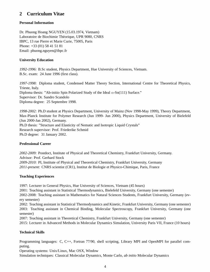
Figure 1: (A) An example diphenyl crystallinemolecule is represented by an ellipsoid particle;(B) a typical equilibrium configuration of the crys-talline phase.
My PhD research focuses on the structure and elasticity propertiesof liquid crystals. Nowadays, the applications of liquid crystalscan be found everywhere, from the laptop screen, digital watch,switchable windows, smartphone screen etc. Liquid crystalphasesare the phases between the solid and liquid phases. They mostof-ten occur in compounds of the rod-like shape or the disc-likeshapeorganic molecules which interact through strongly anisotropic in-termolecular forces. There are three main phases of liquid crystals:nematic, smectic and columnar phases. Nematic phase is the sim-plest phase where the molecules tend to be parallel to a commondirection called the director. In this phase, there is no translationalorder but the orientational order is apparently[1]. This broken sym-metry of space leads to many interesting physics properties. MyPhD work focuses on two properties that are long range structureand elasticity of the nematic phase.
3.1.1 Correlation functions
We construct a coarse-grained model for the liquid crystal,whereeach molecule is represented by an ellipsoid particle [Fig.1(A)].The interaction between particles are modeled by a simple repul-sive pair potential
V12 =
{
4ǫ0(X1212 −X6
12) + ǫ0 : X612 > 1/2,
0 : otherwise(1)
The functionX12 = σ0/(r12 − σ12 + σ0) depends on the distancer12 between particles 1 and 2, and on the shapefunction
σ12(u1,u2, ˆr12) = σ0
{
1− κ
2
[(u1 .r12 + u2 .r12)2
1+ κu1.u2
+(u1 .r12 − u2 .r12)
2
1− κu1.u2
]}
(2)
which approximates the contact distance between two ellipsoids of elongationκ =√
(1 + χ)(1− χ) with ori-entationsu1 andu2 and center - center vectorr12 pointing in the directionr12 =r12/ r12[2]. Having defined thepotential, we carry out Monte Carlo (MC) simulations for theisotropic phase (low densityρ = 0.24/σ3
0 ) and thenematic phase (high densityρ = 0.30/σ3
0). Fig.1(B) shows a snapshot of the system of ellipsoids in the nematicphase. Our primary aim is to study the influence of the isotropic/nematic phase transition on the direct correlationfunctions (DCF) in anisotropic fluids. The DCF is a central quantity in liquid state theories[3], and a study inthe nematic phase is therefore interesting in its own right.The DCFc(u1,u2, r12) is determined from the pairdistribution functions by solving the full Ornstein-Zernike equation[3]
h(u1,u2, r12) = c(u1,u2, r12) +
∫
c(u1,u3, r13)ρ(1)(u3)h(u3,u2, r32)du3dr3, (3)
The total correlation functionh, which describes the total interaction between two particles, and the one-particledistribution functionρ(1), which is the probability for finding a particle, are determined directly from the simulationdata. The DCF has been studied quite intensely in isotropic fluids. In contrast, the determination of this functionin the nematic phase is quite challenge. As mentioned above,in a nematic liquid crystal, the particles remainpositionally disordered, but align preferentially along the director vectorn(r). Therefore, the isotropy of space isspontaneously broken. Consequently, the pair correlationfunctions lose their rotational invariance. This means thatthe pair correlationF (u1,u2, r12) (F stands forh or c) must depend not only on the orientation of each particleui
6

(like in the isotropic phase) but also on the center - center vectorr12. The latter makes the calculation of the functionF to be much more difficult. One of the first tasks of my thesis is to solve this issue, and below I briefly describethe method which I developed to calculate pair correlation functions in the simulated nematic phase. Details of themethod can be found in Ref.[4]. First, a Cartesian coordinate frame where thez axis points in the direction of thedirector vectorn(r) is constructed, and all orientation-dependent functions are expanded in spherical harmonicsYlm(u). This yields
ρ(1) = ρ∑
leven
flYl0, (4)
with the bulk number densityρ, and
F (u1,u2, r) =∑
l1,l2,l,m1,m2,m
Fl1m1l2m2lm(r)Yl1m1(u1)Yl2m2
(u2)Ylm(r). (5)
In uniaxially symmetric phases, only real coefficients withm1 + m2 + m = 0 and evenl1 + l2 + l enter theexpansion. Since our particles have uniaxial symmetry, every single li is even as well. The total correlationfunctionh is defined as
h(u1,u2, r) =ρ(2)(u1,u2, r)
ρ(1)(u1)ρ(1)(u2)− 1, (6)
whereρ(2)(u1,u2, r) is the pair distribution function. In terms of the sphericalharmonic expansion, Eq.6 isrewritten as
ρ(2)l1,m1,l2,m2,l,m
(r) = ρ2(√
4πfl1fl2δm10δm20δl0δm0 +∑
l′1,l′′1,l,l′
2,l′′2
hl′1m1l′2m2lm(r)fl′′
1fl′′
2Γl1l′1l
′′
1
m1m10Γl2l′2l
′′
2
m2m20
)
, (7)
with
Γll′l′′
mm′m′′ =
∫
duY ∗
lm(u)Y ∗
l′m′(u)Y ∗
l′′m′′(u), (8)
and the coefficients of the pair distribution are determineddirectly from the simulation data
ρ(2)l1,m1,l2,m2,l,m
(r) = 4πρ2g(r)〈Y ∗
l1m1(u1)Y
∗
l2m2(u2)Y
∗
lm(u〉δr, (9)
where the average〈· · · 〉δr is performed over all molecule pairs at distances|r1 − r2| = [r, r + δr], andg(r) isthe radial distribution function, i.e., the average total number of molecule pairs divided byN4πρ2δr. Then, thecoefficients of the total correlation functionh are determined by solving the matrix Eq.7. The DCFc is most easilycalculated in Fourier representation. Therefore, we perform a Hankel transformation of all functions in Eq.3 andobtain a new representation in thek-space
hl1,m1,l2,m2,l,m(k) = cl1,m1,l2,m2,l,m(k) + ρ∑
l3,l′3,l′′
3,m3,
l′m′l′′m′′l3
cl1,m1,l3,m3,l′,m′(k)hl′3,m3,l2,m2,l′′,m′′(k) (10)
× fl′′3(−1)m3Γll′l′′
mm′m′′Γl3l′3l
′′
3
m3m30.
The coefficients of the DCF are obtained by solving the matrixequation 10. Fig.2 shows an example of an expansioncoefficient,c202020 , which vanishes in the case of rotational invariance. In theisotropic phase (ρ ≤ 0.283σ−3
0 ),the values are indeed close to zero. In the nematic phase, a nonzero DCF coefficient not only emerges, it mayeven grow quite large. Atρ = 0.3σ−3
0 the values are comparable to those of a symmetry preserving coefficientof similar order. Hence the DCF really reflects the broken symmetry in the nematic phase. To summarize, I havedeveloped a method to calculate DCF without any approximations, and applied it to study the local structure offluids of uniaxial ellipsoids at different densities in the vicinity of the nematic/isotropic phase transition. We findthat the DCF suitably characterizes the local structure, inthe sense that it is short ranged both in the isotropic andthe nematic phase.
3.1.2 Elastic constants
7

Figure 2: Expansion coefficient withli = l = 2 andmi = m = 0 of the direct correlation function inthe molecular frame vsr for different densities. In theisotropic phase, this coefficient must vanish for symmetryreasons. Atρ = 0.283σ−3
0, a small nonzero remnants
are observed due to finite size effects
We then use the DCF to calculate the elastic constants of thenematic phase. Due to the long range orientational orderbreaks a continuous symmetry, the isotropy of space, thereexist soft fluctuation modes-spatial variations of the directorn(r) - which cost no energy in the infinite wavelength limit(i.e., the limit wheren is rotated uniformly) and are other-wise penalized by elastic restoring forces[5]. For symmetryreasons, the latter depend on only three material parametersat large finite wavelengths[1], which are described by an elas-tic free energy functional[6]
F{n(r)} =1
2
∫
dr{K11[▽.n]2 +K22[n.(▽× n]2+
K33[n× (▽× n)]2}, (11)
which has three contributions: the splay, twist, and bendmodes. The parametersKαα (α = 1, 2, 3), called Frankelastic constants, control almost exclusively the structure andthe properties of nematic liquid crystals at mesoscopic lengthscales. It has been derived in Ref.[7], these elastic constantsare expressed in terms of the DCF in thek-space as the fol-lowing
K11 = −kBT
2
∫
∂2c(k,u1,u2)
∂k2x|k=0 × ρ(1)
′
(u1z)ρ(1)′(u2z)u1xu2xdu1du2
K22 = −kBT
2
∫
∂2c(k,u1,u2)
∂k2x|k=0 × ρ(1)
′
(u1z)ρ(1)′(u2z)u1yu2ydu1du2
K33 = −kBT
2
∫
∂2c(k,u1,u2)
∂k2z|k=0 × ρ(1)
′
(u1z)ρ(1)′ (u2z)u1xu2xdu1du2. (12)
We calculate the DCF from the pair distribution functionρ(2) using an upper cutofflmax = 2, 4, and 6, respectively,in the matrix equations 7 and 10, and the elastic constants are calculated from Eq.12. Already the lowest ordercalculation withlmax = 2 gave elastic constants of the correct order of magnitude. Quantitatively reliable resultswere obtained withlmax ≥ 6. In summary, we have presented a reliable method to calculate elastic constants frompair distributions in computer simulations.
3.2 Postdoc research
After my PhD in condensed matter physics, I changed my research field to biophysical chemistry with a focus onproteins. With this, I started to learn molecular dynamics (MD) simulation techniques using protein all-atom mod-els. My first projects were the development and application of computational strategies that allows us to extendwell-established MD techniques to the description of photoinduced nonequilibrium dynamics simulations usingstandard MD program packages. This allows us to study the energy transport[8, 9, 10] and ultrafast conforma-tional dynamics[11, 12, 13] of large systems, which are otherwise very difficult to study by means of semiclas-sical or quantum molecular dynamics methods. To quantitatively compare with picosecond pump-probe Infrared(IR) experiments, I am also involved in the development of accurate methods to simulate 1D and 2D IR spec-tra of peptides[14, 15, 16]. I am also interested in equilibrium fast dynamics such as folding of small peptides.Along this direction, I develop methods to reduce the complexity of peptides in order to capture essential physicsof folding pathways[17, 18, 19, 20, 21, 22], and collaboratewith NMR experiment to determine accurately thestructure of small peptides[23]. Finally, I start to work onthe protein aggregation to understand the fibril growthmechanism[24], and on RNA systems aiming to characterize the structure RNA hairpins[25] and long-range corre-lations in ligand-binding riboswitch[26]. Below, I summary some main results in details.
8

3.2.1 Energy transfer in peptides
(a) Vibrational energy relaxation. In recent years, femtosecond time-resolved infrared spectroscopy has made itpossible to watch the flow of vibrational energy within and between molecules with unprecedented time resolution[27].Particular interest has been payed to biomolecular systems, since the celebrated concept of the relation betweenprotein structure, dynamics, and function ultimately requires an microscopic understanding of the energy flow inthe system. Employing the amide I modes (which mainly involve the stretching of the peptide C= O bond) as aconformational probe, experiments have provided new insight into the structure, fluctuations, and conformationaltransitions of peptides in aqueous solution. Interestingly, the experiments have shown that theν = 1 → 0 popu-lation decay timeT1 of the amide I mode is consistently about 1 ps for all systems considered. This includes themodel systems N-methylacetamid[28], and trialanine[29],various small globular peptides such as apamin, sylla-toxin, and bovine pancreatic tripsin inhibitor[28], and the protein myoglobin[30]. These findings indicate that theamide I population relaxation in peptides (i) is a highly efficient and unusually fast example of vibrational energyredistribution and (ii) represents a generic, sequence-independent feature of peptides.
To describe the vibrational relaxation dynamics, usually asystem-bath approach is employed in which the sys-tem includes the initially excited vibrational mode (and possibly further strongly coupling modes) and the bathcomprises all other degrees of freedom[31]. Second-order time-dependent perturbation theory with respect to thesystembath interaction leads to a reduced density-matrix formulation in which the system is treated numericallyexactly, while the dynamics of the bath enters via equilibrium autocorrelation functions. Being mostly interestedin the vibrational energy relaxation rate1/T1 , well-established assumptions then lead to the widely usedGoldenRule expression. To obtain a realistic modeling of the solvent, furthermore, many workers have pursued a mixedquantum-classical strategy and calculate the bath correlation functions from a classical molecular dynamics simu-lation
Here, my primary aim is to develop an all-atom nonequilibrium molecular dynamics simulation (NEMD)method to directly simulate the vibrational relaxation process using a standard MD program package such asGROMACS[32]. Our method includes the following main steps[8]. First, a quasiclassical sampling of the initialatom positions and velocities is performed to account for the nonequilibrium initial preparation of the system. Tothis end, we represent the solute normal modes{pk, qk} in terms of classical action-angle variables{nk, φk}[33]
qk =√
(2nk + γ) sinφk
pk =√
(2nk + γ) cosφk. (13)
where the factorγ = 1 accounts for the zero-point energy (ZPE) of the mode. To obtain the initial positions andmomenta of the initially excited amide I normal mode, we associate the actionnk with the initial quantum state ofthe amide I mode, e.g.,nk = 1 for the first excited state. The initial actions of the remaining solute modes (whichare at thermal equilibrium) may be sampled from the Boltzmann distributionP (nk) ∝ enkhωk/kT . In all cases,the vibrational phasesφk are picked randomly from the interval[0, 2π] . This way an ensemble of normal-modepositions and momenta are calculated, which present a quasiclassical representation of the quantum initial state ofthe solute molecule. Next, employing these initial conditions, classical MD simulations are performed. To monitorthe vibrational dynamics, the overall rotation and internal motion are separated and a normal-mode analysis isperformed, which yields the time-dependent energy contentof the normal modes. It turns out, however, that theseprocedures are well established only in the case that the vibrational dynamics is well described by small-amplitudemotion around a single equilibrium structure. Consideringflexible biophysical systems far from equilibrium (e.g., afolding peptide) , the single-reference normal-mode approximation must break down. To extend the nonequilibriumdescription of vibrational-energy redistribution to the treatment of molecular systems undergoing large amplitudemotion, we adopt the ideas underlying the instantaneous normal-mode theory[34]. In this formulation at everytime step a normal-mode calculation is performed, which employs the instantaneous position of the trajectory asreference structure, and the instantaneous vibrational energy of the system at timetn+1 is
Evib = V0 +1
2
3N−6∑
k1
hωk(p2k + q2k), (14)
whereqk(tn), pk(tn), andωk(tn) represent the positions, momenta, and frequencies of the instantaneous normalmodes.V0 = V [r(tn)] − 1
2
∑
k hωka2k with ak = (mkhω
3k)
−1/2∑3N
i=1 FiUik being the coordinate shift accounts
9
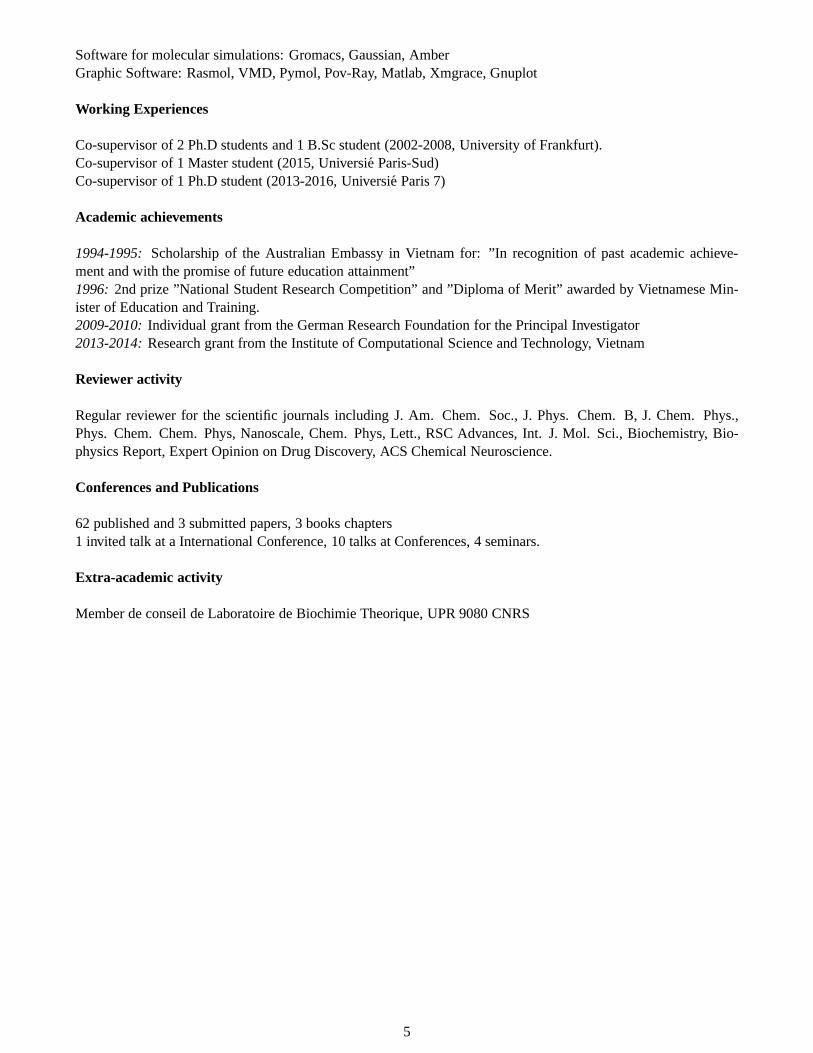
for the non-zero force termFi, andUik is the eigenvector of the normal modes. To demonstrate the performance ofthe method, we study the vibrational energy relaxation process following the excitationν = 0 → 1 of the amide Imode of the model system N-methylacetamid peptide (H3C-COND-CH3).
Figure 3: Vibrational energy relaxation of the amide I mode ofNMA in D2O following ν1 → 0 excitation as obtained for vari-ous quasiclassical initial conditions. For better comparison, the ini-tially included zero-point energy is subtracted. (a) Comparison ofaction-angle initial conditions including zero point energy in all so-lute modes (dashed line), in the amide I mode only (lower solid line),and no solute mode (upper solid line). Note that in the first two casesthe energy content of the amide I mode drops below zero for times≥ 4 ps. (b) Including only 35% of the zero-point energy in all so-lute modes (dashed line) and in the amide I mode only (solid line),respectively, the amide I energy remains positive.
Figure 3(a) shows the time-dependent energycontent of the amide I mode. In the purelyclassical case, where the ZPE is not includedin modes (γ = 0), the decay can be well fit-ted to a biexponential function with the decaytimes t1 = 1.9 ps (80%) andt2 = 13.3 (20%)ps. At longer times, the amide I energy con-verges tokBT ≈ 200 cm−1 which is consis-tent with the equipartition theorem. IncludingZPE in the amide I mode (γ = 1) is seen tolead to a considerable enhancement of the ini-tial relaxation process (t1 = 1.5 ps). Thereis only little change, though, if the ZPE is alsoincluded in the remaining solute modes. Ifwe include only a fraction of ZEP withγ =0.35 to the amide I such that the amide I en-ergy remains larger than the ZPE for all timesunder consideration, we obtaint1 = 1.8 ps( 75%) and t2 = 7.5 ps ( 25%) [Fig.3(b)].In summary, we show that the vibrational en-ergy relaxation rate obtained from the nonequi-librium simulations is in qualitative agreementwith experiment, and our approach thereforemay represent a reasonable and direct descrip-tion of the quantum-mechanical relaxation pro-cess.
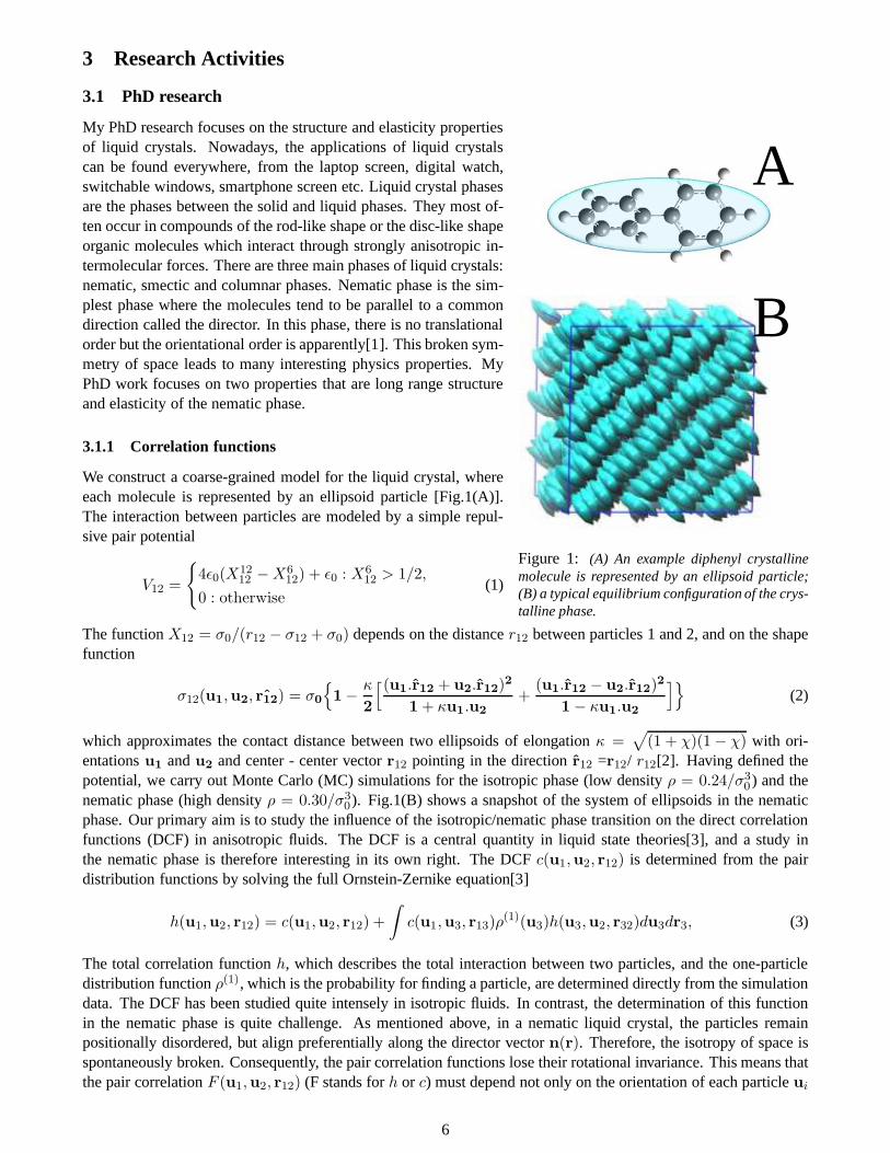
(b) Photoinduced energy transfer.In another work, we investigate energy transport through anα-aminoisobutyricacid-based 310-helix (Aib) dissolved in chloroform in a combined experimental-theoretical approach[9]. The basicidea is that the vibrational energy is locally deposited at the N-terminus of the Aib by ultrafast internal conver-sion of a covalently attached, electronically excited, azobenzene moiety [Fig.4(A)]. Heat flow through the helixis detected with sub-picosecond time resolution by employing vibrational probes as local thermometers at variousdistances from the heat source. This peptide was chosen because it forms exceptionally a stable 310-helix even ina relatively short sequence of 8 amino acids. The apolar solvent chloroform was used to minimize potential lossof the heat flow into the surrounding solvent and moreover mimics the hydrophobic environment in the interior ofa protein. We chose azobenzene molecule because it undergoes ultrafast internal conversion (cis andtrans iso-merization) on a 200-fs timescale. To guide the interpretation of the experiments and obtain a microscopic pictureof the molecular processes underlying energy transport in peptides, we have developed a simple computationalstrategy that allows us to extend well-established MD techniques to the description of photoinduced nonequilib-rium dynamics[11, 12]. To model the laser-induced photoisomerization process, we use a minimal model for thecorresponding potential-energy surfaces that diabatically connects the excited-stateS1 of the cis-isomer with theground stateS0 of the trans-isomer [Fig.4(B)]. The photoexcitation of the system by anultrafast laser pulse ismimicked by instantly switching from the ground-state N=N torsional potential to the excited-state potential. Fol-lowing this nonequilibrium preparation at timet = 0, the system isomerizes along an excited-state N=N potentialwithin 0.2 ps. After isomerization (i.e, for times≥ 0.5 ps), the N=N torsional potential is switched back to itsground state form, and a constant-energy MD simulation is performed up to 100 ps. All simulations were per-formed with the GROMACS program suite[32], using the GROMOS96 force field 43a1[35]. Additional force fieldparameters for the azobenzene unit were derived from density functional theory. Following the nonequilibriumsimulations, the time-dependent observables of interest are obtained via an ensemble average over 800 equilib-
10

rium conformations. We observe that after the photoexcitation at timet = 0, the kinetic energy is deposited intothe azobenzene photoswitch within 0.1 ps. While the excitation of the photoswitch decays on a picosecond timescale, its energy is transferred to the Aib peptide (30%) anddirectly to the solvent (70%). The peptide energyrises within 0.3 ps and remains approximately constant up to10 ps, before it decays with an 20-ps time constant.
A
B
Figure 4:(A) Structure of a stable Aib peptide where one endis attached to an azobenzene molecule which plays a role asa heater. (B) Scheme of theS0 and theS1 potential-energycurves of azobenzene as a function of the N=Ncis − transisomerization coordinateφ. The solid lines represent the adia-batic potentials of a model which is designed to reproduce theexperimentalcis andtrans absorption bands and the ground-statecis− trans energy barrier. The dashed line correspondsto the GROMOS force field potential of the N=N torsion, thedotted line shows a model of the cis-trans photoisomerizationpotential, which simply connects theS1 cis state and theS0
trans state of azobenzene.
The solvent energyEsolv(t) rises with time constants0.5 ps and 20 ps, where the shorter time scale reflectsthe initial momentum transfer of the isomerizing pho-toswitch to its surrounding solvent molecules. Afterthe thermalization of the photoinduced energy, the 20-ps time scale ofEsolv(t) accounts for the subsequentcooling of the peptide. We also observe that the pho-toexcitation does not lead to significant conformationalchanges of the peptide. To elucidate the energy transferalong the peptide chain, we displays the time evolu-tion of the mean kinetic energy for most of the peptidegroups. The kinetic energy of unit 1 (the linker unit tothe photoswitch, Fig.3.2.1) reaches its peak at 0.3 ps,which nicely agrees with the experimental finding thatunit 1 takes up substantial energy within the time reso-lution (0.2 ps). Unit 2 receives 50% of the energy fromunit 1, and unit 3 receives 35% within 1 ps. Accord-ing to the simulation, the terminal unit 9 still receivesa small (20%) amount of the excess energy because ofthe photoexcitation, but this effect is not measurablein the experiment. However, we found that in termsof quantitative numbers, the MD simulation seems tooverestimate the heat diffusion constant by a factor offive.
To understand this discrepancy, we perform exper-iments with a lower energy excitation[10]. To this end,we carry out experiment and simulation with the directexcitation of a peptide C=O oscillator (amide I mode)of unit 1 with 0.2 eV (IR) photons. We should recallthat the energy deposited through the excitation of theazobenzene chromophore is much higher, about 3 eV(UV) photons. The difference in the two excitationmethods has not only a consequence for the amountof energy deposited, but also the form in which it isprovided. The experiments show that heat transportthrough the peptide after excitation with low energy photons is at least 4 times faster than after UV excitation.On the other hand, the heat transport obtained by nonequilibrium MD simulations is largely insensitive to the kindof excitation. Thus, the calculations agree well with the experimental results for the low-frequency case. Thisfinding suggests that the photoinduced energy gets trapped,if it is deposited in high amounts.
3.2.2 Photoinduced conformational rearrangements of peptides
In recent times, a number of experimental techniques have been developed which study biomolecular processessuch as protein folding directly and in a time-resolved manner. For example, there have been various suggestionsto include a molecular photoswitch such as azobenzene into peptides[36]. This guarantees that the light-inducedstructural changes of the chromophore upon photoisomerization around the central N=N double bond are directlytransferred into the peptide chain. Femtosecond time-resolved pump-probe experiments with optical and infrareddetection indicate that the main conformational changes ofthe peptide backbone are completed after only 20 ps,although the subsequent structural equilibration of the peptide continues for about 20 ns. These types of experi-
11
ments, especially in combination with 2D-IR probing, provide a new and promising way to study the folding andunfolding of peptides in unprecedented detail.
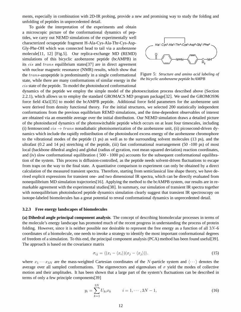
Figure 5: Structure and amino acid labeling ofthe bicyclic azobenzene peptide bcAMPB
To guide the interpretation of the experiments and obtaina microscopic picture of the conformational dynamics of pep-tides, we carry out NEMD simulations of the experimentally wellcharacterized octapeptide fragment H-Ala-Cys-Ala-Thr-Cys-Asp-Gly-Phe-OH which was connected head to tail via a azobenzenemolecule[11, 12] [Fig.5]. Our replica-exchange MD (REMD)simulations of this bicyclic azobenzene peptide (bcAMPB) inits cis and trans equilibrium states[37] are in direct agreementwith nuclear magnetic resonance (NMR) results, which show thatthe trans-azopeptide is predominantly in a single conformationalstate, while there are many conformations of similar energyin thecis state of the peptide. To model the photoinduced conformationaldynamics of the peptide we employ the simple model of the photoexcitation process described above (Section2.2.1), which allows us to employ the standard GROMACS MD program package[32]. We used the GROMOS96force field 43a1[35] to model the bcAMPB peptide. Additionalforce field parameters for the azobenzene unitwere derived from density functional theory. For the initial structures, we selected 200 statistically independentconformations from the previous equilibrium REMD simulations, and the time-dependent observables of interestare obtained via an ensemble average over the initial distribution. Our NEMD simulation draws a detailed pictureof the photoinduced dynamics of the photoswitchable peptide which occurs on at least four timescales, including(i) femtosecondcis → trans nonadiabatic photoisomerization of the azobenzene unit, (ii) picosecond-driven dy-namics which include the rapidly redistribution of the photoinduced excess energy of the azobenzene chromophoreto the vibrational modes of the peptide (1 ps) as well as to thesurrounding solvent molecules (13 ps), and theultrafast (0.2 and 14 ps) stretching of the peptide, (iii) fast conformational rearrangement (50 -100 ps) of mostlocal (backbone dihedral angles) and global (radius of gyration, root mean squared deviation) reaction coordinates,and (iv) slow conformational equilibration ( 500 - 1000 ps) accounts for the subsequent conformational equilibra-tion of the system. This process is diffusion-controlled, as the peptide needs solvent-driven fluctuations to escapefrom traps on the way to the final state. A quantitative comparison to experiment can only be obtained by a directcalculation of the measured transient spectra. Therefore,starting from semiclassical line shape theory, we have de-rived explicit expressions for transient one- and two dimensional IR spectra, which can be directly evaluated fromnonequilibrium MD simulation trajectories[16]. Applyingthe method to the bcAMPB system, our results are in re-markable agreement with the experimental studies[38]. In summary, our simulation of transient IR spectra togetherwith nonequilibrium photoinduced peptide dynamics simulation clearly suggest that transient IR spectroscopy onisotope-labeled biomolecules has a great potential to reveal conformational dynamics in unprecedented detail.
3.2.3 Free energy landscapes of biomolecules
(a) Dihedral angle principal component analysis. The concept of describing biomolecular processes in termsofthe molecule’s energy landscape has promoted much of the recent progress in understanding the process of proteinfolding. However, since it is neither possible nor desirable to represent the free energy as a function of all 3N -6coordinates of a biomolecule, one needs to invoke a strategyto identify the most important conformational degreesof freedom of a simulation. To this end, the principal component analysis (PCA) method has been found useful[39].The approach is based on the covariance matrix
σij = 〈(xi − 〈xi〉)(xj − 〈xj〉)〉. (15)
wherex1 · · · x3N are the mass-weighted Cartesian coordinates of theN -particle system and〈· · · 〉 denotes theaverage over all sampled conformations. The eigenvectors and eigenvalues ofσ yield the modes of collectivemotion and their amplitudes. It has been shown that a large part of the system’s fluctuations can be described interms of only a few principle components[39]
yi =
3N∑
k=1
Ukixk i = 1, · · · , 3N − 1, (16)
12
with U is the (3N × 3N) matrix where theith column contains theith eigenvector of the matrixσ [Eq.15].Although the method has been widely used, it has two serious problems. First, the coordinatesxi in Eq.15describe the internal motion, and must be separated from theoverall motion. However, the completely elimi-nation of overall rotation is impossible in the case of largeamplitude motion such as protein folding/unfolding.
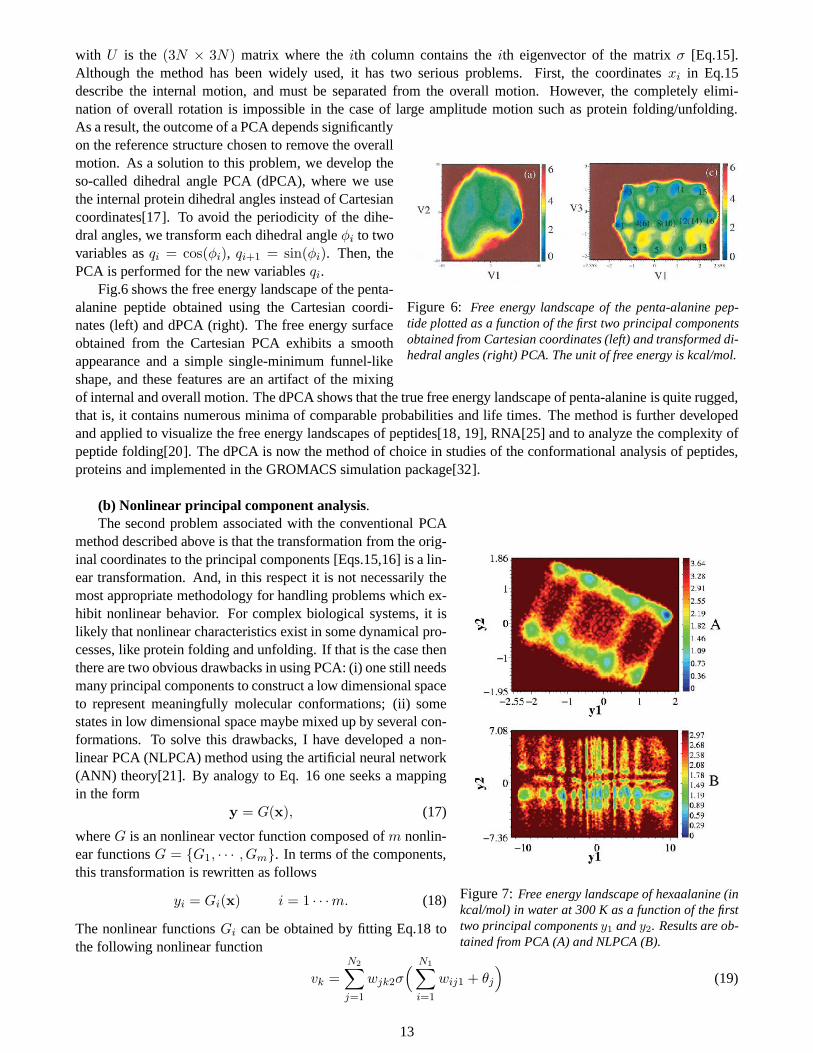
Figure 6: Free energy landscape of the penta-alanine pep-tide plotted as a function of the first two principal componentsobtained from Cartesian coordinates (left) and transformed di-hedral angles (right) PCA. The unit of free energy is kcal/mol.
As a result, the outcome of a PCA depends significantlyon the reference structure chosen to remove the overallmotion. As a solution to this problem, we develop theso-called dihedral angle PCA (dPCA), where we usethe internal protein dihedral angles instead of Cartesiancoordinates[17]. To avoid the periodicity of the dihe-dral angles, we transform each dihedral angleφi to twovariables asqi = cos(φi), qi+1 = sin(φi). Then, thePCA is performed for the new variablesqi.
Fig.6 shows the free energy landscape of the penta-alanine peptide obtained using the Cartesian coordi-nates (left) and dPCA (right). The free energy surfaceobtained from the Cartesian PCA exhibits a smoothappearance and a simple single-minimum funnel-likeshape, and these features are an artifact of the mixingof internal and overall motion. The dPCA shows that the true free energy landscape of penta-alanine is quite rugged,that is, it contains numerous minima of comparable probabilities and life times. The method is further developedand applied to visualize the free energy landscapes of peptides[18, 19], RNA[25] and to analyze the complexity ofpeptide folding[20]. The dPCA is now the method of choice in studies of the conformational analysis of peptides,proteins and implemented in the GROMACS simulation package[32].
(b) Nonlinear principal component analysis.
Figure 7:Free energy landscape of hexaalanine (inkcal/mol) in water at 300 K as a function of the firsttwo principal componentsy1 andy2. Results are ob-tained from PCA (A) and NLPCA (B).
The second problem associated with the conventional PCAmethod described above is that the transformation from the orig-inal coordinates to the principal components [Eqs.15,16] is a lin-ear transformation. And, in this respect it is not necessarily themost appropriate methodology for handling problems which ex-hibit nonlinear behavior. For complex biological systems,it islikely that nonlinear characteristics exist in some dynamical pro-cesses, like protein folding and unfolding. If that is the case thenthere are two obvious drawbacks in using PCA: (i) one still needsmany principal components to construct a low dimensional spaceto represent meaningfully molecular conformations; (ii) somestates in low dimensional space maybe mixed up by several con-formations. To solve this drawbacks, I have developed a non-linear PCA (NLPCA) method using the artificial neural network(ANN) theory[21]. By analogy to Eq. 16 one seeks a mappingin the form
y = G(x), (17)
whereG is an nonlinear vector function composed ofm nonlin-ear functionsG = {G1, · · · , Gm}. In terms of the components,this transformation is rewritten as follows
yi = Gi(x) i = 1 · · ·m. (18)
The nonlinear functionsGi can be obtained by fitting Eq.18 tothe following nonlinear function
vk =
N2∑
j=1
wjk2σ(
N1∑
i=1
wij1 + θj
)
(19)
13
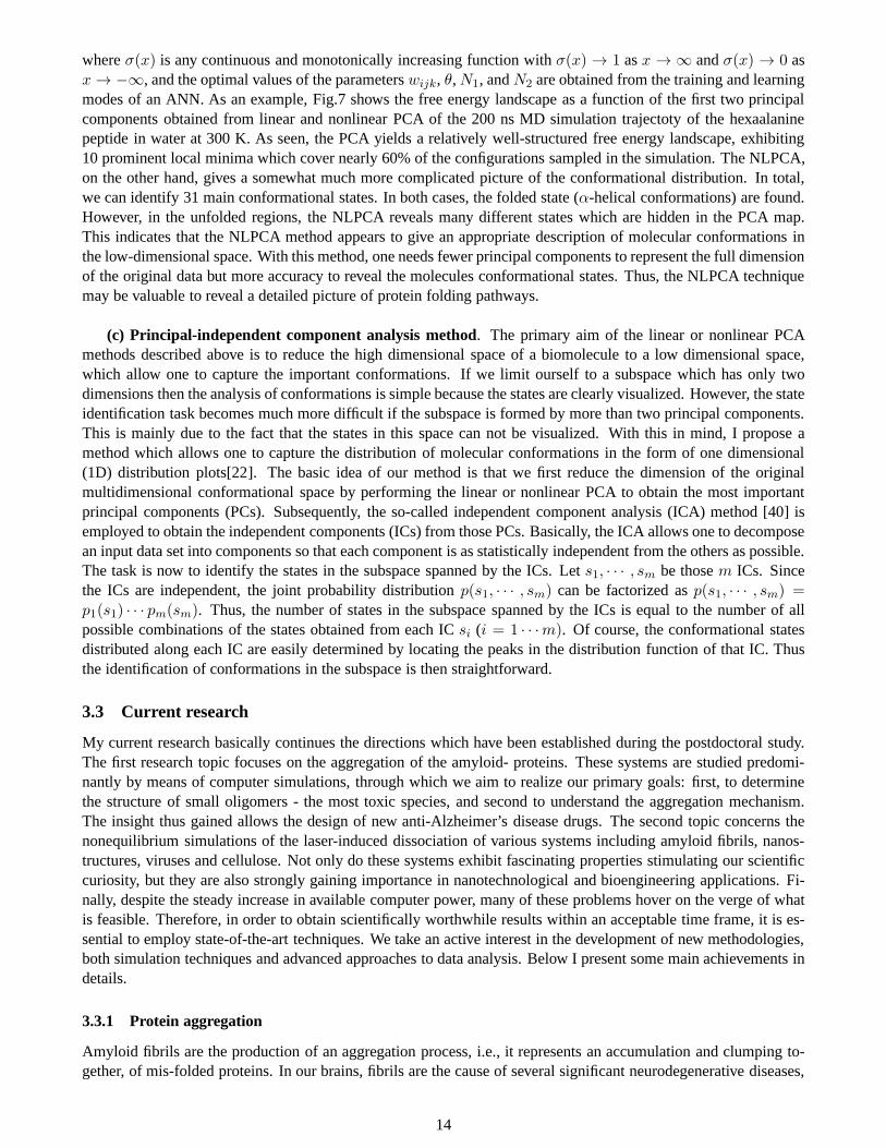
whereσ(x) is any continuous and monotonically increasing function with σ(x) → 1 asx → ∞ andσ(x) → 0 asx → −∞, and the optimal values of the parameterswijk, θ,N1, andN2 are obtained from the training and learningmodes of an ANN. As an example, Fig.7 shows the free energy landscape as a function of the first two principalcomponents obtained from linear and nonlinear PCA of the 200ns MD simulation trajectoty of the hexaalaninepeptide in water at 300 K. As seen, the PCA yields a relativelywell-structured free energy landscape, exhibiting10 prominent local minima which cover nearly 60% of the configurations sampled in the simulation. The NLPCA,on the other hand, gives a somewhat much more complicated picture of the conformational distribution. In total,we can identify 31 main conformational states. In both cases, the folded state (α-helical conformations) are found.However, in the unfolded regions, the NLPCA reveals many different states which are hidden in the PCA map.This indicates that the NLPCA method appears to give an appropriate description of molecular conformations inthe low-dimensional space. With this method, one needs fewer principal components to represent the full dimensionof the original data but more accuracy to reveal the molecules conformational states. Thus, the NLPCA techniquemay be valuable to reveal a detailed picture of protein folding pathways.
(c) Principal-independent component analysis method. The primary aim of the linear or nonlinear PCAmethods described above is to reduce the high dimensional space of a biomolecule to a low dimensional space,which allow one to capture the important conformations. If we limit ourself to a subspace which has only twodimensions then the analysis of conformations is simple because the states are clearly visualized. However, the stateidentification task becomes much more difficult if the subspace is formed by more than two principal components.This is mainly due to the fact that the states in this space cannot be visualized. With this in mind, I propose amethod which allows one to capture the distribution of molecular conformations in the form of one dimensional(1D) distribution plots[22]. The basic idea of our method isthat we first reduce the dimension of the originalmultidimensional conformational space by performing the linear or nonlinear PCA to obtain the most importantprincipal components (PCs). Subsequently, the so-called independent component analysis (ICA) method [40] isemployed to obtain the independent components (ICs) from those PCs. Basically, the ICA allows one to decomposean input data set into components so that each component is asstatistically independent from the others as possible.The task is now to identify the states in the subspace spannedby the ICs. Lets1, · · · , sm be thosem ICs. Sincethe ICs are independent, the joint probability distribution p(s1, · · · , sm) can be factorized asp(s1, · · · , sm) =p1(s1) · · · pm(sm). Thus, the number of states in the subspace spanned by the ICsis equal to the number of allpossible combinations of the states obtained from each ICsi (i = 1 · · ·m). Of course, the conformational statesdistributed along each IC are easily determined by locatingthe peaks in the distribution function of that IC. Thusthe identification of conformations in the subspace is then straightforward.
3.3 Current research
My current research basically continues the directions which have been established during the postdoctoral study.The first research topic focuses on the aggregation of the amyloid- proteins. These systems are studied predomi-nantly by means of computer simulations, through which we aim to realize our primary goals: first, to determinethe structure of small oligomers - the most toxic species, and second to understand the aggregation mechanism.The insight thus gained allows the design of new anti-Alzheimer’s disease drugs. The second topic concerns thenonequilibrium simulations of the laser-induced dissociation of various systems including amyloid fibrils, nanos-tructures, viruses and cellulose. Not only do these systemsexhibit fascinating properties stimulating our scientificcuriosity, but they are also strongly gaining importance innanotechnological and bioengineering applications. Fi-nally, despite the steady increase in available computer power, many of these problems hover on the verge of whatis feasible. Therefore, in order to obtain scientifically worthwhile results within an acceptable time frame, it is es-sential to employ state-of-the-art techniques. We take an active interest in the development of new methodologies,both simulation techniques and advanced approaches to dataanalysis. Below I present some main achievements indetails.
3.3.1 Protein aggregation
Amyloid fibrils are the production of an aggregation process, i.e., it represents an accumulation and clumping to-gether, of mis-folded proteins. In our brains, fibrils are the cause of several significant neurodegenerative diseases,
14
such as Parkinson disease, Alzheimer’s disease (AD)[41, 42]. There is increasing in vivo evidence that the amyloid-β (Aβ) protein and Aβ oligomers are the proximate neurotoxic agents in AD. This protein usually presents in thetwo forms 1 - 40 (Aβ1−40) and 1 - 42 (Aβ1−42) amino acids sequences. Currently, there are no effective treatmentsfor this dreaded disease. As a consequence, extensive studies have been devoted to understand the fundamental as-pects of the aggregation mechanism as well as to develop drugs and alternative approaches for treating AD[42, 43].Below, I summary the main achievements of my research in protein aggregation.
(a) Mechanism of oligomer growth. To monitor the early events that direct assembly of amyloidogenicpeptides we probe the dynamics of formation(Aβ16−22)n−1 +Aβ16−22 ⇀↽ (Aβ16−22)n of (Aβ16−22)n oligomersby adding a monomer to a preformed (Aβ16−22)n−1 (n = 4− 6) oligomer in which the peptides are arranged in anantiparallelβ-sheet conformation[24].
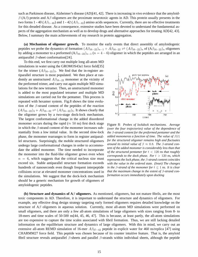
Figure 8: Probes of lockdock mechanisms. Average(over the four trajectories) value of the dependence oftheβ-strand content for the preformed pentamer and theadded monomeras a function of time. The value ofβ(t)for the structured oligomer remains high and fluctuatesaround its initial value ofβ ≈ 0.8. Theβ-strand con-tent of the added monomer is considerably less than thatof the structured pentamer fort < 120 ns that roughlycorresponds to the dock phase. Fort > 120 ns, whichrepresents the lock phase, theβ-strand content coincideswith the value in the ordered state. (Inset) The changesin theβ-strand of the monomer fort ≤ 1 ns. It is clearthat the maximum change in the extent ofβ-strand con-formation occurs immediately upon docking
To this end, we first carry out multiple long all-atom MDsimulations in water using the GROMOS43a1 force field[35]for the trimer (Aβ16−22)3. We find that the in-register an-tiparallel structure is most populated. We then place at ran-domly an unstructuredAβ16−22 monomer at the vicinity ofthe preformed trimer, and carry out again multiple MD simu-lations for the new tetramer. Then, an unstructured monomeris added to the most populated tetramer and multiple MDsimulations are carried out for the pentamer. This process isrepeated with hexamer system. Fig.8 shows the time evolu-tion of theβ-strand content of the peptides of the reaction(Aβ16−22)4 +Aβ16−22 ⇀↽ (Aβ16−22)5. It shows clearly thatthe oligomer grows by a two-stage dock-lock mechanism.The largest conformational change in the added disorderedmonomer occurs during the rapid (≈ 50 ns) first dock stagein which theβ-strand content of the monomer increases sub-stantially from a low initial value. In the second slow-lockphase, the monomer rearranges to form in register antiparal-lel structures. Surprisingly, the mobile structured oligomersundergo large conformational changes in order to accommo-date the added monomer. The time needed to incorporatethe monomer into the fluid-like oligomer grows even whenn = 6, which suggests that the critical nucleus size mustexceed six. Stable antiparallel structure formation exceedshundreds of nanoseconds even though frequent interpeptidecollisions occur at elevated monomer concentrations used inthe simulations. We suggest that the dock-lock mechanismshould be a generic mechanism for growth of oligomers ofamyloidogenic peptides.
(b) Structure and dynamics of Aβ oligomers. As mentioned, oligomers, but not mature fibrils, are the mosttoxic components in AD. Therefore, it is important to understand the structure and dynamics of oligomers. Forexample, any effective drug design strategy targeting early formed oligomers requires detailed knowledge on thestructure of Aβ oligomers in aqueous solution. Currently, most all-atom MDsimulations were performed onsmall oligomers, and there are only a few all-atom simulations of large oligomers with sizes ranging from 8- to18-mers and time scales of 50-500 ns[44, 45, 46, 47]. This is because, at least partly, the all-atom simulationsare too expensive to capture the time scales associated withfibril formation. Thus, we are still lacking detailedinformation on the equilibrium structure and dynamics of large oligomers. With this in mind, we carry out anextensive all-atom REMD simulation of 16-merAβ37−42 peptide in explicit water for 460 ns/replica [47] usingCHARMM27 force field. This peptide was chosen because of its counter intuitive feature. That is, the amyloidfibril structure reveals antiparallelβ-sheets and parallelβ-strands within individual sheets, although the peptide
15
has two opposite charges at the extremities [48]. The extensive conformational sampling allows us to addressimportant questions: what is the equilibrium population distribution of oligomers andβ-sheets? what is the fractionof mixed parallel/antiparallelβ-sheets? what is the critical nucleus size? what is the mechanism of formingordered aggregates? Our simulation shows that the peptide assembly follows the ”condensation-polymerization”mechanism observed experimentally[49]: In the first stage,polymerization driven by hydrophobic collapse takeplaces resulting in various oligomer sizes with little and small β-strand formed. Then, within the amorphousaggregates, two small sheets extend by essentially monomeraddition and further conversion of peptides fromrandom coil toβ-strand can either lead to longerβ-sheetsor newβ-sheet layer.
While this is the longest simulation of such large amyloid oligomer starting from randomized and disorderedchains, this approach is too expensive if one wants to study other large oligomers. Thus, our strategy is to developfurther a coarse-grained lattice model[50], where a residue is represented by a bead located on a corner of a simplecubic lattice, and its side chain is described by an unit vector. The interaction between residues is modeled bythe coarse-grained off-lattice OPEP force field[51]. Our bottom-up approach starts with the determination of thebest lattice force field parameters for the Aβ16−22 dimer by fitting its equilibrium parallel and anti-parallelβ-sheetpopulations to all-atom simulation results. Surprisingly, the calibrated force field is transferable to the trimer ofAβ16−22 and the dimer and trimer of Aβ37−42. The dominant structure of the Aβ16−22 decamer matches the mi-crocrystal structure. In contrast, the Aβ37−42 decamer is largely disordered with mixed by parallel and antiparallelchains, suggesting that the nucleus size is> 10 peptides. Our refined force field coupled to this on-lattice modelshould provide useful insights into the critical nucleation number associated with neurodegenerative diseases[52].
In another work, we carry out long all-atom REMD simulation of the dimeric system formed by the full-lengthAβ1−40 peptide for 400 ns/replica using the CHARMM22* force field[53]. The dimer is chosen because it is thesmallest toxic specie in AD. We find transient configurationswith an unstructured N-terminus and multipleβ-hairpins spanning residues 17-21 and 30-36, but the antiparallel and perpendicular peptide orientation is preferredover the parallel organization. Short-lived conformational states also consist of allα topologies, and one compactpeptide with beta-sheet structure stabilized by a rather extended peptide withα-helical content. Overall, this studyprovides, for the first time, insights into the equilibrium structure ofAβ1−40 dimer in explicit aqueous solution,opening a new avenue for a comprehensive understanding of the impact of pathogenic and protective mutations inearly-stage Alzheimer disease at a molecular level.
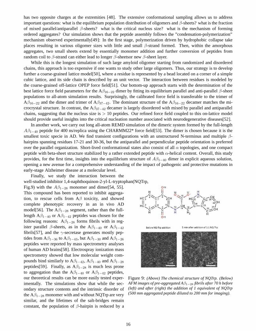
Figure 9: (Above) The chemical structure of NQTrp. (Below)AFM images of pre-aggregated Aβ1−28 fibrils after 70 h before(left) and after (right) the addition of 1 equivalent of NQTrp(500 mm aggregated peptide diluted to 200 mm for imaging).
Finally, we study the interaction between thewell-studied inhibitor 1,4-naphthoquinon-2-yl-L-tryptophan(NQTrp,Fig.9) with theAβ1−28 monomer and dimer[54, 55].This compound has been reported to inhibit aggrega-tion, to rescue cells from Aβ toxicity, and showedcomplete phenotypic recovery in an in vivo ADmodel[56]. The Aβ1−28 segment, rather than the full-length Aβ1−40 or Aβ1−42 peptides was chosen for thefollowing reasons: Aβ1−28 forms fibrils with in reg-ister parallelβ-sheets, as in the Aβ1−40 or Aβ1−42
fibrils[57], and theγ-secretase generates mostly pep-tides from Aβ1−36 to Aβ1−43, but Aβ1−30 and Aβ1−26
peptides were reported by mass spectrometry analysesof human AD brains[58]. Electrospray ionization massspectrometry showed that low molecular weight com-pounds bind similarly to Aβ1−42, Aβ1−40 and Aβ1−28
peptides[59]. Finally, as Aβ1−28 is much less proneto aggregation than the Aβ1−40 or Aβ1−42 peptides,our theoretical results can be more easily tested exper-imentally. The simulations show that while the sec-ondary structure contents and the intrinsic disorder ofthe Aβ1−28 monomer with and without NQTrp are verysimilar, and the lifetimes of the salt-bridges remainconstant, the population ofβ-hairpin is reduced by a
16
factor of 1.5 and the population ofα-helix in the region 17-24 is increased by a factor of two uponNQTrp binding.These two factors, which impact the free energy barrier for nucleation, provide a first explanation for the reportedreduced Aβ1−40 and Aβ1−42 aggregation kinetics in the presence of NQTrp. However, thepopulation of the freeAβ1−28 monomer (does not bind to NQTrp) is rather high (20-25%) at the concentration of 17.5 mM, and thisshows that the affinity of NQTrp is low and hence its inhibitory activity is not very strong[54]. Furthermore, theinteraction of NQTrp with the dimer of Aβ1−28 is very diffuse and not very specific, as most residues display prob-ability contacts between 1 and 3% with the NQTrp molecules. These findings are confirmed by our experimentalresults obtained by fluorescence spectroscopy, FTIR spectroscopy, NMR spectroscopy as well as by cell viabilitystudies[55]. For example, as seen from Fig.9, which displays the AFM images acquired immediately before andafter the addition of NQTrp, there is no significant change, as in both cases images with high concentrations offibrils were observed, even when fibrils were incubated for 6 hwith NQTrp. Taken together, our study suggeststhat the reported anti-AD activity of NQTrp-type moleculesin in vivo models has to involve another mechanism.This study has revealed the potential pitfalls in the development of aggregation inhibitors for amyloidogenic pep-tides, which are of general interest for all the molecules studied in the context of inhibiting the formation of toxicaggregates.
(c) Effects of mutation on the oligomer structure.Because of the disordered structure of the N-terminal in thefibril structure, the role of this region in aggregation, toxicity and pathogenesis was considered for a long time to benegligible, thus remained largely unexploited. Therefore, most of mutation studies have focused on the C-terminalof the Aβ1−40 or the Aβ1−42 peptides. However, several recent studies indicate the possible important of the N-terminal in the Aβ structure, aggregation and toxicity. For examples, Stefansson and co-workers first discovered thesingle-point Ala2→Thr2 (A2T) mutation effects by comparing the complete genome sequences of 1795 Icelanderswith their medical histories[60]. The variant is rare, but it has a huge impact on those fortunate enough to inheriteven a single copy of it. About 0.5% of Icelanders have a protective gene that prevents mental deterioration in oldage. Compared with their countrymen who lack the mutation, Icelanders who carry this mutation are more thanfive times more likely to reach 85 without being diagnosed with AD. In another work, the Italian team leaded byTagliavini has shown that the single-point mutation Ala2→Val2 (A2V) causes dementia in homozygous carriers,whereas heterozygous carriers were found to be protected[61].
To explain these experimental findings, we carry out extensive all-atom REMD simulations (400 ns/replica)for the wild-type (WT) and A2V mutation of the Aβ1−28 monomer, and also for the WT-WT, WT-A2V, WT-A2T dimers of the Aβ1−40 peptide. We show that upon A2V mutation, the population ofβ-hairpins of Aβ1−28
monomer is increased significantly (by a factor of 4), its intrinsic disorder is reduced by a factor of 2, and thefree energy landscape is completely different as compared to the wild type counterpart[62]. Our results also showthat for the homozygous A2V-A2V dimer, the A2V mutation enhances the intermolecular interaction between twoN-terminals, thus reduces the intramolecular interactionbetween N-terminal with CHC and C-terminal of eachmonomer. This means that all three regions N-terminal, CHC,and C-terminal have strong tendency to participateinto the aggregation as compared to WT-WT dimer, leading to the acceleration of the aggregation of A2V chains.In contrast, for the heterozygous WT-A2V dimer, the A2V mutation enhances the intramolecular N-terminal-CHCinteraction, leading to the double-hairpin structure topology. This means that the N-terminal and CHC have weakertendency to form intermolecular contacts as compared to WT-WT dimer, leading to the slowdown of aggregationof the wild-type and mutated A2V chains[63].
In contrast to A2V and A2T mutations, the other single-pointmutations His6→Arg6 (H6R), Asp7→Asn7(D7N) and Asp7→His7 (D7H) are reported to alter the monomer misfolding and produce oligomers that are toxiccompared with the wild-type. To explain this, we carry out MDsimulations for both monomers and dimers ofAβ1−40 and Aβ1−42 WT and mutant counterparts[64, 65, 66]. The long simulations with total time of 3-4µsallows us to understand for the first time the microscopic effects of the mutations. For example, for both Aβ1−40
and Aβ1−42 systems, we did not find an increase ofβ-strand content upon D7N mutation. Rather, we found thatthe enhanced formation rate of Aβ1−40 fibrils comes essentially from the formation of the loop Asp23-Lys28 in themonomer. In contrast, the enhanced rate of Aβ1−42 fibrils does not result from the formation of the loop Asp23-Lys28 in monomer, but may result from the formation of the same loop in dimer, the increased turn propensity ofthe residues 28-30 in monomer, and the higher population ofβ -hairpin conformations involving the residues 19-20with residues 31-32 in the dimer.
17
3.3.2 Nonequilibrium MD simulation of laser-induced dissociation of nanostructures
Recently, new mid-infrared free-electron laser (FEL) having specific oscillation characteristics of a picosecondpulse structure, a tunable wavelength within infrared frequencies and a high photon density has been developedand applied to the amyloid field. By tuning the laser frequency to the amide I bands of the amyloid, experimentswere able to refold amyloid-like fibrils into native form, and to dissociate fibrils of short peptides[67, 68, 69, 70]. These findings are very interesting, however, the microscopics of the dissociation process is largely unknown.To this end, I have developed a comprehensive laser-inducedNEMD simulation method, where a time-dependentelectric field
E(t) = E0 exp[−(t− t0)
2
2σ2] cos[2πcω(t− t0)], (20)
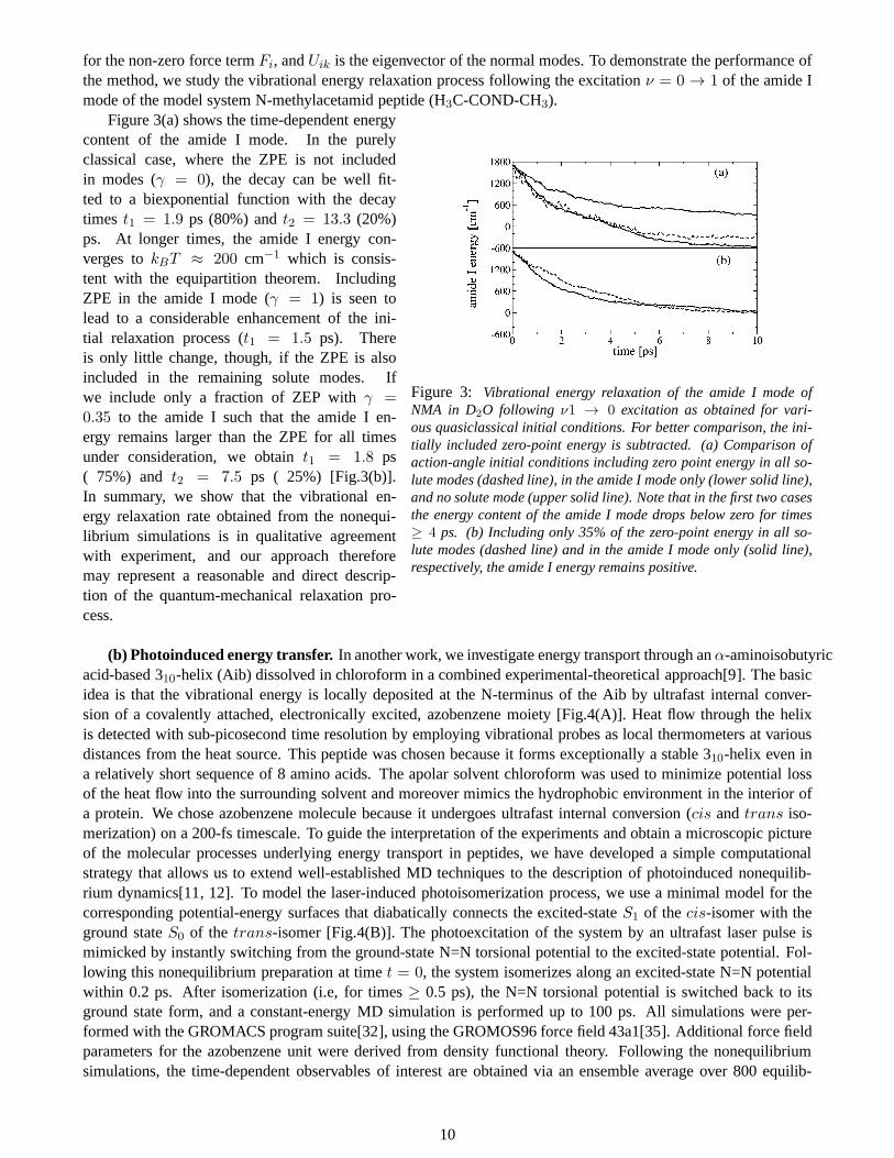
is applied to mimic a laser pulse. Here,E0 represents amplitude of the electric field,σ is the pulse width,t is thetime after the pulse maximumt0, c is the speed of light andω is the frequency. By scanningω, we identify thefrequencyω where the resonance between the vibrational modes of the system and laser frequency takes place, andthis results in quick dissociation of the system. The methodhas been applied to study the dissociation of varioussystems including amyloid fibrils[71], peptide nanotube[72], virus[73] and cellulose.
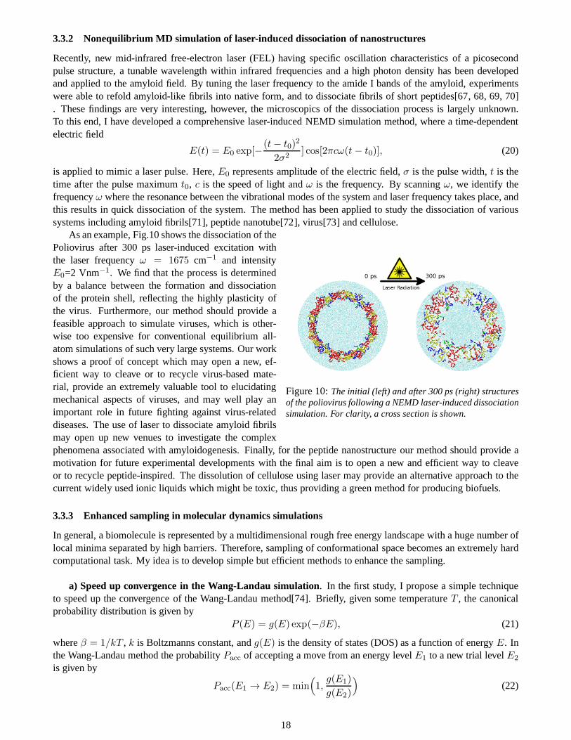
Figure 10:The initial (left) and after 300 ps (right) structuresof the poliovirus following a NEMD laser-induced dissociationsimulation. For clarity, a cross section is shown.
As an example, Fig.10 shows the dissociation of thePoliovirus after 300 ps laser-induced excitation withthe laser frequencyω = 1675 cm−1 and intensityE0=2 Vnm−1. We find that the process is determinedby a balance between the formation and dissociationof the protein shell, reflecting the highly plasticity ofthe virus. Furthermore, our method should provide afeasible approach to simulate viruses, which is other-wise too expensive for conventional equilibrium all-atom simulations of such very large systems. Our workshows a proof of concept which may open a new, ef-ficient way to cleave or to recycle virus-based mate-rial, provide an extremely valuable tool to elucidatingmechanical aspects of viruses, and may well play animportant role in future fighting against virus-relateddiseases. The use of laser to dissociate amyloid fibrilsmay open up new venues to investigate the complexphenomena associated with amyloidogenesis. Finally, for the peptide nanostructure our method should provide amotivation for future experimental developments with the final aim is to open a new and efficient way to cleaveor to recycle peptide-inspired. The dissolution of cellulose using laser may provide an alternative approach to thecurrent widely used ionic liquids which might be toxic, thusproviding a green method for producing biofuels.
3.3.3 Enhanced sampling in molecular dynamics simulations
In general, a biomolecule is represented by a multidimensional rough free energy landscape with a huge number oflocal minima separated by high barriers. Therefore, sampling of conformational space becomes an extremely hardcomputational task. My idea is to develop simple but efficient methods to enhance the sampling.
a) Speed up convergence in the Wang-Landau simulation. In the first study, I propose a simple techniqueto speed up the convergence of the Wang-Landau method[74]. Briefly, given some temperatureT , the canonicalprobability distribution is given by
P (E) = g(E) exp(−βE), (21)
whereβ = 1/kT , k is Boltzmanns constant, andg(E) is the density of states (DOS) as a function of energyE. Inthe Wang-Landau method the probabilityPacc of accepting a move from an energy levelE1 to a new trial levelE2
is given by
Pacc(E1 → E2) = min(
1,g(E1)
g(E2)
)
(22)
18
whereg(E) denotes the current estimate DOS for the system. As the simulation progresses, DOS is updated andthe process is repeated until the histogram of energiesH(E) becomes flat, i.e, the system samples all possibleenergy states. This method has been applied in various contexts, ranging from random spin models and quantumsystems to simple models for proteins. Despite the advantages, it can still be difficult to obtain a reliable DOS withthe Wang-Landau method. With this in mind, my idea is to applya general and controllable method for smoothinga potential energy surface (PES)[75]
Eq(x) =q
β(q − 1)ln(1 + β(q − 1)E(x)), (23)
whereE(x) is the original PES of the system, and the smoothness of PES iscontrolled the parameterq ≥ 1. Onthis smoothed surfaceEq(x) , one begins a standard Wang-Landau calculation to provide acrude, but well-sampledinitial estimate of DOS. In a second step, one restores the true surfaceE(x) and continues to improve the estima-tion of the density of states. Applied the method to a simple Go-type model of 20-residue peptide, the new andoriginal Wang-Landau simulations required on average5.9± 4× 10⁶ and16.6± 2× 10⁶ trial moves, respectively,thus resulting in roughly a threefold speedup. Because of the simple generalized effective potential for the seedingcalculations, the method is general as well as very easy to implement and a wide range of applications is foreseen.
(b) Reduce the number of replicas in the replica-exchange simulation. Currently, the REMD method iswidely used to enhance conformational sampling in biomolecular simulations. However, it has been shown that thenumber of replicasM increase quicklyM ∼
√f , wheref is the number of degrees of freedom of the system. As
a consequence, for large systems such as proteins in explicit solvent, this technique requires many processors andis clearly inappropriate. To this end, I propose a variant ofthe standard REMD method, employing the concept ofsolvent viscosity dependence of dynamics of biomolecules[76]. The physics behind this approach is straightfor-ward: intuitively, the lower the viscosity, the faster a protein chain will diffuse, and the faster it will sample theconformational space. Quantitatively, the Kramers theorysuggested that the rate of protein folding is inversely pro-portional to solvent viscosity, therefore, decreasing theviscosity will increase the folding rate. The basic idea of themethod is to run the system of interest at a normal temperature and normal solvent viscosity, while other replicasrun not only at different temperatures but also at artificially low solvent viscosities. Because both temperature andviscosity are used to enhance sampling, the method requiresfewer replicas than the conventional REMD method.In a simulation the solvent viscosity can be reduced by a factor of λ if mass of solvent molecules is scaled by afactor ofλ2. Of course, the change of mass is probably the easiest task for any biomolecular simulation users. Thatmakes the method is as simple as the conventional REMD methodbut more efficient. I show that (i) given the samenumber of replicas, the new method always converges to equilibrium faster than the conventional REMD simula-tions, (ii) the new method achieves similar sampling quality as the conventional REMD method but could reducethe number of replicas by a factor of 1.5 - 2 at least for the test systems in this work (16-residueβ-hairpin pep-tide in explicit water). For very large systems, the efficiency of the new method will become even more pronounced.
(c) Simulated tempering with on-the-fly weight determination. Another widely used enhanced samplingmethod is the simulated tempering (ST) which does not have limitation to large systems like REMD. However,it does require the determination of a priori unknown weightparameters to ensure a uniform random walk intemperature space and this is non-trivial and very tedious for complex systems. In the ST simulation, temperatureitself becomes a dynamical variable which could take discrete valuesTm (T1 < T2 < · · · < TM ). Probabilitydistribution of a state at temperatureTm and potential energyE is given by the following generalized canonicaldistribution:
WST(E, βm) = exp(βmE + fm), (24)
whereβm = 1/kT . If the weight parametersfm are chosen asfm = ln(∫
dEn(E) exp(βmE)) (n(E) is thedensity of states) then it follows immediately that the distribution of temperature is flat, i.e., a free random walk intemperature space is realized, which in turn induces a random walk in potential energy space and allows the systemto escape from local energy minima. Here, I suggest a simple method to determine these weights on-the-fly basedon the recursion formulae
fn+1 = fn + (βn+1 − βn)(En+1 + En)/2, (25)
whereEn is the average potential energy at temperatureTn at timet. With this scheme, simulated tempering re-quires neither prior trial simulations nor complicated update schemes. The advantage of our method over REMD
19

simulations has been demonstrated with the study of the folding of the 20-residue alanine peptide and the aggre-gation of a trimer formed by the Alzheimers peptide fragmentAβ16−22 using the implicit solvent coarse grainedOPEP force field[77].
The method was then applied to the folding or aggregation of seven proteins with the CHARMM, OPLS, andAMBER protein, and the SPC and TIP3P water force fields[78]. The sampling with ST is found to be more efficientthan with REMD for a much lower CPU cost. For example, starting from unfolded or extended conformations, theWW domain and the Trp-cage peptide fold to their NMR structures with a backbone RMSD of 2.0 and 1A. Re-markably, the ST simulation explores transient non-nativetopologies for Trp-cage that have been rarely discussedby other simulations. Taken together, these results open the door to the study of the configurations of single pro-teins, protein aggregates, and any molecular systems at atomic details in explicit solvent using a single normalCPU. They also demonstrate that our ST scheme can be used withany force field ranging from quantum mechanicsto coarse-grain and atomistic.
(d) Multiple force fields in a single simulated tempering simulation. Two most frequent questions of anymolecular dynamics simulations are: how robust are the results with respect to the force field used and has configu-rational sampling converged? Clearly, the results should be validated using various force fields such as OPLS/ AA,AMBER and CHARMM, and enhanced sampling methods such as replica exchange (RE) or simulated tempering(ST) must be employed. However, repeating RE or ST simulation with different force fields is very time-consumingand rarely done. To this end, I propose a simple and automaticmethod which allows in one single simulationto satisfy three goals: (i) efficient configurational sampling, (ii) force field dependence of the results, and (iii)temperature-dependence of the results modeled by each force field. Let’s consider two independent statesm andnassociated with the configurational spacesxm andxn and the potential energy functionsU andV , respectively. Theprobability distribution of the configurationxm at temperatureTm and potential energyU is given by the followinggeneralized canonical distribution:
P (xm, βm) = e−βmU(xm)+um , (26)
whereβ = 1/kBT (kB is the Boltzmann constant) andum is the weight parameter. The temperature transitionfrom statem to n, while fixing configurationxm, is given by the following Metropolis-like probability[79, 80]:
WST(Tm, Tn) = min[1, e−βnV (xm)+βmU(xm)+(vn−um)], (27)
whereV (xm) is the potential energy ofxm andvn is the weight parameter of staten. As shown in Ref.[81],the acceptance probability, Eq.27, can be reformulated in terms of the instantaneous energy fluctuationsV (xm)−〈V (xm)〉 andU(xm) − 〈U(xm)〉. These fluctuations can result in significant temperature transition probability,even ifU andV are different, and this is the essential physics that makes our method work. Applications to a 1Dsystem and the all-atom chignolin peptide have shown that this new and efficient method should be very useful tostudy the folding of any biomolecules in explicit solvent.
3.3.4 General method to determine structure of amyloid fibrils
As mentioned in Section 2.3.1 it is important to understand the structure oligomers. Many reaction coordinatessuch as the radius of gyration, theβ-sheet size, the oligomer size distribution, the number of contacts, the orderparameterP2, the connectivity length are often used to characterize thestructures, but each variable captures onlyone feature of the self-assembly. The characterization andclassification of the conformations remain difficult forsystems characterized by degenerate states, that is, identical oligomer conformations differing only by the permu-tations of the molecules. To avoid the degeneracy problem, Ipropose a new method allowing the characterizationof the structures of amyloid oligomers having arbitrary shapes and position-orientation arrangements[82]. Themethod takes into account both the intermolecular and intramolecular degrees of freedom, and treats correctly thedegenerate states. Briefly, the intermolecular structuresof an oligomer are described in terms of the combina-tion of double-molecule states. These states describe the position-orientation arrangements of a double-chain inthe presence of the other double-chains, and can be obtainedthrough the PCA method of the inverse distancesbetween side-chain’s centers of mass of two chains of the double-molecule trajectory. The intramolecular struc-tures of an oligomer are described in terms of the combination of single-molecule states. These states describe the
20

structures of a single-chain in the presence of the other chains, and can be obtained through the dPCA method ofthe single-molecule trajectory. The overall structure of the oligomer is given in terms of the product basis of theintermolecular and intramolecular structures. We have shown that our method can be applied to any oligomer sizefor two reasons. First, the identification of the single-molecule and double-molecule states involves the PCA ofsingle- and double-molecule trajectories, and this is straightforward. Second, while the number of combinations ofthe single-molecule or double-molecule states increases with the number of chains, especially for highly flexiblemolecules, the calculation of the combinations is simple and fast, and the identification of the overall structures isa simple matter. In addition, many states can be marginally populated and safely discarded. This method should bea general method to obtain the overall structure of oligomers from simulation trajectory in unprecedented details.
3.3.5 Calculation of the configurational entropy of large biomolecules
Configurational entropy, which measures the number of available configurations that are occupied by a molecule in3D space [83, 84], plays an important role in many chemical and biological processes[84] and structure-based drugdesign[85]. Currently, there are several methods to evaluate configurational entropy, including the thermodynamicintegration, hypothetical scanning technique and the factorization of the multidimensional probability density func-tion into lower-order probability density functions. Although these methods have proven to be quite accurate, theirmain drawbacks are that they tend to be computationally demanding, in particular for large systems such as proteinsin explicit solvent, reducing therefore their possible applications. Our primary aim is to develop simple, fast yetaccurate methods to estimate configurational entropy of large systems. Let us consider a molecule containingNatoms with Cartesian coordinatesx = {xi}, wherexi = (x3i−2, x3i−1, x3i)
T denotes the position of theith atom.Theconfigurational entropyof the molecule, first introduced by Kushick and Karplus [86], is
S = −kB
∫
dxρ(x) ln ρ(x), (28)
kB is Boltzmann’s constant andρ(x) is the canonical probability density distribution function of the system.
(a) Calculation of entropy using variable separation approach. The configurational entropyS, [Eq. 28],can be, in principle, calculated by performing numericallythe multidimensional integrations of the right-hand-sideof Eq. 28. In practice, this is difficult due to high dimensionality of the system. The basis idea of our method isas follows. Physically, the coordinatesx in Eq.28 are strongly correlated as they are imposed by covalent bonds,non-bonded repulsions, and other atomic forces. Mathematically, since those coordinates are just the integrationvariables, a one-to-one linear transformationq = As can be performed, resulting in the new representation of theentropy as follows:
S = −kB
∫
dsρ(s) ln ρ(s) + kB ln |A|. (29)
where|A| is the absolute value of the determinant of the Jacobian square matrixAij = ∂qi/∂si. The key featureof Eq.29 is the dependence of the entropy on the new variabless = s1, · · · , sn which can be obtained via atransformation such that they are quasi-independent, i.e., ρ(s1, · · · , sn) ≈ ρ1(s1) · · · ρn(sn), and entropy can beapproximated as
Sica = −kB
n∑
i
∫
dsiρi(si) ln ρi(si) + kB ln |A|. (30)
The problem now is finding a transformation such that the transformed variablessi are quasi-independent. To thisend, I employ the independent component analysis (ICA) mentioned in the Section 2.2.3 which allows for perform-ing such a transformation [28,29]. This approach takes intoaccount the whole dependence of coordinates, and isvery fast fast even for large systems. Applying the method tothe 16-residueβ-hairpin peptide shows a dramaticimprovement in the entropy,Sica, which is much closer to the exact entropy,S, than other methods such as thequasi-harmonic approach. The method is still very fast evenfor large proteins. Details of the method is describedin Ref.[87].
(b) Calculation of entropy using configurational temperature approach. The well-known quasiharmonic(QH) method approximates the system probability density distribution function by a multivariate Gaussian density
21
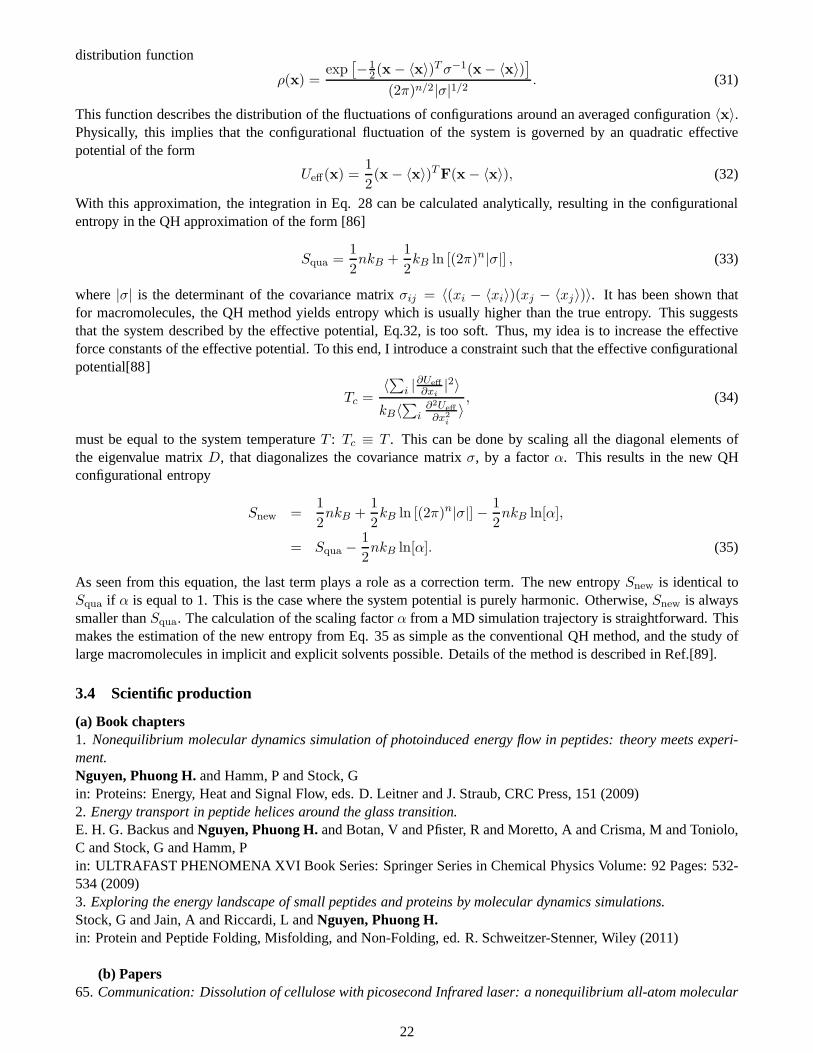
distribution function
ρ(x) =exp
[
−12(x− 〈x〉)Tσ−1(x− 〈x〉)
]
(2π)n/2|σ|1/2 . (31)
This function describes the distribution of the fluctuations of configurations around an averaged configuration〈x〉.Physically, this implies that the configurational fluctuation of the system is governed by an quadratic effectivepotential of the form
Ueff(x) =1
2(x− 〈x〉)TF(x− 〈x〉), (32)
With this approximation, the integration in Eq. 28 can be calculated analytically, resulting in the configurationalentropy in the QH approximation of the form [86]
Squa =1
2nkB +
1
2kB ln [(2π)n|σ|] , (33)
where|σ| is the determinant of the covariance matrixσij = 〈(xi − 〈xi〉)(xj − 〈xj〉)〉. It has been shown thatfor macromolecules, the QH method yields entropy which is usually higher than the true entropy. This suggeststhat the system described by the effective potential, Eq.32, is too soft. Thus, my idea is to increase the effectiveforce constants of the effective potential. To this end, I introduce a constraint such that the effective configurationalpotential[88]
Tc =〈∑i |∂Ueff
∂xi|2〉
kB〈∑
i∂2Ueff
∂x2i
〉, (34)
must be equal to the system temperatureT : Tc ≡ T . This can be done by scaling all the diagonal elements ofthe eigenvalue matrixD, that diagonalizes the covariance matrixσ, by a factorα. This results in the new QHconfigurational entropy
Snew =1
2nkB +
1
2kB ln [(2π)n|σ|] − 1
2nkB ln[α],
= Squa −1
2nkB ln[α]. (35)
As seen from this equation, the last term plays a role as a correction term. The new entropySnew is identical toSqua if α is equal to 1. This is the case where the system potential is purely harmonic. Otherwise,Snew is alwayssmaller thanSqua. The calculation of the scaling factorα from a MD simulation trajectory is straightforward. Thismakes the estimation of the new entropy from Eq. 35 as simple as the conventional QH method, and the study oflarge macromolecules in implicit and explicit solvents possible. Details of the method is described in Ref.[89].
3.4 Scientific production
(a) Book chapters1. Nonequilibrium molecular dynamics simulation of photoinduced energy flow in peptides: theory meets experi-ment.Nguyen, Phuong H.and Hamm, P and Stock, Gin: Proteins: Energy, Heat and Signal Flow, eds. D. Leitner and J. Straub, CRC Press, 151 (2009)2. Energy transport in peptide helices around the glass transition.E. H. G. Backus andNguyen, Phuong H.and Botan, V and Pfister, R and Moretto, A and Crisma, M and Toniolo,C and Stock, G and Hamm, Pin: ULTRAFAST PHENOMENA XVI Book Series: Springer Series inChemical Physics Volume: 92 Pages: 532-534 (2009)3. Exploring the energy landscape of small peptides and proteins by molecular dynamics simulations.Stock, G and Jain, A and Riccardi, L andNguyen, Phuong H.in: Protein and Peptide Folding, Misfolding, and Non-Folding, ed. R. Schweitzer-Stenner, Wiley (2011)
(b) Papers65. Communication: Dissolution of cellulose with picosecond Infrared laser: a nonequilibrium all-atom molecular
22
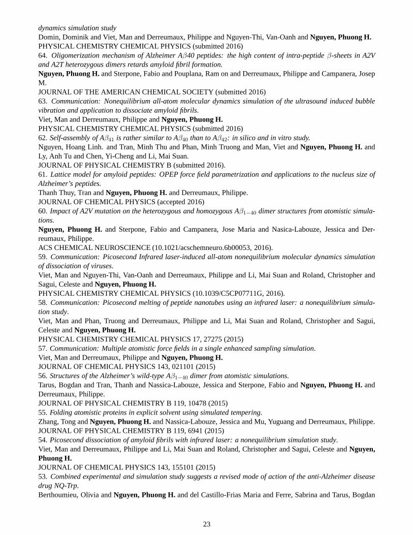
dynamics simulation studyDomin, Dominik and Viet, Man and Derreumaux, Philippe and Nguyen-Thi, Van-Oanh andNguyen, Phuong H.PHYSICAL CHEMISTRY CHEMICAL PHYSICS (submitted 2016)64. Oligomerization mechanism of Alzheimer Aβ40 peptides: the high content of intra-peptideβ-sheets in A2Vand A2T heterozygous dimers retards amyloid fibril formation.Nguyen, Phuong H.and Sterpone, Fabio and Pouplana, Ram on and Derreumaux, Philippe and Campanera, JosepM.JOURNAL OF THE AMERICAN CHEMICAL SOCIETY (submitted 2016)63. Communication: Nonequilibrium all-atom molecular dynamics simulation of the ultrasound induced bubblevibration and application to dissociate amyloid fibrils.Viet, Man and Derreumaux, Philippe andNguyen, Phuong H.PHYSICAL CHEMISTRY CHEMICAL PHYSICS (submitted 2016)62. Self-assembly of Aβ41 is rather similar to Aβ40 than to Aβ42: in silico and in vitro study.Nguyen, Hoang Linh. and Tran, Minh Thu and Phan, Minh Truong and Man, Viet andNguyen, Phuong H.andLy, Anh Tu and Chen, Yi-Cheng and Li, Mai Suan.JOURNAL OF PHYSICAL CHEMISTRY B (submitted 2016).61. Lattice model for amyloid peptides: OPEP force field parametrization and applications to the nucleus size ofAlzheimer’s peptides.Thanh Thuy, Tran andNguyen, Phuong H.and Derreumaux, Philippe.JOURNAL OF CHEMICAL PHYSICS (accepted 2016)60. Impact of A2V mutation on the heterozygous and homozygous Aβ1−40 dimer structures from atomistic simula-tions.Nguyen, Phuong H.and Sterpone, Fabio and Campanera, Jose Maria and Nasica-Labouze, Jessica and Der-reumaux, Philippe.ACS CHEMICAL NEUROSCIENCE (10.1021/acschemneuro.6b00053, 2016).59. Communication: Picosecond Infrared laser-induced all-atom nonequilibrium molecular dynamics simulationof dissociation of viruses.Viet, Man and Nguyen-Thi, Van-Oanh and Derreumaux, Philippe and Li, Mai Suan and Roland, Christopher andSagui, Celeste andNguyen, Phuong H.PHYSICAL CHEMISTRY CHEMICAL PHYSICS (10.1039/C5CP07711G, 2016).58. Communication: Picosecond melting of peptide nanotubes using an infrared laser: a nonequilibrium simula-tion study.Viet, Man and Phan, Truong and Derreumaux, Philippe and Li, Mai Suan and Roland, Christopher and Sagui,Celeste andNguyen, Phuong H.PHYSICAL CHEMISTRY CHEMICAL PHYSICS 17, 27275 (2015)57. Communication: Multiple atomistic force fields in a single enhanced sampling simulation.Viet, Man and Derreumaux, Philippe andNguyen, Phuong H.JOURNAL OF CHEMICAL PHYSICS 143, 021101 (2015)56. Structures of the Alzheimer’s wild-type Aβ1−40 dimer from atomistic simulations.Tarus, Bogdan and Tran, Thanh and Nassica-Labouze, Jessicaand Sterpone, Fabio andNguyen, Phuong H.andDerreumaux, Philippe.JOURNAL OF PHYSICAL CHEMISTRY B 119, 10478 (2015)55. Folding atomistic proteins in explicit solvent using simulated tempering.Zhang, Tong andNguyen, Phuong H.and Nassica-Labouze, Jessica and Mu, Yuguang and Derreumaux, Philippe.JOURNAL OF PHYSICAL CHEMISTRY B 119, 6941 (2015)54. Picosecond dissociation of amyloid fibrils with infrared laser: a nonequilibrium simulation study.Viet, Man and Derreumaux, Philippe and Li, Mai Suan and Roland, Christopher and Sagui, Celeste andNguyen,Phuong H.JOURNAL OF CHEMICAL PHYSICS 143, 155101 (2015)53. Combined experimental and simulation study suggests a revised mode of action of the anti-Alzheimer diseasedrug NQ-Trp.Berthoumieu, Olivia andNguyen, Phuong H.and del Castillo-Frias Maria and Ferre, Sabrina and Tarus, Bogdan
23
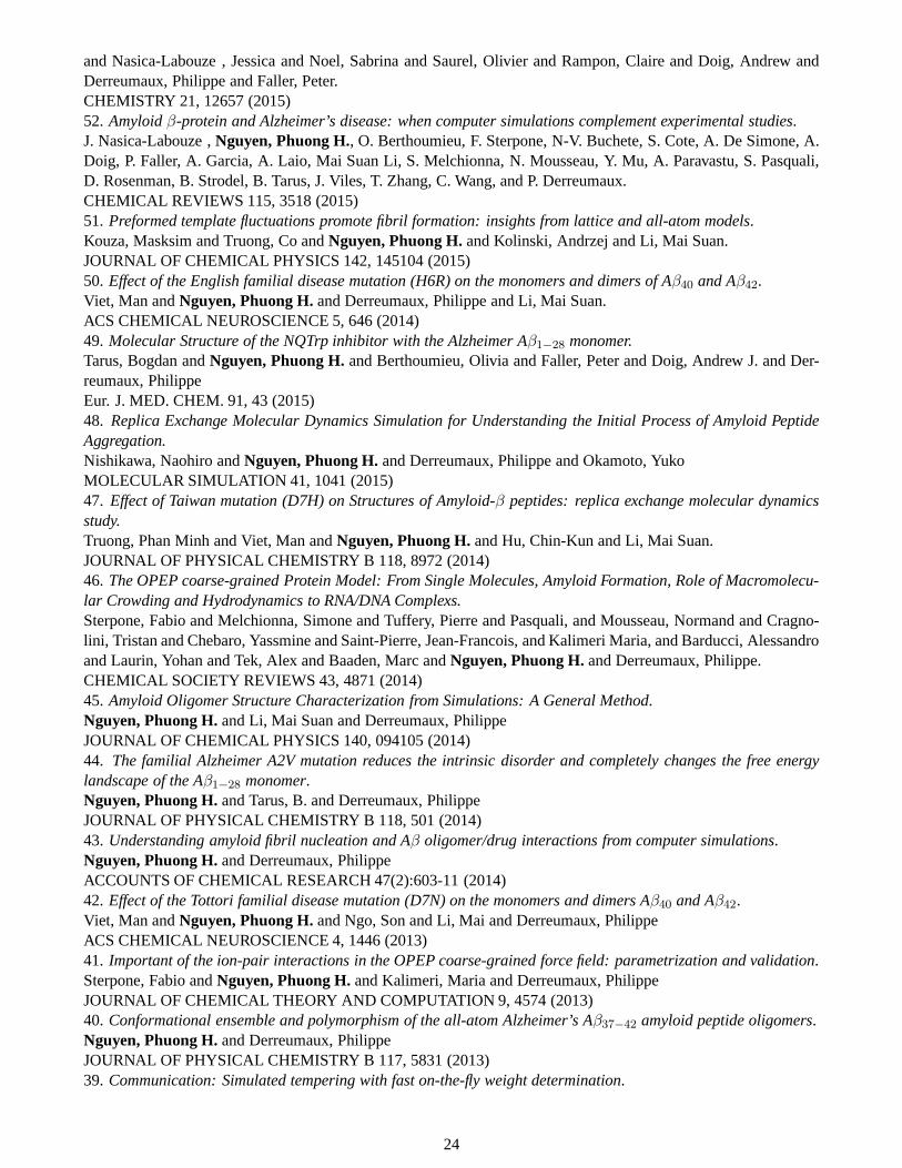
and Nasica-Labouze , Jessica and Noel, Sabrina and Saurel, Olivier and Rampon, Claire and Doig, Andrew andDerreumaux, Philippe and Faller, Peter.CHEMISTRY 21, 12657 (2015)52. Amyloidβ-protein and Alzheimer’s disease: when computer simulations complement experimental studies.J. Nasica-Labouze ,Nguyen, Phuong H., O. Berthoumieu, F. Sterpone, N-V. Buchete, S. Cote, A. De Simone, A.Doig, P. Faller, A. Garcia, A. Laio, Mai Suan Li, S. Melchionna, N. Mousseau, Y. Mu, A. Paravastu, S. Pasquali,D. Rosenman, B. Strodel, B. Tarus, J. Viles, T. Zhang, C. Wang, and P. Derreumaux.CHEMICAL REVIEWS 115, 3518 (2015)51. Preformed template fluctuations promote fibril formation: insights from lattice and all-atom models.Kouza, Masksim and Truong, Co andNguyen, Phuong H.and Kolinski, Andrzej and Li, Mai Suan.JOURNAL OF CHEMICAL PHYSICS 142, 145104 (2015)50. Effect of the English familial disease mutation (H6R) on themonomers and dimers of Aβ40 and Aβ42.Viet, Man andNguyen, Phuong H.and Derreumaux, Philippe and Li, Mai Suan.ACS CHEMICAL NEUROSCIENCE 5, 646 (2014)49. Molecular Structure of the NQTrp inhibitor with the Alzheimer Aβ1−28 monomer.Tarus, Bogdan andNguyen, Phuong H.and Berthoumieu, Olivia and Faller, Peter and Doig, Andrew J. and Der-reumaux, PhilippeEur. J. MED. CHEM. 91, 43 (2015)48. Replica Exchange Molecular Dynamics Simulation for Understanding the Initial Process of Amyloid PeptideAggregation.Nishikawa, Naohiro andNguyen, Phuong H.and Derreumaux, Philippe and Okamoto, YukoMOLECULAR SIMULATION 41, 1041 (2015)47. Effect of Taiwan mutation (D7H) on Structures of Amyloid-β peptides: replica exchange molecular dynamicsstudy.Truong, Phan Minh and Viet, Man andNguyen, Phuong H.and Hu, Chin-Kun and Li, Mai Suan.JOURNAL OF PHYSICAL CHEMISTRY B 118, 8972 (2014)46. The OPEP coarse-grained Protein Model: From Single Molecules, Amyloid Formation, Role of Macromolecu-lar Crowding and Hydrodynamics to RNA/DNA Complexs.Sterpone, Fabio and Melchionna, Simone and Tuffery, Pierreand Pasquali, and Mousseau, Normand and Cragno-lini, Tristan and Chebaro, Yassmine and Saint-Pierre, Jean-Francois, and Kalimeri Maria, and Barducci, Alessandroand Laurin, Yohan and Tek, Alex and Baaden, Marc andNguyen, Phuong H.and Derreumaux, Philippe.CHEMICAL SOCIETY REVIEWS 43, 4871 (2014)45. Amyloid Oligomer Structure Characterization from Simulations: A General Method.Nguyen, Phuong H.and Li, Mai Suan and Derreumaux, PhilippeJOURNAL OF CHEMICAL PHYSICS 140, 094105 (2014)44. The familial Alzheimer A2V mutation reduces the intrinsic disorder and completely changes the free energylandscape of the Aβ1−28 monomer.Nguyen, Phuong H.and Tarus, B. and Derreumaux, PhilippeJOURNAL OF PHYSICAL CHEMISTRY B 118, 501 (2014)43. Understanding amyloid fibril nucleation and Aβ oligomer/drug interactions from computer simulations.Nguyen, Phuong H.and Derreumaux, PhilippeACCOUNTS OF CHEMICAL RESEARCH 47(2):603-11 (2014)42. Effect of the Tottori familial disease mutation (D7N) on themonomers and dimers Aβ40 and Aβ42.Viet, Man andNguyen, Phuong H.and Ngo, Son and Li, Mai and Derreumaux, PhilippeACS CHEMICAL NEUROSCIENCE 4, 1446 (2013)41. Important of the ion-pair interactions in the OPEP coarse-grained force field: parametrization and validation.Sterpone, Fabio andNguyen, Phuong H.and Kalimeri, Maria and Derreumaux, PhilippeJOURNAL OF CHEMICAL THEORY AND COMPUTATION 9, 4574 (2013)40. Conformational ensemble and polymorphism of the all-atom Alzheimer’s Aβ37−42 amyloid peptide oligomers.Nguyen, Phuong H.and Derreumaux, PhilippeJOURNAL OF PHYSICAL CHEMISTRY B 117, 5831 (2013)39. Communication: Simulated tempering with fast on-the-fly weight determination.
24
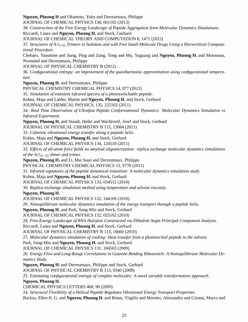
Nguyen, Phuong Hand Okamoto, Yuko and Derreumaux, PhilippeJOURNAL OF CHEMICAL PHYSICS 138, 061102 (2013)38. Construction of the Free Energy Landscape of Peptide Aggregation from Molecular Dynamics Simulations.Riccardi, Laura andNguyen, Phuong H.and Stock, GerhardJOURNAL OF CHEMICAL THEORY AND COMPUTATION 8, 1471 (2012)37. Structures of Aβ1742 Trimers in Isolation and with Five Small-Molecule Drugs Using a Hierarchical Computa-tional Procedure.Chebaro, Yassmine and Jiang, Ping and Zang, Tong and Mu, Yuguang andNguyen, Phuong H.and Mousseau,Normand and Derreumaux, PhilippeJOURNAL OF PHYSICAL CHEMISTRY B (2012)36. Configurational entropy: an improvement of the quasiharmonic approximation using configurational tempera-ture.Nguyen, Phuong H.and Derreumaux, PhilippePHYSICAL CHEMISTRY CHEMICAL PHYSICS 14, 877 (2012)35. Simulation of transient infrared spectra of a photoswitchable peptide.Kobus, Maja and Lieder, Martin andNguyen, Phuong H.and Stock, GerhardJOURNAL OF CHEMICAL PHYSICS, 135, 225102 (2011)34. Real Time Observation of Ultrafast Peptide ConformationalDynamics: Molecular Dynamics Simulation vsInfrared Experiment.Nguyen, Phuong H.and Staudt, Heike and Wachtveitl, Josef and Stock, GerhardJOURNAL OF PHYSICAL CHEMISTRY B 115, 13084 (2011)33. Coherent vibrational energy transfer along a peptide helix.Kobus, Maja andNguyen, Phuong H.and Stock, GerhardJOURNAL OF CHEMICAL PHYSICS 134, 124518 (2011)32. Effects of all-atom force fields on amyloid oligomerization: replica exchange molecular dynamics simulationsof the Aβ16−22 dimer and trimer.Nguyen, Phuong H.and Li, Mai Suan and Derreumaux, PhilippePHYSICAL CHEMISTRY CHEMICAL PHYSICS 13, 9778 (2011)31. Infrared signatures of the peptide dynamical transition: Amolecular dynamics simulation study.Kobus, Maja andNguyen, Phuong H.and Stock, GerhardJOURNAL OF CHEMICAL PHYSICS 133, 034512 (2010)30. Replica exchange simulation method using temperature and solvent viscosity.Nguyen, Phuong H.JOURNAL OF CHEMICAL PHYSICS 132, 144109 (2010)29. Nonequilibrium molecular dynamics simulation of the energy transport through a peptide helix.Nguyen, Phuong H.and Park, Sang-Min and Stock, GerhardJOURNAL OF CHEMICAL PHYSICS 132, 025102 (2010)28. Free-Energy Landscape of RNA Hairpins Constructed via Dihedral Angle Principal Component Analysis.Riccardi, Laura andNguyen, Phuong H.and Stock, GerhardJOURNAL OF PHYSICAL CHEMISTRY B 113, 16660 (2010)27. Molecular dynamics simulation of cooling: Heat transfer from a photoexcited peptide to the solvent.Park, Sang-Min andNguyen, Phuong H.and Stock, GerhardJOURNAL OF CHEMICAL PHYSICS 131, 184503 (2009)26. Energy Flow and Long-Range Correlations in Guanine-Binding Riboswitch: A Nonequilibrium Molecular Dy-namics Study.Nguyen, Phuong H.and Derreumaux, Philippe and Stock, GerhardJOURNAL OF PHYSICAL CHEMISTRY B 113, 9340 (2009)25. Estimating configurational entropy of complex molecules: Anovel variable transformation approach.Nguyen, Phuong H.CHEMICAL PHYSICS LETTERS 468, 90 (2009)24. Structural Flexibility of a Helical Peptide Regulates Vibrational Energy Transport Properties.Backus, Ellen H. G. andNguyen, Phuong H.and Botan, Virgiliu and Moretto, Alessandro and Crisma, Marco and
25
Toniolo, Claudio and Zerbe, Oliver and Stock, Gerhard and Hamm, PeterJOURNAL OF PHYSICAL CHEMISTRY B 112, 15487 (2009)23. Energy transport in peptide helices: A comparison between high- and low-energy excitations.Backus, Ellen H. G. andNguyen, Phuong H.and Botan, Virgiliu and Pfister, Rolf and Moretto, Alessandro andCrisma, Marco and Toniolo, Claudio and Stock, Gerhard and Hamm, PeterJOURNAL OF PHYSICAL CHEMISTRY B 112, 9091 (2008)22. Construction of the free energy landscape of biomolecules via dihedral angle principal component analysis.Altis, Alexandros and Otten, Moritz andNguyen, Phuong H.and Hegger, Rainer and Stock, GerhardJOURNAL OF CHEMICAL PHYSICS 128, 245102 (2008)21. Nonadiabatic vibrational dynamics and spectroscopy of peptides: A quantum-classical description.Kobus, Maja and Gorbunov, Roman D. andNguyen, Phuong H.and Stock, GerhardCHEMICAL PHYSICS 347, 208, 200820. Energy transport in peptide helices.Botan, Virgiliu and Backus, Ellen H. G. and Pfister, Rolf and Moretto, Alessandro and Crisma, Marco and Toniolo,Claudio andNguyen, Phuong H.and Stock, Gerhard and Hamm, PeterPROCEEDINGS OF THE NATIONAL ACADEMY OF SCIENCES OF THE UNITED STATES OF AMERICA104, 12749 (2007)19. Dihedral angle principal component analysis of molecular dynamics simulations.Altis, Alexandros andNguyen, Phuong H.and Hegger, Rainer and Stock, GerhardJOURNAL OF CHEMICAL PHYSICS 126, 244111 (2007)18. Conformational states and folding pathways of peptides revealed by principal-independent component analy-ses.Nguyen, Phuong H.PROTEINS-STRUCTURE FUNCTION AND BIOINFORMATICS 67,579 (2007)17. Structure and dynamics of the homologous series of alanine peptides: A joint molecular dynamics/NMR study.Graf, Juergen andNguyen, Phuong H.and Stock, Gerhard and Schwalbe, HaraldJOURNAL OF THE AMERICAN CHEMICAL SOCIETY 129, 1179 (2007)16. Quantum-classical description of the amide I vibrational spectrum of trialanine.Gorbunov, Roman D. andNguyen, Phuong H.and Kobus, Maja and Stock, GerhardJOURNAL OF CHEMICAL PHYSICS 126, 054509 (2007)15. How complex is the dynamics of peptide folding?Hegger, Rainer and Altis, Alexandros andNguyen, Phuong H.and Stock, GerhardPHYSICAL REVIEW LETTERS 98, 028102 (2007)14. Monomer adds to preformed structured oligomers of Aβ-peptides by a two-stage dock-lock mechanism.Nguyen, Phuong H.and Li, Mai Suan and Stock, Gerhard and Straub, John E. and Thirumalai, D.PROCEEDINGS OF THE NATIONAL ACADEMY OF SCIENCES OF THE UNITED STATES OF AMERICA104, 111 (2007)13. Complexity of free energy landscapes of peptides revealed by nonlinear principal component analysis.Nguyen, Phuong H.PROTEINS-STRUCTURE FUNCTION AND BIOINFORMATICS 65, 898 (2006)12. Photoinduced conformational dynamics of a photoswitchable peptide: A nonequilibrium molecular dynamicssimulation study.Nguyen, Phuong H.and Gorbunov, Roman D. and Stock, GerhardBIOPHYSICAL JOURNAL 91, 1224 (2006)11. Improved Wang-Landau sampling through the use of smoothed potential-energy surfaces.Nguyen, Phuong H.and Mittag, E and Torda, AE and Stock, GerhardJOURNAL OF CHEMICAL PHYSICS 124, 154107 (2006)10. Nonequilibrium molecular dynamics simulation of a photoswitchable peptide.Nguyen, Phuong H.and Stock, GerhardCHEMICAL PHYSICS 323, 36 (2006)9. Free energy landscape and folding mechanism of aβ-hairpin in explicit water: A replica exchange moleculardynamics study.
26

Nguyen, Phuong H.and Stock, G and Mittag, E and Hu, CK and Li, MSPROTEINS-STRUCTURE FUNCTION AND BIOINFORMATICS 61, 795 (2005)8. Structure and energy landscape of a photoswitchable peptide: A replica exchange molecular dynamics study.Nguyen, Phuong H.and Mu, YG and Stock, GPROTEINS-STRUCTURE FUNCTION AND BIOINFORMATICS 60, 485 (2005)7. Reply to the Comment on ”Energy Landscape of a Small Peptide Revealed by Dihedral Angle Principal Compo-nent Analysis.Mu, YG andNguyen, Phuong H.and Stock, GPROTEINS-STRUCTURE FUNCTION AND BIOINFORMATICS 64, 798 (2006)6. Energy landscape of a small peptide revealed by dihedral angle principal component analysis.Mu, YG andNguyen, Phuong H.and Stock, GPROTEINS-STRUCTURE FUNCTION AND BIOINFORMATICS 58, 45 (2005)5. Nonequilibrium molecular-dynamics study of the vibrational energy relaxation of peptides in water.Nguyen, Phuong H.and Stock, GJOURNAL OF CHEMICAL PHYSICS 119, 11350 (2003)4. Local structure in nematic and isotropic liquid crystals.Nguyen, Phuong H.and Schmid, FJOURNAL OF CHEMICAL PHYSICS 119, 1214 (2003)3. The direct correlation function in nematic liquid crystalsfrom computer simulation.Nguyen, Phuong H.and Germano, G and Schmid, FCOMPUTER PHYSICS COMMUNICATIONS 147, 350 (2002)2. Spatial order in liquid crystals: Computer simulations of systems of ellipsoids.Schmid, F andNguyen, Phuong H.MORPHOLOGY OF CONDENSED MATTER: PHYSICS AND GEOMETRY OF SPATIALLY COMPLEX SYS-TEMS, Lecture Notes in Physics 600, 172 (2002)1.Elastic constants from direct correlation functions in nematic liquid crystals: A computer simulation study.Nguyen, Phuong H.and Germano, G and Schmid, FJOURNAL OF CHEMICAL PHYSICS 115, 7227 (2001)
(c) Selected oral communications1. 2002-2008: 8 talks at the Huefeld workshop ”Computer simulation and theory of macromolecules” organizedevery year, Germany.2. 2007: Seminar at the Centre for Self Organising MolecularSystems, University of Leeds, UK.3. 2007: Seminar at the School of Chemical and Biomedical Engineering, Nanyang Technological University,Singapore4. 2011: Seminar at the School of Biological Sciences, Nanyang Technological University, Singapore5. 2012: Speaker at the CECAM workshop ”Anchoring simulations to experiments: challenges for understandingand treating Alzheimer’s disease”, Paris, France.6. 2013: Seminar at the Department of Physics, Freiburg University, Germany7. 2013: Speaker at the ”Journees Modelisation”, Paris, France.8. 2014: Invited speaker at the ”International Conference on Computational Science and Technology”, Warsaw,Poland.
4 Future research projects
4.1 Large-scale nonequilibrium molecular dynamics simulations and experiments to study thefocused ultrasound-induced opening of the blood-brain-barrier
In the near future my research will focus on the study of the ultrasound-induced blood-brain-barrier (BBB) openingfor drug delivery to treat brain diseases. Because of the BBB, designing therapeutic agents for the brain is verychallenging. Indeed, only about 5% of the 7000 known potential molecular drugs can cross the BBB to treatneurodegenerative diseases. For AD, only up to 0.1% of anti-Aβ antibodies administered peripherally can reachthe brain, most remain in the bloodstream. Even small drugs can cross the BBB, but they are usually so dynamic
27
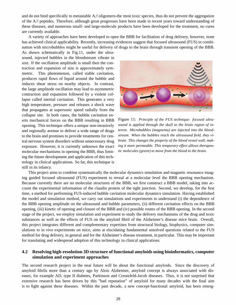
and do not bind specifically to metastable Aβ oligomers-the most toxic species, thus do not prevent the aggregationof the Aβ peptides. Therefore, although great progresses have been made in recent years toward understanding ofthese diseases, and numerous small- and large-molecule products have been developed for the treatment, no curesare currently available.
A variety of approaches have been developed to open the BBB for facilitation of drug delivery, however, nonehas achieved clinical applicability. Recently, increasing evidences suggest that focused ultrasound (FUS) in combi-nation with microbubbles might be useful for delivery of drugs to the brain through transient opening of the BBB.
Figure 11: Principle of the FUS technique: focused ultra-sound is applied through the skull to the brain region of in-terest. Microbubbles (magnenta) are injected into the blood-stream. When the bubbles reach the ultrasound field, they vi-brate. This changes the property of the blood vessel wall, mak-ing it more permeable. This temporary effect allows therapeu-tic molecules (green) to move from the blood to the brain.
As shown schematically in Fig.11, under the ultra-sound, injected bubbles in the bloodstream vibrate insize. If the oscillation amplitude is small then the con-traction and expansion of size is approximately sym-metric. This phenomenon, called stable cavitation,produces rapid flows of liquid around the bubble andinduces shear stress on nearby objects. In contrast,the large amplitude oscillation may lead to asymmetriccontraction and expansion followed by a violent col-lapse called inertial cavitation. This generates a veryhigh temperature, pressure and releases a shock wavethat propagates at supersonic speed radially from thecollapse site. In both cases, the bubble cavitation ex-erts mechanical forces on the BBB resulting in BBBopening. This technique offers a unique non-invasivelyand regionally avenue to deliver a wide range of drugsto the brain and promises to provide treatments for cen-tral nervous system disorders without unnecessary drugexposure. However, it is currently unknown the exactmolecular mechanisms in opening the BBB, thus limit-ing the future development and application of this tech-nology in clinical applications. So far, this technique isstill in its infancy.
This project aims to combine systematically the molecular dynamics simulation and magnetic resonance imag-ing guided focused ultrasound (FUS) experiment to reveal ata molecular level the BBB opening mechanism.Because currently there are no molecular structures of the BBB, we first construct a BBB model, taking into ac-count the experimental information of the claudin protein of the tight junction. Second, we develop, for the firsttime, a method for performing FUS-induced bubble cavitation molecular dynamics simulation. Having establishedthe model and simulation method, we carry out simulations and experiments to understand (i) the dependence ofthe BBB opening amplitude on the ultrasound and bubble parameters, (ii) different cavitation effects on the BBBopening, (iii) kinetic of opening and closure of the BBB and (iv) possible routes of the BBB opening. In the secondstage of the project, we employ simulation and experiment tostudy the delivery mechanisms of the drug and toxicsubstances as well as the effects of FUS on the amyloid fibril of the Alzheimer’s disease mice brain. Overall,this project integrates different and complementary expertises from structural biology, biophysics, computer sim-ulations to in vivo experiments on mice, aims at elucidatingfundamental unsolved questions related to the FUSmethod for drug delivery, in general and for the Alzheimer’sdisease treatment, in particular. This may be importantfor translating and widespread adoption of this technologyin clinical applications.
4.2 Resolving high-resolution 3D structure of functional amyloids using bioinformatics, computersimulation and experiment approaches
The second research project in the near future will be about the functional amyloids. Since the discovery ofamyloid fibrils more than a century ago by Alois Alzheimer, amyloid concept is always associated with dis-eases, for example AD, type II diabetes, Parkinson and Creutzfeld-Jacob diseases. Thus, it is not surprised thatextensive research has been driven by this ”bad reputation”of amyloid for many decades with the final aimis to fight against these diseases. Within the past decade, a new concept-functional amyloid, has been emerg-
28
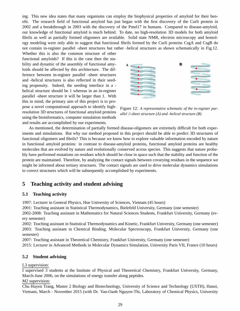
ing. This new idea states that many organisms can employ the biophysical properties of amyloid for their ben-efit. The research field of functional amyloid has just began with the first discovery of the Curli protein in2002 and a breakthrough in 2003 with the discovery of the Pmel17 in humans. Compared to disease-amyloid,our knowledge of functional amyloid is much behind. To date,no high-resolution 3D models for both amyloidfibrils as well as partially formed oligomers are available.Solid state NMR, electron microscopy and homol-ogy modeling were only able to suggest that functional fibrils formed by the Curli proteins CsgA and CsgB donot contain in-register parallel -sheet structures but rather -helical structures as shown schematically in Fig.12.
Figure 12:A representative schematic of the in-register par-allel β-sheet structure (A) and -helical structure (B)
Whether this is also the common structure of otherfunctional amyloids? If this is the case then the sta-bility and dynamic of the assembly of functional amy-loids should be affected by this architecture. The dif-ference between in-register parallel -sheet structuresand -helical structures is also reflected in their seed-ing propensity. Indeed, the seeding interface in a -helical structure should be 1 whereas in an in-registerparallel -sheet structure it will be larger than 1. Withthis in mind, the primary aim of this project is to pro-pose a novel computational approach to identify high-resolution 3D structures of functional amyloid proteinsusing the bioinformatics, computer simulation methodsand results are accomplished by our experiments.
As mentioned, the determination of partially formed disease-oligomers are extremely difficult for both exper-iments and simulations. But why our method proposed in this project should be able to predict 3D structures offunctional oligomers and fibrils? This is because we know howto explore valuable information encoded by naturein functional amyloid proteins: in contrast to disease-amyloid proteins, functional amyloid proteins are healthymolecules that are evolved by nature and evolutionally conserved across species. This suggests that nature proba-bly have performed mutations on residues which should be close in space such that the stability and function of theprotein are maintained. Therefore, by analyzing the contact signals between covarying residues in the sequence wemight be informed about tertiary structures. The contact signals are used to drive molecular dynamics simulationsto correct structures which will be subsequently accomplished by experiments.
5 Teaching activity and student advising
5.1 Teaching activity
1997: Lecturer in General Physics, Hue University of Sciences, Vietnam (45 hours)2001: Teaching assistant in Statistical Thermodynamics, Bielefeld University, Germany (one semester)2002-2008: Teaching assistant in Mathematics for Natural Sciences Students, Frankfurt University, Germany (ev-ery semester)2002: Teaching assistant in Statistical Thermodynamics and Kinetic, Frankfurt University, Germany (one semester)2003: Teaching assistant in Chemical Binding, Molecular Spectroscopy, Frankfurt University, Germany (onesemester)2007: Teaching assistant in Theoretical Chemistry, Frankfurt University, Germany (one semester)2015: Lecturer in Advanced Methods in Molecular Dynamics Simulation, University Paris VII, France (10 hours)
5.2 Student advising
L3 supervision:I supervised 3 students at the Institute of Physical and Theoretical Chemistry, Frankfurt University, Germany,March-June 2006, on the simulations of energy transfer along peptides.M2 supervision:Chu Huyen Trang, Master 2 Biology and Biotechnology, University of Science and Technology (USTH), Hanoi,Vietnam, March - November 2015 (with Dr. Van-Oanh Nguyen-Thi, Laboratory of Chemical Physics, University
29
Paris-Sud). I worked closely with the student on the effectsof force fields on the laser-induced disssociation ofamyloid fibrils.PhD supervision:1. Sang-Min Park, PhD student, Institute of Physical and Theoretical Chemistry, Frankfurt University, Germany,2005 - 2008 (Official advisor: Professor Gerhard Stock). I worked directly with the student on the simulations ofenergy transfer along peptides. We had 2 papers together.2. Jessica Koplin, PhD student, Institute of Physical and Theoretical Chemistry, Frankfurt University, Germany,2002 - 2006 (Official advisor: Professor Gerhard Stock). I worked directly with the student on the simulations ofRNA systems.3. Thanh Thuy Tran, PhD student, Laboratoire de Biochimie Theorique, UPR 9080 CNRS, 2013-2016 (withProfessor Philippe Derreumaux). I work directly with the student on the construction of the coarse-grained proteinmodel for studying of the amyloid protein aggregation. We have had 2 papers together.
6 Administrative and community service activities
- Regular reviewer for the scientific journals including J. Am. Chem. Soc., J. Phys. Chem. B, J. Chem. Phys., Phys.Chem. Chem. Phys, Nanoscale, Chem. Phys, Lett., RSC Advances, Int. J. Mol. Sci., Biochemistry, BiophysicsReport, Expert Opinion on Drug Discovery, ACS Chemical Neuroscience.- Member de conseil de Laboratoire de Biochimie Theorique, UPR 9080 CNRS.
References
[1] P. G. de Gennes and J. Prost.The Physics of Liquid Crystals. Oxford University Press, Oxford, 1993.
[2] B. J. Berne and P. Pechukas.J. Chem. Phys., 56:4213, 1975.
[3] J. P. Hansen and I. R. McDonald.Theory of Simple Liquids. Academic, London, 1986.
[4] P. H. Nguyen and F. Schmid.J. Chem. Phys., 119:1214, 2003.
[5] D. Forster. Hydrodynamic fluctuations, broken symmetry and correlation functions. Frontiers in Physics,Benjamin, Reading, MA, 1975.
[6] C. Frank.Discuss. Faraday Soc., 25:19, 1958.
[7] P. H. Nguyen, G. Germano, and F. Schmid.J. Chem. Phys., 115:7227, 2001.
[8] P. H. Nguyen and G. Stock.J. Chem. Phys., 119:11350, 2003.
[9] V. Botan, E. H. G. Backus, R. Pfister, et al.Proc. Natl. Acad. Sci. USA, 104:1274912754, 2007.
[10] E. H. G. Backus, P. H. Nguyen, V. Botan, et al.J. Phys. Chem. B, 112:9091, 2008.
[11] P. H. Nguyen and G. Stock.Chem. Phys., 323:36, 2006.
[12] P. H. Nguyen, R. D. Gorbunov, and G. Stock.Biophys. J, 91:1224, 2006.
[13] P. H. Nguyen, H. Staudt, J. Wachtveitl, and G. Stock.J. Phys. Chem. B, 115:13084–13092, 2011.
[14] R. D. Gorbunov, P. H. Nguyen, M. Kobus, and G. Stock.J. Chem. Phys., 126:054509, 2007.
[15] M. Kobus, R. D. Gorbunov, P. H. Nguyen, and G. Stock.Chem. Phys., 347:208, 2008.
[16] M. Kobus, M. Lieder, P. H. Nguyen, and G. Stock.J. Chem. Phys., 135:225102, 2011.
[17] Y. Mu, P. H. Nguyen, and G. Stock.Proteins, 58:45–52, 2005.
[18] A. Altis, P. H. Nguyen, R. Hegger, and G. Stock.J. Chem. Phys., 126:244111, 2007.
30
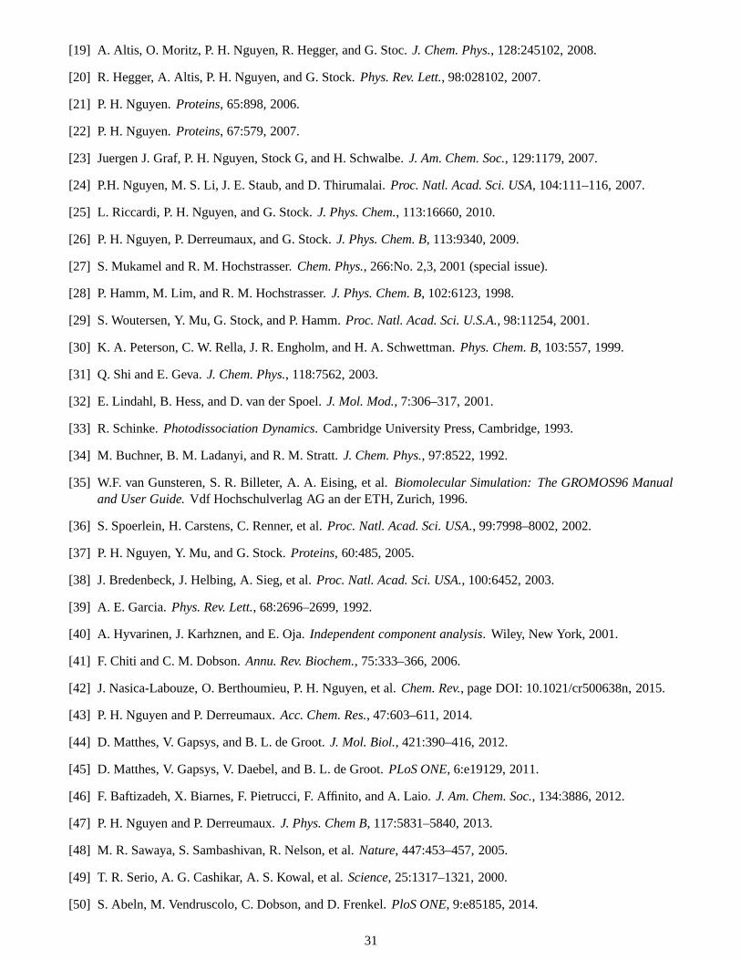
[19] A. Altis, O. Moritz, P. H. Nguyen, R. Hegger, and G. Stoc.J. Chem. Phys., 128:245102, 2008.
[20] R. Hegger, A. Altis, P. H. Nguyen, and G. Stock.Phys. Rev. Lett., 98:028102, 2007.
[21] P. H. Nguyen.Proteins, 65:898, 2006.
[22] P. H. Nguyen.Proteins, 67:579, 2007.
[23] Juergen J. Graf, P. H. Nguyen, Stock G, and H. Schwalbe.J. Am. Chem. Soc., 129:1179, 2007.
[24] P.H. Nguyen, M. S. Li, J. E. Staub, and D. Thirumalai.Proc. Natl. Acad. Sci. USA, 104:111–116, 2007.
[25] L. Riccardi, P. H. Nguyen, and G. Stock.J. Phys. Chem., 113:16660, 2010.
[26] P. H. Nguyen, P. Derreumaux, and G. Stock.J. Phys. Chem. B, 113:9340, 2009.
[27] S. Mukamel and R. M. Hochstrasser.Chem. Phys., 266:No. 2,3, 2001 (special issue).
[28] P. Hamm, M. Lim, and R. M. Hochstrasser.J. Phys. Chem. B, 102:6123, 1998.
[29] S. Woutersen, Y. Mu, G. Stock, and P. Hamm.Proc. Natl. Acad. Sci. U.S.A., 98:11254, 2001.
[30] K. A. Peterson, C. W. Rella, J. R. Engholm, and H. A. Schwettman.Phys. Chem. B, 103:557, 1999.
[31] Q. Shi and E. Geva.J. Chem. Phys., 118:7562, 2003.
[32] E. Lindahl, B. Hess, and D. van der Spoel.J. Mol. Mod., 7:306–317, 2001.
[33] R. Schinke.Photodissociation Dynamics. Cambridge University Press, Cambridge, 1993.
[34] M. Buchner, B. M. Ladanyi, and R. M. Stratt.J. Chem. Phys., 97:8522, 1992.
[35] W.F. van Gunsteren, S. R. Billeter, A. A. Eising, et al.Biomolecular Simulation: The GROMOS96 Manualand User Guide.Vdf Hochschulverlag AG an der ETH, Zurich, 1996.
[36] S. Spoerlein, H. Carstens, C. Renner, et al.Proc. Natl. Acad. Sci. USA., 99:7998–8002, 2002.
[37] P. H. Nguyen, Y. Mu, and G. Stock.Proteins, 60:485, 2005.
[38] J. Bredenbeck, J. Helbing, A. Sieg, et al.Proc. Natl. Acad. Sci. USA., 100:6452, 2003.
[39] A. E. Garcia.Phys. Rev. Lett., 68:2696–2699, 1992.
[40] A. Hyvarinen, J. Karhznen, and E. Oja.Independent component analysis. Wiley, New York, 2001.
[41] F. Chiti and C. M. Dobson.Annu. Rev. Biochem., 75:333–366, 2006.
[42] J. Nasica-Labouze, O. Berthoumieu, P. H. Nguyen, et al.Chem. Rev., page DOI: 10.1021/cr500638n, 2015.
[43] P. H. Nguyen and P. Derreumaux.Acc. Chem. Res., 47:603–611, 2014.
[44] D. Matthes, V. Gapsys, and B. L. de Groot.J. Mol. Biol., 421:390–416, 2012.
[45] D. Matthes, V. Gapsys, V. Daebel, and B. L. de Groot.PLoS ONE, 6:e19129, 2011.
[46] F. Baftizadeh, X. Biarnes, F. Pietrucci, F. Affinito, and A. Laio. J. Am. Chem. Soc., 134:3886, 2012.
[47] P. H. Nguyen and P. Derreumaux.J. Phys. Chem B, 117:5831–5840, 2013.
[48] M. R. Sawaya, S. Sambashivan, R. Nelson, et al.Nature, 447:453–457, 2005.
[49] T. R. Serio, A. G. Cashikar, A. S. Kowal, et al.Science, 25:1317–1321, 2000.
[50] S. Abeln, M. Vendruscolo, C. Dobson, and D. Frenkel.PloS ONE, 9:e85185, 2014.
31

[51] F. Sterpone, S. Melchionna, P. Tuffery, et al.Chem. Rev. Soc., 43:4871, 2015.
[52] T. T. Trand, P. H. Nguyen, and P. Derreumaux.J. Chem. Phys., 144:205103, 2016.
[53] B. Tarus, T. Tran, J. Nassica-Labouze, et al.J. Phys. Chem B, 119:10478–10487, 2015.
[54] B. Tarus, P. H. Nguyen, O. Berthoumieu, et al.Eur. J. MED. CHEM., 91:43–50, 2015.
[55] O. Berthoumieu, P. H. Nguyen, M. del Castillo-Frias, etal. Chemistry- a European journal, DOI:10.1002/chem.201500888, 2015.
[56] R. Scherzer-Attali, R. Pellarin, M. Convertino, et al.PLOS One., 5:e11101, 2010.
[57] E. Mikros, D. Benaki, E. Humpfer, et al.Angew. Chem. Int. Ed. Engl, 40:3603e3605, 2001.
[58] B.D. Moore, P. Chakrabarty, Y. Levites, et al.Alzheimers Res. Ther., 4:18, 2012.
[59] E. Martineau, J.M. de-Guzman, L. Rodionova, et al.J. Am. Soc. Mass. Spectrom, 21:1506e1514, 2010.
[60] T. Jonsson, J. K. Atwal, S. Steinberg, et al.Nature, 488:96–99, 2012.
[61] G. DiFede, M. Catania, M. Morbin, et al.Science, 323:1473–1477, 2009.
[62] P. H. Nguyen, B. Tarus, and P. Derreumaux.J. Phys. Chem. B, 118:501, 2014.
[63] P. H. Nguyen, B. Tarus, and P. Derreumaux. in preparation.
[64] M. H. Viet, P.H. Nguyen, P. Derreumaux, and M.S. Li.ACS Chem. Neuroscience, 5:646, 2014.
[65] M. H. Viet, P.H. Nguyen, N. S. Tung, M.S. Li, and P. P. Derreumaux. ACS Chem. Neuroscience, 4:1446,2013.
[66] P. M. Truong, M. H. Viet, P.H. Nguyen, C. K. Hu, and M.S. Li. J. Phys. Chem. B, 118:8972, 2014.
[67] T. Kawasaki, J. Fujioka, T. Imai, and K. Tsukiyama.The Protein Journal, 31:710–716, 2012.
[68] T. Kawasaki, J. Fujioka, T. Imai, K. Torigoe, and K. Tsukiyama. Lasers in Medical Science, 29:1701–1707,2014.
[69] T. Kawasaki, T. Imai, and K. Tsukiyama.Journal of Analytical Sciences, Methods and Instrumentation,4:9–18, 2014.
[70] T. Kawasaki, T. Yaji, T. Imai, T. Ohta, and K. Tsukiyama.American Journal of Analytical Chemistry, 5:384–394, 2014.
[71] M. H. Viet, P. Derreumaux, M. S. Li, et al.J. Chem. Phys., 143:155101, 2015.
[72] M. H. Viet, P. M. Truong, P. Derreumaux, et al.Phys. Chem. Chem. Phys, 17:27275, 2015.
[73] M. H. Viet, V. O. Nguyen, P. Derreumaux, et al.Phys. Chem. Chem. Phys., 10.1039/C5CP07711G, 2016.
[74] F. Wang and D. P. Landau.Phys. Rev. Lett., 86:2050, 2001.
[75] P. H. Nguyen, E. Mittag, A. E. Torda, and G. Stock.J. Chem. Phys., 124:154107, 2006.
[76] P. H. Nguyen.J. Chem. Phys., 132:144109, 2010.
[77] P. H. Nguyen, Y. Okamoto, and P. Derreumaux.J. Chem. Phys., 138:061102, 2013.
[78] T. Zhang, P. H. Nguyen, J. Nassica-Labouze, Y. Mu, and P.Derreumaux.J. Phys. Chem. B, 119:6941, 2015.
[79] A. P. Lyubartsev, A. A. Martinovski, S. V. Shevkunov, and P. N. Vorontsov-Velyaminov.J. Chem. Phys.,96:1776–1783, 1992.
32
[80] E. Marinari and G. Parisi.Europhys. Lett., 19:451–458, 1992.
[81] M. H. Viet, P. Derreumaux, and P. H. Nguyen.J. Chem. Phys., 143:021101, 2015.
[82] P. H. Nguyen, M. S. Li, and P. Derreumaux.J. Chem. Phys., 140:094105, 2014.
[83] D. Chandler.Introduction to modern statistical mechanics. Oxford university press, New York, 1987.
[84] K. A. Dill. Biochemistry, 29:7133–7155, 1990.
[85] R.S. Spolar and M.T. Record Jr.Science, 263:777, 1994.
[86] M. Karplus and J. N. Kushick.Macromolecules, 14:325, 1981.
[87] P. H. Nguyen.Chem. Phys. Lett., 468:90, 2009.
[88] H. H. Rugh.Phys. Rev. Lett., 78:999, 1997.
[89] P. H. Nguyen and P. Derreumaux.Phys. Chem. Chem. Phys., 14:877–886, 2012.
33
AcknowledgmentsI am very grateful to all my supervisors for special supportsthat they had always gave to me along the years.
I must in particular thanks Dr. Sandro Scandolo who introduced me to the field of computer simulation of con-densed matter physics during my first difficult days in Italy.Moving to Germany, I received best supports frommy supervisor Professor Friederike Schmid, and I am very grateful for her persistence and constructive feedbackthroughout my PhD, particular during those times where I felt defeated by my research. I must thank her for havingtrained me so deeply in the field of soft condensed matter simulations. I especially want to thank Professor GerhardStock for the opportunity he gave me to enter in a new field of research in biophysical chemistry and his continuousencouragement during my postdoctoral.
I especially want to thank all of my collaborators for their expertise, friendship and valuable ideas during mycareer. I especially mention Professor Mai Suan Li who introduced me to the field of protein aggregation, and opena long-term fruitful collaboration. We have shared many interesting discussion on the general topics of physicalchemistry. A special thanks to Professor Peter Hamm for his collaboration on the picosecond Infrared experimentsand Professor Harald Schwalbe for NMR experiments. I would like to express my thanks to Dr. Man Hoang Vietfor working together as well as for discussion and sharing many interesting ideas.
I would like to express my gratitude to Professor Philippe Derreumaux for the opportunity to join the Labora-toire de Biochimie Theorique as CR1 and start fruitful and productive collaboration. I am really lucky to have hadopportunity to work with him as a world-leader in the field of computer simulation of protein folding and proteinaggregation.
Finally, I would like to thank all members of the jury committee for agreeing to judge this work. I would liketo express my thanks to Professor Pedro de Oliveira for his administrative efficiency in the HDR application.
34