molecular mechanisms of action of different … · to examine the structure of the stratum corneum...
TRANSCRIPT

Biochimica et Biophysica Acta 1848 (2015) 1196–1202
Contents lists available at ScienceDirect
Biochimica et Biophysica Acta
j ourna l homepage: www.e lsev ie r .com/ locate /bbamem
Molecular mechanisms of action of different concentrations of ethanol inwater on ordered structures of intercellular lipids and soft keratin in thestratum corneum
Daisuke Horita a,d, Ichiro Hatta c,e, Masato Yoshimoto a, Yuki Kitao a, Hiroaki Todo a, Kenji Sugibayashi a,b,⁎a Faculty of Pharmaceutical Sciences, Josai University, 1-1 Keyakidai, Sakado, Saitama 350-0295, Japanb Life Science Research Center, Josai University, 1-1 Keyakidai, Sakado, Saitama 350-0295, Japanc Department of Research, Nagoya Industrial Science Research Institute, 1-13 Yotsuyadori, Chikusa-ku, Nagoya 464-0819, Japand Research Laboratories, Ikeda Mohando Co., Ltd., 16 Jinden, Kamiichi, Nakaniikawa, Toyama 930-0394, Japane Aichi Synchrotron Radiation Center, Aichi Science & Technology Foundation, 250-3 Minamiyamaguchi-cho, Seto, Aichi 489-0965, Japan
⁎ Corresponding author at: Faculty of PharmaceuticalKeyakidai, Sakado, Saitama 350-0295, Japan.
E-mail address: [email protected] (K. Sugibayashi).
http://dx.doi.org/10.1016/j.bbamem.2015.02.0080005-2736/© 2015 Elsevier B.V. All rights reserved.
a b s t r a c t
a r t i c l e i n f oArticle history:Received 5 August 2014Received in revised form 6 February 2015Accepted 10 February 2015Available online 16 February 2015
Keywords:EthanolStratum corneumIntercellular lipidsCorneocyteX-ray diffractionHairless mouse
Ethanol (EtOH) is one of the bases in topically appliedmedicines that promote the skin permeation of drugs. Al-though the effects of EtOH have been attributed to structural modifications in the stratum corneum, the under-lying mechanisms, especially the influence of different concentrations of EtOH, have not been examinedextensively. Structural modifications in the stratum corneum of hairless mouse due to the application of EtOH/water mixture were herein investigated at the molecular level using synchrotron X-ray diffraction. The resultsrevealed that all EtOH concentrations examined greatly modified the short lamellar structures containing theaqueous layer in intercellular lipids and the structure of keratin fibrils in corneocytes, which can take up hydro-philic compounds. However, the long lamellar and the hydrocarbon-chain packing structureswere unaffected byEtOH. Changes to the short lamellar structures were not proportional to the concentration of EtOH. However, thekeratin fibril structures changed gradually with increasing EtOH concentration. The X-ray diffraction experi-ments enabled the effects of different EtOH concentrations on the morphology of the stratum corneum to beassessed by using a number of experimental samples to avoid variations due to individual differences. The resultsindicated that alterations to the short lamellar structures appeared to be related to the skin permeability of drugswith the application of EtOH/watermixture, andmonotonous structural changes in the keratin fibrils with an in-crease in EtOH concentration may contribute to this permeation as supplement. These results will be useful forthe development of new drug formulations containing EtOH.
© 2015 Elsevier B.V. All rights reserved.
1. Introduction
Ethanol (EtOH) iswidely used as a skin penetration enhancer aswellas a skin disinfectant and tonic. In order for drugs to be effective at theirtarget sites, the active ingredients in topical formulations must pene-trate the skin barrier, which tightly regulates the entry of external sub-stances. This skin barrier is mainly comprised of the stratum corneum,the outermost layer of skin, which markedly restricts the penetrationof drugs. EtOH has been shown to enhance the skin permeation ofdrugs in topical formulations markedly. Estradiol and fentanyl dermalpatches are typical examples of formulations that contain EtOH inorder to enhance their absorption into the skin [1–3]. The enhance-ments reported in the skin permeation of drugs with the application
Sciences, Josai University, 1-1
of topical formulations containing EtOH have been attributed to struc-tural modifications in the stratum corneum. The various mechanismsunderlying the penetration-enhancing effects of EtOH on the stratumcorneum barrier include lipid extraction, increase in lipid fluidity, en-hancement of drug solubility in the lipids of the stratum corneum,change in skin hydration, an altered putative pore pathway, alterationin keratinized proteins, and its effects on solvent drugs [4,5]. However,the effects of EtOH on the structures of soft keratin and intercellularlipids in the stratum corneum have not been extensively examined atthe molecular level.
In the present study, we thus utilized synchrotron X-ray diffractionto examine the structure of the stratum corneum at the molecularlevel aswell as the effects of EtOH/watermixture on the structures of in-tercellular lipids and fibrils in the soft keratin. We previously demon-strated that EtOH/water mixture affected the in vitro permeability ofdrugs through pig skin in an EtOH concentration–dependent manner[6]. The skin permeability of hydrophilic drugs was increased but con-versely decreased by low and high concentrations of EtOH, respectively,

1197D. Horita et al. / Biochimica et Biophysica Acta 1848 (2015) 1196–1202
suggesting that the effects of EtOH/water mixture on the structure ofthe stratum corneum may depend on its volume ratio. In the presentstudy, we measured X-ray diffraction in the stratum corneum ofmouse skin as a function of EtOH concentration. Previous studies havealready used X-ray diffraction in hairless mouse skin [7,8], hairless ratskin [9], and pig skin [10,11]. Although there are slight differences inthe constituents of intercellular lipids among mammalian species, thelamellae have almost the same periodical structure among the mam-mals. As for the skin permeation of chemical compounds, the skin of ro-dents, such as rat and mouse, has higher permeability than pig orhuman skin, whereas the structures of the stratum corneum are similaramong these species. Among these mammals, the X-ray diffraction pro-files of the stratum corneum in hairless mice showed the most distinctdiffraction peaks, and represented the typical characteristics of the stra-tumcorneum inmammals. Therefore,we employedhairlessmouse skinin the present study to detect minute changes in the structure of thestratum corneum upon treatment with EtOH/water mixture. The dif-fraction profile of the stratum corneum has also been investigated inhumans [12,13], and has been found to have similar characteristics tothat in hairless mice.
In this study, we investigated themolecularmechanisms of action ofvarious volume ratios in EtOH/water mixture on intercellular lipids andsoft keratin based on a structural analysis using synchrotron X-ray dif-fraction experiments in stratum corneum treated with the mixture.
2. Materials and methods
2.1. Materials
EtOH (99.5%, HPLC grade), sodium chloride, disodium hydrogenphosphate, and potassium dihydrogen phosphate were obtained fromWako Pure Chemical Ind., Ltd. (Osaka, Japan). Trypsin and trypsin inhib-itor were obtained from Sigma-Aldrich Co., Ltd. (St. Louis, MO, U.S.A.).All other reagents and solvents were of reagent grade or HPLC grade,and used without further purification.
2.2. Animals
Male hairless mice (HR-1, 8–9 weeks old) were obtained fromSaitama Experimental Animals Supply Co., Ltd. (Sugito, Saitama,Japan). All animal studies were conducted according to the recommen-dations of the Institutional Board for Animal Studies, Josai University(Sakado, Saitama, Japan).
S (nm-1)
Log
arith
mic
inte
nsity
(ar
b. u
nits
)
105
104
103
102
0.0 0.5 1.0 1.5 2.0 2.5 3.0
0/100 v/v%
20/80 v/v%
40/60 v/v%
60/40 v/v%
80/20 v/v%
100/0 v/v%
(EtOH/water ratio)
Fig. 1.X-ray diffraction profiles in the hairlessmouse stratum corneum as a function of thevolume ratios in EtOH/watermixture,where n is thenumber of stratumcorneumsamples.
2.3. Sample preparation process
Hairless mouse skin was separated from the abdominal and dorsalregion. After the removal of excess fat, the skin was soaked with0.1% (w/v) trypsin in pH 7.4 phosphate-buffered saline (PBS) at 4 °Cfor 16 h. After being incubated for another 4 h at 37 °C, the stratumcorneum was separated from the skin. It was then treated with0.1% (w/v) trypsin inhibitor and rinsed in distilled water three times.Following this, EtOH/water mixture of 0/100, 20/80, 40/60, 60/40, 80/20 or 100/0 v/v% was applied to the dried stratum corneum for 2 h. Byreducing the content of EtOH/water mixture from stratum corneumtreated by the mixture the EtOH/water mixture content in the stratumcorneum was adjusted to be 20 wt%. An approximately 5-mg piece ofthe sample was placed into a capillary glass tube and immediatelysealed for the X-ray diffraction experiment. We used 25 mice in theexperiment and obtained four samples per mouse. In addition, 13–14samples were used for each experiment; the samples treated by EtOH/water mixture were randomly selected in order to avoid excessive useof a specific region or specific individual.
2.4. X-ray diffraction study
The X-ray diffraction study was performed at the beamline BL40B2(Structural Biology II Beamline) of SPring-8 (Harima, Hyogo, Japan).X-ray diffraction profiles were recorded using an imaging plate system(R-AXIS IV; Rigaku Corporation, Tokyo, Japan) with a 30 cm × 30 cmarea. The X-ray wavelength was 0.0709 nm and sample-to-detectordistance was approximately 500 mm. Reciprocal spacing [S = (2 /λ) × sin θ] was calibrated from the lattice spacing (d = 5.838 nm,where d is the lamellar repeat distance) of silver behenate at room tem-perature, where 2θ is the scattering angle. The exposure time was 15 s.The diffraction pattern was circularly averaged in order to obtainthe radial intensity profile. The profile was obtained from sampleswith almost the same weight of stratum corneum. Photon countingfor the profile was performed at 1152 pixels in the range of S =0.05–3.0 nm−1, and we counted the photon number at each pixel.
3. Results
3.1. Effects of various volume ratios in EtOH/water mixture on the orderedstructure of intercellular lipids
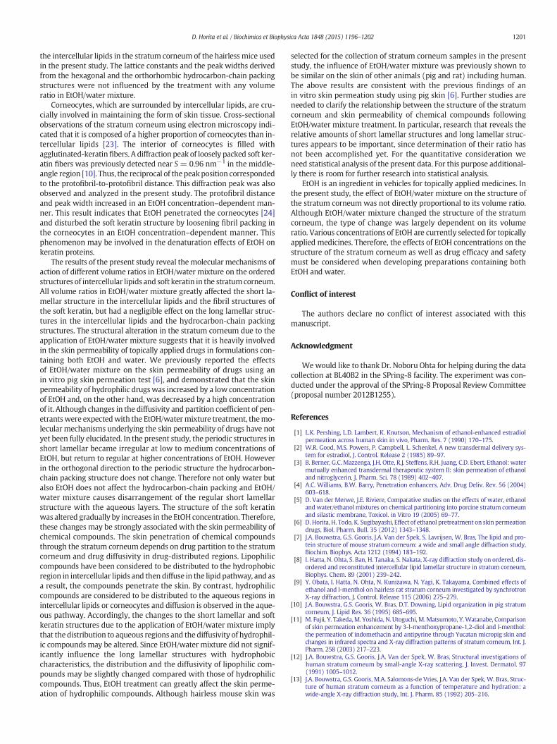
Fig. 1 shows the X-ray diffraction profiles (S = 0.05–3.0 nm−1) ofthe stratum corneumof hairlessmice following treatmentswith variousvolume ratios in EtOH/water mixture.We determined the X-ray diffrac-tion pattern from approximately one dozen stratum corneum sampleswith almost the same volume after the application of EtOH/water mix-ture at each volume ratio. Diffraction peaks were due to the repeat dis-tances of the lamellar structures in the small-angle region aswell as thelattice constants of the lateral hydrocarbon-chain packing structures inthe wide-angle region. Fig. 2a shows the X-ray diffraction profiles inthe small-angle region within the range of S = 0.10–0.40 nm−1. Threepartially overlapping peaks derived from the short and long lamellarstructures were detected within the range of S = 0.12–0.25 nm−1,and these profiles changed according to the volume ratio. In addition,the single diffraction peaks derived from the long and short lamellarstructures were observed near S = 0.30 and S = 0.37, respectively.Fig. 2b shows the X-ray diffraction profiles in the wide-angle regionwithin the range of S = 2.0–3.0 nm−1. Two diffraction peaks nearS=2.4 nm−1 and S=2.7 nm−1 corresponded to the hexagonal and or-thorhombic hydrocarbon-chain packing structures and only the ortho-rhombic hydrocarbon-chain packing structures, respectively. In thisexperiment, the stratum corneum sample from hairless mice showedsplit peaks for the orthorhombic hydrocarbon-chain packing structuresnear S = 2.7 nm−1.

S (nm-1)
Inte
nsity
(arb
.uni
ts)
10,000
8,000
6,000
4,000
2,000
00.10 0.15 0.20 0.25 0.30 0.35 0.40
a0/100 v/v%
20/80 v/v%
40/60 v/v%
60/40 v/v%
80/20 v/v%
100/0 v/v%
(EtOH/water ratio)
S (nm-1)
Inte
nsity
(arb
.uni
ts)
5,000
4,000
3,000
2,000
1,000
02.0 2.2 2.4 2.6 2.8 3.0
b0/100 v/v%
20/80 v/v%
40/60 v/v%
60/40 v/v%
80/20 v/v%
100/0 v/v%
(EtOH/water ratio)
Fig. 2. Small-angle X-ray diffraction profiles (a) and wide-angle X-ray diffraction profiles(b) in the hairless mouse stratum corneum as a function of the volume ratios in EtOH/water mixture, where n is the number of stratum corneum samples.
S (nm-1)
Inte
nsity
(ar
b. u
nits
)
10,000
8,000
6,000
4,000
2,000
00.120 0.145 0.170 0.195 0.220 0.245
aRaw X-ray diffraction profileBest-fitted curveBackground lineLong lamellar structure (2nd)Short lamellar structure (1st)Long lamellar structure (3rd)
S (nm-1)
Inte
nsity
(ar
b. u
nits
)
10,000
8,000
6,000
4,000
2,000
00.120 0.145 0.170 0.195 0.220 0.245
bRaw X-ray diffraction profileBest-fitted curveBackground lineLong lamellar structure (2nd)Short lamellar structure (1st)Long lamellar structure (3rd)
S (nm-1)
Inte
nsity
(ar
b. u
nits
)
2,000
1,600
1,200
800
400
00.26 0.28 0.30 0.32 0.34 0.36
cRaw X-ray diffraction profileBest-fitted curveBackground lineLong lamellar structure (4th)
Fig. 3. Typical curve fitting analysis to small-angle X-ray diffraction profiles of the stra-tum corneum of hairless mice treated with EtOH/water mixture of 60/40 v/v% (a) and100/0 v/v% (b, c). The thin solid line shows the mean of raw small-angle X-ray diffractionprofiles. The thick solid line indicates the best-fitted curve composed of the sum ofGaussian dotted curves and a linear background dotted line.
1198 D. Horita et al. / Biochimica et Biophysica Acta 1848 (2015) 1196–1202
3.2. Analysis of multiple diffraction peaks in the small-angle region
Multiple partially overlapping peaks in the small-angle region in therange of S=0.12–0.25 nm−1 in Fig. 2awere analyzed byfitting toGauss-ian curves. Fig. 3a and b show the results obtained by the Gaussian curvefitting analysis for the small-angle X-ray diffraction profiles of the stra-tum corneum in hairlessmice after treatmentswith EtOH/watermixtureof 60/40 and 100/0 v/v%, respectively. The 2nd order diffraction of thelong lamellar structure, the 1st order diffraction of the short lamellarstructure, and the 3rd order diffraction of the long lamellar structure oc-curred in the region from S = 0.12 to 0.25 nm−1 [12,14]. In order topresent typical examples of the fitting analysis, Fig. 3a and b showthose for EtOH/water mixture of 60/40 and 100/0 v/v%, respectively,where the diffraction profiles fit well to the sumof three Gaussian curvesand a linear background. Fig. 3c shows the results obtained by theGaussian curve fitting analysis for 4th order diffraction peak of the longlamellar structures at EtOH/water mixture of 100/0 v/v%. Although the2nd and 3rd order diffraction peaks of the long lamellar structures over-lapwith other peaks, the 4th order diffraction peakwas obtained as a sin-gle peak with a flat background and has adequate strength for fittinganalysis. Therefore, the 4th diffraction peak was used for the analysis toshow the change of the long lamellar structures more precisely. Table 1shows the repeat distances of the long and short lamellar structuresand the fullwidths at halfmaximumof the diffraction peaks as a functionof the volume ratios in EtOH/water mixture. In order to compare varia-tions in the short and long lamellar structures, the normalized repeat dis-tance and normalized full width at half maximum are shown in Fig. 4.
The application of EtOH/water mixture did not significantly affectthe long lamellar spacing and the peak width. On the other hand, the
short lamellar spacing was minimal and the peak width was maximalafter treatment with EtOH/water mixture of 60/40 v/v%. Thus, EtOH/water mixture was involved in both regular and irregular alteration onthe periodic pattern of the short lamellar structure in an EtOH concen-tration–dependent manner.
3.3. Analysis of diffraction peaks in the wide-angle region
As shown in Fig. 2b, two diffraction peaks (near S = 2.4 nm−1 andnear S = 2.7 nm−1) derived from the hexagonal and orthorhombichydrocarbon-chain packing structures were detected in the wide-

Table 1The repeat distance (a) and full width at half maximum (b) obtained from the 4th orderdiffraction peak for the long lamellar structure and the 1st order diffraction peak for theshort lamellar structure as a function of the volume ratios in EtOH/water mixture.
EtOH/water ratio (v/v %) 0/100 20/80 40/60 60/40 80/20 100/0
a) Repeat distance (nm)Long lamellar structure 13.26 13.30 13.30 13.33 13.30 13.17Short lamellar structure 6.29 6.13 6.13 5.86 5.95 6.42
b) Full width at half maximum (×10−2 nm−1)Long lamellar structure 0.93 0.89 0.89 0.91 0.85 0.88Short lamellar structure 1.84 2.59 2.52 2.83 1.97 1.33
0/100 20/80 40/60 60/40 80/20 100/0
EtOH/water ratio (v/v%)
0.379
0.378
0.377
0.376
0.375
0.374
0.373
0.417
0.416
0.415
0.414
Lat
tice
cons
tant
(nm
)
Fig. 5. Effects of EtOH/water mixture treatment on the lattice constants for the diffractionpeaks at S=2.4 nm−1, S=2.64 nm−1, and S=2.67 nm−1. The open circle, open triangle,and open square show the diffraction peaks at S = 2.4 nm−1, S = 2.64 nm−1, and S =2.67 nm−1, respectively.
1199D. Horita et al. / Biochimica et Biophysica Acta 1848 (2015) 1196–1202
angle region. The peak near S = 2.7 nm−1 was actually composed oftwo peaks. We analyzed these peaks by fitting to two Gaussian curves,similar to that shown in Fig. 3. The split peakwas consequently separat-ed into two peaks (S = 2.64 nm−1 and S = 2.67 nm−1), and the totaldiffraction profile fit well to the sum of the two Gaussian curves. Wethen calculated the lattice spacing and the peak widths from thosediffraction peaks. Figs. 5 and 6 show the effects of the treatmentswith EtOH/water mixture on the lattice spacing and the normalizedfull widths at half maximum, respectively. The lattice spacing andpeak widths for the hexagonal and orthorhombic hydrocarbon-chainpacking structures were almost constant and independent of the EtOHconcentration.
3.4. Effects of various volume ratios in EtOH/water mixture on the softkeratin in corneocytes
We analyzed a broad diffraction peak at approximately S =1.0 nm−1. This broad peak has previously been shown to be derivedfrom the soft keratin, which is loosely packed in corneocytes [10].Fig. 7 shows X-ray diffraction profiles in the region within S =0.5–1.5 nm−1 in the stratum corneum of hairlessmice treatedwith var-ious volume ratios in EtOH/water mixture. To calculate the peak posi-tion and the width, Gaussian curve fitting analysis was again appliedto each diffraction profile. Fig. 8 shows the effects of various volume ra-tios in EtOH/water mixture on the reciprocal of the peak position(protofibril distance) and the full width at half maximum obtainedfrom the diffraction profile for the soft keratin. The protofibril distanceand the peak width increased in an EtOH concentration–dependentmanner. Therefore, EtOH perturbed the soft keratin structure, whichloosened fibril packing.
0/100 20/80 40/60 60/40 80/20 100/0
EtOH/water ratio (v/v%)
Nor
mal
ized
rep
eat d
ista
nce
(an
d )
Nor
mal
ized
ful
l wid
th a
t hal
f m
axim
um(
and
)
1.10
1.05
1.00
0.95
0.90
2.00
1.50
1.00
0.50
0.00
Fig. 4. Effects of EtOH/water mixture treatment on the repeat distances (○ or ●) and thefull widths at half maximum (△ or ▲) for the 4th order diffraction of the long lamellarstructures and the 1st order diffraction of the short lamellar structures. The normalizeddata were calculated by dividing the values of repeat distances and full width at half max-imum with EtOH/water mixture treatments by those for 0/100 v/v%. The open circle oropen triangle and the closed circle or closed triangle show the long lamellar structuresand the short lamellar structures, respectively.
4. Discussion
In the present study, we performed X-ray diffraction experiments onthe stratum corneum of hairless mice to investigate the effects of EtOH/water mixture on its structure. We applied different volume ratios inEtOH/water mixture to the stratum corneum, and then examined itsstructure using X-ray diffraction in the SPring-8 facility. The EtOH/water mixture content in stratum corneum samples was prepared at20 wt% because the water content in the skin surface was estimatedto be 20–30% [15] and mostly stabilizes the lamellar structure at thislevel [16]. There have been difficulties associated with comparing theresults obtained from X-ray diffraction experiments on multiple sam-ples under different conditions because the diffraction profiles of thestratum corneum are greatly affected by individual variations and/orskin sites. Few X-ray diffraction studies have closely examined the
0/100 20/80 40/60 60/40 80/20 100/0
EtOH/water ratio (v/v%)
Nor
mal
ized
ful
l wid
th a
t hal
f m
axim
um 2.00
1.50
1.00
0.50
0.00
Fig. 6. Effects of EtOH/watermixture treatment on the normalized full widths at halfmax-imum for the diffraction peaks at S=2.4 nm−1, S=2.64 nm−1, and S= 2.67 nm−1. Thenormalizeddatawere calculatedbydividing the values of full width at halfmaximumwithEtOH/water mixture treatments by those for 0/100 v/v%. The open circle, open triangle,and open square show the diffraction peak at S = 2.4 nm−1, S = 2.64 nm−1, and S =2.67 nm−1, respectively.

S (nm-1)
Inte
nsity
(ar
b. u
nits
)
900
800
700
600
500
4000.5 0.7 0.9 1.1 1.3 1.5
0/100 v/v%
20/80 v/v%
40/60 v/v%
60/40 v/v%
80/20 v/v%
100/0 v/v%
(EtOH/water ratio)
Fig. 7.Medium-angleX-ray diffraction profiles in thehairlessmouse stratum corneumas afunction of the volume ratios in EtOH/water mixture, where n is the number of stratumcorneum samples.
1200 D. Horita et al. / Biochimica et Biophysica Acta 1848 (2015) 1196–1202
effects of EtOH/water mixture on the structure of the stratum corneum.In this study, we prepared approximately one dozen skin samples perconcentration of EtOH and the X-ray diffraction patterns obtained;therefore, differences due to individual variations and/or skin siteswere avoided and the influence of skin treatments with any volumeratio in EtOH/water mixture on the structure of the stratum corneumcould be compared clearly.
Ordered lamellar structures, such as the long and short lamellarstructures, have periodic structures in the direction of the long axis ofthe hydrocarbon chain. Previous studies reported that the repeat dis-tances of the long and short lamellar structures were approximately13 nm and 6 nm, respectively [12,14]. To detect minute variations inthe lamellar structures, the 1st order diffraction peak for the short la-mellar structures, in particular, must be carefully isolated since thispeak was weaker than that for the long lamellar structures. Gaussiancurve fitting analysis was performed to isolate individual peaks fromeach other. As a result, the repeat distance and the full width at halfmaximum were obtained for the short lamellar structures.
Diffraction peak analyses revealed that EtOH/water mixture influ-enced the short lamellar structures more than the long ones. The obser-vation of the short lamellar structure in hydrated condition was firstreported experimentally by Bouwstra et al. [7,10,12] who have beencareful for proposing the swelling behavior. They have pointed outthat the X-ray diffraction peak for the short lamellar structure becomessharp near the water content of 20 wt% but the swelling effect on theshort lamellar structure was not detectable in human [12] and also inhairless mouse' [7] stratum corneum. On the other hand, in pig stratum
0/100 20/80 40/60 60/40 80/20 100/0
EtOH/water ratio (v/v%)
Cha
nge
to r
ecip
roca
lof
peak
posi
tion
(nm
)
F ull
wid
th a
t hal
f m
axim
um(n
m-1
)1.06
1.05
1.04
1.03
1.02
1.01
1.00
0.32
0.30
0.28
0.26
0.24
0.22
0.20
Fig. 8. Effects of EtOH/water mixture treatment on the reciprocal of the peak position (○)and the full width at half maximum (△) for the soft keratin.
corneum they have observed a weak swelling effect [10], and from acareful study on much attention to the effect of water in hairlessmouse stratum corneum Ohta et al. have found the swelling behaviorwith increasing thewater content [16]. So far in various studies, the dif-fraction peak of short lamellar structure is sometimes undetectable [12,14], sometimes only observed only as a shoulder [7,10] and sometimesgives rise to a clear peak [16]. In addition to these studies much clearlythe swelling behavior in short lamellar structure has been observedfrom the neutron diffraction by Charalambopoulou et al. [17]. The neu-tron diffraction experiment by using heavy water is a powerful tool toobserve the aqueous layer in stratum corneum since the layer ofheavy water can be observed dominantly. It is worthwhile to pointout that the increment of the spacing of short lamellar structurewith in-creasing thewater content in the previous X-ray diffraction study [16] isconsistent with the neutron diffraction study [17]. Furthermore, recent-ly Nakazawa et al. have proposed that based upon the X-ray diffractionstudy on the swelling behavior of short lamellar structure in humanstratum corneum it could involve in adjustment of the water contentin stratum corneum [18]. The above fact indicates that a uniformswelling of short lamellar structure takes place in stratum corneumsince X-ray and neutron diffraction measurement provide evidence forcoherent structural alteration and resultantly uniform swelling of theaqueous layer. The latter fact could be rationally supposed from theswelling behavior under water obtained from the X-ray diffractionstudy on egg lecithin bilayers which are composed of amphiphilic mol-ecules [19] since intercellular lipids in stratum corneum are composedof amphiphilic molecules such as ceramides and fatty acids. Thereforethe aqueous layers in the short lamellar structure could take place inthe face-to-face arrangement of the polar head groups via the aqueouslayer. Hence, appliedwater or EtOHon the stratum corneummight pen-etrate the aqueous layer, and the behavior of the aqueous layer follow-ing the application of various volume ratios in EtOH/water mixturemay depend on differences in the interactions of a water moleculeand an EtOH molecule with the polar head groups of ceramides andfatty acids. The applied EtOH seems to combine with the interface be-tween the aqueous layer and the polar head groups of ceramides andfatty acids by the hydroxyl group of an EtOHmolecule facing the aque-ous layer [20]. As a result, the repeat distance of the short lamellar struc-tures decreasedwith an increase in the EtOH concentration in the rangeof EtOH/water ratio from 0/100 v/v% to 60/40 v/v%, which then con-versely increased when the concentration of EtOH was elevatedabove 60/40 v/v%. The diffraction peak width increased with an in-crease in the EtOH concentration in the range of EtOH/water ratiofrom 0/100 v/v% to 60/40 v/v%, but decreased with further increases inthe EtOH concentration above 60/40%. These results indicate that treat-ment with a low concentration of EtOH disturbs the short lamellarstructures, but on the other hand, treatment with a high concentrationof it causes an aligned structure. In the case of treatment with mostlywater or EtOH, water and EtOHmolecules may exist as structured mol-ecules in highly concentrated water and upon high EtOH, respectively.At intermediate concentrations, water and EtOH may not form struc-tured molecules, so the layer structure may be disrupted. In addition,Kwak et al. reported [21] the effect of various concentrations of EtOHon model membranes of stratum corneum lipids composed of differentlipid classes. They pointed out that lipid model membranes includingceramides were disrupted by the action of EtOH compared with thosewith free fatty acids. This might indicate that there is less ceramide inthe short lamellar structures, being susceptible to EtOH, than in thelong lamellar structures.
On the other hand, the hexagonal and orthorhombic hydrocarbon-chain packing structures observed on a plane perpendicular to thelong axis of the hydrocarbon chainwere previously reported to have lat-tice constants of 0.42 nm, 0.42 nm, and 0.37 nm [22]. Since the diffrac-tion profile with split peaks near S = 2.7 nm−1
fits well to the sum ofthe two Gaussian curves and a background line, there may be at leasttwo types of orthorhombic hydrocarbon-chain packing structure in
1201D. Horita et al. / Biochimica et Biophysica Acta 1848 (2015) 1196–1202
the intercellular lipids in the stratum corneum of the hairless mice usedin the present study. The lattice constants and the peak widths derivedfrom the hexagonal and the orthorhombic hydrocarbon-chain packingstructures were not influenced by the treatment with any volumeratio in EtOH/water mixture.
Corneocytes, which are surrounded by intercellular lipids, are cru-cially involved in maintaining the form of skin tissue. Cross-sectionalobservations of the stratum corneum using electron microscopy indi-cated that it is composed of a higher proportion of corneocytes than in-tercellular lipids [23]. The interior of corneocytes is filled withagglutinated-keratinfibers. A diffraction peak of loosely packed soft ker-atin fibers was previously detected near S = 0.96 nm−1 in the middle-angle region [10]. Thus, the reciprocal of thepeakposition correspondedto the protofibril-to-protofibril distance. This diffraction peak was alsoobserved and analyzed in the present study. The protofibril distanceand peak width increased in an EtOH concentration–dependent man-ner. This result indicates that EtOH penetrated the corneocytes [24]and disturbed the soft keratin structure by loosening fibril packing inthe corneocytes in an EtOH concentration–dependent manner. Thisphenomenon may be involved in the denaturation effects of EtOH onkeratin proteins.
The results of the present study reveal themolecular mechanisms ofaction of different volume ratios in EtOH/water mixture on the orderedstructures of intercellular lipids and soft keratin in the stratumcorneum.All volume ratios in EtOH/water mixture greatly affected the short la-mellar structure in the intercellular lipids and the fibril structures ofthe soft keratin, but had a negligible effect on the long lamellar struc-tures in the intercellular lipids and the hydrocarbon-chain packingstructures. The structural alteration in the stratum corneum due to theapplication of EtOH/water mixture suggests that it is heavily involvedin the skin permeability of topically applied drugs in formulations con-taining both EtOH and water. We previously reported the effectsof EtOH/water mixture on the skin permeability of drugs using anin vitro pig skin permeation test [6], and demonstrated that the skinpermeability of hydrophilic drugswas increased by a low concentrationof EtOH and, on the other hand, was decreased by a high concentrationof it. Although changes in the diffusivity and partition coefficient of pen-etrantswere expectedwith the EtOH/watermixture treatment, themo-lecular mechanisms underlying the skin permeability of drugs have notyet been fully elucidated. In the present study, the periodic structures inshort lamellar became irregular at low to medium concentrations ofEtOH, but return to regular at higher concentrations of EtOH. Howeverin the orthogonal direction to the periodic structure the hydrocarbon-chain packing structure does not change. Therefore not only water butalso EtOH does not affect the hydrocarbon-chain packing and EtOH/water mixture causes disarrangement of the regular short lamellarstructure with the aqueous layers. The structure of the soft keratinwas altered gradually by increases in the EtOHconcentration. Therefore,these changes may be strongly associated with the skin permeability ofchemical compounds. The skin penetration of chemical compoundsthrough the stratum corneum depends on drug partition to the stratumcorneum and drug diffusivity in drug-distributed regions. Lipophiliccompounds have been considered to be distributed to the hydrophobicregion in intercellular lipids and then diffuse in the lipid pathway, and asa result, the compounds penetrate the skin. By contrast, hydrophiliccompounds are considered to be distributed to the aqueous regions inintercellular lipids or corneocytes and diffusion is observed in the aque-ous pathway. Accordingly, the changes to the short lamellar and softkeratin structures due to the application of EtOH/water mixture implythat the distribution to aqueous regions and the diffusivity of hydrophil-ic compounds may be altered. Since EtOH/water mixture did not signif-icantly influence the long lamellar structures with hydrophobiccharacteristics, the distribution and the diffusivity of lipophilic com-pounds may be slightly changed compared with those of hydrophiliccompounds. Thus, EtOH treatment can greatly affect the skin perme-ation of hydrophilic compounds. Although hairless mouse skin was
selected for the collection of stratum corneum samples in the presentstudy, the influence of EtOH/water mixture was previously shown tobe similar on the skin of other animals (pig and rat) including human.The above results are consistent with the previous findings of anin vitro skin permeation study using pig skin [6]. Further studies areneeded to clarify the relationship between the structure of the stratumcorneum and skin permeability of chemical compounds followingEtOH/water mixture treatment. In particular, research that reveals therelative amounts of short lamellar structures and long lamellar struc-tures appears to be important, since determination of their ratio hasnot been accomplished yet. For the quantitative consideration weneed statistical analysis of the present data. For this purpose additional-ly there is room for further research into statistical analysis.
EtOH is an ingredient in vehicles for topically applied medicines. Inthe present study, the effect of EtOH/water mixture on the structure ofthe stratum corneum was not directly proportional to its volume ratio.Although EtOH/water mixture changed the structure of the stratumcorneum, the type of change was largely dependent on its volumeratio. Various concentrations of EtOH are currently selected for topicallyapplied medicines. Therefore, the effects of EtOH concentrations on thestructure of the stratum corneum as well as drug efficacy and safetymust be considered when developing preparations containing bothEtOH and water.
Conflict of interest
The authors declare no conflict of interest associated with thismanuscript.
Acknowledgment
Wewould like to thank Dr. Noboru Ohta for helping during the datacollection at BL40B2 in the SPring-8 facility. The experiment was con-ducted under the approval of the SPring-8 Proposal Review Committee(proposal number 2012B1255).
References
[1] L.K. Pershing, L.D. Lambert, K. Knutson, Mechanism of ethanol-enhanced estradiolpermeation across human skin in vivo, Pharm. Res. 7 (1990) 170–175.
[2] W.R. Good, M.S. Powers, P. Campbell, L. Schenkel, A new transdermal delivery sys-tem for estradiol, J. Control. Release 2 (1985) 89–97.
[3] B. Berner, G.C. Mazzenga, J.H. Otte, R.J. Steffens, R.H. Juang, C.D. Ebert, Ethanol: watermutually enhanced transdermal therapeutic system II: skin permeation of ethanoland nitroglycerin, J. Pharm. Sci. 78 (1989) 402–407.
[4] A.C. Williams, B.W. Barry, Penetration enhancers, Adv. Drug Deliv. Rev. 56 (2004)603–618.
[5] D. Van der Merwe, J.E. Riviere, Comparative studies on the effects of water, ethanoland water/ethanol mixtures on chemical partitioning into porcine stratum corneumand silastic membrane, Toxicol. in Vitro 19 (2005) 69–77.
[6] D. Horita, H. Todo, K. Sugibayashi, Effect of ethanol pretreatment on skin permeationdrugs, Biol. Pharm. Bull. 35 (2012) 1343–1348.
[7] J.A. Bouwstra, G.S. Gooris, J.A. Van der Spek, S. Lavrijsen, W. Bras, The lipid and pro-tein structure of mouse stratum corneum: a wide and small angle diffraction study,Biochim. Biophys. Acta 1212 (1994) 183–192.
[8] I. Hatta, N. Ohta, S. Ban, H. Tanaka, S. Nakata, X-ray diffraction study on ordered, dis-ordered and reconstituted intercellular lipid lamellar structure in stratum corneum,Biophys. Chem. 89 (2001) 239–242.
[9] Y. Obata, I. Hatta, N. Ohta, N. Kunizawa, N. Yagi, K. Takayama, Combined effects ofethanol and l-menthol on hairless rat stratum corneum investigated by synchrotronX-ray diffraction, J. Control. Release 115 (2006) 275–279.
[10] J.A. Bouwstra, G.S. Gooris, W. Bras, D.T. Downing, Lipid organization in pig stratumcorneum, J. Lipid Res. 36 (1995) 685–695.
[11] M. Fujii, Y. Takeda, M. Yoshida, N. Utoguchi, M. Matsumoto, Y.Watanabe, Comparisonof skin permeation enhancement by 3-l-menthoxypropane-1,2-diol and l-menthol:the permeation of indomethacin and antipyrine through Yucatan micropig skin andchanges in infrared spectra and X-ray diffraction patterns of stratum corneum, Int. J.Pharm. 258 (2003) 217–223.
[12] J.A. Bouwstra, G.S. Gooris, J.A. Van der Spek, W. Bras, Structural investigations ofhuman stratum corneum by small-angle X-ray scattering, J. Invest. Dermatol. 97(1991) 1005–1012.
[13] J.A. Bouwstra, G.S. Gooris, M.A. Salomons-de Vries, J.A. Van der Spek, W. Bras, Struc-ture of human stratum corneum as a function of temperature and hydration: awide-angle X-ray diffraction study, Int. J. Pharm. 85 (1992) 205–216.

1202 D. Horita et al. / Biochimica et Biophysica Acta 1848 (2015) 1196–1202
[14] I. Hatta, N. Ohta, K. Inoue, N. Yagi, Coexistence of two domains in intercellular lipidmatrix of stratum corneum, Biochim. Biophys. Acta 1758 (2006) 1830–1836.
[15] I.H. Blank, Factorswhich influence thewater content of the stratum corneum, J. Invest.Dermatol. 18 (1952) 440–443.
[16] N. Ohta, S. Ban, H. Tanaka, S. Nakata, I. Hatta, Swelling of intercellular lipid lamellarstructure with short repeat distance in hairless mouse stratum corneum as studiedby X-ray diffraction, Chem. Phys. Lipids 123 (2003) 1–8.
[17] G.Ch. Charalambopoulou, Th.A. Steriotis, Th. Hauss, A.K. Stubos, N.K. Kanellopoulos,Structural alterations of fully hydrated human stratum corneum, Physica B 350(2004) 603–606.
[18] H. Nakazawa, N. Ohta, I. Hatta, A possible regulation mechanism of water content inhuman stratum corneum via intercellular lipid matrix, Chem. Phys. Lipids 165(2012) 238–243.
[19] Y.K. Levine, M.H.F. Wilkins, Structure of oriented lipid bilayers, Nat. New Biol. 230(1971) 69–72.
[20] T. Adachi, H. Takahashi, K. Ohki, I. Hatta, Interdigitated structure of phospholipid–alcohol systems studied by X-ray diffraction, Biophys. J. 68 (1995) 1850–1855.
[21] S. Kwak, E. Brief, D. Langlais, N. Kitson, M. Lafleur, J. Thewalt, Ethanol perturbs lipidorganization in models of stratum corneum membranes: an investigation combin-ing differential scanning calorimetry, infrared and 2H NMR spectroscopy, Biochim.Biophys. Acta 1818 (2012) 1410–1419.
[22] G.S. Pilgram, A.M. Engelsma-van Pelt, J.A. Bouwstra, H.K. Koerten, Electron diffractionprovides new information on human stratum corneum lipid organization studied inrelation to depth and temperature, J. Invest. Dermatol. 113 (1999) 403–409.
[23] L. Norlén, A. Al-Amoudi, Stratum corneum keratin structure, function, and forma-tion: the cubic rod-packing and membrane templating model, J. Invest. Dermatol.123 (2004) 715–732.
[24] I. Hatta, H. Nakazawa, Y. Obata, N. Ohta, K. Inoue, N. Yagi, Novel method to observesubtle structural modulation of stratum corneum on applying chemical agents,Chem. Phys. Lipids 163 (2010) 381–389.