molecular ferries: membrane carriers that promote ...bsmith3/pdf/cc2003.pdf · molecular ferries:...
TRANSCRIPT

Molecular ferries: membrane carriers that promote phospholipidflip-flop and chloride transport
Bradley D. Smith* and Timothy N. Lambert Department of Chemistry and Biochemistry and the Walther Cancer Research Center, University of NotreDame, Notre Dame, IN 46556, USA
Received (in Cambridge, UK) 27th March 2003, Accepted 3rd June 2003First published as an Advance Article on the web 20th June 2003
The facilitated transport of ionic or polar solutes throughbiological membranes is an essential process for cellular life, anda major technical goal of the pharmaceutical industry. Syn-thetic receptors with affinities for anions are shown to act asmolecular ferries and facilitate the movement of chloride ionsand salts across vesicle and cell membranes. A process thatcompetes with chloride transport is phospholipid translocationor flip-flop. This has led to the development of syntheticscramblases that can alter the transmembrane distribution ofphospholipids and induce biological responses such as mem-brane enzyme activation. The facilitated translocation ofphospholipids with multiply-charged head groups, like phos-phatidylserine, is a difficult supramolecular challenge thatrequires a complementary, multitopic receptor with appropriateamphiphilicity.
IntroductionA typical mammalian cell is around 3 mm in diameter and issurrounded by a 6 nm wide plasma membrane. From the humanperspective, these dimensions are equivalent to a 10 m diameterseminar room surrounded by a shell 20 cm thick. Thisextraordinarily thin membrane is all that separates the cytosolicspace from the extracellular fluid and the lumina of the variousorganelles. The basic structure of a typical cell membrane is aself-assembled bilayer of polar lipids, non-polar lipids, andproteins (Scheme 1). The ratio by weight of protein to lipidvaries from 3.6 in the inner mitochondrial membrane to 0.25 inthe lipid-rich myelin membrane.1 The proteins can act as
receptors, enzymes, channels, and/or pumps which endows themembrane with the capability to: (a) alter its flexibility andmechanical strength, (b) control the concentration of ions andmolecules in the various cellular spaces, (c) conduct bio-chemical reactions, and (d) recognize and communicate withother cell membranes.2 Elucidation of the molecular mecha-nisms that produce these various membrane functions is thegoal of a large number of research laboratories with a widerange of skills and instrumentation. The primary expertise in ourlaboratory is organic supramolecular chemistry and the specificfocus of this article is our recent work on the facilitated transportof anionic solutes through bilayer membranes.
The passage of a charged or polar solute through an unbiasedphospholipid bilayer is very slow. The primary barrier isdiffusion of the solute through the lipophilic membrane interior.A recent molecular dynamics simulation of unassisted diffusionof Na+ and Cl2 through a bilayer membrane shows water andphospholipids accompanying the ion.3 To facilitate transport,cells have evolved to use various supramolecular strategies.Three limiting cases are shown in Scheme 2. The first strategy,
which is employed extensively in our work, uses a mobilecarrier to associate with the polar solute and form a lipophiliccomplex which can then diffuse through the membrane, i.e., a
Bradley D. Smith obtained a B.Sc.(Hons) degree from theUniversity of Melbourne, and a Ph.D. in 1988 from Penn StateUniversity. After postdoctoral training at Oxford Universityand then Columbia University, he moved to the University ofNotre Dame in 1991. He is currently a Professor of Chemistryand Biochemistry with research interests in the fields ofbioorganic and supramolecular chemistry. His group aims todesign and synthesize organic molecules that affect thedynamics of phospholipid bilayer membranes.
Timothy N. Lambert obtained a B.S. degree from the Universityof Texas at Austin, and a Ph.D. in 2001 from New Mexico StateUniversity. He is currently a Senior Research Associate withinthe Walther Cancer Research Center and the Department ofChemistry and Biochemistry at the University of Notre Dame.His current research efforts use the tools of organic synthesisand molecular recognition to invent new technologies forcancer research and treatment. He is particularly interested insolving applied problems through fundamental explorations inchemistry.
Scheme 1 Typical cell membrane.†
Scheme 2 Membrane transport mechanisms.
Th is journa l i s © The Roya l Soc ie ty of Chemist ry 2003
DO
I: 10
.103
9/b
3033
59g
2261CHEM. COMMUN. , 2003, 2261–2268
molecular ferry. The next two cases are channel mechanisms.They differ in the extent of interaction between solute and thechannel walls. In some cases there is extensive interaction suchthat the solute can be visualized as briefly occupying specificbinding sites as it passes through the channel. Such a relaymechanism can exhibit high solute selectivity. In other cases,the channel is sufficiently large that the solute can pass throughas a highly solvated species, and there is only a rough selectionbased on the size of the solvated solute. Both types of channelsystems can promote movement either passively down anelectrochemical gradient, or actively in a specific direction.Furthermore, channels can have sophisticated switching mecha-nisms with chemical, electrical, optical, or mechanical triggers.Transport channels are usually complicated supramolecularassemblies of large transmembrane proteins and it is currently asubstantial challenge to understand their mechanisms of actionat the molecular level. One of the aims of our research is todesign low-molecular-weight mimics of membrane transportsystems. These simplified mimics can be used to addressfundamental mechanistic questions, or they can be developedinto later generation versions that have useful applications.4
When discussing transport across cell membranes it isimportant to bear in mind that the membranes are likely to bepolarized with electrical and various chemical gradients. Thesegradients are maintained by the concerted action of more thantwenty different endogenous membrane transporters. The mostdominant are the ATP-driven ion pumps which control Na+, K+,Ca2+, and H+ gradients. These primary ion gradients aresubsequently used by co-transport (simultaneous passage of twoor more solutes in the same direction) or antiport (simultaneouspassage of two or more solutes in opposite directions) systemsas energy sources to move other ions or polar molecules againsttheir respective concentration gradients. Thus in cells, electro-chemical gradients can drive membrane transport processes thatappear intuitively to be unfavored. For example, consider anerve cell with a typical membrane potential of 260 mV (insidenegative), and a Cl2 concentration gradient of 50 mM internaland 100 mM external. Although there is a concentrationgradient promoting Cl2 transport into the cell, it is over-
whelmed by the electrical potential such that the free energy forinward Cl2 transport is +4 kJ mol21! A useful rule of thumb toremember is that at equilibrium, a univalent ion has a ten-foldconcentration gradient to match a 60 mV transmembraneelectric potential.
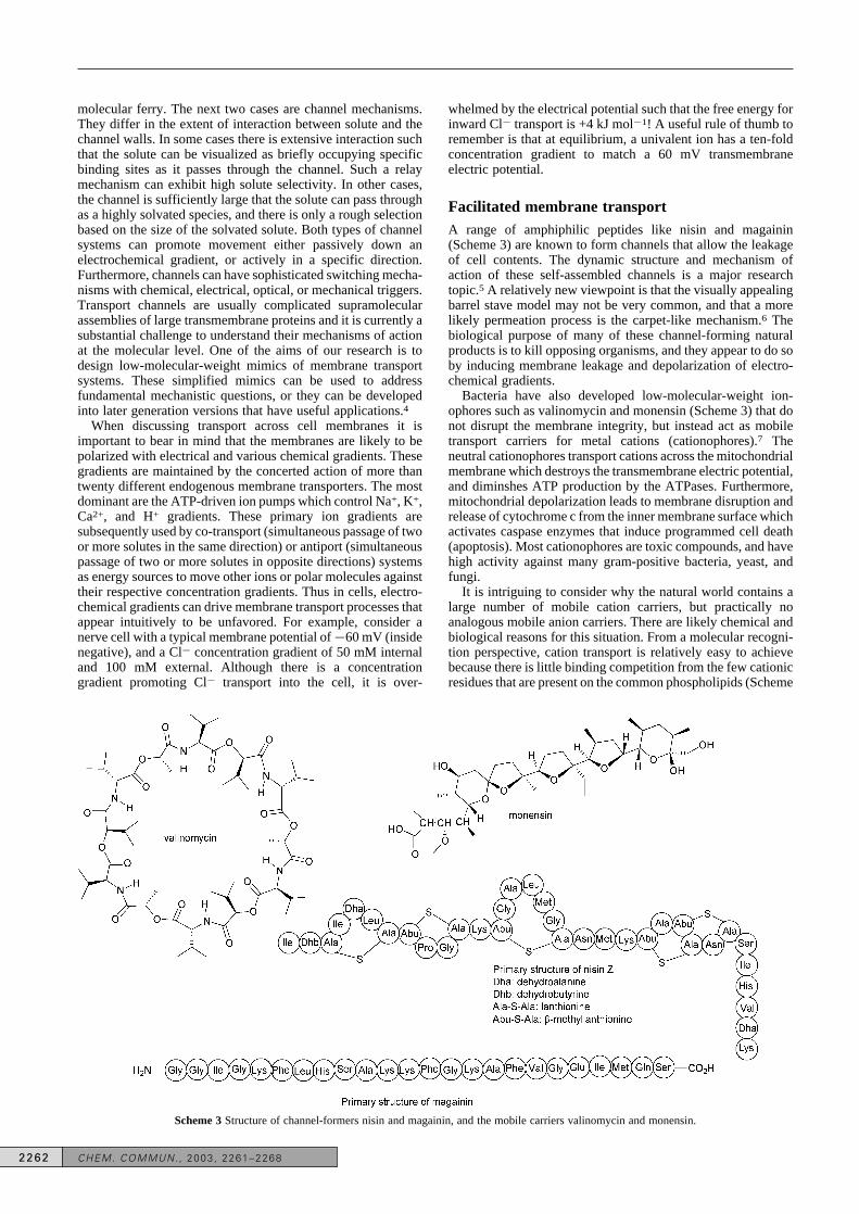
Facilitated membrane transportA range of amphiphilic peptides like nisin and magainin(Scheme 3) are known to form channels that allow the leakageof cell contents. The dynamic structure and mechanism ofaction of these self-assembled channels is a major researchtopic.5 A relatively new viewpoint is that the visually appealingbarrel stave model may not be very common, and that a morelikely permeation process is the carpet-like mechanism.6 Thebiological purpose of many of these channel-forming naturalproducts is to kill opposing organisms, and they appear to do soby inducing membrane leakage and depolarization of electro-chemical gradients.
Bacteria have also developed low-molecular-weight ion-ophores such as valinomycin and monensin (Scheme 3) that donot disrupt the membrane integrity, but instead act as mobiletransport carriers for metal cations (cationophores).7 Theneutral cationophores transport cations across the mitochondrialmembrane which destroys the transmembrane electric potential,and diminshes ATP production by the ATPases. Furthermore,mitochondrial depolarization leads to membrane disruption andrelease of cytochrome c from the inner membrane surface whichactivates caspase enzymes that induce programmed cell death(apoptosis). Most cationophores are toxic compounds, and havehigh activity against many gram-positive bacteria, yeast, andfungi.
It is intriguing to consider why the natural world contains alarge number of mobile cation carriers, but practically noanalogous mobile anion carriers. There are likely chemical andbiological reasons for this situation. From a molecular recogni-tion perspective, cation transport is relatively easy to achievebecause there is little binding competition from the few cationicresidues that are present on the common phospholipids (Scheme
Scheme 3 Structure of channel-formers nisin and magainin, and the mobile carriers valinomycin and monensin.
2262 CHEM. COMMUN. , 2003, 2261–2268

4). Conversely, anion transport is a more difficult supramo-lecular challenge because a putative carrier has to bind theanionic solute at the membrane surface where there is a veryhigh concentration of competing phosphate diester residues. Onthe other hand, it is known that that the interior of a standard,unperturbed bilayer membrane has a net positive charge so if amobile ionophore can move an anionic solute past the polarmembrane surface then passage through the middle of themembrane should be relatively easy.8 Nonetheless, in 1999when we contemplated treating a bilayer membrane with a low-molecular-weight anionophore, we predicted that the mostlikely supramolecular outcome would be transmembrane move-ment of the phospholipid head group. This thought experimentinspired us to learn more about phospholipid flip-flop andtransmembrane phospholipid distribution.
Phosplipid flip-flopUnlike lateral diffusion, which is very rapid, the translocation(or flip-flop) of phospholipids across a model bilayer membraneis known to be a very slow process with a half-life of hours todays (Scheme 5).9 Flip-flop rates in these artificial systems arestrongly dependent on the composition of the polar head-group,and less dependent on the length of the acyl chains.10
Nonetheless, if an artificial membrane is given enough time itwill reach an equilibrated state with a random transmembranedistribution of phospholipids. This is not the case with mostbiological membranes which have an asymmetric distributionof phospholipids (Fig. 1). The degree of asymmetry varies; the
plasma membranes of most eukaryotes maintain a high degreeof asymmetry, whereas a lesser degree of transbilayer asym-metry is observed in the membranes of subcellular organellesand bacterial species. The asymmetric distribution of phospho-lipids is a fundamental feature of normal cell operation. Forexample, the phosphatidylserine (PS) that is normally localizedin the inner monolayer of an animal plasma membrane is vitalnot only for exocytosis and intracellular fusion processes, butalso for lipid–protein interactions and signal transductionpathways.11–14
The asymmetric transmembrane distribution of lipids isgenerated and maintained by a number of phospholipidtranslocases that vary in lipid specificity, energy requirements,and direction of translocation.15 For example, the plasmamembrane phospholipid asymmetry is maintained by thesynchronous action of the aminophospholipid flippase whichselectively pumps PS and phosphatidylethanolamine (PE) to theinner monolayer of the membrane, and a nonspecific, less-active floppase which moves phospholipids to the outermonolayer (Scheme 6). Some of the translocase proteins havebeen isolated, but presently it is not known how translocationworks at the molecular level. The literature contains somespeculation that translocases operate by forming pores andindeed pore forming compounds are known to promotephospholipid flip-flop.16 Furthermore, it has recently beenshown that translocation of phospholipids with singly chargedhead groups can be promoted by single transmembranepeptides.17 Nonetheless, the aminophospholipid flippase isquite head group selective and so the transport mechanism inthis, and related systems, must involve a high degree ofphospholipid head group recognition. A recent crystal structureof a bacterial Lipid A floppase shows a sophisticated chamberstructure that appears to cycle through a dynamic three-stagebinding–translocation–release mechanism.18
Scheme 4 Structures of the common phospholipids.
Scheme 5 Phospholipid translocation or flip-flop.
Fig. 1 Distribution of phospholipids across an erythrocyte membrane.
CHEM. COMMUN. , 2003, 2261–2268 2263

A loss of normal phospholipid asymmetry is often associatedwith abnormal cell function. For example, the appearance of PSin the outer monolayer of membranes correlates with cell deathand clearance by phagocytosis.19 Inactivation of the amino-phospholipid flippase alone is not sufficient to expose PS; asimultaneous Ca2+-dependent nonspecific scrambling processis necessary. PS-exposing apoptotic cells are cleared from thebloodstream by macrophages following a specific recognitionevent between PS receptors on the macrophage and theexternalized PS. Similarly, aging erythrocytes and plateletsslowly externalize PS, culminating in engulfment by macro-phages.20 Another consequence of phospholipid randomizationis blood coagulation. The binding of blood cells to proteinsinvolved in the coagulation cascade is dependent on PSexposure. The tenase and prothrombin complexes bind to thepatches of anionic lipid on the cell surface, and through a seriesof activation steps the fibrin matrix of the clot is formed.21
Thus, we had two initial reasons for developing synthetictranlocation mimics. The first motivation was to learn whatminimal supramolecular requirements would induce transloca-tion of the various phospholipid head groups across a bilayermembrane. The second more practical motivation was togenerate a series of synthetic scramblases that would promotethe appearance of PS on the surface of cell surfaces, andhopefully produce selective biological and therapeutic re-sponses.
As a starting point, we chose to develop a translocase forphosphatidylcholine (PC), since it has a zwitterionic head groupand seemed a more straightforward transport challenge. There isexperimental evidence that the primary site of hydration in thePC head group is the phosphate diester residue and not thediffuse tetraalkylammonium cation.22 We reasoned that if anappropriate uncharged anion receptor could form a hydrogen
bonded complex with the phosphodiester, then the lipophilicreceptor/head group complex would more readily diffusethrough the membrane. We chose to initially examine simplederivatives of tris(aminoethyl)amine (tren), namely sulfona-mide 1 and amide 2, as well as control ester 3 (Scheme 7).23–25
Compounds 1 and 2 were previously shown by Reinhoudt andcoworkers to act as receptors for anions like dihydrogenphosphate.26 The compounds also have a nice balance of watersolubility and amphiphilicity so they can be readily formulatedfor transport studies.
We employ the NBD/dithionite quenching assay to measurethe rate with which fluorescent NBD-labeled phospholipids aretranslocated across vesicle membranes (Schemes 8 and 9).27
The assay starts with surface differentiated vesicles preparedwith NBD-lipids (0.5 mol%) in either the membrane outermonolayer (exo-labeled) or inner monolayer (endo-labeled).Upon treatment with sodium dithionite (Na2S2O4), the NBD
Scheme 6 Enzyme promoted flip-flop in an erythrocyte membrane.
Scheme 8 Fluorescent assay for flip-flop.
Scheme 7 Tren-based phospholipid scramblase candidates.
2264 CHEM. COMMUN. , 2003, 2261–2268

fluorescence is quenched due to chemical reduction of the nitrogroup. Vesicle membranes are effectively impermeable todithionite, therefore, only NBD-lipid located in the outer leafletis chemically quenched. At any given time, the percentage ofNBD-lipid located in the outer monolayer can be determinedfrom the drop in fluorescence intensity when a portion of thevesicles is subjected to dithionite quenching. The systemprogresses to an equilibrated state with the outer monolayercontaining about 60% NBD-lipid. Reported half-lives forinward translocation correspond to the time taken to reach 80%of the NBD-lipid in the outer monolayer
We found that sulfonamide 1 was especially good atpromoting NBD-PC translocation, whereas the amide 2 andester 3 were not effective (Table 1).23 In light of the success
with sulfonamide 1, the inability of amide 2 to promotetranslocation was puzzling because it is also a hydrogen bonddonor (although less acidic than 1). Structural insight wasgained from X-ray structures of 1 and 2 (Fig. 2).25 The structure
of 1 shows that it is preorganized with an open binding pocketfor an anionic residue such as the phosphate diester group in PC,
but the X-ray structure of 2 shows that the anti-amide bondconformation forces the three aryl groups to block this anionbinding pocket. Additional structural and kinetic studiesindicate that the supramolecular complex in Scheme 10 is the
species that is translocated through the membrane. Thesulfonamide 1 can also transport natural PC across cellmembranes. This is most vividly demonstrated by the echino-cyte rescue experiment shown in Scheme 11. It is known that
treatment of blood cells with dilaurylphosphatidylcholine(DLPC) induces a change in morphology from discocyte toechinocyte.28 A return to the discocyte shape occurs as the PCslowly equilibrates across the bilayer. We found that the rate ofshape return is strongly enhanced upon addition of sulfonamide1, but less so by amide 2, and hardly at all by ester 3.24
The translocation property of sulfonamide 1 has been used asa biological tool to study the peroxisome proliferator-activatedreceptor g (PPARg).29 Upon binding specific lipid ligands,PPARg undergoes a structural rearrangement that releasestranscriptional inhibitors and recruits transcriptional co-activa-tors. Oxidized hexadecyl azelaoyl PC (azPC) was observed toincrease PPARg activity. Furthermore, expression of PPAR-responsive genes was observed to increase in the presence of 1,suggesting that the azPC must translocate through the cellmembrane.
More recently we have embarked on a fruitful collaborationwith Professor A. Davis and his coworkers at Bristol University.Together we have synthesized and evaluated the phospholipidtranslocation abilities of various steroid derivatives that havebeen structurally altered to have anion binding pockets. Inparticular, the cholate scaffold is well-suited for membranetransport,‡ because its facial amphiphilicity provides sufficientwater solubility, yet the compound can also partition readilyinto bilayer membranes.30 Initially we prepared and examinedthe 3-acetoxybis(urea) 4 and showed that it was about five timesmore effective at NBD-PC translocation across vesicle and cellmembranes than the tren sulfonamide 1.31 A combination ofkinetic and structrural studies indicated that 4 forms astoichiometric complex with the PC head group (Scheme 12)which promotes its diffusion across the membrane. However,compound 4 has little effect on the translocation of anionic PSwhich is the eventual goal of this research.
We needed to design a version of 4 with PS-translocationabilities. We reasoned that at neutral pH, cationic versions of 4should form charge-neutral complexes with the anionic PS
Scheme 9 NBD-labeled phospholipids.
Table 1 Half-lives (min) for NBD-phospholipid translocation into vesi-cles
Scramblase NBD-PC NBD-PE NBD-PS
1 5 5 > 1202 > 180 > 180 > 1803 > 180 > 180 > 180
Fig. 2 X-Ray structures of 1·H2O (top) and 2 (bottom).
Scheme 10 Proposed complex between 1 and PC head group.
Scheme 11 Echinocyte rescue experiment.
CHEM. COMMUN. , 2003, 2261–2268 2265

group, and so we prepared and evaluated structurally relatedcompounds 5 and 6 (Scheme 13).32 We were intrigued to find
that these two compounds have remarkably different transloca-tion abilities. Vesicle translocation experiments showed thatanalogue 6 can only weakly translocate NDB-labeled phospha-tidic acid (PA) and phosphatidylglycerol (PG), anionic phos-pholipids with singly charged head groups, and cannottranslocate NDB-labeled PS, PC and PE, phospholipids withmultiply charged head groups (Table 2). On the other hand,
compound 5 can effectively translocate all types of anionicNBD-labeled phospholipids across vesicle membranes. More-over, compound 5 can also translocate endogenous PS acrosserythrocyte membranes. The amount of PS on the erythrocyte
surface was measured by flow cytometry analysis in conjunc-tion with fluorescein labeled PS-binding protein Annexin V(Annexin V-FITC).33 As shown in Fig. 3, the addition of
compound 5 increased the fraction of cells that bound AnnexinV-FITC from 1% to 39%. This effect was magnified to 80%when the cells were pretreated with N-ethylmaleimide whichinhibits the endogenous PS flippase from pumping the PS backto the inner monolayer of the cell membrane.13
We believe that the difference in translocation propertiesbetween 5 and 6 provides useful information about the difficultyof translocating PS compared to PA or PG. In essence, thedifference can be interpreted in terms of the Hofmeister effect,a commonly observed partitioning selectivity that is attributedto differences in solvation.34 In the case of PA or PG, it isrelatively easy to desolvate their singly charged head groups(using compounds 5 or 6 for example) and induce membranetranslocation. However, it is intrinsically harder for phospho-lipids with multiply-charged head groups, like PS, to diffusethrough the lipophilic interior of a bilayer membrane. At neutralpH, the PS head group has three charges that sum to a netnegative charge (Scheme 4). Translocation of the PS head groupis facilitated if some or all of these residues are desolvated.10
When compound 6 binds to the PS head group, the resultingsupramolecular complex is highly amphiphilic and so does notreadily translocate. However, the ditopic binding pocket ofcompound 5 is large enough to simultaneously contact thephosphate and the carboxylate residues, which leads to moreeffective dehydration of the PS head group and subsequenttranslocation (Scheme 14).
The idea that it is easier for a scramblase to translocate asingly charged phospholipid compared to a multiply chargedphospholipid is consistent with a recent report that single,membrane spanning helical peptides can translocate PA andPG, but not PS and PC.17 The hypothesis predicts that PA andPG can be translocated by structurally simple, monotopicreceptors that can desolvate their relatively small head groups(even a single proton may suffice35). However, larger, multi-topic receptors are needed to bind and translocate the multiply-charged PS head group.36 It seems that an important evolution-ary property of PS is the fact that it is intrinsically difficult to
Scheme 12 Cholate bis(urea) 4 and its supramolecular complex with aphosphate diester.
Scheme 13 Cationic cholate derivatives.
Table 2 Half-lives (min) for NBD-Phospholipid translocation into vesi-cles.
Scramblase NBD-PS NBD-PC NBD-PE NBD-PG NBD-PA
4 30 30 30 < 1 < 15 30 20 30 8 < 16 > 180 ì180 ì180 120 120None ì180 ì180 ì180 > 180 > 180
Fig. 3 Flow cytometry analysis of Annexin V-FITC binding to normal orNEM-pretreated erythrocytes.
Scheme 14 Putative ditopic complex between 5 and the PS head group.
2266 CHEM. COMMUN. , 2003, 2261–2268

translocate. This makes it easier for cells to maintain asym-metric PS transmembrane distributions which they can then useas platforms for various recognition and signaling processes.Such asymmetries would be significantly harder to preservewith PA or PG as a substitute for PS.
With a functioning PS scramblase in hand we are now in aposition to test if it can induce biological activities. To date wehave only examined the ability of 5 to affect the bloodcoagulation process.32 Specifically, we have measured theeffect of 5 to alter the conversion of prothrombin to thrombin onthe erythrocyte surface. We find that eyrthocytes treated with 5can increase the amount of thrombin by a factor of four. Variouscontrol experiments indicate that this is due to the increasedamount of PS on the cell surface which facilitates assembly ofthe precursor prothrombinase complex. In the future we willexamine other biological processes. For example, it is knownthat external PS levels can influence phagocytosis, aging, andabnormal adherence, whereas internal PS levels can influencecell signaling, enzyme activation, exocytosis, intracellularfusion, and membrane mechanical stability.11–15
Chloride transportOnce we established that mobile anionophores can translocatephospholipids across a membrane, we were curious to see ifthey can also transport small anions such as Cl2.‡ Our interestalso has a practical motivation since it is possible that mobileanionophores could be used as antibiotics or as Cl2 transportersfor diseases such as cystic fibrosis.37
As a starting point we chose to examine the ability of cholate4 to promote the efflux of Cl2 from vesicles.38 A chlorideselective electrode was used to monitor Cl2 levels outside thevesicles; a straightforward assay that gives very reproducibledata. We found that compound 4 can promote Cl2 efflux fromvesicles, but not the leakage of fluorescent dyes like carboxy-fluorescein. The rate of Cl2 efflux was dependent on theidentity of the external anion in the order, NO3
2 > HCO32 >
SO422 (Fig. 4), but was independent of cation identity, i.e., Na+
~ K+ ~ Cs+. This is strong evidence that the transport processis an anion antiport mechanism (Scheme 15) and that 4 acts asa mobile carrier. The compound does not produce defects in themembrane because a vesicle system with Cl2 external andSO4
22 internal leads to a negative potential inside as expectedwith an antiport mechanism and a transport selectivity of Cl2over SO4
22. In terms of potential applications, we have recentlydiscovered analogues of 4 that are able to raise the rate of Cl2transport across a layer of epithelial cells.38 The utility of thisresult is the focus of future studies.
Chloride salt transportThe prodigiosins are a rare example of natural, mobile carriersfor Cl2, and they work by co-transporting H+/Cl2 in the samedirection.39 A mechanistic consideration of salt co-transporterssuggests a number of reasons why they may be quite effectiveas membrane transport agents. (1) A neutral anionophore mustuse an anion antiport mechanism or otherwise transport must bedriven by a transmembrane potential gradient. Thus, a potentialdrawback with an antiport process is the requirement for asuitable counter anion for back transport. As seen with cholate4 the rate of chloride efflux from vesicles is very dependent onthe identity of the entering anion (Fig. 4). (2) With a salt co-transporter it is a neutral complex that partitions into themembrane which can be energetically quite favorable. This isrelated to the reason why weak acids and bases are often gooddrug candidates; they have high membrane permeabilitybecause at neutral pH a small fraction of the material isuncharged and can readily pass through the membrane inte-rior.
Our Cl2 efflux experiments with salt-binding macrobicycle 7(Scheme 16)40 show that it is indeed an outstanding Cl2
transporter and more active than the cholate 4.41 Sincecompound 7 uses a co-transport mechanism (Scheme 17), therate of Cl2 efflux from vesicles is not dependent on the identityof the external ions (Fig. 5). This mechanistic difference maybecome important in certain transport applications like drugdelivery. For example, if the goal is to use a mobile anionophoreto transport an anionic drug into a cell, then an antiporter like 4may not be effective because the cell cytoplasm does notcontain a lipophilic anion that can be simultaneously trans-ported in the reverse direction, or it cannot overwhelm anunfavorable membrane potential. However, this would notlikely be the case with salt co-transporter 7, whose ability todeliver salts into cells should be independent of the identity ofcell contents and membrane potential.
Fig. 4 Cl2 release from vesicles upon addition of 4 as a function of externalanion. The vesicles were lysed after 300 s.
Scheme 15 Anion antiport mechanism.
Scheme 16 Receptor 7 can bind salts as contact ion-pairs.
CHEM. COMMUN. , 2003, 2261–2268 2267

SummaryOur work shows that small, anion receptor molecules withappropriate amphiphilicity can bind and transport Cl2 acrossvesicle and cell membranes. A process that competes with Cl2transport is facilitated phospholipid translocation or flip-flop,however, it should be possible to enhance either pathway byappropriate molecular design. For example, one way ofincreasing Cl2 transport is to develop a salt transporter like 7that operates by a co-transport mechanism. The facilitatedtranslocation of PS, a biologically important phospholipid witha multiply-charged head group, is a difficult supramolecularchallenge that requires a complementary, multitopic receptorwith appropriate amphiphilicity. Our initial success at thrombinactivation makes us hopeful that effective synthetic PSscramblases will induce a range of useful biological andpharmaceutical effects.
We are very grateful for funding support from the USNational Institutes of Health, US National Science Foundation,and the Walther Cancer Research Center. We also acknowledgethe excellent technical and intellectual contributions of the othermembers of our research group, especially J. Middleton Boon,Atanas V. Koulov, Joseph M. Mahoney, and Ramesh Shukla.
Notes and references† Taken from Biology, 5th edn., ed. N. Campbell, J. Reece and L. Mitchell,Benjamin Cummings, New York, 1999. Reprinted with permission fromPearson Education, Inc.‡ Synthetic membrane transporters are presently being developed by anumber of research groups, and the designs of Regen and Kobuke alsoemploy cholate scaffolds. For a recent summary, see reference 4.
1 W. H. Evans and J. M. Graham, Membrane Structure and Function, IRLPress, Oxford, 1989.
2 E. Sackman, in Handbook of Biological Physics, Volume 1, ed. R.Lipowsky and E. Sackman, Elsevier, Amsterdam, 1995, ch. 1.
3 M. A. Wilson and A. Pohorille, J. Am. Chem. Soc., 1996, 118,6580–6587.
4 J. M. Boon and B. D. Smith, Curr. Opin. Chem. Biol., 2002, 6,749–756.
5 K. Matsuzaki, Biochim. Biophys. Acta, 1998, 1376, 391–400.6 Y. R. Vandenburg, B. D. Smith, E. Biron and N. Voyer, Chem.
Commun., 2002, 1694–1695.7 M. Dobler, Ionophores and Their Structures, Wiley, New York,
1981.8 J. R. Clarke, Adv. Colloid Interface Sci., 2001, 89, 263–281.9 J. M. Boon and B. D. Smith, Med. Res. Rev., 2002, 22, 251–281.
10 R. Homan and H. J. Pownall, Biochim. Biophys. Acta, 1988, 938,155–166.
11 N. Kato, M. Nakanishi and N. Hirashima, Biochemistry, 2002, 41,8068–8074.
12 S. Manno, Y. Takakuwa and N. Mohandas, Proc. Natl. Acad. Sci. USA,2002, 99, 1943–1948.
13 A. J. Verkleij and J. A. Post, J. Membr. Biol., 2000, 178, 1–10.14 R. F. A. Zwaal and A. J. Schroit, Blood, 1997, 89, 1121–1132.15 D. L. Daleke and J. V. Lyles, Biochim. Biophys. Acta, 2000, 1486,
108–127.16 E. Fattal, S. Nir, R. A. Parente and F. C. Szoka, Biochemistry, 1994, 33,
6721–6731.17 M. A. Kol, A. N. C. Van Laak, D. T. S. Rikjers, J. A. Killian, A. I. P. M.
De Kroon and B. De Kruijff, Biochemistry, 2003, 42, 231–237.18 G. Chang and C. B. Roth, Science, 2001, 293, 1793–1800.19 V. A. Fadok, D. L. Bratton, D. M. Rose, A. Pearson, R. A. B. Ezekewitz
and P. M. Henson, Nature, 2000, 405, 85–90.20 F. E. Boas, L. Forman and E. Beutler, Proc. Natl. Acad. Sci. USA, 1998,
95, 3077–3081.21 E. M. Bevers, P. Comfurius, J. L. M. L. Van Rijn, H. C. Hemker and R.
F. A. Zwaal, Eur. J. Biochem., 1982, 122, 429–436.22 Y. S. Tsai, S. M. Ma, H. Kamaya and I. Ueda, Mol. Pharmacol., 1987,
31, 623–630.23 J. M. Boon and B. D. Smith, J. Am. Chem. Soc., 1999, 121,
11924–11925.24 J. M. Boon and B. D. Smith, J. Am. Chem. Soc., 2001, 123,
6221–6226.25 J. M. Boon, T. N. Lambert, B. D. Smith, A. M. Beatty, V. Ugrinova and
S. N. Brown, J. Org. Chem., 2002, 67, 2168–2174.26 S. Valiyaveettil, J. F. J. Engbersen, W. Verboom and D. N. Reinhoudt,
Angew. Chem., Int. Ed. Engl., 1993, 32, 900–901.27 J. C. McIntyre and R. G. Sleight, Biochemistry, 1991, 30,
11819–11827.28 J. E. Ferrell, K. Lee and W. H. Huestis, Biochemistry, 1985, 24,
2849–2857.29 S. S. Davies, A. V. Pontsler, G. K. Marathe, K. A. Harrison, R. C.
Murphy, J. C. Hinshaw, G. D. Prestwich, A. St Hilaire, S. M. Prescott,G. A. Zimmerman and T. M. McIntyre, J. Biol. Chem., 2001, 276,16015–16023.
30 M. Treyer, P. Walde and T. Oberholzer, Langmuir, 2002, 18,1043–1050.
31 T. N. Lambert, J. M. Boon, B. D. Smith, M. N. Pérez-Payán and A. P.Davis, J. Am. Chem. Soc., 2002, 124, 5276–5277.
32 J. M. Boon, T. N. Lambert, A. L. Sisson, A. P. Davis and B. D. Smith,J. Am. Chem. Soc., 2003, in press.
33 I. Vermes, C. Haanen, H. Steffens-Nakken and C. Reutelingsperger, J.Immunol. Methods, 1995, 184, 39–51.
34 B. A. Moyer and P. V. Bonnesen, in Supramolecular Chemistry ofAnions, ed. A. Bianchi, K. Bowman-James and E. Garcia-Espana,Wiley-VCH, New York, 1997, pp. 377–416.
35 S. J. Eastman, M. J. Hope and P. R. Cullis, Biochemistry, 1991, 30,1740–1745.
36 T. N. Lambert and B. D. Smith, Coord. Chem. Rev., 2003, 240,129–141.
37 B. J. Rosenstein and P. L. Zeitlin, Lancet, 1998, 351, 277–282.38 A. V. Koulov, T. N. Lambert, R. Suklah, M. Jain, J. M. Boon, B. D.
Smith, H. Li., D. N. Sheppard, J. Joos, J. P. Clare and A. P. Davis,Angew. Chem., Int. Ed., submitted.
39 K. Tanigaki, T. Sato, Y. Tanaka, T. Ochi, A. Nishikawa, K. Nagai, H.Kawaashima and S. Ohkuma, FEBS Lett., 2002, 524, 37–42.
40 J. M. Mahoney, A. M. Beatty and B. D. Smith, J. Am. Chem. Soc., 2001,123, 5847–5848.
41 A. V. Koulov, J. M. Mahoney and B. D. Smith, Org. Biomol. Chem.,2003, 1, 27–29.
Scheme 17 Salt efflux from vesicles using co-transporter 7.
Fig. 5 Cl2 efflux upon addition of 7 to vesicles containing 500 mM of NaCland dispersed in 375 mM Na2SO4 (squares) or 375 mM Cs2SO4
(triangles).
2268 CHEM. COMMUN. , 2003, 2261–2268