molecular changes in diabetic foot ulcers - trt · 2019-11-17 · molecular changes in diabetic...
TRANSCRIPT
Molecular changes in diabetic foot ulcers
Ching-Jen Wang a,*, Jih-Yang Ko a, Yur-Ren Kuo b, Ya-Ju Yang a
aThe Department of Orthopedic Surgery, Kaohsiung Chang Gung Memorial Hospital and Chang Gung University College of Medicine,
Kaohsiung, TaiwanbThe Department of Plastic and Reconstructive Surgery, Kaohsiung Chang Gung Memorial Hospital and Chang Gung University College of
Medicine, Kaohsiung, Taiwan
d i a b e t e s r e s e a r c h a n d c l i n i c a l p r a c t i c e 9 4 ( 2 0 1 1 ) 1 0 5 – 1 1 0
a r t i c l e i n f o
Article history:
Received 24 March 2011
Accepted 13 June 2011
Published on line 13 July 2011
Keywords:
Diabetic foot ulcer
Shockwave
Hyperbaric oxygen
Molecular changes
a b s t r a c t
Aim: This study investigated the molecular changes of extracorporeal shockwave therapy
(ESWT) and hyperbaric oxygen therapy (HBOT) in chronic diabetic foot ulcers.
Methods: A cohort study consisted of 39 patients (44 ulcers) in the ESWT group and 38
patients (40 ulcers) in the HBOT group with similar demographic characteristics. The ESWT
group received shockwave therapy twice per week for total six treatments. The HBOT group
received hyperbaric oxygen therapy daily for total 20 treatments. Biopsy was performed
from the periphery of the ulcer before and after treatment. The specimens were immuno-
stained, and the positive immuno-activities of vWF, VEGF, eNOS, PCNA, EGF and TUNEL
expressions were examined and quantified microscopically.
Results: Significant increases in vWF, VEGF, eNOS, PCNA and EGF expressions and a
decrease in TUNEL expression were noted after ESWT (P < 0.05), whereas the changes after
HBOT were statistically not significant (P > 0.05). The differences of vWF, VEGF, eNOS, PCNA,
EGF and TUNEL expressions between the two groups were comparable before treatment
(P > 0.05), however, the differences became statistically significant after treatment (P < 0.05)
favoring the ESWT group.
Conclusion: ESWT showed significant increases in angiogenesis and tissue regeneration over
HBOT in diabetic foot ulcers.
# 2011 Elsevier Ireland Ltd. All rights reserved.
C o n t e n t s l i s t s a v a i l a b l e a t S c i e n c e D i r e c t
Diabetes Researchand Clinical Practice
journal homepage: www.elsevier.com/locate/diabres
1. Introduction
The international consensus and practical guidelines on the
management of chronic diabetic foot ulcers (DFU) suggest
multi-disciplinary approaches including control of diabetes,
orthotic shoe wear, off loading device, wound care and surgery
in selected cases [1–6]. However, treatment of DFU remains
challenging because of unsatisfactory results from surgical
and non-surgical treatments [5,7]. Many adjunctive therapies
are designed to improve the care of DFU including negative
* Corresponding author at: The Department of Orthopedic surgery, KaohHsiang 833, Kaohsiung, Taiwan. Tel.: +886 7 733 5279; fax: +886 7 733
E-mail addresses: [email protected] (C.-J. Wang), kojy@[email protected] (Y.-J. Yang).
0168-8227/$ – see front matter # 2011 Elsevier Ireland Ltd. All rights
doi:10.1016/j.diabres.2011.06.016
pressure wound therapy, ultrasound, recombinant human
platelet-derived growth factor-BB (rPDGF-BB), acellular matrix
product [7–12]. Recently, extracorporeal shockwave therapy
(ESWT) and hyperbaric oxygen therapy (HBOT) were shown
effective in burn, acute and chronic wounds and diabetic
ulcers [13–18], and the therapeutic benefits were attributed to
the improvements in topical blood perfusion and cell activity
[13–15]. Despite good clinical results, the exact working
mechanisms of ESWT and HBOT in DFU were not thoroughly
understood. We hypothesized that the effects of ESWT and
siung Chang Gung Memorial Hospital, 123 Ta-Pei Road, Niao-Sung 5515.adm.cgmh.org.tw (J.-Y. Ko), [email protected] (Y.-R. Kuo),
reserved.

d i a b e t e s r e s e a r c h a n d c l i n i c a l p r a c t i c e 9 4 ( 2 0 1 1 ) 1 0 5 – 1 1 0106
HBOT in DFU may be linked to molecular changes in
angiogenesis and tissue regeneration. This study investigated
the molecular changes after ESWT and HBOT in chronic
diabetic foot ulcers.
2. Patients and methods
The Institutional Review Board approved this study (Clinical-
Trial.gov, NCT01219127). The inclusion criteria included
patients with chronic non-healing diabetic foot ulcers for
more than 3 months duration. Exclusion criteria included
patients with cardiac arrhythmia or a pacemaker, pregnancy
and patients with malignancy.
A cohort study consisted of 39 patients with 44 diabetic foot
ulcers in the ESWT group and 38 patients with 40 ulcers in the
HBOT group. Both groups showed comparable demographic
characteristics. The ESWT group received shockwave therapy
twice per week for total six treatments. The HBOT group
received hyperbaric oxygen therapy daily for total 20 treat-
ments. The technical details of ESWT and HBOT were
described in previous study [15]. Biopsy was performed from
the periphery of the ulcer before and after treatment. The
specimens were subjected to immunohistochemical analysis.
To evaluate the effects of neo-angiogenesis and tissue
regeneration, immunohistochemical stains were performed
with respective reagents for von Willebrand factor (vWF),
vascular endothelial growth factor (VEGF), endothelial nitric
oxide synthase (eNOS), proliferating cell nuclear antigen
(PCNA), epidermal growth factor (EGF) and terminal deox-
ynucletidyl transferase mediated UTP nick end labeling
Table 1 – The values of vWF, VEGF, eNOS, PCNA, EGF and TUN
Before treatmentMean � SD (range
vWF ESWT 13.5 � 9.2 (0–20)
HBOT 19.4 � 9.58 (5–28)
P-Value-2 0.413
VEGF ESWT 39.3 � 7.68 (26–50)
HBOT 44.1 � 8.4 (33–55)
P-Value-2 0.155
eNOS ESWT 22.4 � 6.53 (13–36)
HBOT 27.5 � 7.6 (24–41)
P-Value-2 0.149
PCNA ESWT 27.8 � 10 (14–40)
HBOT 28.9 � 8.2 (20–45)
P-Value-2 0.781
EGF ESWT 29.1 � 10.5 (11–44)
HBOT 32.5 � 13.8 (5–53)
P-Value-2 0.651
TUNEL ESWT 67.6 � 9.1 (49–76)
HBOT 63.0 � 12.3 (45–81)
P-Value-2 0.427
vWF, von Willebrand factor; VEGF, Vessel endothelial growth factor; eNO
antigen; EGF, epidermal growth factor TUNEL, transference-mediated dig
before and after treatment within the same group. P-value-2, compariso
(TUNEL). The molecular changes of vWF, VEGF, eNOS, PCNA,
EGF and TUNEL expressions before and after treatment were
examined and quantified microscopically with immunohisto-
chemical analysis.
2.1. Immunohistochemical analysis
Immunohistochemical analyses were performed to evaluate
the effects of ESWT and HBOT on the molecular changes in
neo-angiogenesis and tissue regeneration. Specimens were
further analyzed with immunohistochemical stains for vWF,
VEGF, eNOS, PCNA, EGF and TUNEL. The specimens were fixed
in 4% PBS-buffered formaldehyde and embedded in paraffin
wax. Specimens were then cut longitudinally into 5-mm thick
sections and transferred to poly-lysine-coated slides. Sections
of the specimens were immunoassayed with specific reagents
for vWF, VEGF, eNOS, PCNA, EGF and TUNEL to identify new
vessel formation, cell activities including cell apoptosis (Santa
Cruz Biotechnology Inc, CA, USA). Immuno-reactivity in the
specimens was demonstrated using a horseradish peroxidase
(HRP)-30-, 30-diaminobenzidine (DAB) cell and tissue staining
kit (R&D Systems, Inc. Minneapolis, MN, USA). Immuno-
activities were quantified from five images of the same
specimen using a Zeiss Axioskop 2 plus microscope (Carl
Zeiss, Gottingen, Germany). All images of each specimen were
captured using a Cool CCD camera (SNAP-Pro c.f. Digital kit;
Media Cybernetics, Sliver Spring, MD, USA). Images were
analyzed using an Image-Pro1 Plus image-analysis software
(Media Cybernetics, Sliver Spring, MD, USA). The percentage of
immuno-labeled positive cells over the total cells in each area
was counted and the average results were used for analysis.
EL expressions before and after treatment.
)After treatment
Mean � SD (range)P-Value-1
49.7 � 10.7 (43–62) 0.038
21.17 � 11.7 (5–30) 0.893
0.024
65.7 � 13.5 (36–91) <0.001
45.8 � 4.1 (35–60) 0.090
<0.001
54.64 � 16.3 (17–80) 0.001
37.1 � 22.1 (20–87) 0.400
0.009
60.5 � 21.9 (13-98) 0.001
36.7 � 11.9 (23–68) 0.061
0.001
68.9 � 11.1 (51-88) 0.002
40.8 � 14.8 (25–72) 0.051
<0.001
31.9 � 16.4 (12–68) 0.005
57.6 � 23.7 (24–95) 0.161
0.001
S, endothelial nitric oxide synthase; PCNA, proliferation cell nuclear
oxigenin-deoxy-UTP nick end labeling; P-value-1, comparison of data
n of data between ESWT and HBOT.

d i a b e t e s r e s e a r c h a n d c l i n i c a l p r a c t i c e 9 4 ( 2 0 1 1 ) 1 0 5 – 1 1 0 107
2.2. Statistical analysis
The data before and after treatment within the same group
were compared statistically using a paired t-test. The
differences between the two groups were compared statisti-
cally using Mann–Whitney U-test. The statistical significance
was set at P < 0.05.
3. Results
The results of immunohistochemical analysis for vWF, VEGF,
eNOS, PCNA, EGF and TUNEL expressions are summarized in
Table 1. The microscopic features of immunohistochemical
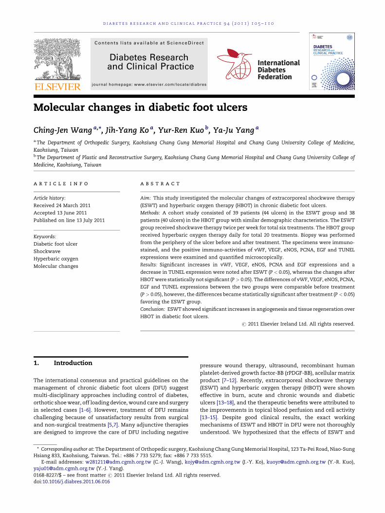
stains are shown in Fig. 1 for vWF and VEGF, Fig. 2 for eNOS
and PCNA and Fig. 3 for EGF and TUNEL expression. Significant
increases in vWF, VEGF, eNOS, PCNA and EGF, and a decrease
in TUNEL expressions were noted after treatment with ESWT
(P < 0.05), whereas the molecular changes after HBOT were
statistically not significant. The differences of vWF, VEGF,
eNOS, PCNA, EGF, and TUNEL expressions between the two
groups were comparable before treatment (P > 0.05), however,
the differences became statistically significant (P < 0.05) after
treatment favoring the ESWT.
Fig. 1 – Microscopic features of vWF and VEGF expressions sho
whereas the changes after HBOT are not significant (B).
4. Discussion
Wound healing stages include inflammation, proliferation,
epithelization, and remodeling. Non-healing wounds occur
when this process is interrupted or out of sequence, often the
case in diabetic ulcers [5]. At a molecular level, failure of
wound healing may result either from deficient supply or
functional inhibition of growth factors such as those investi-
gated during this study. The current study demonstrated
significant increases in vWF, VEGF, eNOS, PCNA and EGF and a
decrease in TUNEL expression after treatment with ESWT as
compared to HBOT in chronic diabetic ulcers. It appears that
ESWT significantly increased angiogenesis (vWF, VEGF and
eNOS) and cell proliferation (PCNA and EGF) and decreased cell
apoptosis (TUNEL) leading to tissue regeneration and wound
repair. These findings are supported by other studies that
ESWT enhanced skin flap survival and diabetic wound healing
via increasing angiogenesis and topical blood perfusion in
animals [14]. The results of the current study suggest that
ESWT may have the ability to improve wound healing by
increasing angiogenesis and cell activity in the wound
environment, normalizing the rate of apoptosis, and making
positive changes to growth factor and cytokine levels. This
wed significant increases after treatment with ESWT (A),
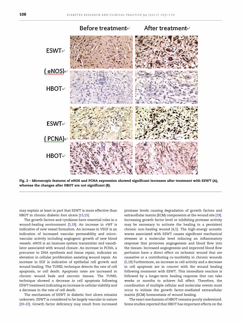
Fig. 2 – Microscopic features of eNOS and PCNA expression showed significant increases after treatment with ESWT (A),
whereas the changes after HBOT are not significant (B).
d i a b e t e s r e s e a r c h a n d c l i n i c a l p r a c t i c e 9 4 ( 2 0 1 1 ) 1 0 5 – 1 1 0108
may explain at least in part that ESWT is more effective than
HBOT in chronic diabetic foot ulcers [13,15].
The growth factors and cytokines have essential roles in a
wound-healing environment [5,19]. An increase in vWF is
indicative of new vessel formation. An increase in VEGF is an
indication of increased vascular permeability and micro-
vascular activity including angiogenic growth of new blood
vessels. eNOS is an immune system transmitter and vasodi-
lator associated with wound closure. An increase in PCNA, a
precursor to DNA synthesis and tissue repair, indicates an
elevation in cellular proliferation assisting wound repair. An
increase in EGF is indicative of epithelial cell growth and
wound healing. The TUNEL technique detects the rate of cell
apoptosis, or cell death. Apoptosis rates are increased in
chronic wound beds and necrotic tissues. The TUNEL
technique showed a decrease in cell apoptosis following
ESWT treatment indicating an increase in cellular viability and
a decrease in the rate of cell death.
The mechanism of ESWT in diabetic foot ulcers remains
unknown. ESWT is considered to be largely vascular in nature
[20–22]. Growth factor deficiency may result from increased
protease levels causing degradation of growth factors and
extracellular matrix (ECM) components at the wound site [19].
Increasing growth factor level or inhibiting protease activity
may be necessary to activate the healing in a persistent
chronic non-healing wound [4,5]. The high-energy acoustic
waves associated with ESWT causes significant mechanical
stresses at a molecular level inducing an inflammatory
response that promotes angiogenesis and blood flow into
the tissues. Increased angiogenesis and improved blood flow
perfusion have a direct effect on ischemic wound that are
causative or a contributing co-morbidity in chronic wounds
[5,19] Furthermore, an increase in cell activity and a decrease
in cell apoptosis are in concert with the wound healing
following treatment with ESWT. This immediate reaction is
followed by a longer-term healing response that can take
weeks or months to achieve full effect. Therefore, the
coordination of multiple cellular and molecular events must
occur to initiate the growth factor-mediated extracellular
matrix (ECM) homeostasis of wound healing.
The exact mechanism of HBOT remains poorly understood.
Some studies reported that HBOT has important effects on the
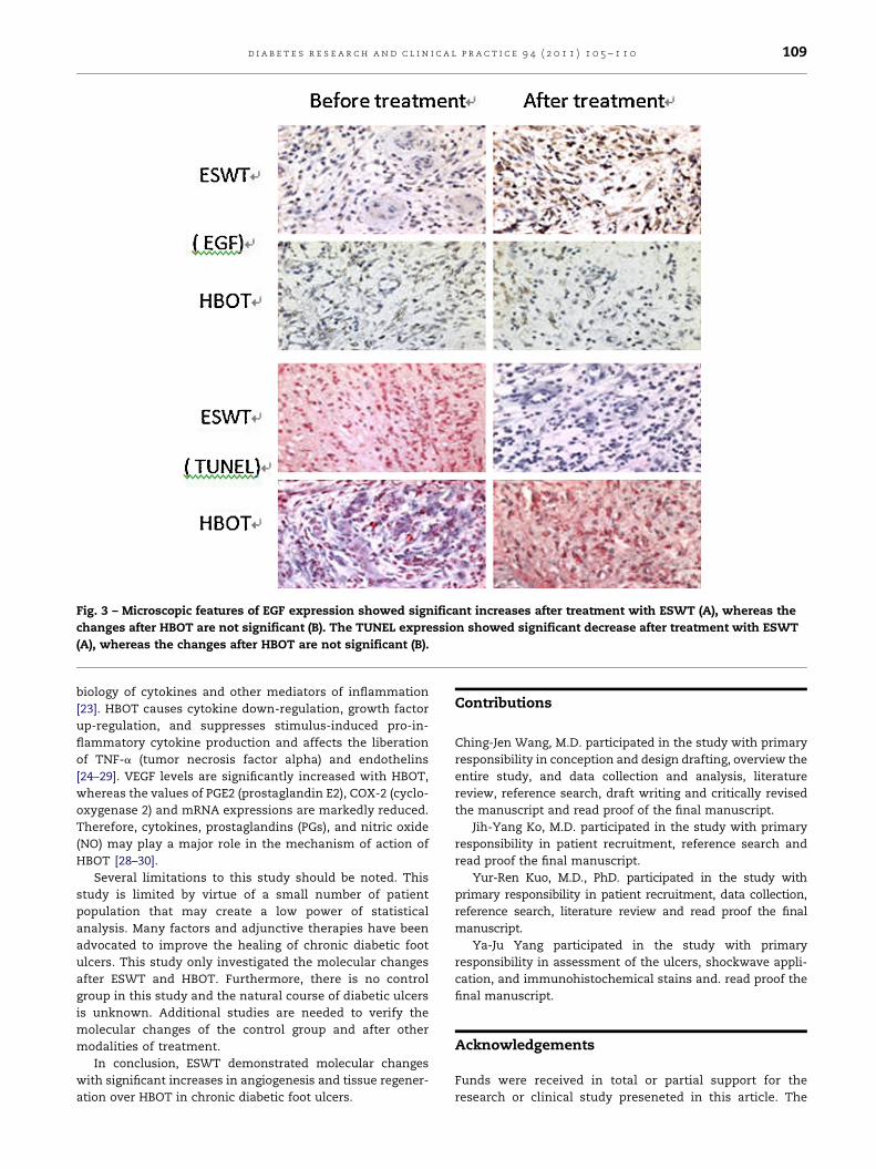
Fig. 3 – Microscopic features of EGF expression showed significant increases after treatment with ESWT (A), whereas the
changes after HBOT are not significant (B). The TUNEL expression showed significant decrease after treatment with ESWT
(A), whereas the changes after HBOT are not significant (B).
d i a b e t e s r e s e a r c h a n d c l i n i c a l p r a c t i c e 9 4 ( 2 0 1 1 ) 1 0 5 – 1 1 0 109
biology of cytokines and other mediators of inflammation
[23]. HBOT causes cytokine down-regulation, growth factor
up-regulation, and suppresses stimulus-induced pro-in-
flammatory cytokine production and affects the liberation
of TNF-a (tumor necrosis factor alpha) and endothelins
[24–29]. VEGF levels are significantly increased with HBOT,
whereas the values of PGE2 (prostaglandin E2), COX-2 (cyclo-
oxygenase 2) and mRNA expressions are markedly reduced.
Therefore, cytokines, prostaglandins (PGs), and nitric oxide
(NO) may play a major role in the mechanism of action of
HBOT [28–30].
Several limitations to this study should be noted. This
study is limited by virtue of a small number of patient
population that may create a low power of statistical
analysis. Many factors and adjunctive therapies have been
advocated to improve the healing of chronic diabetic foot
ulcers. This study only investigated the molecular changes
after ESWT and HBOT. Furthermore, there is no control
group in this study and the natural course of diabetic ulcers
is unknown. Additional studies are needed to verify the
molecular changes of the control group and after other
modalities of treatment.
In conclusion, ESWT demonstrated molecular changes
with significant increases in angiogenesis and tissue regener-
ation over HBOT in chronic diabetic foot ulcers.
Contributions
Ching-Jen Wang, M.D. participated in the study with primary
responsibility in conception and design drafting, overview the
entire study, and data collection and analysis, literature
review, reference search, draft writing and critically revised
the manuscript and read proof of the final manuscript.
Jih-Yang Ko, M.D. participated in the study with primary
responsibility in patient recruitment, reference search and
read proof the final manuscript.
Yur-Ren Kuo, M.D., PhD. participated in the study with
primary responsibility in patient recruitment, data collection,
reference search, literature review and read proof the final
manuscript.
Ya-Ju Yang participated in the study with primary
responsibility in assessment of the ulcers, shockwave appli-
cation, and immunohistochemical stains and. read proof the
final manuscript.
Acknowledgements
Funds were received in total or partial support for the
research or clinical study preseneted in this article. The

d i a b e t e s r e s e a r c h a n d c l i n i c a l p r a c t i c e 9 4 ( 2 0 1 1 ) 1 0 5 – 1 1 0110
funding source was from Chang Gung Research Fund
(CMRPG880221).
Conflict of interest
The authors declared that they have no conflict of interest.
One author (CJW) has served as a member of the scientific
advisory committee of Sanuwave until Nov. 2010. The
remaining authors declared no conflict of interest.
r e f e r e n c e s
[1] Schaper NC, Apelqvist J, Bakker K. The internationalconsensus and practical guidelines on the managementand prevention of the diabetic foot. Curr Diab Rep2003;3(6):475–9 [PMID: 14611743].
[2] Armstrong DG, Lavery LA, Harkless LB. Validation of adiabetic wound classification system. The contribution ofdepth, infection, and ischemia to risk of amputation.Diabetes Care 1998;21:855–9 [PMID: 9589255].
[3] Jeffcoate WJ, Macfarlane RM, Fletcher EM. The descriptionand classification of diabetic foot lesions. Diabet Med1993;10:676–9 [PMID: 8403832].
[4] Jeffcoate WJ, Harding KG. Diabetic foot ulcers. Lancet2003;361(9368):1545–51 [PMID: 12737879].
[5] Macfarlane RM, Jeffcoate WJ. Factors contributing topresentation of diabetic foot ulcers. Diabet Med1997;14(10):867–70 [PMID: 9371480].
[6] Treece KA, Macfarlane RM, Pound N, Game FL, Jeffcoate WJ.Validation of a system of foot ulcer classification indiabetes mellitus. Diabet Med 2004;21:987–91 [PMID:15317603].
[7] Ruffieux P, Hommel L, Saurat JH. Long-term assessment ofchronic leg ulcer treatment by autologous skin grafts.Dermatology 1997;195:77–80 [PMID: 9267750].
[8] Landau Z. Topical hyperbaric oxygen and low energy laserfor the treatment of diabetic foot ulcers. Arch OrthopTrauma Surg 1998;117:156–8 [PMID: 9521521].
[9] Ennis WJ, Foremann P, Mozen N, Massey J, Conner-Kerr T,Meneses P. Ultrasound therapy for recalcitrant diabetic footulcers: results of a randomized, double-blind, controlled,multicenter study. Ostomy Wound Manage 2005;51(8):24–39[PMID: 16234574].
[10] Margolis DJ, Bartus C, Hoffstad O, Malay S, Berlin JA.Effectiveness of recombinant human platelet-derivedgrowth factor for the treatment of diabetic neuropathic footulcers. Wound Repair Regen 2005;13(6):531–6 [PMID:16283867].
[11] Steed DL. Clinical evaluation of recombinant humanplatelet-derived growth factor for the treatment of lowerextremity ulcers. Plast Reconstr Surg 2006;117(7Suppl.):143S–9S [PMID: 16799381].
[12] Reyzelman A, Crews RT, Moore JC, Moore L, Mukker JS,Offutt S, et al. Clinical effectiveness of an acellular dermalregenerative tissue matrix compared to standard woundmanagement in healing diabetic foot ulcers: a prospectiverandomized, multicentre study. Int Wound J 2009;6(3):196–208 [PMID: 19368581].
[13] Wang CJ, Kuo YR, Wu RW, Liu RT, Hsu CS, Wang FS, et al.Extracorporeal shockwave treatment for chronic diabeticfoot ulcers. J Surg Res 2009;152:96–103 [PMID: 18619622].
[14] Kuo YR, Wang CT, Wang FS, Chiang YC, Wang CJ.Extracorporeal shockwave therapy enhanced woundhealing via increasing topical blood perfusion and tissueregeneration in a rat model of STZ-induced diabetes.Wound Repair Regen 2009;17(4):522–30 [PMID: 19614917].
[15] Wang CJ, Wu RW, Yang YJ. Treatment of diabetic footulcers: a comparative study of extracorporeal shockwavetherapy and hyperbaric oxygen therapy. Diab Res Clin Pract2011;92(2):187–93 [PMID: 21310502].
[16] Meirer R, Kamelger FS, Piza-Katzer H. Shock wave therapy:an innovative treatment method for partial thicknessburns. Burns 2005;31:921–2 [PMID: 16199297].
[17] Meirer R, Kamelger FS, Huemer GM, Wanner S, Piza-KatzerH. Extracorporeal shock wave may enhance skin flapsurvival in an animal model. Br J Plast Surg 2005;58(1):53–7[PMID: 15629167].
[18] Schaden W, Thiele R, Kolpl C, Pusch M, Nissan A, AttingerCE, et al. Shock wave therapy for acute and chronic softtissue wounds: a feasible study. J Surg Res 2007;143(1):1–12[PMID: 17904157].
[19] Yager D, Nwomeh B. The Proteolytic Environment ofchronic wounds. Wound Repair Regen 1999;7:433–41 [PMID:10633002].
[20] Wang CJ, Huang HY, Pai CH. Shock wave enhancedneovascularization at the bone tendon junction. A study ina dog model. J Foot Ankle Surg 2002;41(1):16–22 [PMID:11858601].
[21] Wang CJ, Wang FS, Yang KD, Weng LH, Hsu CC, Huang CS,et al. Shock wave induces neovascularization at thetendon-bone junction. A study in rabbits. J Orthop Res2003;21:984–9 [PMID: 14554209].
[22] Wang CJ. Biological Mechanism of MusculoskeletalShockwaves. Int Soc Musculoskeletal Shockwave TherNewsletter 2004;1(1):8(1).
[23] Boykin Jr JV. The nitric oxide connection: hyperbaricoxygen therapy, becaplermin, and diabetic ulcermanagement. Adv Skin Wound Care 2000;13:169–74 [PMID:11075012].
[24] Faglia E, Favales F, Aldeghi A, Calia P, Quarantiello A, OrianiG, et al. Adjunctive systemic hyperbaric oxygen therapy intreatment of severe prevalently ischemic diabetic footulcer. A randomized study. Diabetes care 1996;19:1338–43[PMID: 8941460].
[25] Kalani M, Jorneskog G, Naderi N, Lind F, Brismar K.Hyperbaric oxygen (HBO) therapy in treatment of diabeticfoot ulcers. Long-term follow-up. J Diabetes Complications2002;16(2):153–8 [PMID: 12039398].
[26] Senior C. Treatment of diabetic foot ulcers with hyperbaricoxygen. J Wound Care 2000;9:193–7 [PMID: 11933305].
[27] Kessler L, Bilbault P, Ortega F, Grasso C, Passemard R,Stephan D, et al. Hyperbaric oxygen accelerates the healingrate of nonischemic chronic diabetic foot ulcers: aprospective randomized study. Diabetes Care 2003;26:2378–82 [PMID: 12882865].
[28] Al-Waili NS, Butler GJ. Effects of hyperbaric oxygen oninflammatory response to wound and trauma: possiblemechanism of action. Scientific World J 2006;6:425–41[PMID: 16604253].
[29] Buras J. Basic mechanisms of hyperbaric oxygen in thetreatment of ischemia-reperfusion injury. Int AnesthesiolClin 2000;3:91–109 [PMID: 10723671].
[30] Abidia A, Laden G, Kuhan G, Johnson BF, Wilkinson AR,Renwick PM, et al. The role of hyperbaric oxygen therapy inischaemic diabetic lower extremity ulcers: a double-blindrandomised-controlled trial. Eur J Vasc Endovasc Surg2003;25:513–8 [PMID: 12787692].