molecular biology of bladder cancer - pdfs.semanticscholar.org · unitat de biologia cel.lular i...
TRANSCRIPT
Bladder cancer is a major cause of health expensesand it presents formidable clinical challenges. Twotypes of tumors have been identified, papillary andnon-papillary. The former are mainly characterizedby FGFR3 and chromosome 9 alterations and a lowfrequency of Tp53 alterations. The latter are charac-terized by a high frequency of alterations in genesin the p53 and Rb pathways. Chromosome 9 alter-ations, specially in 9q, are crucial to bladder cancerdevelopment and occur in both types of tumors.Progression of some superficial tumors (mainlyTaG3 and T1G3) to high-grade, invasive, carcino-mas provides evidence of some overlap between thetwo pathways. Distinct gene expression profileshave been identified in superficial and invasive tu-mors. The stage is now ready for the clinical appli-cation of this knowledge.
Key words Bladder • FGFR3 • Genetic alterations • p53Rb • Urothelial carcinoma
Luis NM, López-Knowles E, Real FX (2007) Molecular biolo-gy of bladder cancer. Clin Transl Oncol 9:5-12
INTRODUCTION
Bladder cancer is a major health burden in theWestern world. In 2002, approximately 139,000 newbladder cancer cases were diagnosed in Europe, themale:female ratio being 3.7:1. In the same year, ap-proximately 51,000 patients died from this disease1.In Spain, it is the 5th leading cause of cancer-related
death in men and the 13th in women. Bladder cancergenerates the highest medical cost per patient and itis the fifth most expensive cancer in terms of totalmedical care expenditures.The two best established lifestyle/environmental riskfactors for bladder cancer are smoking and occupa-tional exposure to aromatic amines (i.e. dye, textile,leather, chemical and rubber industries)2. Therefore,this tumor is a largely preventable disease.
BLADDER TUMORS
Approximately 95% of malignant bladder tumors areurothelial cell carcinomas (UCC) which can be clas-sified as papillary (most common type, tend to growslowly towards the lumen), solid (less frequent, infil-trate the bladder wall and are more agressive) orCarcinoma In Situ (CIS, a very agressive kind of can-cer that involves only the inner lining of the bladder).
Staging and grading
The bladder is a hollow and distensible organ thatsits on the pelvic floor and collects urine from thekidneys. A scheme of the bladder wall is representedin figure 1.Tumors are classified according to depth of invasioninto superficial (Ta and T1) and invasive (T2, T3, T4).pTa tumors are defined as those which have an exo-phytic «fingerlike» growth and do not grow beyondthe urothelium; pT1 tumors invade the lamina pro-pria but not the muscularis propria; pT2 tumors asthose invading the muscularis propria; pT3 tumors asthose invading perivesical tissue; and pT4 tumors asthose invading other organ structures. Grading is used to evaluate the cytological and/orgrowth pattern characteristics of the tumor and it isan important predictor of its biologic potential3. 1. Flat and papillary hyperplasia. Flat hyperplasiaconsists of a markedly thickened mucosa without cy-tologic atypia. Papillary hyperplasia is characterizedby urothelium of variable thickness exhibiting aslightly undulating growth.
Molecular biology of bladder cancerNuno M. Luisa, Elena López-Knowlesa and Francisco X. Reala,b
aUnitat de Biologia Cel.lular i Molecular. Institut Municipal d’Investigació Mèdica. Barcelona. Spain.bUniversitat Pompeu Fabra. Barcelona. Spain.
*Supported by an unrestricted educational grantfrom Sanofi-Aventis.
Correspondence: F.X. Real.Unitat de Biologia Cel.lular i Molecular.Institut Municipal d’Investigació Mèdica.Carrer del Dr. Aiguader, 88.08003 Barcelona. Spain.E-mail: [email protected]
EDUCATIONAL SERIES Blue series*Clin Transl Oncol (2007) 9:5-12
DOI 10.1007/s12094-007-0003-x
(5-12) CTO-Blue series Molecular biology...qxp 09/02/2007 18:36 Página 5

6 N.M. Luis et al.: Molecular biology of bladder cancer
2. Flat lesions with atypia can be classified as reactiveatypia, dysplasia or CIS. Dysplasia indicates cytologicand architectural abnormalities. 3. Papillary neoplasms can be classified as papilloma,papillary neoplasm of low malignancy potential(PUNLMP), low grade papillary carcinoma and highgrade papillary carcinoma. In papilloma, normal ap-pearing urothelium lines papillary fronds. PUNLMPrefers to thickened urothelium in the absence of cyto-logic features of malignancy. Low grade papillarycarcinoma exhibits an overall orderly appearanceand has minimal anomalies in architecture and/orcytologic features. High grade papillary carcinomahas a disorderly appearance with marked architec-tural and cytologic abnormalities.
Natural history of bladder tumors: two pathways,clonal origin
UCC is a heterogeneous disease with a variable natu-ral history. Low-grade Ta tumors have a very lowprogression rate and rarely present a threat to the pa-tient. At the other extreme, high-grade tumors implya high risk of progression and cancer death.
Two pathways
It has been proposed that there may be at least twoseparate pathways leading to bladder cancer, papillaryand nonpapillary4. Papillary lesions arise in hyperplas-tic urothelium whereas invasive tumors arise fromdysplastic urothelium. The overlap between both path-ways is reflected by progression of superficial papillarytumors to high grade invasive carcinomas in somecases. Papillary tumors are commonly associated with
alterations in genes involved in the RAF/MEK/ERKpathway (i.e. FGFR3)5,6 and in PIK3CA7. By contrast,non-papillary invasive tumors are commonly associ-ated with p53 and pRb pathway alterations (fig. 2).
Clonal origin
The development of multiple tumors in either a syn-chronous or metachronous manner in the same pa-tient is a common characteristic of UCC. Two theo-ries have been proposed to account for these findings:tumors may be monoclonal in origin (multifocal tu-mors evolving from a single transformed cell) or theymay be oligoclonal (a change in the urothelium giv-ing rise to multiple clones of initiated cells subse-quently evolving into independent tumors). Currentevidence supports the notion that most tumors aremonoclonal but up to 30% of tumors may indeed beoligoclonal8,9.
Genetic alterations in bladder cancer
For many years, it has been clear that UCC representsa complex disease and that it is a paradigm of tumorprogression. The current status of knowledge on themolecular biology of bladder cancer is presented,pointing out the differences between papillary and in-vasive bladder tumors.
MOLECULAR GENETICS OF PAPILLARYBLADDER TUMORS
Fibroblast growth factor receptor 3 (FGFR3)
FGFRs (1-4) regulate proliferation, differentiation, an-giogenesis and embryonic development. They displaydistinct tissue expression, ligand specificity, signal
Fig. 1. Bladder tumors are classified on the basisof the degree of involvement of the bladder walland the nuclear grade.Ta tumors do not invadethe basal membrane and T1 tumors do not invademuscle.This classification is of prognosticand therapeutic importance (Adapted fromThe University of Michigan, Department of Urologyat the Michigan Urology Center).
(5-12) CTO-Blue series Molecular biology...qxp 09/02/2007 18:36 Página 6
N.M. Luis et al.: Molecular biology of bladder cancer 7
pathway activation, and biological effects. FGFRmRNAs are commonly subject to alternative splicing.There are two major isoforms of FGFR3 transcriptsthat are generated from a mutually exclusive splicingevent in which the second half of the third Ig-like do-main is encoded by either the 151 bp of exon 8(FGFR3b, predominant in epithelia) or the 145 bp ofexon 9 (FGFR3c, predominant in mesenchymal cells).Inherited skeletal dysplasias have been linked to acti-vating germline point mutations in FGFR1, 2 and 3.Germline point mutations in FGFR3 cause the shortlimb syndromes of achondroplasia, hypochondropla-sia and thanatophoric dysplasia. Mutations lead toconstitutive receptor activation; increasing activity isassociated with increasing severity of the disease10,11.Activating FGFR3 mutations occur in 50-60% of UCCand are associated with papillary, low grade, superficialtumors5. Somatic mutations are restricted to a fewcodons, essentially the same that are mutated in thegermline in short limb syndromes. Mutations in the ex-tracellular domain (i.e. codons 248, 249) or within thetransmembrane domain and its vicinity (i.e. codons372, 375, 382, 393) lead to receptor dimerization in theabsence of ligand; mutations in the intracellular do-main (codon 652) confer constitutive kinase activity.Mutations are associated with an increased risk of re-currence and a lower risk of progression among pa-tients with superficial tumors, indicating that they are amarker of tumors with good prognosis12.
Ras genes
H-ras, K-ras, and N-ras code for monomeric GTPasesthat can activate the RAF/MEK/ERK and PI3K/AKT/
PTEN cascades. Oncogenic Ras mutations make theGTPase insensitive to the action of activating proteins(GAPs) and lock it in the GTP-bound, active state. InUCC, point mutations have been identified in codons12, 13 and 61 in H-ras and in N-ras at an overall fre-quency of 10-20%. FGFR3 and Ras mutations are mu-tually exclusive, possibly because they both activatethe MAPK pathway. Ras mutations do not seem to beassociated with stage or grade13. Amplification of the gene coding for Raf1, a kinase in-volved in Ras protein signalling, has been associatedto grade, stage and poor survival and overexpressionhas been associated to grade, but not to stage14,15.
PIK3CA and TSC1
The phosphatidylinositol-3-kinase (PI3K) pathway iscrucial to many aspects of cell growth and survival.Class IA proteins are most important in proliferationand tumorigenesis; they are constituted by a het-erodimer of p110, the catalytic subunit, and an adap-tor subunit (p85, p50 or p55). PIK3CA, which encodesp110α, maps to 3q26, a region amplified in UCC.PIK3CA mutations have been reported in 20% of tu-mors, in association with low stage and grade tumorsand with FGFR3 mutations7. There is some evidencethat UCC can also overexpress PI3K in comparisonwith adjacent normal epithelium.TSC1 (9q34) encodes hamartin, a GAP that negativelyregulates cell cycle progression. Its action is blockedby AKT and activation of the PI3K pathway. Inac-tivating TSC1 mutations are found in 12% of UCC andLOH at 9q34 occurs in 32% of cases16,17. Stage andgrade do not seem associated with these alterations.
Hyperplasia Low-grade non-invasive papillary tumorRAF/MEK/Erk
PI3K
15%80%9p–/9q–
20%Normal
urothelium
P53/pRbHigh-grade invasive tumor
> 50%
Metastasis
N-cad, E-cadMMPs, VEGF, COX2
9p–/9q–
CIS/dysplasia
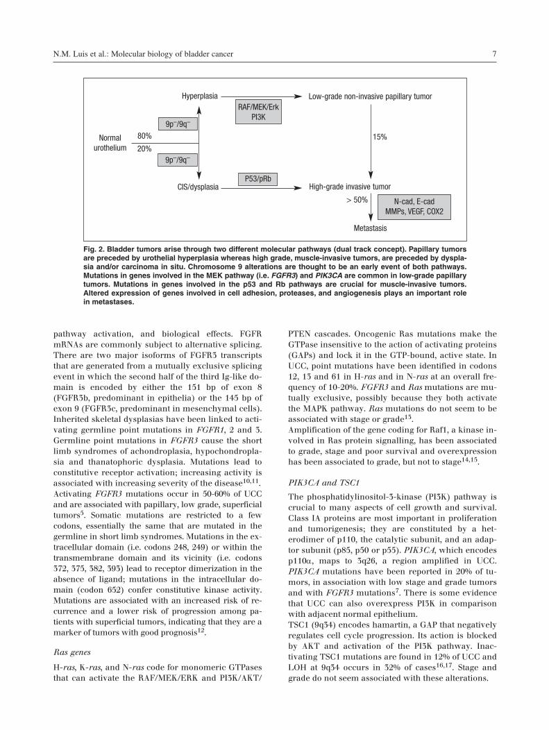
Fig. 2. Bladder tumors arise through two different molecular pathways (dual track concept). Papillary tumorsare preceded by urothelial hyperplasia whereas high grade, muscle-invasive tumors, are preceded by dyspla-sia and/or carcinoma in situ. Chromosome 9 alterations are thought to be an early event of both pathways.Mutations in genes involved in the MEK pathway (i.e. FGFR3) and PIK3CA are common in low-grade papillarytumors. Mutations in genes involved in the p53 and Rb pathways are crucial for muscle-invasive tumors.Altered expression of genes involved in cell adhesion, proteases, and angiogenesis plays an important rolein metastases.
(5-12) CTO-Blue series Molecular biology...qxp 09/02/2007 18:36 Página 7
8 N.M. Luis et al.: Molecular biology of bladder cancer
MOLECULAR GENETICS OF INVASIVETUMORS
Virtually all human tumors deregulate either the Rbor p53 pathway or both and this is also the case forinvasive UCC.
Rb pathway alterations
The Rb pathway is responsible for regulating the pas-sage from G1 to S phase. Phosphorylation of pRb byCDK4/6 and CDK2 dissociates the pRb-repressor com-plex, leading to the release of bound E2F from pRb.«Free» E2F is then active and drives the transcriptionof S-phase genes encoding for proteins required for G1to S phase transition and DNA replication.Mutations of the Rb1 gene have been described inUCC but the studies are not extensive due to the large
gene size. There is controversy about the prognosticvalue of pRb expression in tumor tissues18-20.p16, encoded by the INK4A/ARF locus, is a CDK4/6inhibitor which blocks the phosphorylation of pRb.The INK4A/ARF locus plays a central role in tumorsuppression and it is inactivated in approximately50% of human cancers. mRNA levels of p16INK4A andp14ARF are undetectable in normal urothelium andincrease with stage and grade21. At the protein level,loss of p16 has been associated to minimally invasivebladder cancer. p16INK4A and p14ARF methylationhave been proposed as biomarkers of stage, clinicaloutcome, and prognosis. E2F1 expression has been correlated with prolifera-tion in UCC, having a growth promoting effect. TheE2F3 locus is amplified in invasive tumors22 and itsproduct is overexpressed in 33% of UCC23.
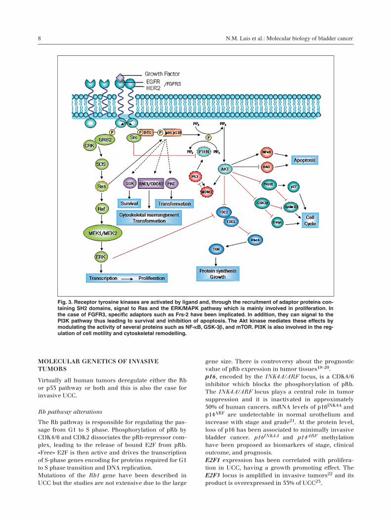
Fig. 3. Receptor tyrosine kinases are activated by ligand and, through the recruitment of adaptor proteins con-taining SH2 domains, signal to Ras and the ERK/MAPK pathway which is mainly involved in proliferation. Inthe case of FGFR3, specific adaptors such as Frs-2 have been implicated. In addition, they can signal to thePI3K pathway thus leading to survival and inhibition of apoptosis. The Akt kinase mediates these effects bymodulating the activity of several proteins such as NF-κκB, GSK-3ββ, and mTOR. PI3K is also involved in the reg-ulation of cell motility and cytoskeletal remodelling.
(5-12) CTO-Blue series Molecular biology...qxp 09/02/2007 18:36 Página 8
Cyclin D1 overexpression occurs frequently in UCCand has been proposed to be associated with thegrowth of low-grade papillary tumors24.
p53 pathway alterations
The p53 pathway plays a major role in the responseto DNA damage, oncogenic stress, and other types ofcellular stress. p53 induces G1 arrest by upregulatingp21, G2 arrest by upregulating GADD45, 14-3-3σ andp21, and apoptosis by upregulating genes such asBax, NOXA, and PUMA. In addition, it can repressgenes such as c-myc to promote G1 arrest and cyclinB1 to promote G2 arrest25.The Tp53 gene is frequently altered in UCC: 270 dif-ferent mutations have been registered in the IARCdatabase26 up to October 2006, of which 262 (97%)are in exons 4 to 9. The most common mutations aremissense (72.5%), 12.2% and 5.55% being nonsenseand silent, respectively. The main hotspots arecodons 285, 248, 280, 175 and 213. Tp53 mutations aresignificantly more frequent among high grade andstage tumors; their prevalence ranging from 14-52%depending on the T and G subgroup considered27.Overal, Tp53 mutations and p53 nuclear overexpres-sion are uncommon in TaG1/G2 tumors and are fre-quent in high grade/invasive tumors28. HDM2, a p53target gene and regulator, is gained/amplified in ap-proximately 9% of high grade tumors. p63 and p73 are members of the p53 family. p63 mapsto 3q27-29, a region that is amplified in UCC; this lo-cus encodes multiple proteins resulting from alterna-tive splicing that transactivate p53 responsive ele-ments or act as dominant negatives towards p53 andp73. p63 is lost in most invasive tumors. A decrease ofthe levels of p73 has also been associated to bladdercancer stage29. Two additional important genes in the p53 pathwayare Aurora A kinase (STK6) and p21. Overexpressionof Aurora A (20q13), often associated with gene am-plification, leads to increased degradation of p53,causing down-regulation of checkpoint-responsepathways and facilitating oncogenic transformation.Expression of p21 is reduced in muscle invasive tu-mors compared to non-invasive tumors30.A few studies have combined the analysis of p53, p16,p21 and pRb expression in bladder tumors and de-scribed that they act in cooperative or synergisticways to promote progression: p53/p16, p53/p21,p53/pRb or p53/p21/pRb18,31-34.
PTEN
This gene maps to 10q23 and it encodes a lipid phos-phatase that acts as a negative regulator of PI3K path-way by hydrolyzing 3,4,5-PIP3 to 4,5-PIP2. PTENphysically interacts with p53 in the nucleus, leading
to p53 stabilization and increased transcriptional ac-tivity. PTEN is mutated or deleted in 14% of invasivebladder cancers with 40% LOH at 10q35,36. PTENdownregulation has been described in 13% of tumors,mainly muscle-invasive tumors.
GENOMIC LEVEL ALTERATIONS
Chromosomal aberrations can be primary, related tothe cause of a tumor, or secondary, involved in pro-gression. Deletions and gains/amplifications con-tribute to altered expression of tumor suppressor ge-nes and oncogenes, respectively. Higher rates ofgenomic alterations are present in pT1 than in papil-lary pTa tumors. The most consistent alterations inadvanced-stage UCC are gains of 1q, 8q and 20q andlosses of 8p, chromosome 11 and 9. Array CGH analy-sis has been applied to the study of UCC37-39. A sum-mary of the most common alterations reported, clas-sified on the basis of their chromosomal location,follows.
Alterations common to both pathways(chromosome 9)
The q arm of chromosome 9 is lost both in low andhigh grade tumors, suggesting it is a primary event inthe genesis of bladder cancer40,41. Losses at 9q cover3 major deleted regions (9q22, 9q32-33, and 9q34) andone or several tumor suppressor genes may be locat-ed in them. Candidate genes therein include Netrin,TSC1, PTCH and DBCCR117,42-44. Allelic loss at 9qhas been reported as an early occurrence in the de-velopment of bladder cancer but it has also been as-sociated with invasive disease and with disease re-currence in superficial bladder tumors. Deletions of9p are also common in bladder tumors and affectmainly 9p21, where the INK4A/ARF locus maps45.
Other alterations – invasive tumors
Alterations in chromosome 8 often involve loss of thep arm, gain of the q arm and amplification of a smallregion at 8p12. LOH in 8p is associated with a moreaggressive tumor phenotype indicating the possiblepresence of a tumor suppressor gene. The minimalregion of 8p21-22 contains several candidate genes:TRAIL-R2, DBC2 and LZTS146,47. A commonly gai-ned region in 8q contains c-myc. 8p12 amplicons con-tain FGFR1.5p amplification is one of the few alterations occur-ring more frequently in muscle invasive tumors thanin early invasive cancers48. The most common site ofamplification (5p12) contains TRIO; 5p13-12 has alsobeen defined as a critical region of allele losses asso-ciated with tumor progression and a marker of ad-verse prognosis independent of stage and grade49.
N.M. Luis et al.: Molecular biology of bladder cancer 9
(5-12) CTO-Blue series Molecular biology...qxp 09/02/2007 18:36 Página 9
10 N.M. Luis et al.: Molecular biology of bladder cancer
Amplification of 6p22 affects up to 20% of high grade,invasively growing tumors39. This region has beennarrowed down to 1.6 Mb at 6p22.3 and contains po-tential oncogenes such as SOX4, CDKAL, DEK, ID4,and E2F, the latter being a strong pathogenic candi-date23,50-52.The regions containing EGFR1 (7p12) and EGFR2(17q11) are amplified in 4.6% and 3.4% of UCC, res-pectively; protein overexpression has been describedin 48%53 and 41%54 of tumors, respectively. Amplifi-cation/overexpression of EGFR is associated with tu-mor proliferation, aggressive behaviour and poorprognosis55. Other common alterations in UCC are gains of 1q and20q, amplifications of 11q13 and 12q14 (candidategenes cyclin D1 and HDM2, respectively) and lossesof 11p.
METHYLATION
As in other tumors, promoter methylation constitutesa common mechanism of silencing of tumor suppres-sor genes in UCC56. The frequencies of methylation ofthe best studied genes are: cadherin-1 (36%), RAS-as-sociated domain family (RASSF1A) (35%), CDH13(29%), secreted Frizzled-related protein 1 (sFRP1) (29%),FHIT (16%), retinoic acid receptor β (15%), p16INK4A
(7%), and death-associated kinase (4%)57. Hyperme-thylation of APC, p14ARF and RASSF1A has also beendescribed in exfoliated cells in the urine of patientswith UCC. A recent study showed that methylation ofpromoter regions of p16, p14, E-cadherin, RARβ2,RASSF1a and GSTP1 occurs in both normal and CISsamples from patients with UCC and increases withprogression58. sFRP gene silencing by methylationhas been shown to be associated to invasive bladdercancer and to overall survival59.
EXPRESSION ANALYSES WITH MICROARRAYS
The first study analyzing gene expression in UCCarrays showed different gene expression profiles insuperficial and invasive tumors60. The same groupsubsequently identified expression profiles distin-guishing stages, as well as a similar expression pro-file between CIS and invasive tumors61. Data frommicroarrays with 10368 cDNAs allowed to identify 25genes able to classify tumors as superficial or inva-sive with 90.5% accuracy62. This classifier had an82.5% accuracy when used on the data set fromDyrskjot et al. The gene-classifiers reported byBlaveri and Dyrskjot had no genes in common. Otherstudies have also reported minimal overlap for thegenes identified for clinically similar tumors.Sánchez-Carbayo et al separated superficial from in-
vasive tumors with 82.2% accuracy and stratified tu-mors on the basis of clinical outcome with 82% (alltumors) or 90% accuracy (when considering only in-vasive tumors)63.
APOPTOSIS
In the bladder, as in other tissues, failure of the regu-latory genes involved in apoptosis may result in sur-vival of cells with genomic abnormalities, tumorigen-esis and resistance to anticancer agents. Low levels ofFAS and FASL have been associated with highergrade, stage and a poor prognosis64. Overexpressionof the antiapoptotic protein BCL-2, involved in mito-chondrial permeabilization, is associated with p53overexpression and with poor outcome65. Survivin,an inhibitor of caspase-3 and caspase-7, is detectablein urine and has been proposed as a biomarker forthe detection of bladder cancer66. FHIT protein hasbeen shown absent or reduced in 61% of UCC and itsexpression correlated with pathological and clinicalstatus67.
INVASION AND METASTASIS
Cadherins are the main mediators of cell-cell adhe-sion in epithelia. Loss of E-cadherin has been de-scribed at higher frequency in high-grade, invasive,UCC than in low-grade papillary tumors68. Hyperme-thylation of CpG dinucleotides in the promoter ofCDH1 (encoding E-cadherin) occurs frequently inUCC69. The status of E-cadherin has been proposedas an independent prognostic indicator for diseaseprogression.
CONCLUDING REMARKS
The information accumulated in the last few years onthe molecular changes associated with papillary andinvasive bladder tumors allows a more accurate mo-lecular classification of UCC. This information mayhelp not only in the prediction of patient outcome butin the selection of treatment, as well. Bladder canceris one of the solid tumors in which molecular studiesmay soon become part of standard clinical practice.
ACKNOWLEDGEMENTS
Work in the authors’ laboratory was funded, in part,by grants C03/010 and G03/174 from Instituto deSalud Carlos III, Ministerio de Sanidad, Madrid.N.M.L. is recipient of a Ph.D. grant from Fundação deCiência e Tecnologia, Portugal, and E.L. was support-ed by a Predoctoral Fellowship from the Ramón Are-ces Foundation.
(5-12) CTO-Blue series Molecular biology...qxp 09/02/2007 18:36 Página 10
N.M. Luis et al.: Molecular biology of bladder cancer 11
References
1. Ferlay J, Bray F, Pisani P, Parkin DM.GLOBOCAN 2002 (2004) Cancer Inciden-ce, Mortality and Prevalence WorldwideIARC CancerBase No. 5 version 2.0.: IARCPress, Lyon
2. Silverman DT, Devessa SS, Moore LE,Rothman, N (2006) Bladder cancer. Can-cer epidemiology and prevention, 3rdedn. Oxford University Press, New York,pp 1101-1127
3. Eble J, Sauter G, Epstein JI, Sesterhenn IA(2004) Pathology and Genetics of Tu-mours of the Urinary System and MaleGenital Organs. IARC Press, Lyon, France
4. Pugh RC (1973) Proceedings: The Patho-logy of Cancer of the Bladder. Cancer 32:1267-1274
5. Billerey C, Chopin D, Aubriot-Lorton MHet al (2001) Frequent FGFR3 Mutations inPapillary Non-Invasive Bladder (PTa)Tumors. Am. J. Pathol. 158:1955-1959
6. Cappellen D, De Oliveira C, Ricol D et al(1999) Frequent Activating Mutations ofFGFR3 in Human Bladder and CervixCarcinomas. Nat Genet 23:18-20
7. Lopez-Knowles E, Hernandez S, Malats Net al (2006) PIK3CA Mutations Are anEarly Genetic Alteration Associated WithFGFR3 Mutations in Superficial PapillaryBladder Tumors. Cancer Res 66:7401-7404
8. van Tilborg AA, de Vries A, de Bont M etal (2000) Molecular Evolution of MultipleRecurrent Cancers of the Bladder. HumMol Genet 9:2973-2980
9. Hafner C, Knuechel R, Stoehr R, Hart-mann A (2002) Clonality of Multifocal Uro-thelial Carcinomas: 10 Years of MolecularGenetic Studies. Int J Cancer 101:1-6
10. Naski MC, Wang Q, Xu J, Ornitz DM(1996) Graded Activation of FibroblastGrowth Factor Receptor 3 by MutationsCausing Achondroplasia and Thanato-phoric Dysplasia. Nat Genet 13:233-237
11. Webster MK, D'Avis PY, Robertson SC,Donoghue DJ (1996) Profound Ligand-Independent Kinase Activation of Fibro-blast Growth Factor Receptor 3 by the Ac-tivation Loop Mutation Responsible for aLethal Skeletal Dysplasia, ThanatophoricDysplasia Type II. Mol Cell Biol 16:4081-4087
12. Hernandez S, Lopez-Knowles E, Lloreta Jet al (2006) Prospective Study of FGFR3Mutations As a Prognostic Factor inNonmuscle Invasive Urothelial BladderCarcinomas. J Clin Oncol 24:3664-3671
13. Jebar AH, Hurst CD, Tomlinson DC et al(2005) FGFR3 and Ras Gene MutationsAre Mutually Exclusive Genetic Events inUrothelial Cell Carcinoma. Oncogene24:5218-5225
14. Mhawech-Fauceglia P, Fischer G, Beck Aet al (2006) Raf1, Aurora-A/STK15 and E-Cadherin Biomarkers Expression inPatients With PTa/PT1 Urothelial BladderCarcinoma; a Retrospective TMA Study of246 Patients With Long-Term Follow-Up.Eur J Surg Oncol 32:439-444
15. Simon R, Richter J, Wagner U et al (2001)High-Throughput Tissue Microarray Ana-lysis of 3p25 (RAF1) and 8p12 (FGFR1)Copy Number Alterations in UrinaryBladder Cancer. Cancer Res 61:4514-4519
16. Adachi H, Igawa M, Shiina H et al (2003)Human Bladder Tumors With 2-Hit Mu-tations of Tumor Suppressor Gene TSC1and Decreased Expression of P27. J Urol170:601-604
17. Knowles MA, Habuchi T, Kennedy W,Cuthbert-Heavens D (2003) Mutation
Spectrum of the 9q34 Tuberous SclerosisGene TSC1 in Transitional Cell Carcino-ma of the Bladder. Cancer Res 63:7652-7656
18. Shariat SF, Tokunaga H, Zhou J et al(2004) P53, P21, PRB, and P16 ExpressionPredict Clinical Outcome in CystectomyWith Bladder Cancer. J Clin Oncol 22:1014-1024
19. Cordon-Cardo C (2004) P53 and RB: Sim-ple Interesting Correlates or Tumor Mar-kers of Critical Predictive Nature? J ClinOncol 22:975-977
20. Cordon-Cardo C, Wartinger D, Petrylak Det al (1992) Altered Expression of theRetinoblastoma Gene-Product - Prognos-tic Indicator in Bladder-Cancer. J NatlCancer Inst 84:1251-1256
21. Frere-Belda MA, Gil Diez de Medina S,Daher A et al (2004) Profiles of the 2INK4a Gene Products, P16 and P14ARF, inHuman Reference Urothelium andBladder Carcinomas, According to PRband P53 Protein Status. Hum Pathol 35:817-824
22. Oeggerli M, Tomovska S, Schraml P et al(2004) E2F3 Amplification and Overex-pression Is Associated With Invasive Tu-mor Growth and Rapid Tumor Cell Pro-liferation in Urinary Bladder Cancer.Oncogene 23:5616-5623
23. Feber A, Clark J, Goodwin G et al (2004)Amplification and Overexpression ofE2F3 in Human Bladder Cancer. Oncoge-ne 23:1627-1630
24. Lee CC, Yamamoto S, Morimura K et al(1997) Significance of Cyclin D1 Overex-pression in Transitional Cell Carcinomasof the Urinary Bladder and Its CorrelationWith Histopathologic Features. Cancer79:780-789
25. Vogelstein B, Lane D, Levine AJ (2000)Surfing the P53 Network. Nature 408:307-310
26. Olivier M, Eeles R, Hollstein M et al(2002) The IARC TP53 Database: New On-line Mutation Analysis and Recommen-dations to Users. Hum Mutat 19:607-614
27. Malats N, Bustos A, Nascimento CM et al(2005) P53 As a Prognostic Marker forBladder Cancer: a Meta-Analysis and Re-view. Lancet Oncol. 6:678-686
28. Kelsey KT, Hirao T, Schned A, et al (2004)A Population-Based Study of Immunohis-tochemical Detection of P53 Alteration inBladder Cancer. Br J Cancer 90:1572-1576
29. Puig P, Capodieci P, Drobnjak M et al(2003) P73 Expression in Human Normaland Tumor Tissues: Loss of P73alpha Ex-pression Is Associated With Tumor Pro-gression in Bladder Cancer. Clin CancerRes 9:5642-5651
30. Stein JP, Ginsberg DA, Grossfeld GD et al(1998) Effect of P21WAF1/CIP1 Expres-sion on Tumor Progression in BladderCancer. J Natl Cancer Inst 90:1072-1079
31. Chatterjee SJ, Datar R, Youssefzadeh D etal (2004) Combined Effects of P53, P21,and PRb Expression in the Progression ofBladder Transitional Cell Carcinoma. JClin Oncol 22:1007-1013
32. Cote RJ, Dunn MD, Chatterjee SJ et al(1998) Elevated and Absent PRb Expres-sion Is Associated With Bladder CancerProgression and Has Cooperative EffectsWith P53. Cancer Res 58:1090-1094
33. Garcia del Muro X, Condom E, Vigues Fet al (2004) P53 and P21 ExpressionLevels Predict Organ Preservation andSurvival in Invasive Bladder CarcinomaTreated With a Combined-Modality Ap-proach. Cancer 100:1859-1867
34. Hitchings AW, Kumar M, Jordan S et al(2004) Prediction of Progression in PTaand PT1 Bladder Carcinomas With P53,P16 and PRb. Br J Cancer 91:552-557
35. Cappellen D, Gil Diez de Medina S, Cho-pin D et al (1997) Frequent Loss of Hete-rozygosity on Chromosome 10q in Mus-cle-Invasive Transitional Cell Carcinomasof the Bladder. Oncogene 14:3059-3066
36. Wang DS, Rieger-Christ K, Latini JM et al(2000) Molecular Analysis of PTEN andMXI1 in Primary Bladder Carcinoma. IntJ Cancer 88:620-625
37. Blaveri E, Brewer JL, Roydasgupta R et al(2005) Bladder Cancer Stage and Outco-me by Array-Based Comparative Geno-mic Hybridization. Clin Cancer Res 11:7012-7022
38. Hurst CD, Fiegler H, Carr P et al (2004)High-Resolution Analysis of Genomic CopyNumber Alterations in Bladder Cancer byMicroarray-Based Comparative GenomicHybridization. Oncogene 23: 2250-2263
39. Veltman JA, Fridlyand J, Pejavar S et al(2003) Array-Based Comparative GenomicHybridization for Genome-Wide Scre-ening of DNA Copy Number in BladderTumors. Cancer Res 63:2872-2880
40. Hartmann A, Schlake G, Zaak D et al(2002) Occurrence of Chromosome 9 andP53 Alterations in Multifocal Dysplasiaand Carcinoma in Situ of Human UrinaryBladder. Cancer Res 62:809-818
41. Hopman AH, Moesker O, Smeets AW et al(1991) Numerical Chromosome 1, 7, 9,and 11 Aberrations in Bladder CancerDetected by in Situ Hybridization. CancerRes 51:644-651
42. Aboulkassim TO, Larue H, Lemieux P etal (2003) Alteration of the PATCHEDLocus in Superficial Bladder Cancer.Oncogene 22:2967-2971
43. Amira N, Cancel-Tassin G, Bernardini Set al (2004) Expression in Bladder Transi-tional Cell Carcinoma by Real-TimeQuantitative Reverse Transcription Poly-merase Chain Reaction Array of 65 Genesat the Tumor Suppressor Locus 9q34.1-2:Identification of 5 Candidates Tumor Sup-pressor Genes. Int J Cancer 111:539-542
44. Wright KO, Messing EM, Reeder JE(2004) DBCCR1 Mediates Death in Cul-tured Bladder Tumor Cells. Oncogene 23:82-90
45. Williamson MP, Elder PA, Shaw ME et al(1995) P16 (CDKN2) Is a Major DeletionTarget at 9p21 in Bladder Cancer. HumMol Genet 4:1569-1577
46. Adams J, Cuthbert-Heavens D, Bass S,Knowles MA (2005) Infrequent Mutationof TRAIL Receptor 2 (TRAIL-R2/DR5) inTransitional Cell Carcinoma of the Blad-der With 8p21 Loss of Heterozygosity. Can-cer Lett 220:137-144
47. Knowles MA, Aveyard JS, Taylor CF et al(2005) Mutation Analysis of the 8p Can-didate Tumour Suppressor Genes DBC2(RHOBTB2) and LZTS1 in BladderCancer. Cancer Lett 225:121-130
48. Richter J, Beffa L, Wagner U et al (1998)Patterns of Chromosomal Imbalances inAdvanced Urinary Bladder Cancer Detec-ted by Comparative Genomic Hybridiza-tion. Am J Pathol 153:1615-1621
49. Bohm M, Wieland I, Schmidt C et al(2002) Loss of Heterozygosity on Chro-mosome 5p13-12 Predicts Adverse Prog-nosis in Advanced Bladder Cancer Inde-pendent of Tumor Stage and Grade. JUrol 168:2655-2658
50. Aaboe M, Birkenkamp-Demtroder K,Wiuf C et al (2006) SOX4 Expression in
(5-12) CTO-Blue series Molecular biology...qxp 09/02/2007 18:36 Página 11
Bladder Carcinoma: Clinical Aspects andin Vitro Functional Characterization. Can-cer Res 66:3434-3442
51. Evans AJ, Gallie BL, Jewett MA et al(2004) Defining a 0.5-Mb Region of Ge-nomic Gain on Chromosome 6p22 inBladder Cancer by Quantitative-MultiplexPolymerase Chain Reaction. Am J Pathol164:285-293
52. Wu Q, Hoffmann MJ, Hartmann FH,Schulz WA (2005) Amplification andOverexpression of the ID4 Gene at 6p22.3in Bladder Cancer. Mol Cancer 4:16
53. Neal DE, Sharples L, Smith K et al (1990)The Epidermal Growth Factor Receptorand the Prognosis of Bladder Cancer.Cancer 65:1619-1625
54. Ohta JI, Miyoshi Y, Uemura H et al (2001)Fluorescence in Situ Hybridization Eva-luation of C-ErbB-2 Gene Amplificationand Chromosomal Anomalies in BladderCancer. Clin. Cancer Res 7:2463-2467
55. Chow NH, Chan SH, Tzai TS (2001) Ex-pression Profiles of ErbB Family Recep-tors and Prognosis in Primary Transi-tional Cell Carcinoma of the UrinaryBladder. Clin. Cancer Res 7:1957-1962
56. Wolff EM, Liang G, Jones PA (2005)Mechanisms of Disease: Genetic andEpigenetic Alterations That Drive BladderCancer. Nat Clin Pract Urol 2:502-510
57. Maruyama R, Toyooka S, Toyooka KO etal (2001) Aberrant Promoter MethylationProfile of Bladder Cancer and ItsRelationship to Clinicopathological Fea-tures. Cancer Res 61:8659-8663
58. Dhawan D, Hamdy FC, Rehman I et al(2006) Evidence for the Early Onset ofAberrant Promoter Methylation inUrothelial Carcinoma. J Pathol 209:336-343.
59. Marsit CJ, Karagas MR, Andrew A et al(2005) Epigenetic Inactivation of SFRPGenes and TP53 Alteration Act Jointly AsMarkers of Invasive Bladder Cancer.Cancer Res 65:7081-7085
60. Thykjaer T, Workman C, Kruhoffer M etal (2001) Identification of Gene Expres-sion Patterns in Superficial and InvasiveHuman Bladder Cancer. Cancer Res 61:2492-2499
61. Dyrskjot L, Thykjaer T, Kruhoffer M et al(2003) Identifying Distinct Classes ofBladder Carcinoma Using Microarrays.Nat Genet 33:90-96
62. Blaveri E, Simko JP, Korkola JE et al(2005) Bladder Cancer Outcome and Sub-type Classification by Gene Expression.Clin Cancer Res 11:4044-4055
63. Sanchez-Carbayo M, Socci ND, Lozano Jet al (2006) Defining Molecular Profiles ofPoor Outcome in Patients With Invasive
Bladder Cancer Using OligonucleotideMicroarrays. J Clin Oncol 24:778-789
64. Yamana K, Bilim V, Hara N et al (2005)Prognostic Impact of FAS/CD95/APO-1 inUrothelial Cancers: Decreased Expres-sion of Fas Is Associated With DiseaseProgression. Br J Cancer 93:544-551
65. Ong F, Moonen LM, Gallee MP et al(2001) Prognostic Factors in TransitionalCell Cancer of the Bladder: an EmergingRole for Bcl-2 and P53. Radiother Oncol61:169-175
66. Shariat SF, Casella R, Khoddami SM et al(2004) Urine Detection of Survivin Is aSensitive Marker for the NoninvasiveDiagnosis of Bladder Cancer. J Urol 171:626-630
67. Baffa R, Gomella LG, Vecchione A et al(2000) Loss of FHIT Expression in Tran-sitional Cell Carcinoma of the UrinaryBladder. Am J Pathol 156:419-424
68. Garcia del Muro X, Torregrosa A, MunozJ et al (2000) Prognostic Value of the Ex-pression of E-Cadherin and Beta-Cateninin Bladder Cancer. Eur J Cancer 36:357-362
69. Ribeiro-Filho LA, Franks J, Sasaki M et al(2002) CpG Hypermethylation of Promo-ter Region and Inactivation of E-CadherinGene in Human Bladder Cancer. MolCarcinog 34:187-198
12 N.M. Luis et al.: Molecular biology of bladder cancer
(5-12) CTO-Blue series Molecular biology...qxp 09/02/2007 18:36 Página 12