molecular aspects of biomineralization of the echinoderm endoskeleton
TRANSCRIPT
Molecular Aspects of Biomineralization of the Echinoderm Endoskeleton
Christopher E. Killian* and Fred H. Wilt*
Department of Molecular and Cell Biology, University of California, Berkeley, 142 Life Sciences Addition, Berkeley, California 94720-3200
Received December 17, 2007
Contents
1. Introduction 44632. Embryonic Development of the Endoskeleton 44633. The Larval Spicules 4466
3.1. Mineral and the Endoskeleton 44663.2. Integral Matrix Proteins 44663.3. Other Biomineralization Related Genes 44683.4. Biosynthesis and Secretion 4469
4. Biomineralization in Adults 44705. Evolution of Integral Matrix Proteins 44716. Concluding Remarks 44727. Acknowledgments 44738. References 4473
1. IntroductionDuring the latter half of the 19th century, there was an
explosion of new discovery in the biological sciences, fueledmainly by better microscopes and the spirit of rationalinquiry, which was dominant in intellectual circles in theUnited Kingdom and Europe. A marine station, the StazioneZoologica, was founded in Naples in 1870. A cohort of thebest zoologists from Germany and other European countriesavailed themselves of the opportunity to study unknownmarine creatures. Echinoderms, especially sea urchins,became favorite material for investigations of cell biologyand embryology, primarily because the material was abun-dant, the eggs were often optically clear, and fertilizationand development could be followed using the improvedmicroscopes. Some of the most important biological discov-eries of the time were made using this material. Richard Foland the Hertwig brothers demonstrated that sperm and eggunited to form the zygote, and others showed the embryoformed by division of cells that arose from the egg. Thelarvae that arose from these fertilized eggs formed calcareousinternal skeletons, called spicules, that were elaborate andwhose forms were species specific. In a series of very famousexperiments begun at Naples in 1889, Theodor Boverishowed that it is the nucleus and chromosomes that dictatedevelopment of the embryo. The main character used in thatanalysis was the morphology of the skeleton that formed inhybrids between two species. A sketch of interesting historyof this early period of biology can be found in a recent articleby Laubichler and Davidson.1
Adult echinoderms also possess calcareous skeletal ele-ments, such as tests, plates, spines, pedicellariae, and teeth.These elements are abundant in the fossil record. Biologists,chemists, materials scientists, and paleontologists have all
been fascinated by the extraordinary ability of echinodermsto carry out biomineralization, constructing beautiful, light,strong skeletal elements.
There is a large body of literature and recent reviews onvarious aspects of biomineralization in echinoderms, espe-cially sea urchins, some of which will be cited in appropriateplaces. Wide ranging reviews of biomineralization in marineinvertebrates can be found in the books by Lowenstam andWeiner,2 Simkiss and Wilbur,3 and Dove et al.4 Thesesources consider some topics, such as biomechanics andmaterials properties of skeletal elements, that we are unableto cover here. In this review, we shall first present the basicoutlines of spicule formation and biomineralization, and thenproceed to emphasize (1) more recent discoveries on thestructure and composition of the mineral and organic matrixof the spicule and the genes that encode them, (2) what isknown of the function of matrix proteins in the depositionand properties of the spicule, (3) recent work on biominer-alization in tissues of the adult, and (4) recent work on thegenes involved in biomineralization and their evolution.
Echinoderms are the only nonchordate phylum that pos-sesses an endoskeleton. Though many creatures possess hardskeletons or dentition and integument, these shells, carapaces,and spicules are secreted to form hard structures outside thebody proper, that is, they are exoskeletons. Only vertebrates,invertebrate chordates, and echinoderms form their endosk-eletons within the outer cellular covering of the organism.Echinoderms utilize primarily calcium carbonate in the formof calcite for skeleton building, while vertebrates utilizeprimarily carbonated apatite (calcium phosphate). Adults ofall the echinoderm classes possess calcareous endoskeletalstructures, but detailed study of the formation of the skeletonand its structure in larvae and adults has been carried outalmost exclusively in the Echinoidea (sea urchins and sanddollars). Embryos of the brittle stars (Ophiuroidea) are theonly other echinoderm class that possesses embryonicskeletons. The little that is known of the skeletons of otherechinoderm classes has been recently reviewed.5
2. Embryonic Development of the EndoskeletonSea urchin embryos represent excellent material for the
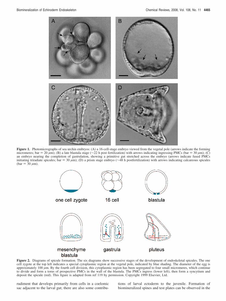
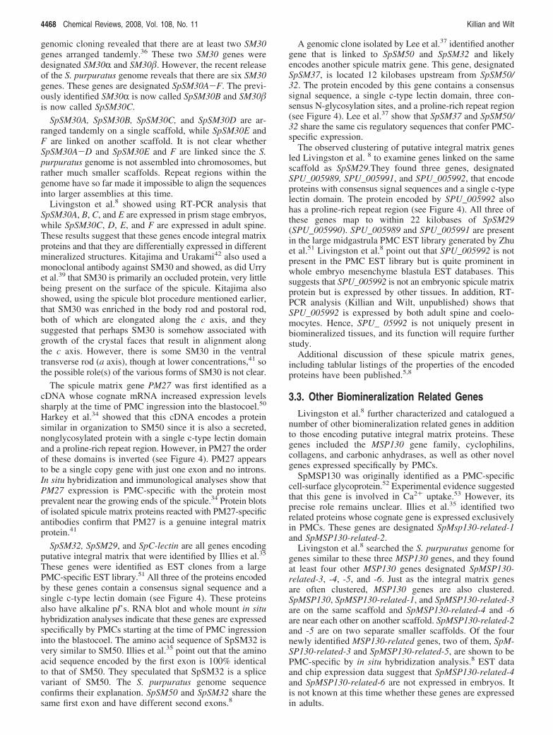
study of biomineralized structure formation. There is abun-dant material; a single female of some species can shed 10mL of packed eggs, producing about 1.5 × 10⁷ embryos.The embryos of many species are very clear, so observationof the process in living embryos is not difficult, and theprocesses can be observed in real time (reviewed by Wiltand Ettensohn6). Figure 1 shows some light micrographs ofsea urchin embryos at various stages of larval development.
Skeletal elements of embryos can be obtained in largequantities, and vestiges of adherent soft tissue are removed* E-mail addresses: [email protected]; [email protected].
Chem. Rev. 2008, 108, 4463–4474 4463
10.1021/cr0782630 CCC: $71.00 2008 American Chemical SocietyPublished on Web 09/27/2008
by treatment with NaClO. The cells that secrete the skeleton,micromeres and their descendants, which are called primarymesenchyme cells (PMCs), can be cultured in Vitro, wherethey recapitulate the processes they carry out in the intact
embryo.7 The genome of the purple sea urchin, Strongylo-centrotus purpuratus, has been completely sequenced andpartially annotated,8,9 and the network of genes that directthe establishment and development of the PMCs has beenelucidated.10,11
The embryonic development leading to spicule forma-tion has been intensively studied and recently reviewed.6,12,13
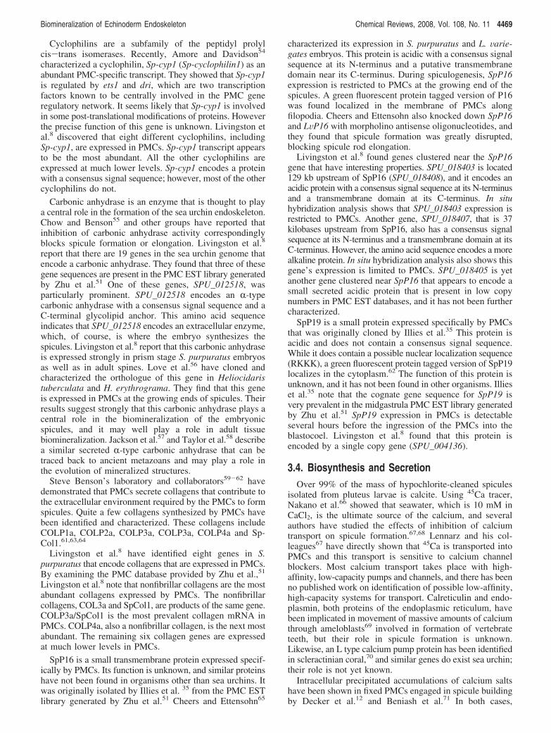
In brief, development of the spicule can be divided intofive phases: micromere formation, PMC formation, syn-cytium formation, skeleton deposition, and skeletonremodeling and elaboration [see Figure 2]. Micromeresare four small cells that arise during the fourth cell divisionat the vegetal pole of the cleaving zygote. This quartetdivides once more to give rise to four large and four smalldaughter cells. The larger daughters will continue to dividethree or four (depending on the species) more times toform a torus of cells at the vegetal pole of the hollowblastula.
During the next phase, which usually occurs between 8and 24 h postfertilization, depending on the temperature andthe species, the micromere descendants, that is PMCs, detachthemselves from the wall and ingress into the hollow blastula,forming a migratory population of 32-64 PMCs. The PMCsadopt stereotypical locations within the blastula as othercomplex gastrulation movements are occurring to producea primitive gut. Adjacent PMCs fuse with one anotherforming a continuous multicellular syncytium [see Figure1C]. Within a short time, usually only a few hours, small,rhombohedral, birefringent granules can be detected in thesyncytium located in the future ventrolateral position of thelarva. Spicules form by continued deposition of calcareousmaterial in three radii emanating from this initial granule,elongating in the crystallographic a-axes, then bending atnearly right angles and continuing to elongate in the c-axis.During late embryonic and early larval development theendoskeleton will become many times larger and morecomplex. During this time, the skeletal elements will growby deposition of mineral at the tips of pre-existent spiculesand also by initiation of new skeletal rods, de novo,presumably from mesenchyme cells separated from thesyncytium. When embryonic development has ceased and afeeding larva has formed, the larva will grow as part of aplanktonic community. The scenario we have just describedapplies to indirectly developing euechinoid species, whichconstitutes the majority of sea urchin species. The only otherechinoderm group that forms endoskeletal spicules in theembryo are the Ophiuroidea, the brittle stars. All the otherclasses do, however, form mineralized structures in the adult,which we shall describe later.
Experiments during the halcyon days of experimentalembryology (cf. Wilt and Ettensohn6) showed that thedetailed morphology of the spicules, for example, whetherplain or fenestrated, is a property of the PMCs. However,the overall form of the spicule, indeed whether it is formedat all, depends upon signals that emanate from the adjacentepithelial wall of the blastula. It has recently been shownthat the growth factor VEGF (vascular endothelial growthfactor) is secreted by a small number of cells in theectodermal wall and that the PMCs express the receptor forVEGF.14 Initiation of biomineralization depends upon thissignaling.
Formation of mineralized tissues of the adult begins duringthe planktonic phase of larval existence and is quite complex.All the major organ systems form initially in a juvenile
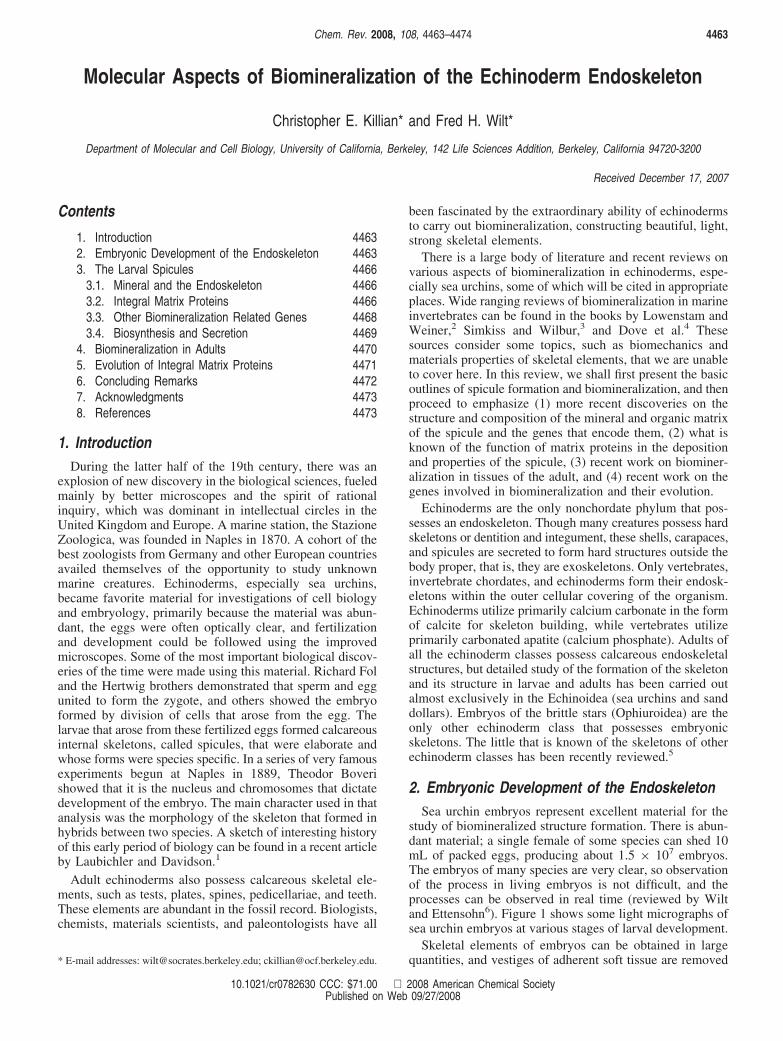
Christopher Killian was born in New York City. He attended Franklin andMarshall College and received an A.B. degree in 1981. He then attendedGeorgetown University and received a Ph.D. degree from the Departmentof Biology in 1985. His graduate studies in the laboratory of David Nishiokafocused on biochemical and cellular changes during sea urchin fertilizationand early development. Christopher joined Fred Wilt’s laboratory at theUniversity of California, Berkeley, in 1985 as a postdoctoral researcher.He has remained as a research associate in Fred Wilt’s laboratory atBerkeley. His studies in the Wilt laboratory were centered initially on themolecular details of tissue differentiation in the sea urchin embryo. Hisresearch interests have refocused on the cellular and molecular mech-anisms underlying the process of biomineralization during sea urchinspicule formation. Using biochemical, cellular, molecular, and genomicapproaches, he and others in Fred Wilt’s laboratory have identified andcharacterized a number of the integral matrix proteins and the cognategenes that are involved in sea urchin biomineralization.
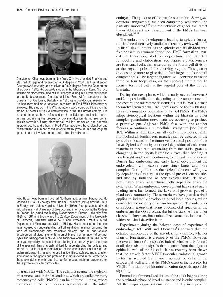
Fred H. Wilt was born in the small Northern Indiana town, Nappanee. Hereceived a B.A. in Zoology from Indiana University (1956) and the Ph.D.in Biology from Johns Hopkins University (1959). After postdoctoral workin biochemistry at University of Liverpool and in embryology at the Collegede France, he joined the Biology Department at Purdue University from1960 to 1964 and then joined the Zoology Department at the Universityof California, Berkeley, where he is now Professor Emeritus in theDepartment of Molecular and Cellular Biology. His main research interestshave focused on understanding cell differentiation in embryos using thetools of biochemistry and molecular biology, and he has studieddevelopment of visual pigments in amphibians, the formation of erythro-cytes and hemoglobin in chicks, and early development of the sea urchinembryo, especially its endoskeleton. During the past 20 years, the focusof the research has gradually shifted to understanding the cellular andmolecular basis of biomineralization of the endoskeletal spicule of seaurchin embryos. His research group has identified, isolated, and character-ized some of the genes and proteins that are involved in the formation ofthese skeletal elements and that confer unusual material properties onthese protein-calcite composites.
4464 Chemical Reviews, 2008, Vol. 108, No. 11 Killian and Wilt
rudiment that develops primarily from cells in a coelomicsac adjacent to the larval gut; there are also some contribu-
tions of larval ectoderm to the juvenile. Formation ofbiomineralized spines and test plates can be observed in the
Figure 1. Photomicrographs of sea urchin embryos: (A) a 16-cell-stage embryo viewed from the vegetal pole (arrows indicate the formingmicromeres; bar ) 20 µm); (B) a late blastula stage (∼22 h post fertilization) with arrows indicating ingressing PMCs (bar ) 30 µm); (C)an embryo nearing the completion of gastrulation, showing a primitive gut stretched across the embryo (arrows indicate fused PMCsinitiating triradiate spicules; bar ) 30 µm); (D) a prism stage embryo (∼48 h postfertilization) with arrows indicating calcareous spicules(bar ) 30 µm).
Figure 2. Diagrams of spicule formation. The six diagrams show successive stages of the development of endoskeletal spicules. The onecell zygote at the top left indicates a special cytoplasmic region at the vegetal pole, indicated by blue shading. The diameter of the egg isapproximately 100 µm. By the fourth cell division, this cytoplasmic region has been segregated to four small micromeres, which continueto divide and form a torus of prospective PMCs in the wall of the blastula. The PMCs ingress (lower left), then form a syncytium anddeposit the spicule (red). This figure is adapted from ref 119 by permission. Copyright 1999 Elsevier, Ltd.
Biomineralization of Echinoderm Endoskeleton Chemical Reviews, 2008, Vol. 108, No. 11 4465
rudiment, usually forming in conjunction with or close tolarval spicules.
During settling of the larva and metamorphosis, the larvaltissues undergo autolysis and the juvenile urchin beginsfurther growth and development attached to a substrate onthe floor of the ocean or intertidal zone.
3. The Larval Spicules
3.1. Mineral and the EndoskeletonThe sea urchin embryonic and adult endoskeleton is
comprised of magnesian calcite (usually about 5% MgCO3
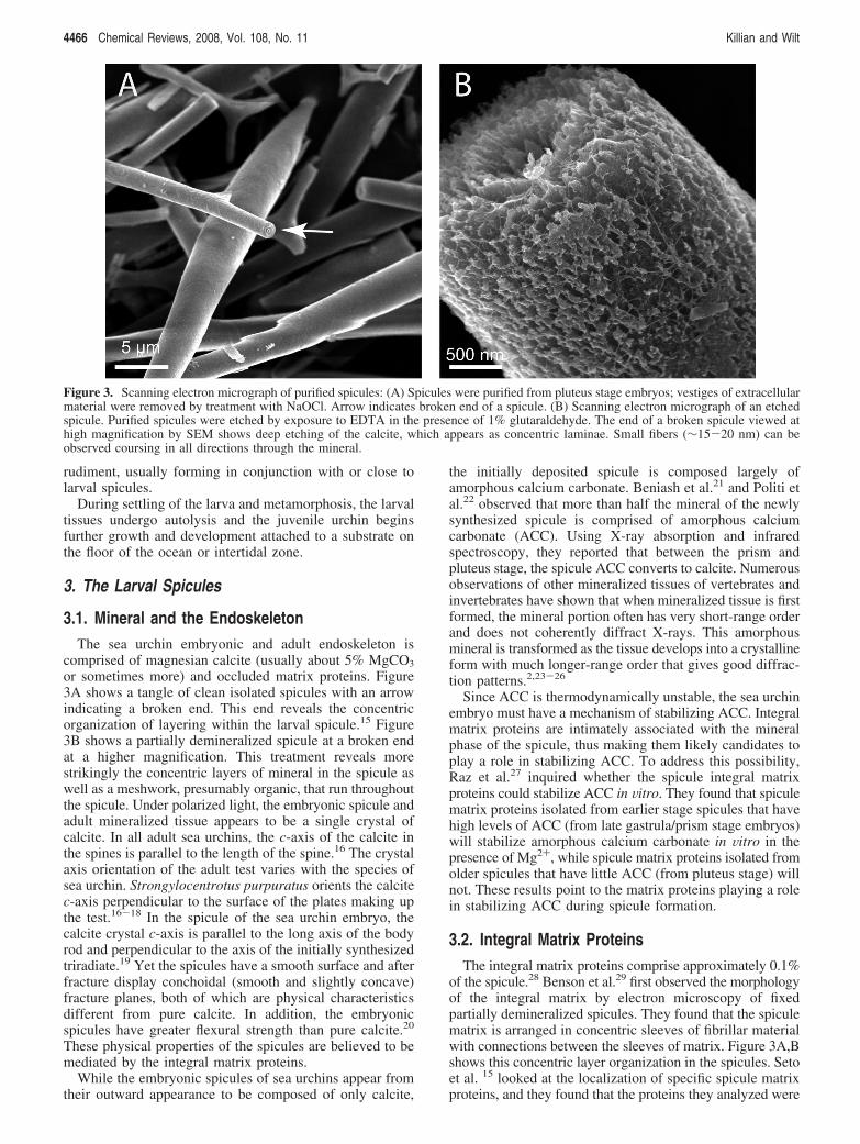
or sometimes more) and occluded matrix proteins. Figure3A shows a tangle of clean isolated spicules with an arrowindicating a broken end. This end reveals the concentricorganization of layering within the larval spicule.15 Figure3B shows a partially demineralized spicule at a broken endat a higher magnification. This treatment reveals morestrikingly the concentric layers of mineral in the spicule aswell as a meshwork, presumably organic, that run throughoutthe spicule. Under polarized light, the embryonic spicule andadult mineralized tissue appears to be a single crystal ofcalcite. In all adult sea urchins, the c-axis of the calcite inthe spines is parallel to the length of the spine.16 The crystalaxis orientation of the adult test varies with the species ofsea urchin. Strongylocentrotus purpuratus orients the calcitec-axis perpendicular to the surface of the plates making upthe test.16-18 In the spicule of the sea urchin embryo, thecalcite crystal c-axis is parallel to the long axis of the bodyrod and perpendicular to the axis of the initially synthesizedtriradiate.19 Yet the spicules have a smooth surface and afterfracture display conchoidal (smooth and slightly concave)fracture planes, both of which are physical characteristicsdifferent from pure calcite. In addition, the embryonicspicules have greater flexural strength than pure calcite.20
These physical properties of the spicules are believed to bemediated by the integral matrix proteins.
While the embryonic spicules of sea urchins appear fromtheir outward appearance to be composed of only calcite,
the initially deposited spicule is composed largely ofamorphous calcium carbonate. Beniash et al.21 and Politi etal.22 observed that more than half the mineral of the newlysynthesized spicule is comprised of amorphous calciumcarbonate (ACC). Using X-ray absorption and infraredspectroscopy, they reported that between the prism andpluteus stage, the spicule ACC converts to calcite. Numerousobservations of other mineralized tissues of vertebrates andinvertebrates have shown that when mineralized tissue is firstformed, the mineral portion often has very short-range orderand does not coherently diffract X-rays. This amorphousmineral is transformed as the tissue develops into a crystallineform with much longer-range order that gives good diffrac-tion patterns.2,23-26
Since ACC is thermodynamically unstable, the sea urchinembryo must have a mechanism of stabilizing ACC. Integralmatrix proteins are intimately associated with the mineralphase of the spicule, thus making them likely candidates toplay a role in stabilizing ACC. To address this possibility,Raz et al.27 inquired whether the spicule integral matrixproteins could stabilize ACC in Vitro. They found that spiculematrix proteins isolated from earlier stage spicules that havehigh levels of ACC (from late gastrula/prism stage embryos)will stabilize amorphous calcium carbonate in Vitro in thepresence of Mg2+, while spicule matrix proteins isolated fromolder spicules that have little ACC (from pluteus stage) willnot. These results point to the matrix proteins playing a rolein stabilizing ACC during spicule formation.
3.2. Integral Matrix ProteinsThe integral matrix proteins comprise approximately 0.1%
of the spicule.28 Benson et al.29 first observed the morphologyof the integral matrix by electron microscopy of fixedpartially demineralized spicules. They found that the spiculematrix is arranged in concentric sleeves of fibrillar materialwith connections between the sleeves of matrix. Figure 3A,Bshows this concentric layer organization in the spicules. Setoet al. 15 looked at the localization of specific spicule matrixproteins, and they found that the proteins they analyzed were
Figure 3. Scanning electron micrograph of purified spicules: (A) Spicules were purified from pluteus stage embryos; vestiges of extracellularmaterial were removed by treatment with NaOCl. Arrow indicates broken end of a spicule. (B) Scanning electron micrograph of an etchedspicule. Purified spicules were etched by exposure to EDTA in the presence of 1% glutaraldehyde. The end of a broken spicule viewed athigh magnification by SEM shows deep etching of the calcite, which appears as concentric laminae. Small fibers (∼15-20 nm) can beobserved coursing in all directions through the mineral.
4466 Chemical Reviews, 2008, Vol. 108, No. 11 Killian and Wilt
widely distributed throughout the mineral of the spicules.They suggested that the spicule matrix is woven finely aroundthe microcrystalline domains of calcite.
Benson et al.28 and Venkatesan and Simpson30 using one-dimensional SDS-PAGE identified eight to ten proteins ascomprising the integral spicule matrix. Higher resolutionstudies by Killian and Wilt31 used 35S-methionine-radiola-beled spicule matrix proteins and analyzed the integral matrixproteins with 2-D gel electrophoresis. These studies foundthat the spicule matrix was actually comprised of more thanfour dozen proteins. Most of the proteins are acidic, but thereare also a number of proteins with alkaline isoelectric points.
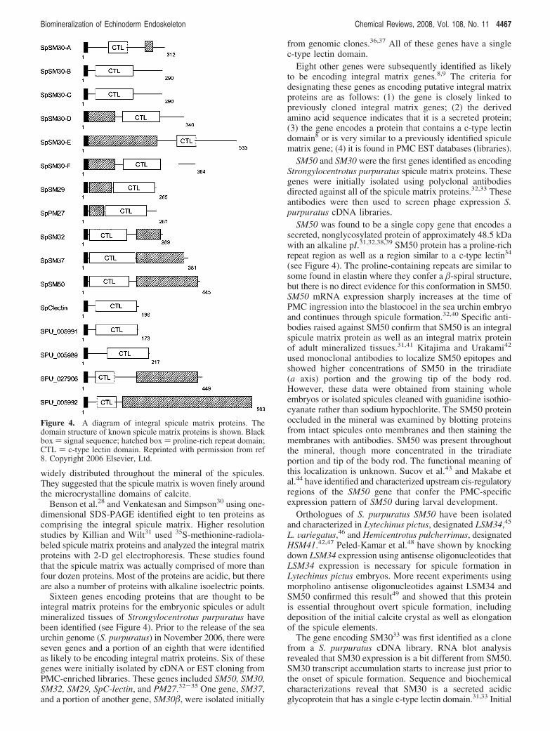
Sixteen genes encoding proteins that are thought to beintegral matrix proteins for the embryonic spicules or adultmineralized tissues of Strongylocentrotus purpuratus havebeen identified (see Figure 4). Prior to the release of the seaurchin genome (S. purpuratus) in November 2006, there wereseven genes and a portion of an eighth that were identifiedas likely to be encoding integral matrix proteins. Six of thesegenes were initially isolated by cDNA or EST cloning fromPMC-enriched libraries. These genes included SM50, SM30,SM32, SM29, SpC-lectin, and PM27.32-35 One gene, SM37,and a portion of another gene, SM30�, were isolated initially
from genomic clones.36,37 All of these genes have a singlec-type lectin domain.
Eight other genes were subsequently identified as likelyto be encoding integral matrix genes.8,9 The criteria fordesignating these genes as encoding putative integral matrixproteins are as follows: (1) the gene is closely linked topreviously cloned integral matrix genes; (2) the derivedamino acid sequence indicates that it is a secreted protein;(3) the gene encodes a protein that contains a c-type lectindomain8 or is very similar to a previously identified spiculematrix gene; (4) it is found in PMC EST databases (libraries).
SM50 and SM30 were the first genes identified as encodingStrongylocentrotus purpuratus spicule matrix proteins. Thesegenes were initially isolated using polyclonal antibodiesdirected against all of the spicule matrix proteins.32,33 Theseantibodies were then used to screen phage expression S.purpuratus cDNA libraries.
SM50 was found to be a single copy gene that encodes asecreted, nonglycosylated protein of approximately 48.5 kDawith an alkaline pI.31,32,38,39 SM50 protein has a proline-richrepeat region as well as a region similar to a c-type lectin34
(see Figure 4). The proline-containing repeats are similar tosome found in elastin where they confer a �-spiral structure,but there is no direct evidence for this conformation in SM50.SM50 mRNA expression sharply increases at the time ofPMC ingression into the blastocoel in the sea urchin embryoand continues through spicule formation.32,40 Specific anti-bodies raised against SM50 confirm that SM50 is an integralspicule matrix protein as well as an integral matrix proteinof adult mineralized tissues.31,41 Kitajima and Urakami42
used monoclonal antibodies to localize SM50 epitopes andshowed higher concentrations of SM50 in the triradiate(a axis) portion and the growing tip of the body rod.However, these data were obtained from staining wholeembryos or isolated spicules cleaned with guanidine isothio-cyanate rather than sodium hypochlorite. The SM50 proteinoccluded in the mineral was examined by blotting proteinsfrom intact spicules onto membranes and then staining themembranes with antibodies. SM50 was present throughoutthe mineral, though more concentrated in the triradiateportion and tip of the body rod. The functional meaning ofthis localization is unknown. Sucov et al.43 and Makabe etal.44 have identified and characterized upstream cis-regulatoryregions of the SM50 gene that confer the PMC-specificexpression pattern of SM50 during larval development.
Orthologues of S. purpuratus SM50 have been isolatedand characterized in Lytechinus pictus, designated LSM34,45
L. Variegatus,46 and Hemicentrotus pulcherrimus, designatedHSM41.42,47 Peled-Kamar et al.48 have shown by knockingdown LSM34 expression using antisense oligonucleotides thatLSM34 expression is necessary for spicule formation inLytechinus pictus embryos. More recent experiments usingmorpholino antisense oligonucleotides against LSM34 andSM50 confirmed this result49 and showed that this proteinis essential throughout overt spicule formation, includingdeposition of the initial calcite crystal as well as elongationof the spicule elements.
The gene encoding SM3033 was first identified as a clonefrom a S. purpuratus cDNA library. RNA blot analysisrevealed that SM30 expression is a bit different from SM50.SM30 transcript accumulation starts to increase just prior tothe onset of spicule formation. Sequence and biochemicalcharacterizations reveal that SM30 is a secreted acidicglycoprotein that has a single c-type lectin domain.31,33 Initial
Figure 4. A diagram of integral spicule matrix proteins. Thedomain structure of known spicule matrix proteins is shown. Blackbox ) signal sequence; hatched box ) proline-rich repeat domain;CTL ) c-type lectin domain. Reprinted with permission from ref8. Copyright 2006 Elsevier, Ltd.
Biomineralization of Echinoderm Endoskeleton Chemical Reviews, 2008, Vol. 108, No. 11 4467
genomic cloning revealed that there are at least two SM30genes arranged tandemly.36 These two SM30 genes weredesignated SM30R and SM30�. However, the recent releaseof the S. purpuratus genome reveals that there are six SM30genes. These genes are designated SpSM30A-F. The previ-ously identified SM30R is now called SpSM30B and SM30�is now called SpSM30C.
SpSM30A, SpSM30B, SpSM30C, and SpSM30D are ar-ranged tandemly on a single scaffold, while SpSM30E andF are linked on another scaffold. It is not clear whetherSpSM30A-D and SpSM30E and F are linked since the S.purpuratus genome is not assembled into chromosomes, butrather much smaller scaffolds. Repeat regions within thegenome have so far made it impossible to align the sequencesinto larger assemblies at this time.
Livingston et al.8 showed using RT-PCR analysis thatSpSM30A, B, C, and E are expressed in prism stage embryos,while SpSM30C, D, E, and F are expressed in adult spine.These results suggest that these genes encode integral matrixproteins and that they are differentially expressed in differentmineralized structures. Kitajima and Urakami42 also used amonoclonal antibody against SM30 and showed, as did Urryet al.39 that SM30 is primarily an occluded protein, very littlebeing present on the surface of the spicule. Kitajima alsoshowed, using the spicule blot procedure mentioned earlier,that SM30 was enriched in the body rod and postoral rod,both of which are elongated along the c axis, and theysuggested that perhaps SM30 is somehow associated withgrowth of the crystal faces that result in alignment alongthe c axis. However, there is some SM30 in the ventraltransverse rod (a axis), though at lower concentrations,41 sothe possible role(s) of the various forms of SM30 is not clear.
The spicule matrix gene PM27 was first identified as acDNA whose cognate mRNA increased expression levelssharply at the time of PMC ingression into the blastocoel.50
Harkey et al.34 showed that this cDNA encodes a proteinsimilar in organization to SM50 since it is also a secreted,nonglycosylated protein with a single c-type lectin domainand a proline-rich repeat region. However, in PM27 the orderof these domains is inverted (see Figure 4). PM27 appearsto be a single copy gene with just one exon and no introns.In situ hybridization and immunological analyses show thatPM27 expression is PMC-specific with the protein mostprevalent near the growing ends of the spicule.34 Protein blotsof isolated spicule matrix proteins reacted with PM27-specificantibodies confirm that PM27 is a genuine integral matrixprotein.41
SpSM32, SpSM29, and SpC-lectin are all genes encodingputative integral matrix that were identified by Illies et al.35
These genes were identified as EST clones from a largePMC-specific EST library.51 All three of the proteins encodedby these genes contain a consensus signal sequence and asingle c-type lectin domain (see Figure 4). These proteinsalso have alkaline pI’s. RNA blot and whole mount in situhybridization analyses indicate that these genes are expressedspecifically by PMCs starting at the time of PMC ingressioninto the blastocoel. The amino acid sequence of SpSM32 isvery similar to SM50. Illies et al.35 point out that the aminoacid sequence encoded by the first exon is 100% identicalto that of SM50. They speculated that SpSM32 is a splicevariant of SM50. The S. purpuratus genome sequenceconfirms their explanation. SpSM50 and SpSM32 share thesame first exon and have different second exons.8
A genomic clone isolated by Lee et al.37 identified anothergene that is linked to SpSM50 and SpSM32 and likelyencodes another spicule matrix gene. This gene, designatedSpSM37, is located 12 kilobases upstream from SpSM50/32. The protein encoded by this gene contains a consensussignal sequence, a single c-type lectin domain, three con-sensus N-glycosylation sites, and a proline-rich repeat region(see Figure 4). Lee et al.37 show that SpSM37 and SpSM50/32 share the same cis regulatory sequences that confer PMC-specific expression.
The observed clustering of putative integral matrix genesled Livingston et al. 8 to examine genes linked on the samescaffold as SpSM29.They found three genes, designatedSPU_005989, SPU_005991, and SPU_005992, that encodeproteins with consensus signal sequences and a single c-typelectin domain. The protein encoded by SPU_005992 alsohas a proline-rich repeat region (see Figure 4). All three ofthese genes map to within 22 kilobases of SpSM29(SPU_005990). SPU_005989 and SPU_005991 are presentin the large midgastrula PMC EST library generated by Zhuet al.51 Livingston et al.8 point out that SPU_005992 is notpresent in the PMC EST library but is quite prominent inwhole embryo mesenchyme blastula EST databases. Thissuggests that SPU_005992 is not an embryonic spicule matrixprotein but is expressed by other tissues. In addition, RT-PCR analysis (Killian and Wilt, unpublished) shows thatSPU_005992 is expressed by both adult spine and coelo-mocytes. Hence, SPU_ 05992 is not uniquely present inbiomineralized tissues, and its function will require furtherstudy.
Additional discussion of these spicule matrix genes,including tablular listings of the properties of the encodedproteins have been published.5,8
3.3. Other Biomineralization Related GenesLivingston et al.8 further characterized and catalogued a
number of other biomineralization related genes in additionto those encoding putative integral matrix proteins. Thesegenes included the MSP130 gene family, cyclophilins,collagens, and carbonic anhydrases, as well as other novelgenes expressed specifically by PMCs.
SpMSP130 was originally identified as a PMC-specificcell-surface glycoprotein.52 Experimental evidence suggestedthat this gene is involved in Ca2+ uptake.53 However, itsprecise role remains unclear. Illies et al.35 identified tworelated proteins whose cognate gene is expressed exclusivelyin PMCs. These genes are designated SpMsp130-related-1and SpMSP130-related-2.
Livingston et al.8 searched the S. purpuratus genome forgenes similar to these three MSP130 genes, and they foundat least four other MSP130 genes designated SpMSP130-related-3, -4, -5, and -6. Just as the integral matrix genesare often clustered, MSP130 genes are also clustered.SpMSP130, SpMSP130-related-1, and SpMSP130-related-3are on the same scaffold and SpMSP130-related-4 and -6are near each other on another scaffold. SpMSP130-related-2and -5 are on two separate smaller scaffolds. Of the fournewly identified MSP130-related genes, two of them, SpM-SP130-related-3 and SpMSP130-related-5, are shown to bePMC-specific by in situ hybridization analysis.8 EST dataand chip expression data suggest that SpMSP130-related-4and SpMSP130-related-6 are not expressed in embryos. Itis not known at this time whether these genes are expressedin adults.
4468 Chemical Reviews, 2008, Vol. 108, No. 11 Killian and Wilt
Cyclophilins are a subfamily of the peptidyl prolylcis-trans isomerases. Recently, Amore and Davidson54
characterized a cyclophilin, Sp-cyp1 (Sp-cyclophilin1) as anabundant PMC-specific transcript. They showed that Sp-cyp1is regulated by ets1 and dri, which are two transcriptionfactors known to be centrally involved in the PMC generegulatory network. It seems likely that Sp-cyp1 is involvedin some post-translational modifications of proteins. Howeverthe precise function of this gene is unknown. Livingston etal.8 discovered that eight different cyclophilins, includingSp-cyp1, are expressed in PMCs. Sp-cyp1 transcript appearsto be the most abundant. All the other cyclophilins areexpressed at much lower levels. Sp-cyp1 encodes a proteinwith a consensus signal sequence; however, most of the othercyclophilins do not.
Carbonic anhydrase is an enzyme that is thought to playa central role in the formation of the sea urchin endoskeleton.Chow and Benson55 and other groups have reported thatinhibition of carbonic anhydrase activity correspondinglyblocks spicule formation or elongation. Livingston et al.8
report that there are 19 genes in the sea urchin genome thatencode a carbonic anhydrase. They found that three of thesegene sequences are present in the PMC EST library generatedby Zhu et al.51 One of these genes, SPU_012518, wasparticularly prominent. SPU_012518 encodes an R-typecarbonic anhydrase with a consensus signal sequence and aC-terminal glycolipid anchor. This amino acid sequenceindicates that SPU_012518 encodes an extracellular enzyme,which, of course, is where the embryo synthesizes thespicules. Livingston et al.8 report that this carbonic anhydraseis expressed strongly in prism stage S. purpuratus embryosas well as in adult spines. Love et al.56 have cloned andcharacterized the orthologue of this gene in Heliocidaristuberculata and H. erythrograma. They find that this geneis expressed in PMCs at the growing ends of spicules. Theirresults suggest strongly that this carbonic anhydrase plays acentral role in the biomineralization of the embryonicspicules, and it may well play a role in adult tissuebiomineralization. Jackson et al.57 and Taylor et al.58 describea similar secreted R-type carbonic anhydrase that can betraced back to ancient metazoans and may play a role inthe evolution of mineralized structures.
Steve Benson’s laboratory and collaborators59-62 havedemonstrated that PMCs secrete collagens that contribute tothe extracellular environment required by the PMCs to formspicules. Quite a few collagens synthesized by PMCs havebeen identified and characterized. These collagens includeCOLP1a, COLP2a, COLP3a, COLP3a, COLP4a and Sp-Col1.61,63,64
Livingston et al.8 have identified eight genes in S.purpuratus that encode collagens that are expressed in PMCs.By examining the PMC database provided by Zhu et al.,51
Livingston et al.8 note that nonfibrillar collagens are the mostabundant collagens expressed by PMCs. The nonfibrillarcollagens, COL3a and SpCol1, are products of the same gene.COLP3a/SpCol1 is the most prevalent collagen mRNA inPMCs. COLP4a, also a nonfibrillar collagen, is the next mostabundant. The remaining six collagen genes are expressedat much lower levels in PMCs.
SpP16 is a small transmembrane protein expressed specif-ically by PMCs. Its function is unknown, and similar proteinshave not been found in organisms other than sea urchins. Itwas originally isolated by Illies et al. 35 from the PMC ESTlibrary generated by Zhu et al.51 Cheers and Ettensohn65
characterized its expression in S. purpuratus and L. Varie-gates embryos. This protein is acidic with a consensus signalsequence at its N-terminus and a putative transmembranedomain near its C-terminus. During spiculogenesis, SpP16expression is restricted to PMCs at the growing end of thespicules. A green fluorescent protein tagged version of P16was found localized in the membrane of PMCs alongfilopodia. Cheers and Ettensohn also knocked down SpP16and LVP16 with morpholino antisense oligonucleotides, andthey found that spicule formation was greatly disrupted,blocking spicule rod elongation.
Livingston et al.8 found genes clustered near the SpP16gene that have interesting properties. SPU_018403 is located129 kb upstream of SpP16 (SPU_018408), and it encodes anacidic protein with a consensus signal sequence at its N-terminusand a transmembrane domain at its C-terminus. In situhybridization analysis shows that SPU_018403 expression isrestricted to PMCs. Another gene, SPU_018407, that is 37kilobases upstream from SpP16, also has a consensus signalsequence at its N-terminus and a transmembrane domain at itsC-terminus. However, the amino acid sequence encodes a morealkaline protein. In situ hybridization analysis also shows thisgene’s expression is limited to PMCs. SPU_018405 is yetanother gene clustered near SpP16 that appears to encode asmall secreted acidic protein that is present in low copynumbers in PMC EST databases, and it has not been furthercharacterized.
SpP19 is a small protein expressed specifically by PMCsthat was originally cloned by Illies et al.35 This protein isacidic and does not contain a consensus signal sequence.While it does contain a possible nuclear localization sequence(RKKK), a green fluorescent protein tagged version of SpP19localizes in the cytoplasm.62 The function of this protein isunknown, and it has not been found in other organisms. Illieset al.35 note that the cognate gene sequence for SpP19 isvery prevalent in the midgastrula PMC EST library generatedby Zhu et al.51 SpP19 expression in PMCs is detectableseveral hours before the ingression of the PMCs into theblastocoel. Livingston et al.8 found that this protein isencoded by a single copy gene (SPU_004136).
3.4. Biosynthesis and SecretionOver 99% of the mass of hypochlorite-cleaned spicules
isolated from pluteus larvae is calcite. Using 45Ca tracer,Nakano et al.66 showed that seawater, which is 10 mM inCaCl2, is the ultimate source of the calcium, and severalauthors have studied the effects of inhibition of calciumtransport on spicule formation.67,68 Lennarz and his col-leagues67 have directly shown that 45Ca is transported intoPMCs and this transport is sensitive to calcium channelblockers. Most calcium transport takes place with high-affinity, low-capacity pumps and channels, and there has beenno published work on identification of possible low-affinity,high-capacity systems for transport. Calreticulin and endo-plasmin, both proteins of the endoplasmic reticulum, havebeen implicated in movement of massive amounts of calciumthrough ameloblasts69 involved in formation of vertebrateteeth, but their role in spicule formation is unknown.Likewise, an L type calcium pump protein has been identifiedin scleractinian coral,70 and similar genes do exist sea urchin;their role is not yet known.
Intracellular precipitated accumulations of calcium saltshave been shown in fixed PMCs engaged in spicule buildingby Decker et al.12 and Beniash et al.71 In both cases,
Biomineralization of Echinoderm Endoskeleton Chemical Reviews, 2008, Vol. 108, No. 11 4469
considerable processing and fixation preceded visualization,leaving open the question of artifact. Importantly, Beniashet al.71 showed that the visualized granules were probablyamorphous calcium carbonate and that heating converted thegranules to calcite. Recent studies72 from the Wilt laboratoryused a vital fluorescent dye, calcein, to tag calcium precipi-tates in PMCs in Vitro that were secreting spicules. Theyobserved punctate labeling of cells after a brief pulse of dye.After washing of the cells and continued culture, theyobserved that the cellular fluorescence had disappeared whilethe newly secreted spicule became labeled. This is consistentwith a precursor of intracellular ACC as the proximate sourcefor the secreted calcium that is incorporated into the spicule.
In contrast to intracellular calcium being localized inpunctate deposits visible in the light microscope (i.e.,approximately 200 nm or more), immunostaining of spiculematrix proteins SpSM30B and SpSM50 show broad swatchesof perinuclear intracellular localization without any evidenceof punctate appearance.41 Immuno-electron microscope stud-ies73 have shown that both of these proteins are present athigh levels in the Golgi apparatus and also in small (<50nm) intracellular post-Golgi transport vesicles. Hence, cal-cium and these two matrix proteins are localized in cellularcompartments of different sizes and are probably transportedand delivered vectorially via different trafficking vehicles.The secreted calcium and these two matrix proteins are alsodirected to different positions in the spicule; this has beenconfirmed by following green fluorescent protein taggedSpSM30B and SpSM50.67,68,72 The tagged matrix proteinsare secreted and retained near the PMC that synthesizes them,while secreted calcium, followed by calcein labeling, movesquickly through the syncytium to the extending tip of thespicule. Ingersoll and Wilt74 also showed that inhibition ofmetalloproteases interfered with secretion of both calciumand matrix protein; however the mechanism of action ofproteases in this regard is unknown.75,76
We should add that the secretion of calcium and matrixproteins is targeted. These components end up in the spicule;hence, trafficking must be vectorial. PMCs secrete manyother proteins and proteoglycans,61 as well as the previouslymentioned nonfibrillar collagens, that populate the blastocoelbut are not found in spicules.
4. Biomineralization in AdultsThe five extant classes of echinoderms all possess a system
of internal skeletal support comprised of calcareous plates,or ossicles, formed by the mesodermally derived dermis. Thechordates are the only other animal phylum with skeletalsupport enclosed by a covering epithelium. The sea urchins(Class Echinoidea) have been most studied, and they possessthe most extensive endoskeleton. In addition to the test andspines, which may appear to be on the surface but are coveredby a thin epidermis, the adults possess elaborate teeth andsmall appendages called pedicellariae. Even the tube feethave calcareous ossicles at their distal tips and small spiculesembedded in the walls of the tube foot.
Though the process of metamorphosis has been describedin the classical zoological literature, little is known aboutthe origins of the adult endoskeleton.77 The developingjuvenile, known as the echinus rudiment, forms primarilyfrom coelomic tissue adjacent to the gut of the larva. MamikoYajima has carried out experiments designed to determinewhich cells in the larva give rise to stem cells in the adultthat differentiate into adult biomineralized tissues. Initial
studies78 demonstrated that both the additional skeletalelements of late larval development and the developingspines, tube feet, and test plates of the juvenile are formedby cells that stain with monoclonal antibodies directed againstcell-surface-specific antigens found in the PMCs of theembryo and larva. On the other hand, the skeletogenic cellsof the developing juvenile, while morphologically similarto PMCs, only gradually display the antigen just prior toovert calcification; this argues that fully differentiated PMCdescendants are not involved in biomineralization of juvenileskeletal elements. There was no evidence that alreadydifferentiated, brightly staining PMCs were responsible forforming adult structures.
This work was followed up79 by transplantation of cellsbetween two species (P. depressus and H. pulcherrimus) thatpossess distinguishable skeletal phenotypes. Both skeleto-genic PMCs and another mesenchymal population calledsecondary mesenchyme cells (SMCs) were transplanted.Transplanted PMCs only conferred a donor phenotype onskeletons of early larvae, but transplantation of SMCsconferred a skeleton of donor phenotype in late larvae andmetamorphosing animals. This is consistent with previouswork80,81 showing that if PMCs are removed from an earlyembryo, the SMCs can transdifferentiate and form a com-plete, normal larval skeleton. Yajima marked cells bytransplanting PMCs from embryos containing a transgenefor green fluorescent protein, a reliable and widely used celllineage tracer. When tagged PMCs were transplanted, theycould be seen engaged in skeletogenesis in developing larvae,but after metamorphosis, they were undetectable. In contrast,tagged SMCs participated in late larval skeletogenesis andcould also be demonstrated participating in skeletogenesisduring metamorphosis. These results were confirmed by PCRanalysis and make a strong case for participation, perhapsexclusive, of SMC descendants in development of biomin-eralized skeletal elements of the adult. This hypothesis wassupported by experiments using embryos of the sand dollar,Peronella japonica.82 When the micromeres, which form thePMCs but not SMCs, are removed, the larva did not formarms with spicules, but after metamorphosis normal juvenileswith adult skeletal elements were formed.
Earlier studies on the SpSM50 and SpSM30B genes showedthat they are expressed in mineralized tissues of the adultbut not in nonmineralized tissues.83-85 Ameye et al.86
showed by using transmission immuno-electron microscopythat SpSM30B and SpSM50 are expressed in scleroblasts(sometimes called calcoblasts) of test plates and developingpedicellariae and in the odontoblasts of teeth. The proteinswere localized in Golgi stacks, Golgi-derived vesicles, andthe matrix of several adult mineralized tissues. Subsequentwork87 using scanning electron microscopy combined withimmunolabeling clearly showed expression of SpSM30B inspine trabeculae.
Politi and her collaborators88 studied regeneration of thespine. They characterized the newly regenerated materialafter gentle etching of the material in water. Electronmicroscopy and Fourier transform infrared spectroscopyshowed that the newly forming tip of the regenerate iscomposed of hydrated, amorphous, precipitated calciumcarbonate. During regeneration, the hydrated ACC is trans-formed to an anhydrous form of ACC, which in turn istransformed to the more stable crystalline calcite. Thisimportant observation is consistent with what is known aboutspicule formation in the embryo, and the progression of
4470 Chemical Reviews, 2008, Vol. 108, No. 11 Killian and Wilt
hydrated amorphous calcium carbonate through an anhydrousstate, which is then transformed to calcite, may be awidespread scenario of biomineralization. Calcite is thelowest energy state of precipitated calcium carbonate, so theconversion of ACC to calcite is favored thermodynamically.The details of these changes at the atomic level are understudy. There are several interesting and puzzling questions:What is the nature of conversion of hydrated to nonhydratedACC and what is the fate of the released water? Do organiccomponents play an important role in this conversion? Howdoes perfect crystallinity become initiated and propagatedin the anhydrous ACC, and what are the roles of organicmolecules in this conversion? The answers to these questionsare unknown. What is known is that hydrated ACC canpersist in natural materials for a very long time, presumablybecause of the activity of matrix proteins.24 Anhydrous ACCfound in spicules and regenerating spines has a structure closeto calcite but still is sufficiently disordered to preventcoherent diffraction of X-rays.22 Its transformation to calciteis also regulated and occurs over hours or days rather thanthe seconds taken when pure ACC is formed and transformsto calcite in the laboratory.
The tooth of the adult sea urchin is composed of highmagnesium calcite. The tooth contains several differentcalcitic structural elements. It is continuously abraded at itsdistal (adoral) tip. The histology of the tooth was studiedusing light and electron microscopy in pioneering studiesby Markel and his colleagues.89,90 The structure, composition,and mechanical properties have been described by Wang etal.91 The tooth is a complex assemblage of single-crystalcalcite plates, needles of calcite, and very high magnesium(∼40%) microcrystals of calcite. There is also an organicmatrix surrounding, and possibly embedded within, thevarious elements of the tooth. It is astonishing that the wholetooth behaves as two single crystals when viewed in the lightmicroscope with polarized light.
Arthur Veis and his colleagues have carried out ahistological analysis92,93 of this complex organ. The prolif-erative soft tissue, called the plumula, is found in theproximal portion of the tooth. Mineralization begins here,but then ceases. The cells of the plumula continuouslymigrate distally along the forming tooth, forming a cellularsyncytium that deposits channels of extracellular membra-nous material. In the tooth proper, just distal to the plumula,these channels become mineralized with high magnesium(∼10-15%) calcite; subsequently, adjacent plates becomeconnected with very high magnesium (up to 40%) polycrys-talline calcite, which binds the plates together.90 Thecrystallographic axes of these elements are well-aligned sothat the assemblage diffracts as a single crystal. Smallcavities, ranging from 10-220 nm diameter, possibly filledwith hydrated extracellular material, are scattered throughoutthe mature tooth.94 The tooth is a continuously growing andcomplex structure. The cells and some mineral form in theplumula. The highly mineralized distal, incisal edge of thetooth is constantly abraded and lost by the scraping thatoccurs during feeding. Even the highly cellular plumula iscomposed of different histological and functional zones. Veisand his colleagues speculate that the mineral formed in theproliferative zone of the plumula may serve an entirelydifferent purpose than the calcified elements of the tooth,possibly providing skeletal support for the soft plumula ratherthan dentition.
The Veis group has used micro-computer-tomographictechniques to map the distribution of calcium and magnesiumin the tooth structures,95 as well as the mineralized supportingorgan, Aristotle’s lantern.96 They have also extracted water-soluble proteins, as well as proteins released by demineral-ization of the tooth, and fractionated them. There are a verylarge number of proteins, and of particular interest is thefinding of proteins with phosphorylated serine and a highcontent of aspartic acid in mineralized plates of the tooth.97
Another novel form of mapping of Ca, Mg, and protein wascarried out by secondary ion mass spectroscopy (SIMS), andit was shown that aspartic acid fragments colocalized wellwith very high magnesium containing calcite.98
Recent studies from the Weizmann laboratory99 show thatthe formation of calcite needles of the tooth utilizes an ACCprecursor phase, and the formation of the calcite plates mayinvolve a similar mechanism since ACC can be identifiedin the center of plates. The ACC of the developing needle isgradually transformed to calcite. So the strategy of utilizationof ACC as precursor to biomineralized calcite can now beextended to include the spicule of the embryo, the regenerat-ing spine, and the continuously growing tooth.
Various antibodies directed against vertebrate dentin andbone proteins and antibodies directed against SM30 andSM50 proteins of embryonic spicules have been employedto detect cross-reactions with proteins of the tooth. Crossreactivity with vertebrate dentin matrix protein and the SM50protein have been demonstrated. As mentioned previously,Ameye et al.86 demonstrated cross-reacting SM30- andSM50-like epitopes in mineralizing portions of the tooth.Recently the Veis laboratory has constructed cDNA librariesof plumula and tooth tissue.100 These libraries probablycontain cDNA that encode a very large variety of proteins,not all of them necessarily related to biomineralization. Theyidentified a homologue of a vertebrate protein called mortalin,which is a member of the hsp70 group of proteins that servea variety of functions in humans and other vertebrates. Theprotein is expressed in odontoblasts and matrix of theplumula in the region where syncytium formation takes place.The use of cDNA libraries101 of plumula and mineralizedtooth tissues portends good progress for identification andcharacterization of tooth matrix proteins.
5. Evolution of Integral Matrix ProteinsThe putative sea urchin integral matrix proteins identified
so far all contain a single c-type lectin domain. There maywell be other types of proteins that will be identified as alsocomprising the integral matrix. However, the prevalence ofc-type lectins raises the question: why have c-type lectinsbeen co-opted by sea urchins for use during biomineraliza-tion? This question has intriguing evolutionary implications.Bottjer et al.102 point out that the calcitic stereom (as thefenestrated skeleton of the adult is called) present in seaurchins represents a synapomorphy (common trait) that ispresent in all echinoderms and that these integral matrixproteins may be a window into how echinoderms first formedmineralized tissues. Paleogenomics, the study of ancientgenomes through the analysis of extant organisms, is anapproach to this problem. However, sequences of the integralmatrix proteins do not yet exist for direct examination ofthis question. Nevertheless, comparisons of sea urchinintegral matrix proteins to those of animals in other phylaare possible and offer a few preliminary clues.8,101
Biomineralization of Echinoderm Endoskeleton Chemical Reviews, 2008, Vol. 108, No. 11 4471
Biomineralization in vertebrates is widely studied and anumber of integral matrix proteins that are involved in theformation of vertebrate teeth and bones have been identified.The literature is too extensive to review here. However,among the most widely studied matrix proteins are the so-called secreted calcium binding phosphoproteins (SCPPs).SCPPs include dentin sialophosphoproteins (DSPPs), secretedphosphoproteins 1 (SPP1), small integrin-binding ligandN-linked glycoprotein (SIBLING), and secreted protein,acidic, rich in cysteine (SPARC). Kawasaki and Weiss103
and Fisher et al.104 have looked at SCPP protein sequences,as well as the genomic organization of their cognate genes.They have hypothesized that despite the diverse amino acidcomposition of the encoded proteins, genomic organizationof SCPP genes points to a common origin. They proffer theidea that this rapidly evolving class of proteins is derivedfrom a SPARC gene that was present in protostomes anddeuterostomes (see Kawasaki and Weiss105 for review).
Sea urchins do not utilize SCPPs for biomineralizationdespite the fact that they do have a SPARC gene. SpSPARQis utilized for other functions in sea urchins.8 Are theresimilarities in the role of SCPPs in vertebrates and c-typelectins in sea urchins? It is unclear whether c-type lectinsand SCPPs mediate mineralization in the same way. Howeverthe two classes of proteins do have some shared physicalcharacteristics. C-type lectins have regions that tolerate largeamounts of sequence variability while still maintaining thec-type lectin fold.106 SCPPs also tolerate wide sequencevariability, yet keep their structural integrity.103 Perhapsgenes that are able to diversify quickly but retain structuralintegrity were more easily coopted to act as integral matrixproteins.
During the Cambrian expansion of metazoan body plandiversity some 544 million years ago (Mya), there was therelatively sudden appearance of skeletons and carapaces.107,108
Since mineralized skeletons appear over such a relativelybrief period of time, the hypothesis has been put forth thatthe Cambrian soft-bodied organisms must have “recruited”proteins that were already serving other functions. In otherwords, variants of existing proteins were used to help regulateformation of mineralized composites that could be used forstructural function.107,109
A number of reports107,110,111 have suggested that seawaterchemistry may have dictated the choice of mineralogy ofthe skeletons of clades when mineralized structures firstappeared during the Cambrian period. Porter110 found a closecorrelation of seawater Mg2+/Ca2+ ratios with the mineral-ogy of calcium carbonate of the mineralized skeleton firstacquired by various clades. Seawater from the Ediacaran(635-542 Mya) and Nemakit-Dalynian (542-525 Mya)periods favored the initial formation of aragonite (anotherstable form of crystalline CaCO3) skeletons, while seawaterfrom the Tommotian (525-521 Mya) and the Atdabanian(521-519 Mya) through the Toyonian (519-513 Mya)period favored the initial formation of calcite skeletons. Oncea clade formed a skeleton, changes in the Mg2+/Ca2+ ratiohad no effect on the mineralogy of the skeleton formed.Porter110 suggests that the initial skeleton formation inparticular taxa was a response to the environment. Once theskeleton was formed, subsequent biomineralization was anintracellular process, which is less influenced by the environ-ment. Echinoderm skeleton formation fits in with this theoryput forth by Porter and others. The echinoderms first formedcalcitic skeletons 520 Mya during the Atdabanian period,
which was during a time when the seawater chemistryfavored the initial formation of calcitic skeletons.
The sea urchin genome contains genes that encode over ahundred small c-type lectins that have one or two c-typelectin domains.112,113 These sorts of proteins are involvedin innate immunity and function as opsonins. Opsonins bindparticles and enhance the phagocytosis of the particle. Smithet al.114 report that sea urchin coelomic fluid contains a largenumber of small c-type lectins. The coelomocytes are thecells of the sea urchin adult that are at the center of the seaurchin immune response.114-116 In addition, RT-PCR analy-ses show that the cognate transcripts of the putative integralmatrix proteins SpSM30F and SpSM29 are found in coelo-mocytes.8 The protein encoded by SpSM30F is also similarin structure to the collectins.112 The collectins are a familyof proteins that are a component of the complement system,which results in the opsonization of foreign particles.117
These observations suggest an unexplored tie betweenproteins involved in biomineralization and innate immunityin the sea urchin. Dubois et al.118 have also suggested thatcells of mesothelial origin could possibly serve both immuneand skeleton-forming roles.
Taken together, these various observations raise thepossibility that the early echinoderms first co-opted rapidlydiversifying c-type lectins of the immune system to regulatecalcite mineralization during times of changing seawaterchemistry in the early Cambrian period. Comparisons of genesequences encoding integral matrix proteins from otherechinoderms, when they become available, should helpclarify whether this hypothesis is viable.
6. Concluding RemarksThe developing sea urchin offers splendid opportunities
to observe biomineralization in real time. The backgroundof biological information about the development of theskeleton in these embryos is excellent, and there is asubstantial literature on the formation of the spicule. Thetest and spines of the adult have also been studied, andsimilar occluded matrix molecules seem to be found in theadult and embryo. The introduction of modern molecularbiology and genomics brings more power to studies ofbiomineralization in this system.
What do we know from study of this system? The numberof matrix molecules and their biochemical complexity isgreater than originally envisaged. Limited functional studiesof these matrix proteins is now possible using molecularbiological techniques, and at least one of them, SM50, hasbeen shown to be essential for spicule formation. We canexpect this approach to be fruitful for study of other matrixmolecules. The relationship of matrix to the material proper-ties of the spicule is of great interest; progress has been slow,but application of newer techniques of structural analysisshould give us some insight in the near future.
Comparisons of echinoderm biomineralization with ver-tebrates now make clear that from the point of view ofoccluded matrix molecules, the bones and teeth of vertebratesare constructed from a different suite of organic molecules.Detailed comparisons of echinoderms with mollusk shellsshould be of great interest and can now be done withadditional genomic information. We predict that comparativestudies that utilize genomics will clarify the evolution ofbiomineralization of animal skeletons.
4472 Chemical Reviews, 2008, Vol. 108, No. 11 Killian and Wilt
7. AcknowledgmentsWork from the authors’ laboratory has been supported by
the National Institutes of Health (Grant DE 13735) and theNational Science Foundation (Grant 444724).
8. References(1) Laubichler, M. D.; Davidson, E. H. DeV. Biol. 2008, 314, 1.(2) Lowenstam, H. A.; Weiner, S. On Biomineralization; Oxford
University Press: New York, 1989.(3) Simkiss, K.; Wilbur, K. Biomineralization: Cell Biology and Mineral
Deposition; Academic Press: San Diego, CA, 1989.(4) Biomineralization; Dove, P. M., DeYoreo, J. J., Weiner, S., Eds.;
2003; Vol. 54.(5) Wilt, F. H.; Killian, C. E.; Livingston, B. T. Differentiation 2003,
71, 237.(6) Wilt, F. H.; Ettensohn, C. A. In Handbook of Biomineralization;
Baeuerlein, E., Ed.; Wiley-VCH: Weinheim, Germany, 2007.(7) Okazaki, K. Am. Zool. 1975, 15, 567.(8) Livingston, B. T.; Killian, C. E.; Wilt, F.; Cameron, A.; Landrum,
M. J.; Ermolaeva, O.; Sapojnikov, V.; Maglott, D. R.; Buchanan,A. M.; Ettensohn, C. A. DeV. Biol. 2006, 300, 335.
(9) Sea Urchin Genome Sequencing Consortium; Sodergren, E.; Wein-stock, G. M.; Davidson, E. H.; Cameron, R. A.; Gibbs, R. A.;Angerer, R. C.; Angerer, L. M.; Arnone, M. I.; Burgess, D. R.; Burke,R. D.; Coffman, J. A.; Dean, M.; Elphick, M. R.; Ettensohn, C. A.;Foltz, K. R.; Hamdoun, A.; Hynes, R. O.; Klein, W. H.; Marzluff,W.; McClay, D. R.; Morris, R. L.; Mushegian, A.; Rast, J. P.; Smith,L. C.; Thorndyke, M. C.; Vacquier, V. D.; Wessel, G. M.; Wray,G.; Zhang, L.; Elsik, C. G.; Ermolaeva, O.; Hlavina, W.; Hofmann,G.; Kitts, P.; Landrum, M. J.; Mackey, A. J.; Maglott, D.; Panopoulou,G.; Poustka, A. J.; Pruitt, K.; Sapojnikov, V.; Song, X.; Souvorov,A.; Solovyev, V.; Wei, Z.; Whittaker, C. A.; Worley, K.; Durbin,K. J.; Shen, Y.; Fedrigo, O.; Garfield, D.; Haygood, R.; Primus, A.;Satija, R.; Severson, T.; Gonzalez-Garay, M. L.; Jackson, A. R.;Milosavljevic, A.; Tong, M.; Killian, C. E.; Livingston, B. T.; Wilt,F. H.; Adams, N.; Belle, R.; Carbonneau, S.; Cheung, R.; Cormier,P.; Cosson, B.; Croce, J.; Fernandez-Guerra, A.; Geneviere, A.-M.;Goel, M.; Kelkar, H.; Morales. J.; Mulner-Lorillon, O.; Robertson,A. J.; Goldstone, J. V.; Cole, B.; Epel, D.; Gold, B.; Hahn, M. E.;Howard-Ashby, M.; Scally, M.; Stegeman, J. J.; Allgood, E. L.; Cool,J.; Judkins, K. M.; McCafferty, S. S.; Musante, A. M.; Obar, R. A.;Rawson, A. P.; Rossetti, B. J.; Gibbons, I. R.; Hoffman, M. P.; Leone,A.; Istrail, S.; Materna, S. C.; Samanta, M. P.; Stolc, V.; Tongprasit,W.; Tu, Q.; Bergeron, K.-F.; Brandhorst, B. P.; Whittle, J.; Berney,K.; Bottjer, D. J.; Calestani, C.; Peterson, K.; Chow, E.; Yuan, Q. A.;Elhaik, E.; Graur, D.; Reese, J. T.; Bosdet, I.; Heesun, S.; Marra,M. A.; Schein, J.; Anderson, M. K.; Brockton, V.; Buckley, K. M.;Cohen, A. H.; Fugmann, S. D.; Hibino, T.; Loza-Coll, M.; Majeske,A. J.; Messier, C.; Nair, S. V.; Pancer, Z.; Terwilliger, D. P.; Agca,C.; Arboleda, E.; Chen, N.; Churcher, A. M.; Hallbook, F.; Hum-phrey, G. W.; Idris, M. M.; Kiyama, T.; Liang, S.; Mellott, D.; Mu,X.; Murray, G.; Olinski, R. P.; Raible, F.; Rowe, M.; Taylor, J. S.;Tessmar-Raible, K.; Wang, D.; Wilson, K. H.; Yaguchi, S.; Gaas-terland, T.; Galindo, B. E.; Gunaratne, H. J.; Juliano, C.; Kinukawa,M.; Moy, G. W.; Neill, A. T.; Nomura, M.; Raisch, M.; Reade, A.;Roux, M. M.; Song, J. L.; Su, Y.-H.; Townley, I. K.; Voronina, E.;Wong, J. L.; Amore, G.; Branno, M.; Brown, E. R.; Cavalieri, V.;Duboc, V.; Duloquin, L.; Flytzanis, C.; Gache, C.; Lapraz, F.; Lepage,T.; Locascio, A.; Martinez, P.; Matassi, G.; Matranga, V.; Range,R.; Rizzo, F.; Rottinger, E.; Beane, W.; Bradham, C.; Byrum, C.;Glenn, T.; Hussain, S.; Manning, G.; Miranda, E.; Thomason, R.;Walton, K.; Wikramanayke, A.; Wu, S.-Y.; Xu, R.; Brown, C. T.;Chen, L.; Gray, R. F.; Lee, P. Y.; Nam, J.; Oliveri, P.; Smith, J.;Muzny, D.; Bell, S.; Chacko, J.; Cree, A.; Curry, S.; Davis, C.; Dinh,H.; Dugan-Rocha, S.; Fowler, J.; Gill, R.; Hamilton, C.; Hernandez,J.; Hines, S.; Hume, J.; Jackson, L.; Jolivet, A.; Kovar, C.; Lee, S.;Lewis, L.; Miner, G.; Morgan, M.; Nazareth, L. V.; Okwuonu, G.;Parker, D.; Pu, L.-L.; Thorn, R.; Wright, R. Science 2006, 314, 941.
(10) Oliveri, P.; Carrick, D. M.; Davidson, E. H. DeV. Biol. 2002, 246,209.
(11) Oliveri, P.; Davidson, E. H. Curr. Opin. Genet. DeVelop. 2004, 14,351.
(12) Decker, G. L.; Morrill, J. B.; Lennarz, W. J. DeVelopment 1987, 101,297.
(13) Wilt, F. H. Zool. Sci. 2002, 19, 253.(14) Duloquin, L.; Lhomond, G.; Gache, C. DeVelopment 2007, 134, 2293.(15) Seto, J.; Zhang, Y.; Hamilton, P.; Wilt, F. J. Struct. Biol. 2004, 148,
123.(16) Raup, D. M. In Physiology of Echinodermata; Boolootian, R. A. Ed.;
John Wiley and Sons: New York, 1966.
(17) Kirchner, G. Zool. Jahrb., Abt. Anat. Ontog. Tiere 1929, 51, 299.(18) Raup, D. M. Syst. Zool. 1962, 11, 97.(19) Okazaki, K.; Inoue, S. DeV., Growth Differ. 1976, 18, 413.(20) Emlet, R. Biol. Bull. 1982, 163, 264.(21) Beniash, E.; Aizenberg, J.; Addadi, L.; Weiner, S. Proc. R. Soc.
London [Biol.] 1997, 264, 461.(22) Politi, Y.; Levi-Kalisman, Y.; Raz, S.; Wilt, F.; Addadi, L.; Weiner,
S.; Sagi, I. AdV. Funct. Mater. 2006, 16, 1289.(23) Addadi, L.; Raz, S.; Weiner, S. AdV. Mater. 2003, 15, 959.(24) Aizenberg, J.; Weiner, S.; Addadi, L. Connect. Tissue Res. 2003,
44, 20.(25) Arnold, S.; Plate, U.; Weismann, H. P.; Stratman, U.; Kohl, H.;
Hohling, H. J. J. Microsc. 2001, 202, 488.(26) Marxen, J. C.; Becker, W.; Finke, D.; Hasse, B.; Epple, M. J. Mollus.
Stud. 2003, 69, 113.(27) Raz, S.; Hamilton, P. C.; Wilt, F. H.; Weiner, S.; Addadi, L. AdV.
Funct. Mater. 2003, 13, 1.(28) Benson, S. C.; Benson, N. C.; Wilt, F. J. Cell Biol. 1986, 102, 1878.(29) Benson, S.; Jones, E. M.; Benson, N. C.; Wilt, F. Exp. Cell Res.
1983, 148, 249.(30) Venkatesan, M.; Simpson, R. T. Exp. Cell Res. 1986, 166, 259.(31) Killian, C. E.; Wilt, F. H. J. Biol. Chem. 1996, 271, 9150.(32) Benson, S.; Sucov, H.; Stephens, L.; Davidson, E.; Wilt, F. DeV.
Biol. 1987, 120, 499.(33) George, N. C.; Killian, C. E.; Wilt, F. H. DeV. Biol. 1991, 147, 334.(34) Harkey, M. A.; Klueg, K.; Sheppard, P.; Raff, R. A. DeV. Biol. 1995,
168, 549.(35) Illies, M. R.; Peeler, M. T.; Dechtiaruk, A. M.; Ettensohn, C. A.
DeV. Genes EVol. 2002, 212, 419.(36) Akasaka, K.; Frudakis, T. N.; Killian, C. E.; George, N. C.; Yamasu,
K.; Khaner, O.; Wilt, F. H. J. Biol. Chem. 1994, 269, 20592.(37) Lee, Y.-H.; Britten, R. J.; Davidson, E. H. DeV., Growth Differ. 1999,
41, 303.(38) Katoh-Fukui, Y.; Noce, T.; Ueda, T.; Fujiwara, Y.; Hashimoto, N.;
Higashinakagawa, T.; Killian, C. E.; Livingston, B. T.; Wilt, F. H.;Benson, S. C.; Sucov, H. M.; Davidson, E. H. DeV. Biol. 1991, 145,201.
(39) Sucov, H. M.; Benson, S.; Robinson, J. J.; Britten, R. J.; Wilt, F.;Davidson, E. H. DeV. Biol. 1987, 120, 507.
(40) Killian, C. E.; Wilt, F. H. DeV. Biol. 1989, 133, 148.(41) Urry, L. A.; Hamilton, P. C.; Killian, C. E.; Wilt, F. H. DeV. Biol.
2000, 225, 201.(42) Kitajima, T.; Urakami, H. DeV., Growth Differ. 2000, 42, 295.(43) Sucov, H. M.; Hough Evans, B.; Franks, R. R.; Britten, R. J.;
Davidson, E. H. Genes DeV. 1988, 2, 1238.(44) Makabe, K.; Kirchhamer, C. V.; Britten, R. J.; Davidson, E. H.
DeVelopment 1995, 121, 1957.(45) Livingston, B. T.; Shaw, R.; Bailey, A.; Wilt, F. DeV. Biol. 1991,
148, 473–480.(46) Guss, K. A.; Ettensohn, C. A. DeVelopment 1997, 124, 1899.(47) Katoh-Fukui, Y.; Noce, T.; Ueda, T.; Fujima, Y.; Hashimoto, N.;
Higashinakagawa, T. Int. J. DeV. Biol 1992, 36, 353.(48) Peled-Kamar, M.; Hamilton, P.; Wilt, F. H. Exp. Cell Res. 2002,
272, 56.(49) Wilt, F.; Croker, L.; Killian, C. E.; McDonald, K. InVert. Biol 2008,
in press.(50) Harkey, M. A.; Whiteley, H. R.; Whiteley, A. H. DeV. Biol. 1988,
125, 381.(51) Zhu, X.; Mahairas, G.; Illies, M.; Cameron, R. A.; Davidson, E. H.;
Ettensohn, C. A. DeVelopment 2001, 128, 2615.(52) Leaf, D. S.; Anstrom, J. A.; Chin, J. E.; Harkey, M. A.; Showman,
R. M.; Raff, R. A. DeV. Biol. 1987, 121, 29.(53) Carson, D. D.; Farach, M. C.; Earles, D. S.; Decker, G. L.; Lennarz,
W. J. Cell 1985, 41, 639.(54) Amore, G.; Davidson, E. H. DeV. Biol. 2006, 292, 555.(55) Chow, G.; Benson, S. C. Exp. Cell Res. 1979, 124, 451.(56) Love, A. C.; Andrews, M. E.; Raff, R. A. EVol. DeV. 2007, 9, 51.(57) Jackson, D. J.; Macis, L.; Reitner, J.; Degnan, B. M.; Worheide, G.
Science 2007, 316, 1893.(58) Taylor, M. W.; Thacker, R. W.; Hentschel, U. Science 2007, 316,
1854.(59) Benson, S.; Smith, L.; Wilt, F.; Shaw, R. Exp. Cell Res. 1990, 188,
141–146.(60) Blankenship, J.; Benson, S. Exp. Cell Res. 1984, 152, 98.(61) Wessel, G. M.; Etkin, M.; Benson, S. DeV. Biol. 1991, 148, 261.(62) Ettensohn, C. A.; Guss, K. A.; Hodor, P. G.; Malinda, K. M. In
ReproductiVe Biology of InVertebrates, Progress in DeVelopmentalBiology; Collier, J. R. Ed.; John Wiley and Sons: New York, 1997;Vol. VII.
(63) Angerer, L. M.; Chambers, S. A.; Yang, Q.; Vankatesan, M.; Angerer,R. C.; Simpson, R. T. Genes DeV. 1988, 2, 239.
(64) Exposito, J. Y.; Suzuki, H.; Geourjon, C.; Garrone, R.; Solursh, M.;Ramirez, F. J. Biol. Chem. 1994, 269, 13167.
Biomineralization of Echinoderm Endoskeleton Chemical Reviews, 2008, Vol. 108, No. 11 4473
(65) Cheers, M. S.; Ettensohn, C. A. DeV. Biol. 2005, 283, 384.(66) Nakano, E.; Okazaki, K.; Iwamatsu, T. Biol. Bull. 1963, 125, 125.(67) Hwang, S. P.; Lennarz, W. J. Exp. Cell Res. 1993, 205, 383.(68) Mitsunaga, K.; Makihara, R.; Fujino, Y.; Yasumasu, I. Differentiation
1986, 30, 197.(69) Hubbard, M. J. Eur. J. Biochem. 1996, 239, 611.(70) Zoccola, D.; Tambutte, E.; Senegas-Balas, F.; Michiels, J.-F.; Failla,
J.-P.; Jaubert, J.; Allemand, D. Gene 1999, 227, 157.(71) Beniash, E.; Addadi, L.; Weiner, S. J. Struct. Biol. 1999, 125, 50.(72) Wilt, F. H.; Killian, C. E.; Hamilton, P. C.; Croker, L. Exp. Cell
Res. 2008, 314, 1744.(73) Ingersoll, E. P.; McDonald, K. L.; Wilt, F. H. J. Exp. Zool. 2003,
300A, 101.(74) Ingersoll, E. P.; Wilt, F. H. DeV. Biol. 1998, 196, 95.(75) Huggins, L. DeV., Growth Differ. 2001, 43, 383.(76) Roe, J. L.; Park, H. R.; Strittmatter, W. J.; Lennarz, W. J. Exp. Cell
Res. 1989, 181, 542.(77) Smith, M. M.; Smith, L. C.; Cameron, R. A.; Urry, L. A. J. Morphol.
2008, 269, 713.(78) Yajima, M.; Kiyomoto, M. Biol. Bull. 2006, 211, 183.(79) Yajima, M. DeV. Biol. 2007, 307, 272.(80) Ettensohn, C. A.; McClay, D. R. DeV. Biol. 1988, 125, 396.(81) Fukushi, T. Bull. Mar. Biol. Station Asamushi 1962, 11, 20.(82) Yajima, M. EVol. DeV. 2007, 9, 257–266.(83) Drager, B. J.; Harkey, M. A.; Iwata, M.; Whiteley, A. H. DeV. Biol.
1989, 133, 14.(84) Kitajima, T.; Tomita, M.; Killian, C. E.; Akasaka, K.; Wilt, F. H.
DeV., Growth Differ. 1996, 38, 687.(85) Richardson, W.; Kitajima, T.; Wilt, F.; Benson, S. DeV. Biol. 1989,
132, 266.(86) Ameye, L.; Hermann, R.; Killian, C.; Wilt, F.; Dubois, P. J. His-
tochem. Cytochem. 1999, 47, 1189.(87) Ameye, L.; De Becker, G.; Killian, C.; Wilt, F.; Kemps, R.; Kuypers,
S.; Dubois, P. J. Struct. Biol. 2001, 134, 56.(88) Politi, Y.; Arad, T.; Klein, E.; Weiner, S.; Addadi, L. Science 2004,
306, 1161.(89) Markel, K.; Gorny, P.; Abraham, K. Fortsch. Zool. 1977, 24, 103.(90) Markel, K.; Roser, U.; Machenstedt, M.; Klostermann, M. Zoomor-
phology 1986, 106, 232.(91) Wang, R. Z.; Addadi, L.; Weiner, S. Philos. Trans. R. Soc. Lond. B
1997, 352, 469.(92) Veis, A.; Dahl, T.; Barss, J.; Stock, S. In Echinoderms: Munchen;
Heinzeller, T., Nebelsick, J. H.; Eds.; Taylor and Francis: London,2004.
(93) Veis, D. J.; Albinger, T. M.; Chohisy, J.; Rahima, M.; Sabsay, B.;Veis, A. J. Exp. Zool. 1986, 240, 35.
(94) Robach, J. S.; Stock, S. R.; Veis, A. J. Struct. Biol. 2005, 151, 18.
(95) Stock, S. R.; Barss, J.; Dahl, T.; Veis, A.; Almer, J. D. J. Struct.Biol.2002, 139, 1.
(96) Stock, S. R.; Nagaraja, S.; Barss, J.; Dahl, T.; Veis, A. J. Struct.Biol. 2003, 141, 9.
(97) Veis, A.; Barss, J.; Dahl, T.; Rahima, M.; Stock, S. Microsc. Res.Tech. 2002, 59, 342.
(98) Robach, J. S.; Stock, S. R.; Veis, A. J. Struct. Biol. 2006, 155, 87.(99) Ma, Y.; Weiner, S.; Addadi, L. AdV. Funct. Mater 2007, 17, 2693.
(100) Alvares, K.; Dixit, S. N.; Lux, E.; Barss, J.; Veis, A. J. Exp. Zool.2007, 308B, 357.
(101) Wilt, F. H.; Killian, C. E. Met. Ions Life Sci. 2008, 4, 37.(102) Bottjer, D. J.; Davidson, E. H.; Peterson, K. J.; Cameron, R. A.
Science 2006, 314, 956.(103) Kawasaki, K.; Weiss, K. M. Proc Nat. Acad. Sci. U.S.A. 2003, 100,
4060.(104) Fisher, L. W.; Torchia, D. A.; Fohr, B.; Young, M. F.; Fedarko, N. S.
Biochem. Biophys. Res. Commun. 2001, 280, 460.(105) Kawasaki, K.; Weiss, K. M. J. Exp. Zool. 2006, 306B, 295.(106) McMahon, S. A.; Miller, J. I.; Lawton, J. A.; Kerkow, D. E.; Hodes,
A.; Marti-Renom, M. A.; Doulatov, S.; Narayanan, E.; Sali, A.;Miller, J. F.; Ghosh, P. Nat. Struct. Mol. Biol. 2005, 12, 886.
(107) Knoll, A. H. ReV. Mineral. Geochem. 2003, 54, 329.(108) Valentine, J. W. On the Origin of Phyla; University of Chicago Press:
Chicago, IL, 2004.(109) Westbroek, P.; Marin, F. Nature 1998, 392, 861.(110) Porter, S. M. Science 2007, 316, 1302.(111) Stanley, S. M.; Hardie, L. A. Palaeogeogr., Palaeoclimatol., Palaeo-
ecol. 1998, 144, 3.(112) Hibino, T.; Loza-Coll, M.; Messier, C.; Majeske, A. J.; Cohen, A. H.;
Terwilliger, D. P.; Buckley, K. M.; Brockton, V.; Nair, S. V.; Berney,K.; Fugmann, S. D.; Anderson, M. K.; Pancer, Z.; Cameron, R. A.;Smith, L. C.; Rast, J. P. DeV. Biol. 2006, 300, 349.
(113) Rast, J. P.; Smith, L. C.; Loza-Coll, M.; Hibino, T.; Litman, G. W.Science 2006, 314, 952.
(114) Smith, L. C.; Rast, J. P.; Brockton, V.; Terwilliger, D. P.; Nair, S. V.;Buckley, K. M.; Majeske, A. J. InVertebr. SurViVal J. 2006, 3, 25.
(115) Gross, P. S.; Al-Sharif, W. Z.; Clow, L. A.; Smith, L. C. DeV. Comp.Immunol. 1999, 23, 429.
(116) Rast, J. P.; Pancer, Z.; Davidson, E. H. Curr. Top. Microbiol.Immunol. 2000, 248, 3.
(117) Gupta, G.; Surolia, A. Bioessays 2007, 29, 452.(118) Dubois, P.; Ghyoot, M.; Jangoux, M. In Echinoderm Research 1995;
Emson, R. H., Smith, A. B., Campbell, A. C. Eds.; Balkema:Rotterdam, The Netherlands, 1995.
(119) Wilt, F. H. J. Struct. Biol. 1999, 126, 216.
CR0782630
4474 Chemical Reviews, 2008, Vol. 108, No. 11 Killian and Wilt