molecular and cellular basis of disease (mcbd). cell injury
TRANSCRIPT
Molecular and Cellular Basis of Disease (MCBD)
Cell InjuryCell Injury
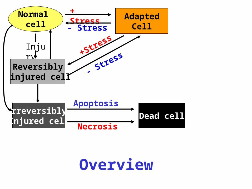
AdaptedCell
+ Stress
Injury
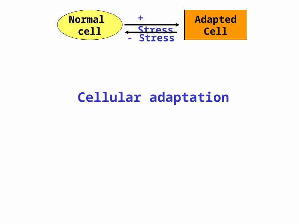
Normal cell
Reversibly injured cell
Irreversibly Injured cell
Dead cell
+Stress
Apoptosis
Necrosis
- Stress
- Stress
Overview

AdaptedCell
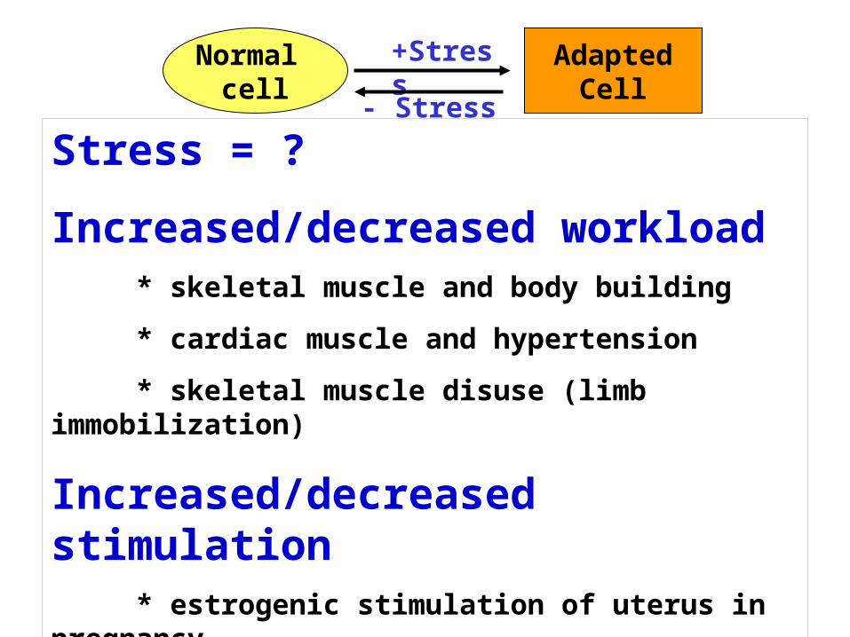
+ StressNormal cell
- Stress
Cellular adaptation
AdaptedCell
+StressNormal cell
Stress = ?
Increased/decreased workload* skeletal muscle and body building
* cardiac muscle and hypertension
* skeletal muscle disuse (limb immobilization)
Increased/decreased stimulation* estrogenic stimulation of uterus in pregnancy
* estrogen/prolactin stimulation of breast (lactation)
* denervation of muscle
- Stress
AdaptedCell
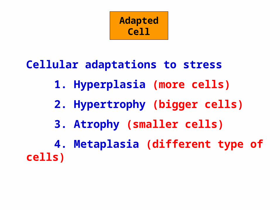
Cellular adaptations to stress
1. Hyperplasia (more cells)
2. Hypertrophy (bigger cells)
3. Atrophy (smaller cells)
4. Metaplasia (different type of cells)
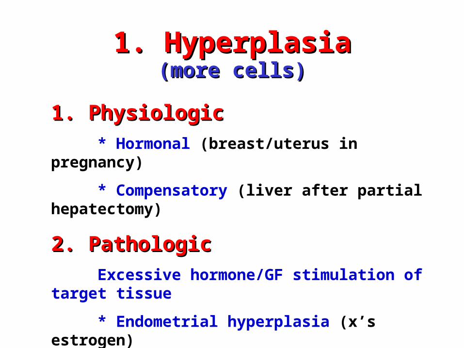
1. Hyperplasia1. Hyperplasia(more cells)(more cells)
1. Physiologic1. Physiologic
* Hormonal (breast/uterus in pregnancy)
* Compensatory (liver after partial hepatectomy)
2. Pathologic2. Pathologic
Excessive hormone/GF stimulation of target tissue
* Endometrial hyperplasia (x’s estrogen)
* Benign prostatic hyperplasia (x’s androgens)
* Connective tissue cells in wound healing
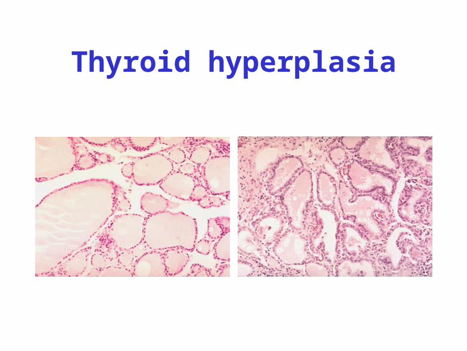
Thyroid hyperplasia
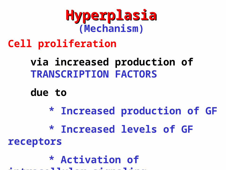
HyperplasiaHyperplasia(Mechanism)
Cell proliferation
via increased production of TRANSCRIPTION FACTORS
due to
* Increased production of GF
* Increased levels of GF receptors
* Activation of intracellular signaling
Results in larger organ
AdaptedCell
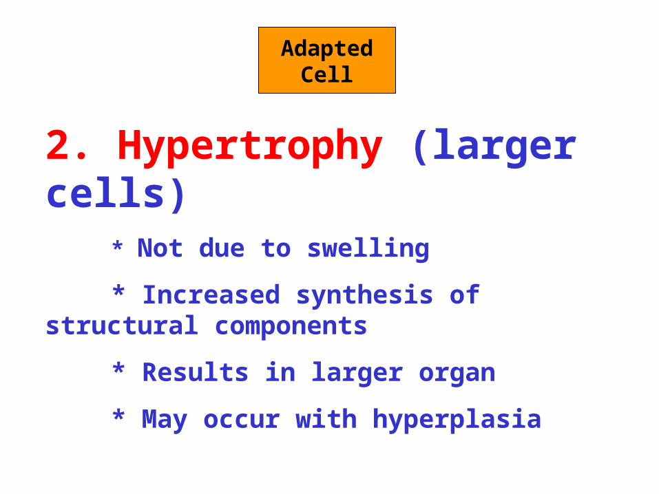
2. Hypertrophy (larger cells)* Not due to swelling
* Increased synthesis of structural components
* Results in larger organ
* May occur with hyperplasia
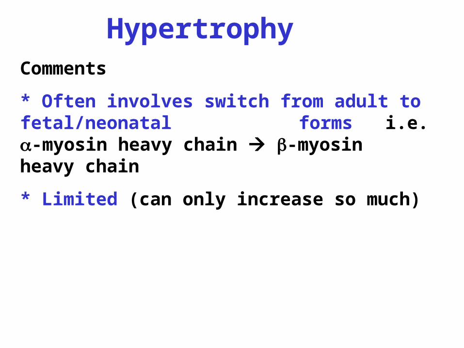
HypertrophyComments
* Often involves switch from adult to fetal/neonatal forms i.e. -myosin heavy chain -myosin heavy chain
* Limited (can only increase so much)
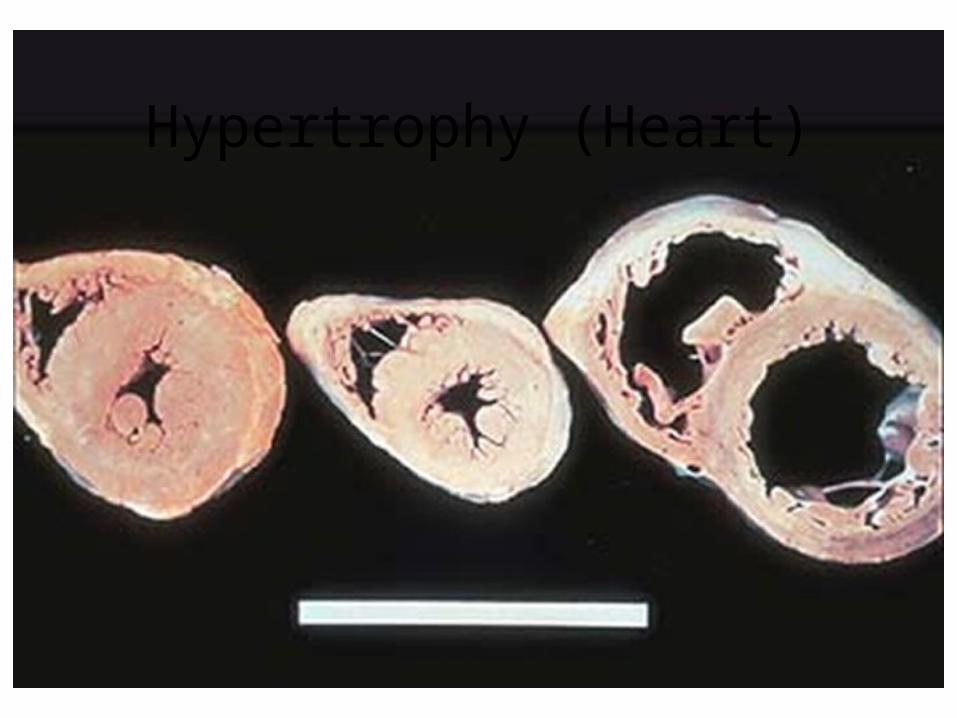
Hypertrophy (Heart)
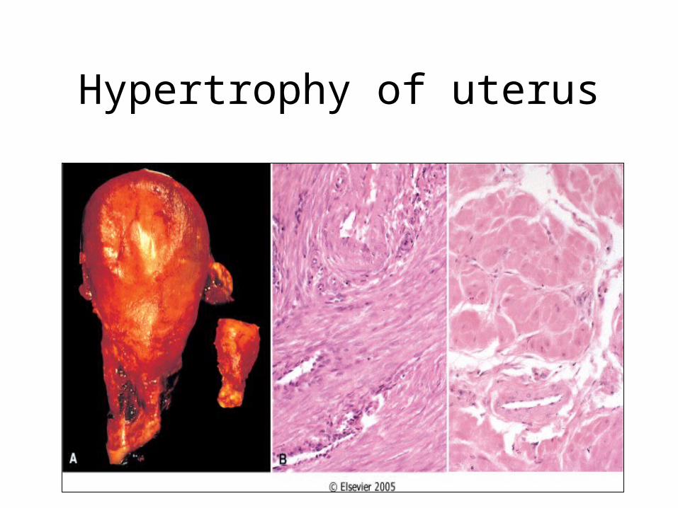
Hypertrophy of uterus
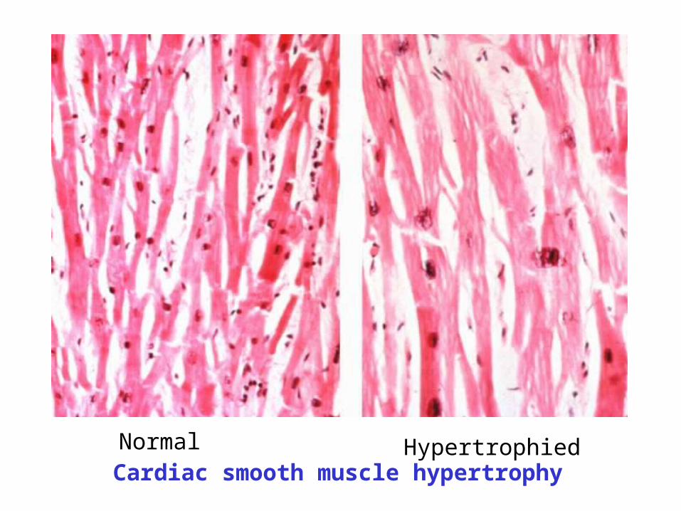
Normal HypertrophiedCardiac smooth muscle hypertrophy
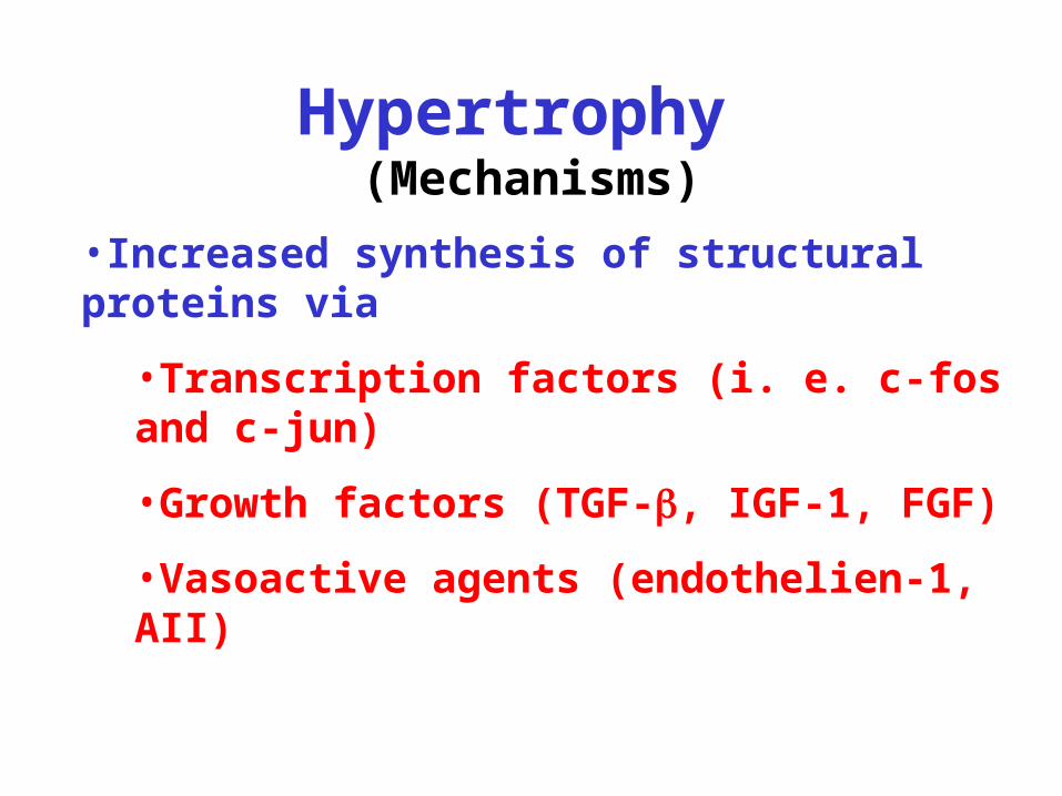
Hypertrophy (Mechanisms)
•Increased synthesis of structural proteins via
•Transcription factors (i. e. c-fos and c-jun)
•Growth factors (TGF-, IGF-1, FGF)
•Vasoactive agents (endothelien-1, AII)
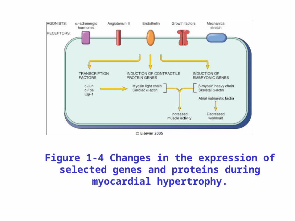
Figure 1-4 Changes in the expression of selected genes and proteins during myocardial hypertrophy.
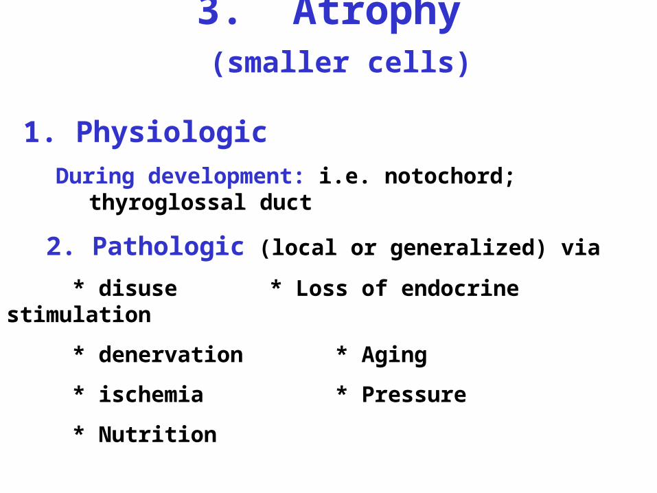
3. Atrophy (smaller cells)
1. Physiologic
During development: i.e. notochord; thyroglossal duct
2. Pathologic (local or generalized) via
* disuse * Loss of endocrine stimulation
* denervation * Aging
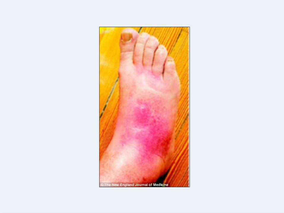
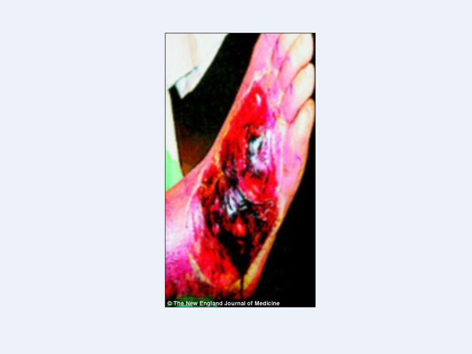
* ischemia * Pressure
* Nutrition
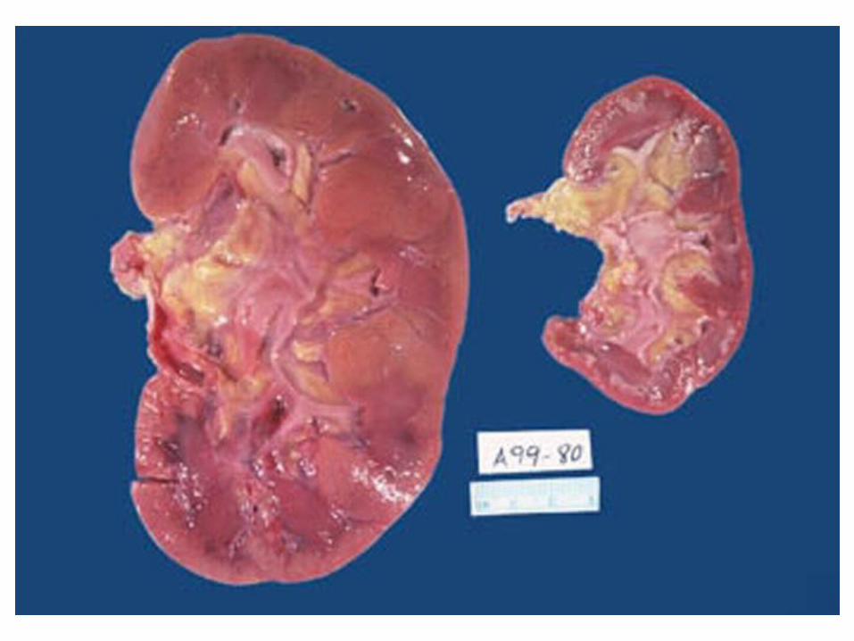
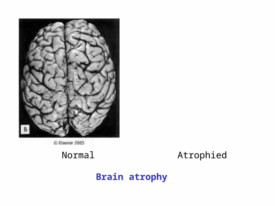
Normal Atrophied
Brain atrophy
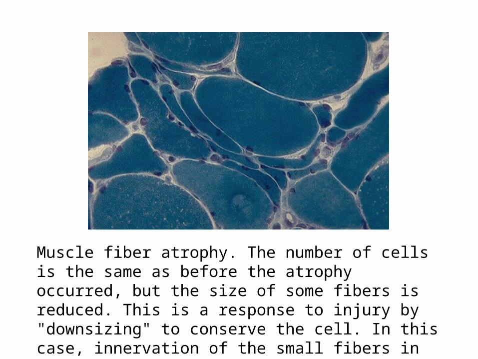
Muscle fiber atrophy. The number of cells is the same as before the atrophy occurred, but the size of some fibers is reduced. This is a response to injury by "downsizing" to conserve the cell. In this case, innervation of the small fibers in the center was lost. This is a trichrome stain.
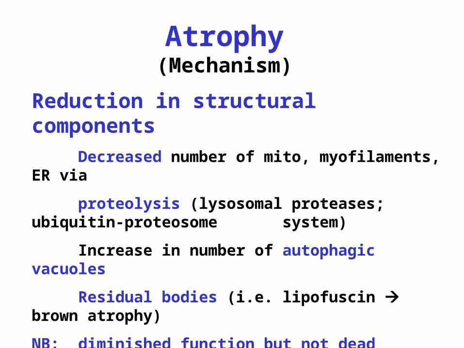
Atrophy(Mechanism)
Reduction in structural components
Decreased number of mito, myofilaments, ER via
proteolysis (lysosomal proteases; ubiquitin-proteosome system)
Increase in number of autophagic vacuoles
Residual bodies (i.e. lipofuscin brown atrophy)
NB: diminished function but not dead
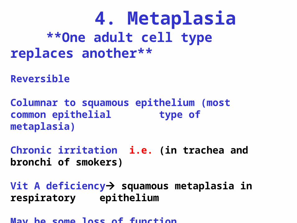
4. Metaplasia **One adult cell type replaces another**
Reversible
Columnar to squamous epithelium (most common epithelial type of metaplasia)
Chronic irritation i.e. (in trachea and bronchi of smokers)
Vit A deficiency squamous metaplasia in respiratory epithelium
May be some loss of function
May predispose to maligancy
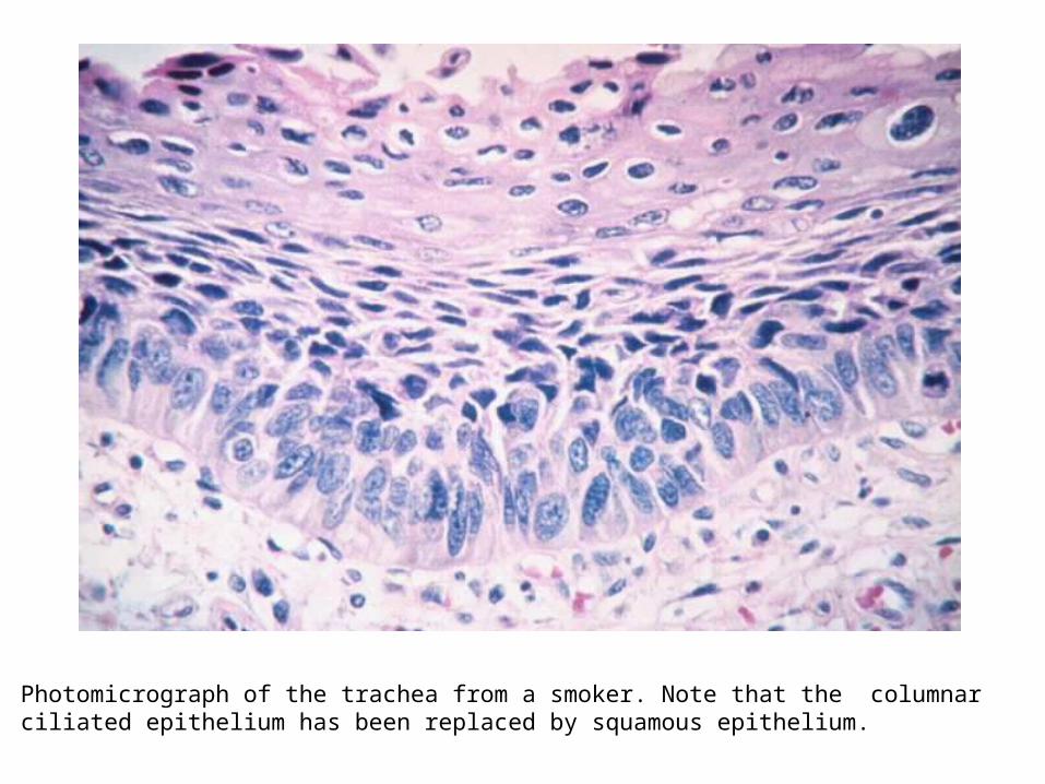
Photomicrograph of the trachea from a smoker. Note that the columnar ciliated epithelium has been replaced by squamous epithelium.
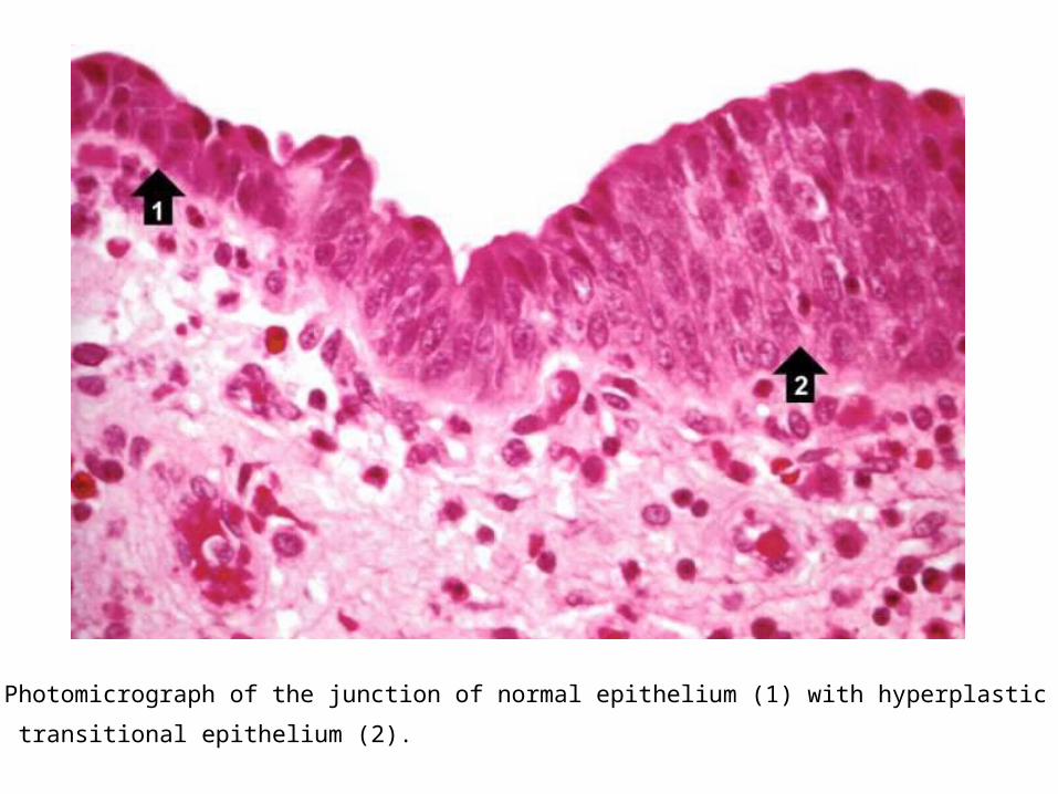
Photomicrograph of the junction of normal epithelium (1) with hyperplastic
transitional epithelium (2).
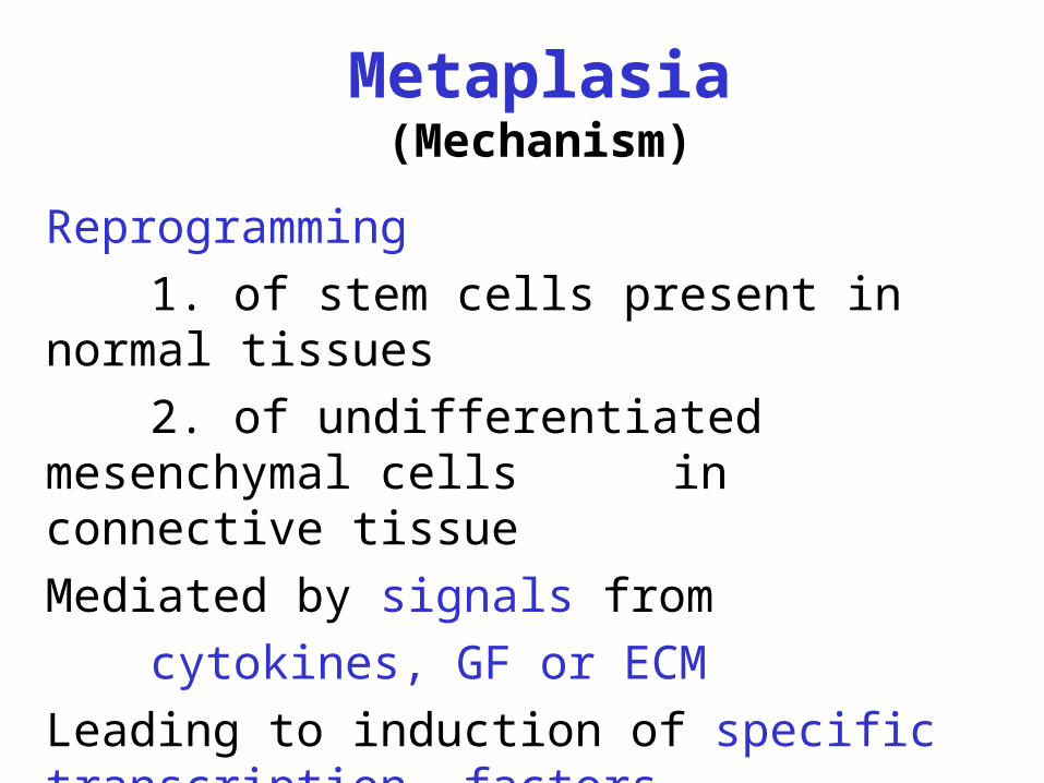
Metaplasia(Mechanism)
Reprogramming
1. of stem cells present in normal tissues
2. of undifferentiated mesenchymal cells in connective tissue
Mediated by signals from
cytokines, GF or ECM
Leading to induction of specific transcription factors
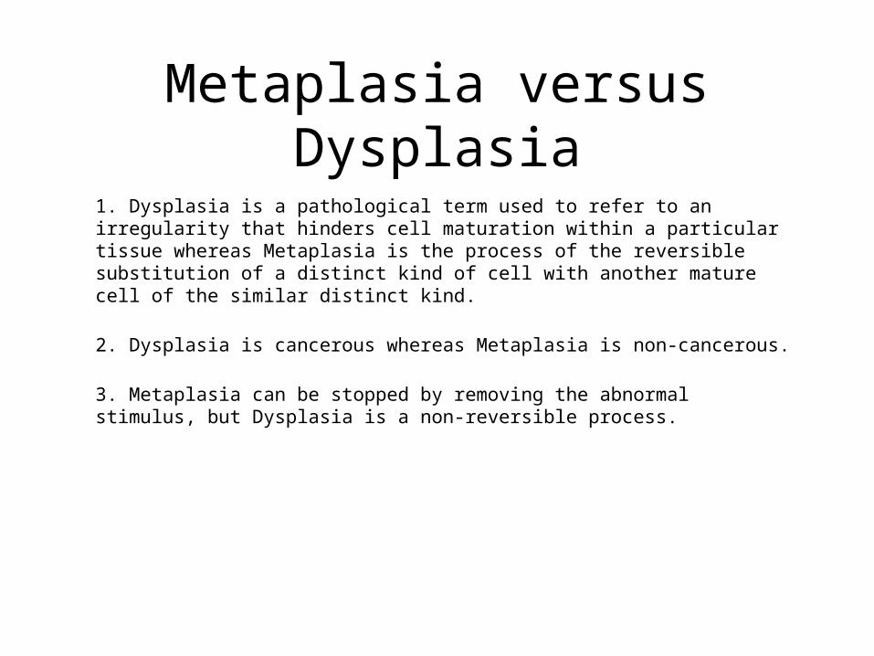
Metaplasia versus Dysplasia
1. Dysplasia is a pathological term used to refer to an irregularity that hinders cell maturation within a particular tissue whereas Metaplasia is the process of the reversible substitution of a distinct kind of cell with another mature cell of the similar distinct kind.
2. Dysplasia is cancerous whereas Metaplasia is non-cancerous.
3. Metaplasia can be stopped by removing the abnormal stimulus, but Dysplasia is a non-reversible process.