modern dry eye assessment the routine - … · modern dry eye assessment – the routine ... 4....
TRANSCRIPT
London SECO/AOP 2013 - Purslow
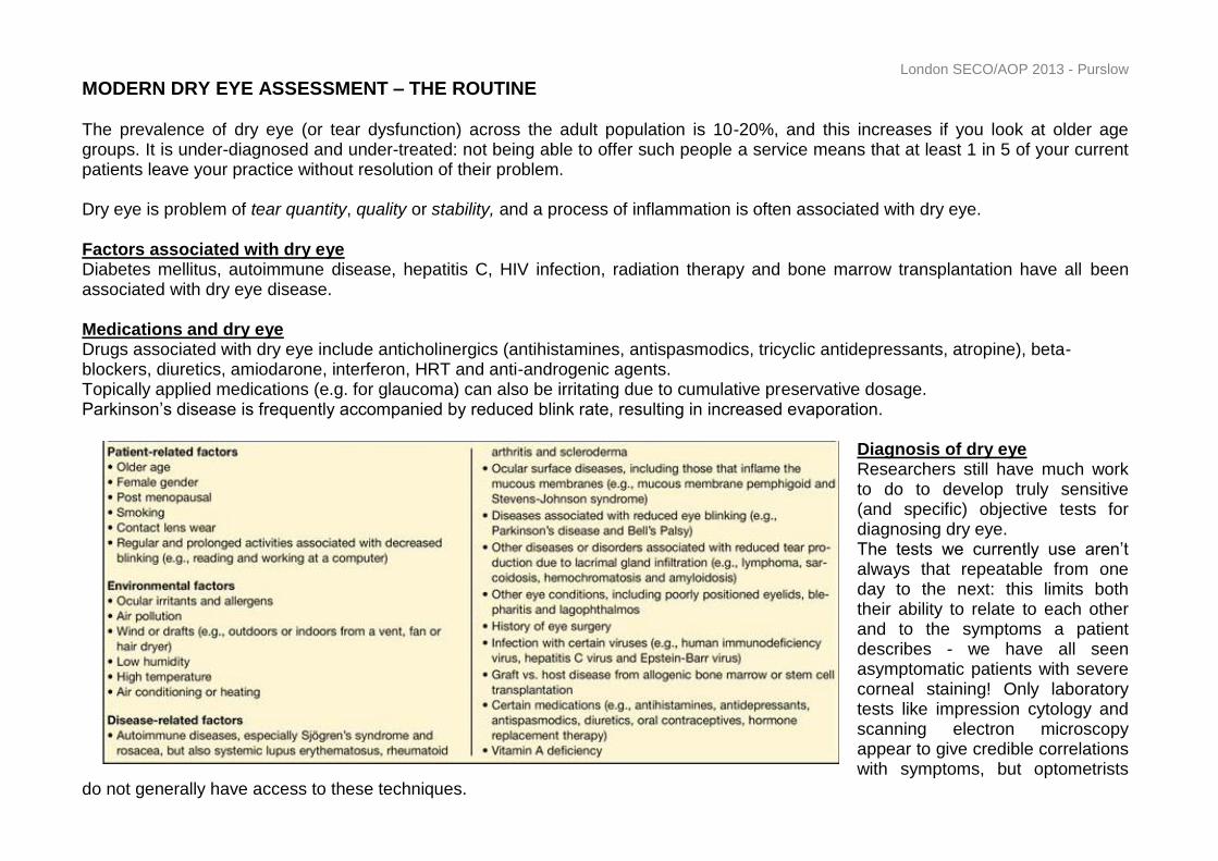
MODERN DRY EYE ASSESSMENT – THE ROUTINE The prevalence of dry eye (or tear dysfunction) across the adult population is 10-20%, and this increases if you look at older age groups. It is under-diagnosed and under-treated: not being able to offer such people a service means that at least 1 in 5 of your current patients leave your practice without resolution of their problem. Dry eye is problem of tear quantity, quality or stability, and a process of inflammation is often associated with dry eye. Factors associated with dry eye Diabetes mellitus, autoimmune disease, hepatitis C, HIV infection, radiation therapy and bone marrow transplantation have all been associated with dry eye disease. Medications and dry eye Drugs associated with dry eye include anticholinergics (antihistamines, antispasmodics, tricyclic antidepressants, atropine), beta-blockers, diuretics, amiodarone, interferon, HRT and anti-androgenic agents. Topically applied medications (e.g. for glaucoma) can also be irritating due to cumulative preservative dosage. Parkinson’s disease is frequently accompanied by reduced blink rate, resulting in increased evaporation.
Diagnosis of dry eye Researchers still have much work to do to develop truly sensitive (and specific) objective tests for diagnosing dry eye. The tests we currently use aren’t always that repeatable from one day to the next: this limits both their ability to relate to each other and to the symptoms a patient describes - we have all seen asymptomatic patients with severe corneal staining! Only laboratory tests like impression cytology and scanning electron microscopy appear to give credible correlations with symptoms, but optometrists
do not generally have access to these techniques.
London SECO/AOP 2013 - Purslow
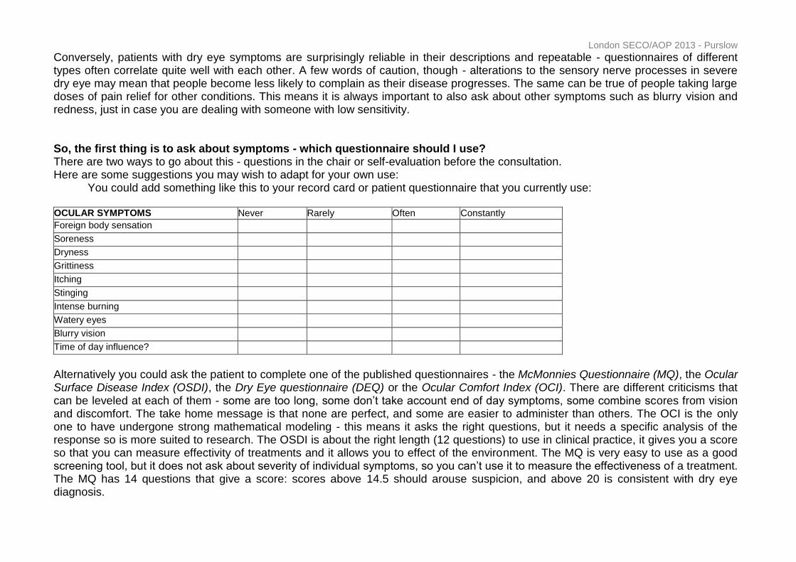
Conversely, patients with dry eye symptoms are surprisingly reliable in their descriptions and repeatable - questionnaires of different types often correlate quite well with each other. A few words of caution, though - alterations to the sensory nerve processes in severe dry eye may mean that people become less likely to complain as their disease progresses. The same can be true of people taking large doses of pain relief for other conditions. This means it is always important to also ask about other symptoms such as blurry vision and redness, just in case you are dealing with someone with low sensitivity. So, the first thing is to ask about symptoms - which questionnaire should I use? There are two ways to go about this - questions in the chair or self-evaluation before the consultation. Here are some suggestions you may wish to adapt for your own use: You could add something like this to your record card or patient questionnaire that you currently use: OCULAR SYMPTOMS Never Rarely Often Constantly
Foreign body sensation Soreness Dryness Grittiness Itching Stinging Intense burning Watery eyes Blurry vision Time of day influence?
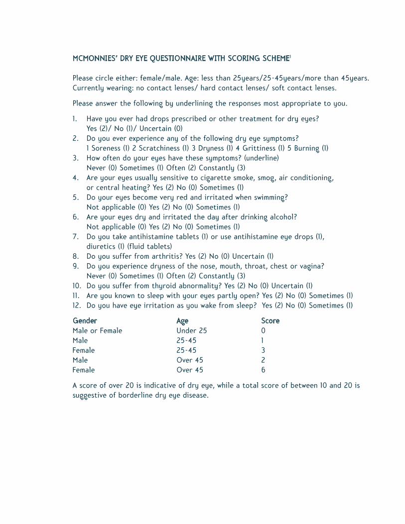
Alternatively you could ask the patient to complete one of the published questionnaires - the McMonnies Questionnaire (MQ), the Ocular Surface Disease Index (OSDI), the Dry Eye questionnaire (DEQ) or the Ocular Comfort Index (OCI). There are different criticisms that can be leveled at each of them - some are too long, some don’t take account end of day symptoms, some combine scores from vision and discomfort. The take home message is that none are perfect, and some are easier to administer than others. The OCI is the only one to have undergone strong mathematical modeling - this means it asks the right questions, but it needs a specific analysis of the response so is more suited to research. The OSDI is about the right length (12 questions) to use in clinical practice, it gives you a score so that you can measure effectivity of treatments and it allows you to effect of the environment. The MQ is very easy to use as a good screening tool, but it does not ask about severity of individual symptoms, so you can’t use it to measure the effectiveness of a treatment. The MQ has 14 questions that give a score: scores above 14.5 should arouse suspicion, and above 20 is consistent with dry eye diagnosis.
London SECO/AOP 2013 - Purslow
You can also simplify the MQ further down to just two questions, and it would still be a good screening tool: 1. How often do your eyes have symptoms of dryness?
a. Never b. Sometimes c. Often d. Constantly
2. How often do your eyes have symptoms of scratchiness a. Never b. Sometimes c. Often d. Constantly
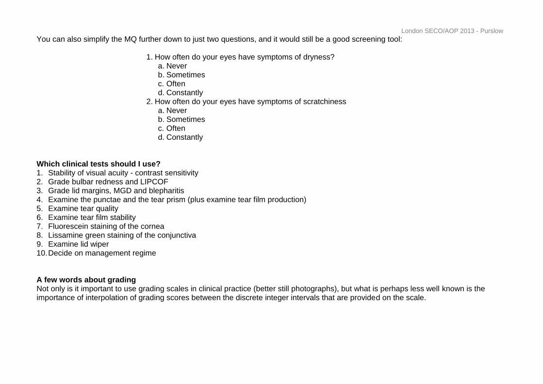
Which clinical tests should I use? 1. Stability of visual acuity - contrast sensitivity 2. Grade bulbar redness and LIPCOF 3. Grade lid margins, MGD and blepharitis 4. Examine the punctae and the tear prism (plus examine tear film production) 5. Examine tear quality 6. Examine tear film stability 7. Fluorescein staining of the cornea 8. Lissamine green staining of the conjunctiva 9. Examine lid wiper 10. Decide on management regime A few words about grading Not only is it important to use grading scales in clinical practice (better still photographs), but what is perhaps less well known is the importance of interpolation of grading scores between the discrete integer intervals that are provided on the scale.
London SECO/AOP 2013 - Purslow
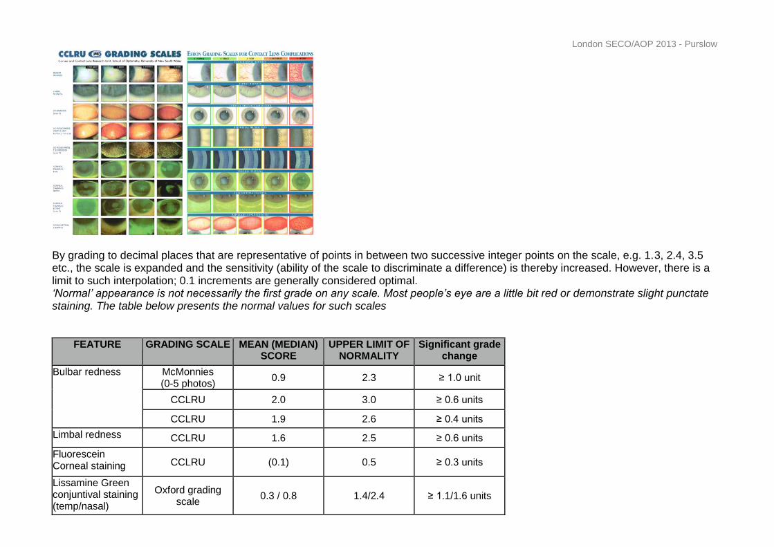
By grading to decimal places that are representative of points in between two successive integer points on the scale, e.g. 1.3, 2.4, 3.5 etc., the scale is expanded and the sensitivity (ability of the scale to discriminate a difference) is thereby increased. However, there is a limit to such interpolation; 0.1 increments are generally considered optimal. ‘Normal’ appearance is not necessarily the first grade on any scale. Most people’s eye are a little bit red or demonstrate slight punctate staining. The table below presents the normal values for such scales
FEATURE GRADING SCALE MEAN (MEDIAN) SCORE
UPPER LIMIT OF NORMALITY
Significant grade change
Bulbar redness McMonnies (0-5 photos)
0.9 2.3 ≥ 1.0 unit
CCLRU 2.0 3.0 ≥ 0.6 units
CCLRU 1.9 2.6 ≥ 0.4 units
Limbal redness CCLRU 1.6 2.5 ≥ 0.6 units
Fluorescein Corneal staining CCLRU (0.1) 0.5 ≥ 0.3 units
Lissamine Green conjuntival staining (temp/nasal)
Oxford grading scale
0.3 / 0.8 1.4/2.4 ≥ 1.1/1.6 units
London SECO/AOP 2013 - Purslow
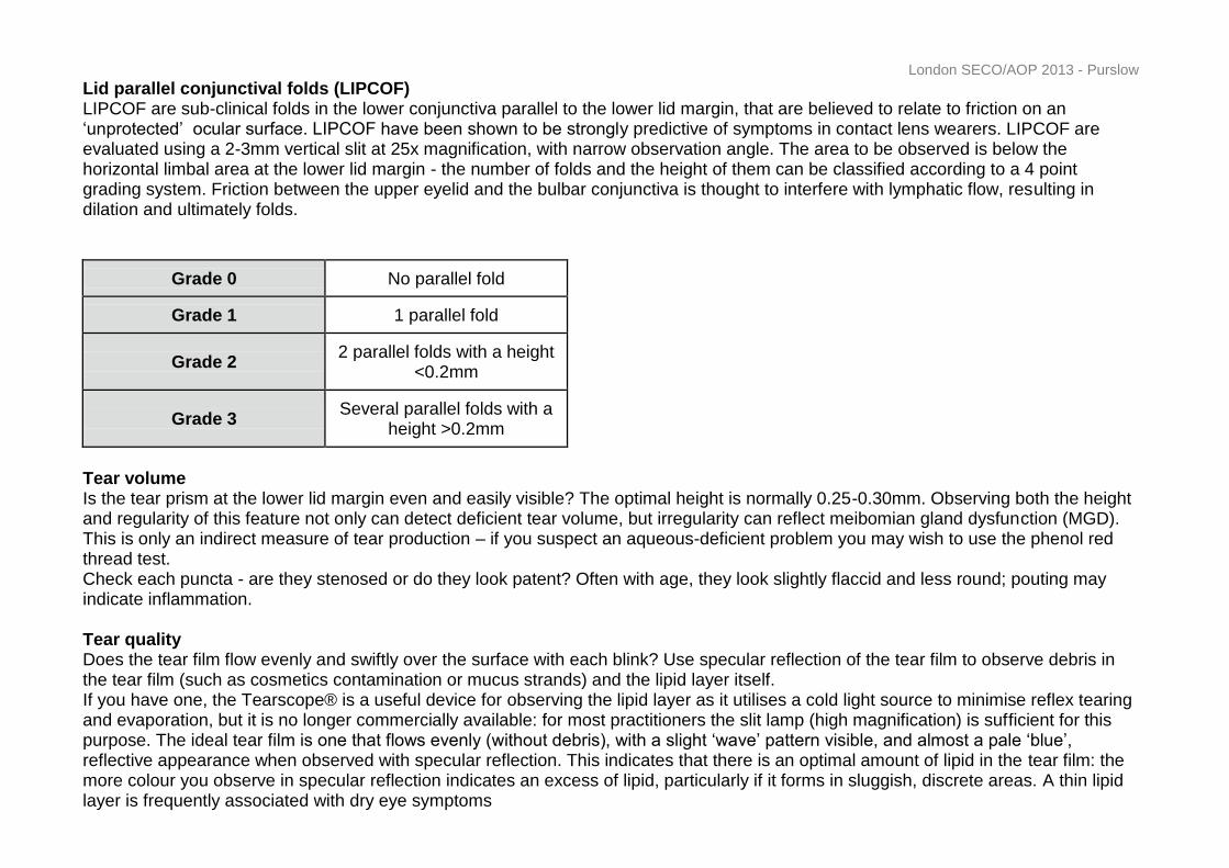
Lid parallel conjunctival folds (LIPCOF) LIPCOF are sub-clinical folds in the lower conjunctiva parallel to the lower lid margin, that are believed to relate to friction on an ‘unprotected’ ocular surface. LIPCOF have been shown to be strongly predictive of symptoms in contact lens wearers. LIPCOF are evaluated using a 2-3mm vertical slit at 25x magnification, with narrow observation angle. The area to be observed is below the horizontal limbal area at the lower lid margin - the number of folds and the height of them can be classified according to a 4 point grading system. Friction between the upper eyelid and the bulbar conjunctiva is thought to interfere with lymphatic flow, resulting in dilation and ultimately folds.
Grade 0 No parallel fold
Grade 1 1 parallel fold
Grade 2 2 parallel folds with a height
<0.2mm
Grade 3 Several parallel folds with a
height >0.2mm
Tear volume Is the tear prism at the lower lid margin even and easily visible? The optimal height is normally 0.25-0.30mm. Observing both the height and regularity of this feature not only can detect deficient tear volume, but irregularity can reflect meibomian gland dysfunction (MGD). This is only an indirect measure of tear production – if you suspect an aqueous-deficient problem you may wish to use the phenol red thread test. Check each puncta - are they stenosed or do they look patent? Often with age, they look slightly flaccid and less round; pouting may indicate inflammation. Tear quality Does the tear film flow evenly and swiftly over the surface with each blink? Use specular reflection of the tear film to observe debris in the tear film (such as cosmetics contamination or mucus strands) and the lipid layer itself. If you have one, the Tearscope® is a useful device for observing the lipid layer as it utilises a cold light source to minimise reflex tearing and evaporation, but it is no longer commercially available: for most practitioners the slit lamp (high magnification) is sufficient for this purpose. The ideal tear film is one that flows evenly (without debris), with a slight ‘wave’ pattern visible, and almost a pale ‘blue’, reflective appearance when observed with specular reflection. This indicates that there is an optimal amount of lipid in the tear film: the more colour you observe in specular reflection indicates an excess of lipid, particularly if it forms in sluggish, discrete areas. A thin lipid layer is frequently associated with dry eye symptoms
London SECO/AOP 2013 - Purslow
Tear stability The tear film should remain stable for at least 10-20 seconds with little or no break-up. If blinking is prevented, the tear film will thin and eventually expose the ocular surface. If this break up is assessed with sodium fluorescein dye added to the tear fluid, the time to break-up will generally be shorter than that obtained with the more preferred non-invasive methods. A one-position keratometer can be used but this will only observe tear thinning and break-up in the central area; a projected grid is required to monitor the whole corneal surface, either using the Tearscope® or a topographer. The patient is asked to blink normally then hold their eye open for as long as possible - the number of seconds before the grid distorts in any area indicates break up and directly reflects tear film stability. This is called the non-invasive break-up time (NIBUT). If the patient needs to blink before break-up occurs, then this is recorded as the maximum inter-blinking interval. In normal eyes, NIBUT is typically 30 seconds or more. Lid margin disease A very common cause of dry eye symptoms, often a chronic condition which needs long-term management, but treatments can be effective. Firstly, look for redness and swelling, particularly at the edge closer to the eye. Check for foamy or brimming tears along the lid margin Next evert the upper and lower lids - look for redness in a columnar fashion - following the paths of the glands Observe the meibomian gland openings, and the lashes for signs of anterior blepharitis. Corneal staining with Fluorescein Some degree of corneal staining is a normal finding in up to 79% of corneas and 71% of conjunctivae in normal eyes. However, signs of corneal staining can also tell us that the cornea is compromised. Interestingly, it is still not absolutely clear how fluorescein acts on the ocular surface. It is commonly accepted that it penetrates only interruptions or gaps in the epithelium and can only permeate into intracellular spaces and underlying tissues via damaged cells. However, there is some evidence to suggest that cells do not have to be damaged to be stained by fluorescein. ‘Moisten and shake’ for a Fluoret is the right approach - too much and you may get quenching where it reaches its own natural limit of fluorescence. For dry eye assessment, it is best to apply the fluoret strip to the lower tarsal plate,
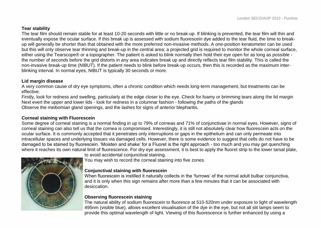
to avoid accidental conjunctival staining. You may wish to record the corneal staining into five zones. Conjunctival staining with fluorescein When fluorescein is instilled it naturally collects in the ‘furrows’ of the normal adult bulbar conjunctiva, and it is only when this sign remains after more than a few minutes that it can be associated with desiccation. Observing fluorescein staining The natural ability of sodium fluorescein to fluoresce at 510-520nm under exposure to light of wavelength 495nm (visible blue), allows excellent visualisation of the dye in the eye, but not all slit lamps seem to provide this optimal wavelength of light. Viewing of this fluorescence is further enhanced by using a
London SECO/AOP 2013 - Purslow
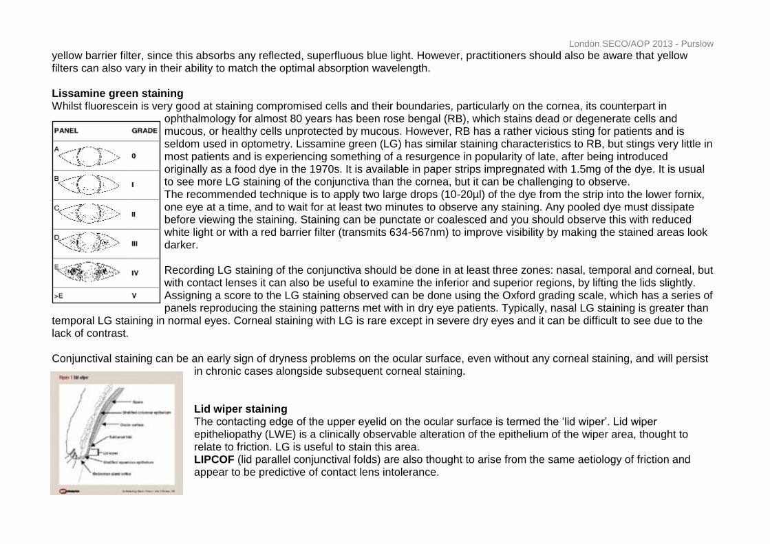
yellow barrier filter, since this absorbs any reflected, superfluous blue light. However, practitioners should also be aware that yellow filters can also vary in their ability to match the optimal absorption wavelength. Lissamine green staining Whilst fluorescein is very good at staining compromised cells and their boundaries, particularly on the cornea, its counterpart in
ophthalmology for almost 80 years has been rose bengal (RB), which stains dead or degenerate cells and mucous, or healthy cells unprotected by mucous. However, RB has a rather vicious sting for patients and is seldom used in optometry. Lissamine green (LG) has similar staining characteristics to RB, but stings very little in most patients and is experiencing something of a resurgence in popularity of late, after being introduced originally as a food dye in the 1970s. It is available in paper strips impregnated with 1.5mg of the dye. It is usual to see more LG staining of the conjunctiva than the cornea, but it can be challenging to observe. The recommended technique is to apply two large drops (10-20µl) of the dye from the strip into the lower fornix, one eye at a time, and to wait for at least two minutes to observe any staining. Any pooled dye must dissipate before viewing the staining. Staining can be punctate or coalesced and you should observe this with reduced white light or with a red barrier filter (transmits 634-567nm) to improve visibility by making the stained areas look darker. Recording LG staining of the conjunctiva should be done in at least three zones: nasal, temporal and corneal, but with contact lenses it can also be useful to examine the inferior and superior regions, by lifting the lids slightly. Assigning a score to the LG staining observed can be done using the Oxford grading scale, which has a series of panels reproducing the staining patterns met with in dry eye patients. Typically, nasal LG staining is greater than
temporal LG staining in normal eyes. Corneal staining with LG is rare except in severe dry eyes and it can be difficult to see due to the lack of contrast. Conjunctival staining can be an early sign of dryness problems on the ocular surface, even without any corneal staining, and will persist
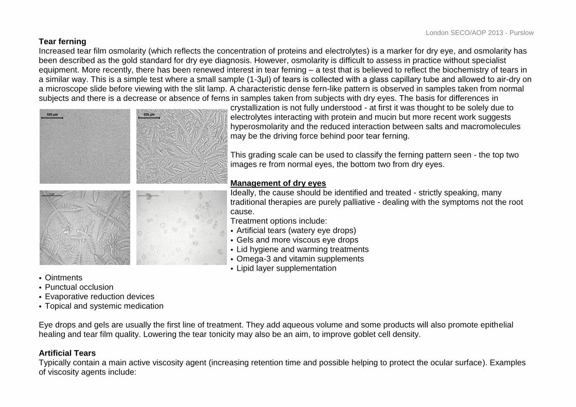
in chronic cases alongside subsequent corneal staining. Lid wiper staining The contacting edge of the upper eyelid on the ocular surface is termed the ‘lid wiper’. Lid wiper epitheliopathy (LWE) is a clinically observable alteration of the epithelium of the wiper area, thought to relate to friction. LG is useful to stain this area. LIPCOF (lid parallel conjunctival folds) are also thought to arise from the same aetiology of friction and appear to be predictive of contact lens intolerance.
London SECO/AOP 2013 - Purslow
Tear ferning Increased tear film osmolarity (which reflects the concentration of proteins and electrolytes) is a marker for dry eye, and osmolarity has been described as the gold standard for dry eye diagnosis. However, osmolarity is difficult to assess in practice without specialist equipment. More recently, there has been renewed interest in tear ferning – a test that is believed to reflect the biochemistry of tears in a similar way. This is a simple test where a small sample (1-3μl) of tears is collected with a glass capillary tube and allowed to air-dry on a microscope slide before viewing with the slit lamp. A characteristic dense fern-like pattern is observed in samples taken from normal subjects and there is a decrease or absence of ferns in samples taken from subjects with dry eyes. The basis for differences in
crystallization is not fully understood - at first it was thought to be solely due to electrolytes interacting with protein and mucin but more recent work suggests hyperosmolarity and the reduced interaction between salts and macromolecules may be the driving force behind poor tear ferning. This grading scale can be used to classify the ferning pattern seen - the top two images re from normal eyes, the bottom two from dry eyes. Management of dry eyes Ideally, the cause should be identified and treated - strictly speaking, many traditional therapies are purely palliative - dealing with the symptoms not the root cause. Treatment options include:
• Artificial tears (watery eye drops) • Gels and more viscous eye drops • Lid hygiene and warming treatments • Omega-3 and vitamin supplements • Lipid layer supplementation
• Ointments • Punctual occlusion • Evaporative reduction devices • Topical and systemic medication Eye drops and gels are usually the first line of treatment. They add aqueous volume and some products will also promote epithelial healing and tear film quality. Lowering the tear tonicity may also be an aim, to improve goblet cell density. Artificial Tears Typically contain a main active viscosity agent (increasing retention time and possible helping to protect the ocular surface). Examples of viscosity agents include:
London SECO/AOP 2013 - Purslow
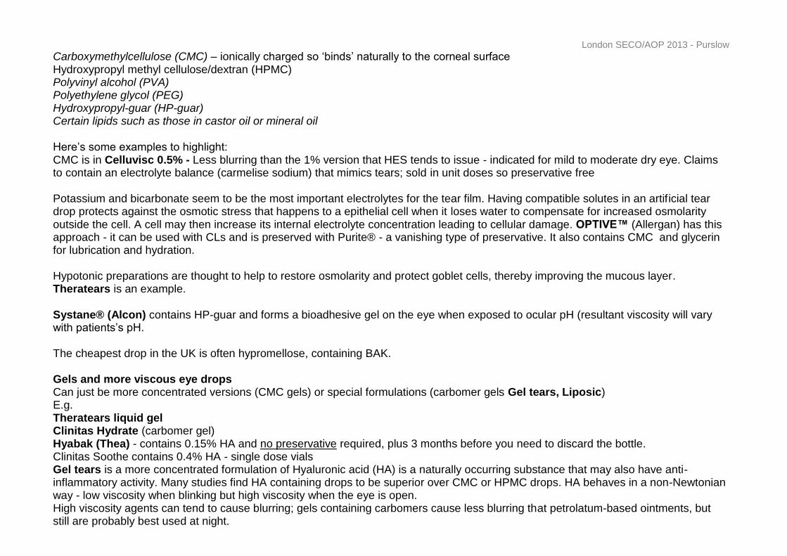
Carboxymethylcellulose (CMC) – ionically charged so ‘binds’ naturally to the corneal surface Hydroxypropyl methyl cellulose/dextran (HPMC) Polyvinyl alcohol (PVA) Polyethylene glycol (PEG) Hydroxypropyl-guar (HP-guar) Certain lipids such as those in castor oil or mineral oil Here’s some examples to highlight: CMC is in Celluvisc 0.5% - Less blurring than the 1% version that HES tends to issue - indicated for mild to moderate dry eye. Claims to contain an electrolyte balance (carmelise sodium) that mimics tears; sold in unit doses so preservative free Potassium and bicarbonate seem to be the most important electrolytes for the tear film. Having compatible solutes in an artificial tear drop protects against the osmotic stress that happens to a epithelial cell when it loses water to compensate for increased osmolarity outside the cell. A cell may then increase its internal electrolyte concentration leading to cellular damage. OPTIVE™ (Allergan) has this approach - it can be used with CLs and is preserved with Purite® - a vanishing type of preservative. It also contains CMC and glycerin for lubrication and hydration. Hypotonic preparations are thought to help to restore osmolarity and protect goblet cells, thereby improving the mucous layer. Theratears is an example. Systane® (Alcon) contains HP-guar and forms a bioadhesive gel on the eye when exposed to ocular pH (resultant viscosity will vary with patients’s pH. The cheapest drop in the UK is often hypromellose, containing BAK. Gels and more viscous eye drops Can just be more concentrated versions (CMC gels) or special formulations (carbomer gels Gel tears, Liposic) E.g. Theratears liquid gel Clinitas Hydrate (carbomer gel) Hyabak (Thea) - contains 0.15% HA and no preservative required, plus 3 months before you need to discard the bottle. Clinitas Soothe contains 0.4% HA - single dose vials Gel tears is a more concentrated formulation of Hyaluronic acid (HA) is a naturally occurring substance that may also have anti-inflammatory activity. Many studies find HA containing drops to be superior over CMC or HPMC drops. HA behaves in a non-Newtonian way - low viscosity when blinking but high viscosity when the eye is open. High viscosity agents can tend to cause blurring; gels containing carbomers cause less blurring that petrolatum-based ointments, but still are probably best used at night.

London SECO/AOP 2013 - Purslow
A few words about preservatives… The more severe the dry eye, the more you should avoid preservatives is my general advice, but the preserved tears are normally well tolerated in mild dry eye when used no more than 4-6x each day. Some preservatives have the potential to be more toxic than some, particularly the very effective ones (e.g. BAK) Lipid layer supplementation Refresh Endura (Allergan) contains castor oil and is intended to restore the lipid layer - this can be particularly useful in MGD. Paraffin ointment can be used at night to help the lipid layer. Clarymist Liposomal spray is now called Eye logic spray. Optrex also has a version - Optrex eye mist. These are sprayed onto closed eye lids; liposomes migrate to the lid margin and a small amount replenishes the lipid layer. They are more expensive but can be kept for up to 3 years and has a sterile spray mechanism (no preservative required). Clinitas Ultra 3 contains phospholipid Ointments Used when a high protection is required, e.g. recurrent corneal erosion Liquid paraffin, soft paraffin E.g. Lacrilube Reducing evaporation 1. Moisture spectacles/ goggles - to reduce tear evaporation by increasing humidity around the eye (side-shields or swimming goggles) 2. Bandage contact lenses Lid hygiene and MGD treatment Eye bag Thea range of products - Bleph range cleaning wipes, solution, gel cleanser, Blephasteam goggles (NEW) Lid care Supranettes Eye mask SteriLid
London SECO/AOP 2013 - Purslow
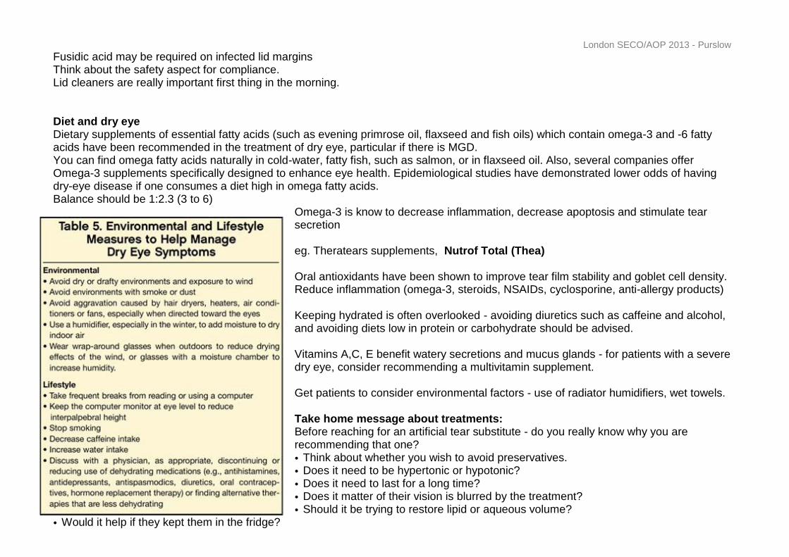
Fusidic acid may be required on infected lid margins Think about the safety aspect for compliance. Lid cleaners are really important first thing in the morning. Diet and dry eye Dietary supplements of essential fatty acids (such as evening primrose oil, flaxseed and fish oils) which contain omega-3 and -6 fatty acids have been recommended in the treatment of dry eye, particular if there is MGD. You can find omega fatty acids naturally in cold-water, fatty fish, such as salmon, or in flaxseed oil. Also, several companies offer Omega-3 supplements specifically designed to enhance eye health. Epidemiological studies have demonstrated lower odds of having dry-eye disease if one consumes a diet high in omega fatty acids. Balance should be 1:2.3 (3 to 6)
Omega-3 is know to decrease inflammation, decrease apoptosis and stimulate tear secretion eg. Theratears supplements, Nutrof Total (Thea) Oral antioxidants have been shown to improve tear film stability and goblet cell density. Reduce inflammation (omega-3, steroids, NSAIDs, cyclosporine, anti-allergy products) Keeping hydrated is often overlooked - avoiding diuretics such as caffeine and alcohol, and avoiding diets low in protein or carbohydrate should be advised. Vitamins A,C, E benefit watery secretions and mucus glands - for patients with a severe dry eye, consider recommending a multivitamin supplement. Get patients to consider environmental factors - use of radiator humidifiers, wet towels. Take home message about treatments: Before reaching for an artificial tear substitute - do you really know why you are recommending that one?
• Think about whether you wish to avoid preservatives. • Does it need to be hypertonic or hypotonic? • Does it need to last for a long time? • Does it matter of their vision is blurred by the treatment? • Should it be trying to restore lipid or aqueous volume?
• Would it help if they kept them in the fridge?

London SECO/AOP 2013 - Purslow
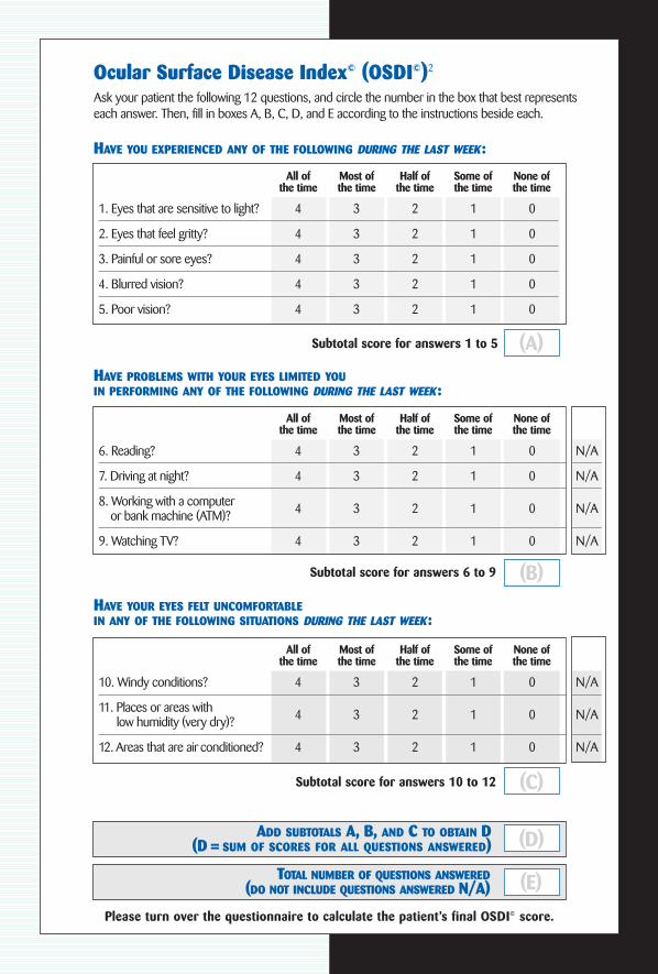
Ocular Surface Disease Index© (OSDI©)2
Ask your patient the following 12 questions, and circle the number in the box that best representseach answer. Then, fill in boxes A, B, C, D, and E according to the instructions beside each.
HAVE YOU EXPERIENCED ANY OF THE FOLLOWING DURING THE LAST WEEK:
All ofthe time
Most ofthe time
Half ofthe time
Some ofthe time
None ofthe time
1. Eyes that are sensitive to light? 4 3 2 1 0
2. Eyes that feel gritty? 4 3 2 1 0
3. Painful or sore eyes? 4 3 2 1 0
4. Blurred vision? 4 3 2 1 0
5. Poor vision? 4 3 2 1 0
Subtotal score for answers 1 to 5
HAVE PROBLEMS WITH YOUR EYES LIMITED YOUIN PERFORMING ANY OF THE FOLLOWING DURING THE LAST WEEK:
All ofthe time
Most ofthe time
Half ofthe time
Some ofthe time
None ofthe time
6. Reading? 4 3 2 1 0 N/A
7. Driving at night? 4 3 2 1 0 N/A
8. Working with a computer 4 3 2 1 0 N/Aor bank machine (ATM)?
9. Watching TV? 4 3 2 1 0 N/A
Subtotal score for answers 6 to 9
HAVE YOUR EYES FELT UNCOMFORTABLEIN ANY OF THE FOLLOWING SITUATIONS DURING THE LAST WEEK:
All ofthe time
Most ofthe time
Half ofthe time
Some ofthe time
None ofthe time
10. Windy conditions? 4 3 2 1 0 N/A
11. Places or areas with 4 3 2 1 0 N/Alow humidity (very dry)?
12. Areas that are air conditioned? 4 3 2 1 0 N/A
Subtotal score for answers 10 to 12
Please turn over the questionnaire to calculate the patient’s final OSDI© score.
(A)
(B)
(C)
(D)
(E)
ADD SUBTOTALS A, B, AND C TO OBTAIN D(D = SUM OF SCORES FOR ALL QUESTIONS ANSWERED)
TOTAL NUMBER OF QUESTIONS ANSWERED(DO NOT INCLUDE QUESTIONS ANSWERED N/A)
77089 9/20/04 10:31 PM Page 1
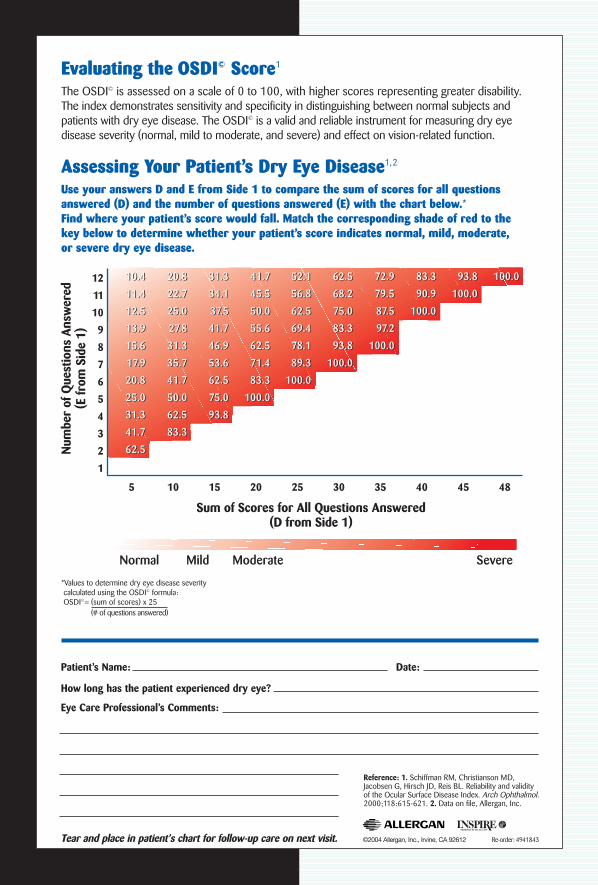
Evaluating the OSDI© Score1
The OSDI© is assessed on a scale of 0 to 100, with higher scores representing greater disability.The index demonstrates sensitivity and specificity in distinguishing between normal subjects andpatients with dry eye disease. The OSDI© is a valid and reliable instrument for measuring dry eye disease severity (normal, mild to moderate, and severe) and effect on vision-related function.
Assessing Your Patient’s Dry Eye Disease1,2
Use your answers D and E from Side 1 to compare the sum of scores for all questionsanswered (D) and the number of questions answered (E) with the chart below.*Find where your patient’s score would fall. Match the corresponding shade of red to the key below to determine whether your patient’s score indicates normal, mild, moderate, or severe dry eye disease.
Patient’s Name: Date:
How long has the patient experienced dry eye?
Eye Care Professional’s Comments:
Tear and place in patient’s chart for follow-up care on next visit.
Num
ber
of Q
uest
ions
Ans
wer
ed(E
from
Sid
e 1)
Sum of Scores for All Questions Answered (D from Side 1)
12
11
10
9
8
7
6
5
4
3
2
1
5 484540353025201510
10.4
11.4
12.5
13.9
15.6
1 7.9
20.8
25.0
31.3
41.7
62.5
20.8
22.7
25.0
27.8
31.3
35.7
41.7
50.0
62.5
83.3
31.3
34.1
37.5
41.7
46.9
53.6
62.5
75.0
93.8
41.7
45.5
50.0
55.6
62.5
71.4
83.3
100.0
52.1
56.8
62.5
69.4
78.1
89.3
100.0
62.5
68.2
75.0
83.3
93.8
100.0
72.9
79.5
87.5
97.2
100.0
83.3
90.9
100.0
93.8
100.0
100.0
Normal Mild Moderate Severe
10.4
11.4
12.5
13.9
15.6
1 7.9
20.8
25.0
31.3
41.7
62.5
20.8
22.7
25.0
27.8
31.3
35.7
41.7
50.0
62.5
83.3
31.3
34.1
37.5
41.7
46.9
53.6
62.5
75.0
93.8
41.7
45.5
50.0
55.6
62.5
71.4
83.3
100.0
52.1
56.8
62.5
69.4
78.1
89.3
100.0
62.5
68.2
75.0
83.3
93.8
100.0
72.9
79.5
87.5
97.2
100.0
83.3
90.9
100.0
93.8
100.0
100.0
*Values to determine dry eye disease severitycalculated using the OSDI© formula:OSDI©= (sum of scores) x 25
(# of questions answered)
Reference: 1. Schiffman RM, Christianson MD,Jacobsen G, Hirsch JD, Reis BL. Reliability and validity of the Ocular Surface Disease Index. Arch Ophthalmol.2000;118:615-621. 2. Data on file, Allergan, Inc.
©2004 Allergan, Inc., Irvine, CA 92612 Re-order: 4941843
77089 9/20/04 10:31 PM Page 2