modalities of tonometry and their accuracy with respect to corneal thickness and irregularities
TRANSCRIPT
J Optom, Vol. 1, No. 2, October-December 2008
Modalities of Tonometry and their Accuracy with Respect to Corneal Thickness and Irregularities Carlos Gustavo V. De Moraes1, Tiago S. Prata1, Jeffrey Liebmann2,3 and Robert Ritch1,4
ABSTRACTRecent advances in tonometry have led to the development of a number of devices with differing clinical applications. Their role in cases of abnormal corneal thickness and surface irregularities is par-ticularly important, as inaccurate estimation of the true intraocular pressure (IOP) in such cases may lead to suboptimal evaluation. The purpose of the present review was to evaluate the accuracy of the most widely used devices in cases of corneal thickness and sur-face irregularities, based on a survey of the published literature. The analysis was based on a Medline search focusing mainly on papers that have evaluated the devices’ accuracy with respect to corneal thickness and irregularities. Nine device types (Goldmann tonometer, Tono-Pen, Perkins tonometer, Ocular Response Analyzer, non-contact tonometer, pneumatonometer, I-Care rebound tonometer, Pascal dynamic con-tour tonometer (DCT) and Phosphene tonometer) were described in detail. Moreover, the physical principles and major utility of each tonometer were compared. Each of the many different commercially available tonometers has specific advantages and disadvantages. New non-invasive tech-nologies are getting closer to a precise estimation of the true IOP. However, among all tonometers, none is highly accurate when both corneal thickness and surface irregularities are present. Fifty years after its development, Goldmann tonometry remains the gold standard to which all other devices are compared. (J Optom 2008;1:43-49 ©2008 Spanish Council of Optometry)
KEY WORDS: tonometry accuracy; intraocular pressure; corneal thickness; corneal irregularities.
RESUMENAvances recientes en el campo de la tonometría han propiciado el desarrollo de diversos dispositivos con diferentes aplicaciones clíni-cas. Resulta de particular importancia el papel que juegan en el caso de espesores corneales anormales o en presencia de irregularidades en la superficie de la córnea, puesto que, en esos casos, una medida imprecisa de la presión intraocular (PIO) real puede traer como consecuencia una valoración incorrecta del caso. El objetivo de esta revisión es evaluar el grado de precisión de los dis-positivos más utilizados hoy en día en casos en los que existen irregulari-dades en la superficie de la córnea o en el espesor corneal. Para ello, se llevó a cabo una búsqueda de datos entre los artículos publicados. El análisis estuvo basado en una búsqueda bibliográfica realizada dentro de Medline, centrándose principalmente en aquellos artícu-
los científicos que habían estudiado el grado de precisión del dis-positivo en función del espesor corneal del paciente y de la presencia o no de irregularidades corneales. Se describen en detalle nueve tipos de dispositivos: tonómetro de Goldmann, Tono-Pen, tonómetro de Perkins, Ocular Response Analyzer, tonómetro sin contacto, neumotonómetro, tonómetro de rebote I-Care, tonómetro de contorno dinámico Pascal y tonómetro de fosfeno. Además, se presentan de forma comparada los principios físicos en los que está basado cada tonómetro y las situaciones en las que cada uno resulta de mayor utilidad. Cada uno de los distintos tonómetros disponibles en el mercado presenta ventajas y desventajas respecto al resto. Las nuevas tec-nologías no invasivas están logrando estimaciones de la PIO cada vez más cercanas al valor real de la PIO. Sin embargo, de entre todos los tonómetros analizados, no hay ninguno que sea sumamente pre-ciso sea cual sea el espesor corneal y en presencia de irregularidades en la superficie de la córnea. Trascurridos cincuenta años desde su aparición, la tonometría de Goldmann sigue siendo la técnica de referencia, con la que han de compararse el resto de dispositivos. (J Optom 2008;1:43-49 ©2008 Consejo General de Colegios de Ópticos-Optometristas de España)
PALABRAS CLAVE: exactitud tonometría; presión intraocular; espesor corneal; irregularidades corneales.
INTRODUCTION
The term “intraocular pressure” (IOP) describes the tension exerted by the aqueous humor in the intraocular tissues as a result of the balance between its production and drainage. Since there is currently no safe invasive method of measuring the IOP intraocularly, it is really “estimated” rather than “measured” in clinical practice. However, because of its widely accepted use in the literature and in practice, we will use the term “measurement” in this review.
The importance of measuring IOP comes primarily from the concept that it is the most significant risk factor for the development and progression of glaucoma,1-5 the most impor-tant cause of irreversible blindness worldwide.6 Nevertheless, precise IOP measurement is subject to some confounding variables, such as circadian variation7-9 and the influence of corneal biomechanical properties.10-13 Furthermore, as refractive procedures become more popular and increases the indication of different modalities of keratoplasty, there is a growing interest on whether changes in corneal structure may influence IOP measurements and how it could affect the management of these patients once they are referred to a glaucoma practice.
Most commercially available tonometers estimate IOP based on corneal applanation or indentation. These devices consider that the force exerted on the external corneal sur-face reflects the pressure at the level of the endothelium and,
From the 1Einhorn Clinical Research Center, New York Eye and Ear Infirmary, New York, NY, USA. 2Manhattan Eye, Ear and Throat Hospital, New York; NY, USA. 3New York University School of Medicine, New York, NY, USA. 4Department of Ophthalmology, New York Medical College, Valhalla, NY, USA.Received: 11 August 2008Revised: 2 October 2008Accepted: 9 October 2008Corresponding author: Robert Ritch. Professor and Chief, Glaucoma Service, Department of Ophthalmology, The New York Eye and Ear Infirmary, 310 East 14th Street, New York, NY 10003, USA e-mail: [email protected]
doi:10.3921/joptom.2008.43
REVIEW J Optom 2008;1:43-49
J Optom, Vol. 1, No. 2, October-December 2008
44 Tonometry and Cornea: De Moraes CG et al.
therefore, the pressure in the anterior chamber and vitreous cavity (see equation 1). Where (F) is the force applied to the outer corneal surface area (A), (Pcp) is the pressure related to the corneal properties, and (tIOP) is the true IOP.
(equation 1) F / A = Pcp + tIOP
In that case, for a given ideal standard infinitely thin and spherical corneal area, the external pressure would equal the true IOP. This assumption presupposes that all indi-viduals have identical corneal thickness and viscoelasticity. However, differences in these properties exist across indi-viduals depending on their age, race, corneal abnormalities, or even between fellow eyes.11-13 Thus, the accuracy of IOP measurement depends on the corneal thickness, curvature and biomechanical properties.
In this review, we will describe the most commonly used tonometers, their applicability, advantages, disadvantages, and the differences among them in terms of accuracy of measurement. To that end, we have divided them into two groups: applanation and non-applanation tonometers.
APPLANATION TONOMETERS
Goldmann Tonometer (Haag Streit, Koeniz, Switzerland)Goldmann applanation tonometry (GAT), developed in
the 1950s, is based on the Imbert-Fick law, which states that “the pressure in a sphere filled with fluid and surrounded by an infinitely thin and flexible membrane is measured by the counter-pressure which just flattens the membrane to a plane.”14 However, such a hypothetical membrane does not fit the corneal model. Hence, Goldmann and Schmidt14 sug-gested that this device would be more precise in patients hav-ing an average central corneal thickness (CCT) of between 500 and 525 μm.
Particular attention was paid to CCT after the Ocular Hypertension Treatment Study (OHTS) found that eyes with thinner CCT are at increased risk of developing glau-coma.2 One hypothesis was that thinner corneas may lead to underestimation of the real IOP; that is, the pressure that is causing glaucoma damage is actually higher than that meas-ured by GAT. Despite that, GAT is still the most commonly used tonometer in clinical practice worldwide and thus remains the “gold standard” for tonometry.
The clinician should be particularly careful regarding value reliability when measuring IOP in eyes with signifi-cantly thinner (<525 μm) or thicker (>555 μm) corneas. In eyes with decreased CCT, GAT tends to underestimate IOP, while in eyes with increased CCT, this measuring technique tends to overestimate IOP. While controversial, the same seems to be valid for corneas that are flatter or steeper than usual.15,16
The suggested inaccuracy of GAT measurements has raised an extensive debate in the literature, particularly in cases of irregular corneas (keratoconus) and following surgical procedures (penetrating keratoplasty and refractive surgery). In keratoconus, high astigmatism, and stromal scarring, GAT may show greater variability and lower accura-cy.17-19 Brooks et al.18 found that GAT measurements yielded
significantly lower values at the apex of the cone when com-pared to flatter or thicker areas of the cornea, which resulted in an overall underestimation of the true IOP. In keratoconus patients, GAT IOP seems to be 5.3±2.2 mmHg lower than that yielded by non-applanation tonometry, which seemed to provide measurements closer to the true IOP.20
Ismail et al.19 reported that in eyes that had undergone penetrating keratoplasty, GAT measurements may be less precise than non-applanation tonometry. Meyenberg et al.21 suggested that GAT could slightly underestimate IOP in postkeratoplasty eyes (3.1±2.5 mm), a fact which was con-firmed in cases in which the procedure preserves the deeper corneal layers.22
Regarding the effect of refractive surgery on GAT IOP measurements, there seems to be an agreement on the apparent IOP-lowering effect of the different modalities of surgery.23-27 Kirwan et al.26 found that the mean GAT IOP decreased 3.7±2.3 mmHg following LASIK, and a similar decrease was observed following LASEK. Moreover, photore-fractive keratectomy (PRK) seems to induce a smaller GAT IOP underestimation than LASIK.27 It should be emphasized that the IOP might not truly decrease following these proce-dures, but rather it is underestimated as a result of changes in the corneal structure (e.g., decreased thickness, presence of fluid or scarring tissue).
Attempts have been made to establish specific formulas to calculate the influence of CCT on IOP measurement, but there is no consensus about its use in practice.28 The simple concept that IOP is being over- or underestimated depend-ing on these variables should suffice when estimating the subject’s IOP in clinical setting.
Since GAT is taken nowadays as the gold standard, all further comparisons of the different types of tonometers will be based on GAT in this review.
Tono-Pen XL (Mentor O&O Inc., Norwell, MA, U.S.A.)This is a light-weight contact electronic applanation
tonometer, which is portable and easy to calibrate and oper-ate. Its digital readout minimizes user bias, and due to its small contact area (2.36 mm2 compared to 7.35 mm2 in GAT), it is recommended for IOP measurements in irregular corneas.29,30 It is also useful when there is poor patient coop-eration, allowing measurements in both supine and sitting positions. A minimum of four measurements is necessary before the device can yield an average value. It also provides a coefficient of variation (COV) which ideally should be less then 5% for a measurement to be considered accurate according to manufacturer information.
However, studies have shown the Tono-Pen to over- or underestimate IOP without a consistent pattern. Salvetat et al.30 found that Tono-Pen tended to underestimate GAT in 0.5±4.5 mmHg. The authors also reported that compared with GAT, the Tono-Pen showed a tendency to underestimate IOP in eyes with lower IOP (<24 mmHg), and overestimate IOP in those eyes with higher IOP (>24 mmHg). In eyes with increased CCT (>584 μm), the Tono-Pen tended to produce, consistently, higher IOP readings than GAT. Similarly, Broman et al.31 suggested that the
Tonometry and Cornea: De Moraes CG et al. 45
J Optom, Vol. 1, No. 2, October-December 2008
Tono-Pen would overall underestimate the true IOP based on the assumption that this would be closer to the average IOP measured by three different tonometers. With regard to irregular corneas, Mollan et al.32 evaluated the perform-ance of four different tonometers in eyes with keratoconus and found that Tono-Pen overestimated GAT in 3.6±10.1 mmHg. Moreover, in this group of patients, the Tono-Pen gave overestimated IOP values for lower IOPs and under-estimated ones for higher IOPs (always compared to GAT readings), whereas it seems to be less dependent on CCT in keratoconus than GAT. In postkeratoplasty eyes, GAT and Tono-Pen showed a good agreement (mean difference 0.14 mmHg) and Tono-Pen IOP was largely independent of corneal thickness.33 In summary, Tono-Pen measurements should be interpreted cautiously especially in eyes with increased CCT, whereas it might prove helpful in irregular corneas due to its smaller contact area.
Perkins Handheld Tonometer (Medtronic Solan, Jacksonville, FL, USA)
The Perkins applanation tonometer is a portable version of the GAT, also requiring topical instillation of fluorescein. Portable, handheld tonometers have the advantage of being easily transported from site to site for screening examinations and for those patients for whom the use of a chin rest proves difficult. They are especially useful for the determination of the daily curve of IOP (supine position). There is a close agreement between the Perkins tonometer and GAT,34,35
with a mean difference of 1.0 mmHg between the two tonometers.34 Also, since breath-holding (required for GAT measurements, taken in sitting position) and thus thorax compression may cause transitory elevations of IOP, the Perkins tonometer may provide more reliable measurements in these cases of falsely elevated IOP.36
Due to its high agreement with GAT and with intra- cameral measurements readings, Perkins tonometry could be considered the gold standard for portable tonometry.37
Ocular Response Analyzer (Reichert Ophthalmic Instruments, Depew, New York, USA)
The ocular response analyzer (ORA) uses an air-pressure-triggered, dynamic, bi-directional corneal applanation method to measure corneal biomechanical parameters. An air pulse on the cornea causes its movement inward, and creates a slight concavity. The device makes two measurements of the corneal response to the air pulse—the force necessary to flatten the cornea as the pressure of the air pulse rises, and the force at which the cornea flattens again after the air pump shuts off. The difference between the two measurements (inward and outward applanation) is termed corneal hysteresis. Based on this initial evaluation, the device provides 4 different parameters: Goldmann-correlated IOP, corneal-compensated IOP (IOPcc), corneal resistance factor (CRF) and corneal hysteresis (CH).38 Among these parameters, CH is the one that has been most evaluated in previous studies and is considered an indicator of the viscoelastic properties of the cornea.31 CH is weakly cor-related with CCT, is almost constant throughout the day,39 and seems unassociated with refractive error or axial length.40,41
ORA measurements show good within-session repeat-ability in normal volunteers.42 Mean values of IOPcc, CH and CRF in a study including healthy patients (CCT=557 ± 36μm; GAT=14.8±3 mmHg) were 16.2±4.1 mmHg, 10.6 ±2.3 mmHg, 10.9±2.4 mmHg, respectively.43 Regarding the device’s IOPcc, it seems to provide higher IOP readings than GAT in normal subjects. Moreover, IOPcc measurements could provide an estimate of IOP that is less influenced by corneal properties than that provided by GAT.31,39-44 Patients with lower CCT and CH values tend to have higher IOPcc values, compared to GAT results. Conversely, patients with higher CCT and CH values tend to get lower IOPcc val-ues.43,44 Furthermore, Medeiros et al.13 demonstrated that IOPcc was not correlated with CCT or corneal curvature, but it was positively associated with age. Even though the overall difference between GAT IOP and IOPcc was not sig-nificant, it tended to be bigger for increasing CCT values.
Glaucoma patients appear to have lower CH values with ORA than normal subjects, and lower CH values have been associated with progressive visual field worsening.45 Lower CH was observed in glaucoma patients with acquired pits of the optic nerve head compared to patients who did not. This could suggest that corneal biomechanical properties may reflect viscoelastic properties of the lamina cribrosa, for example.46
In keratoconus eyes, both CH and CRF seem to be lower than in a normal population (10.6±2.2 versus 8.7 ± 2.2 mmHg, and 10.0±2.5 versus 6.9±2.4 mmHg, respec-tively).32 There is also a significant decrease of both CH and CRF following LASIK,47 which could probably account for the changes observed in GAT measurements as described previously.
Although interesting initial results have been published with regard to various diseases, there is still a need to better understand their clinical value. ORA has shown to provide reproducible and stable values within different day-time measurements,48,49 which were independent from the IOP fluctuation detected by GAT.48 These findings suggest a promissory role of ORA in understanding the effect of dif-ferent corneal conditions on tonometry.
Non-contact Tonometer or Air-puff Tonometer (Reichert Ophthalmic Instruments, Depew, New York, USA)
Grolman created this non-contact applanation tonometer in the 1950s aiming to make it available for optometrists to perform tonometry measurements. Briefly, an air-puff causes a transient applanation of the cornea, while an infrared light beam is reflected by the flattened surface. The amount of light reflected during the applanation period is compared with the time the air-puff took to cause applanation, allow-ing this device to provide an electronic measurement of the IOP. It also provides the ocular pulse amplitude and tono-graphic measurements that estimate the aqueous outflow efficiency of the trabecular meshwork according to manufac-turer information. Historically, non-contact tonometers were not considered to be the most accurate way to measure IOP. There were concerns that low pressures were overestimated and high pressures underestimated. The oldest versions
J Optom, Vol. 1, No. 2, October-December 2008
46 Tonometry and Cornea: De Moraes CG et al.
of this tonometer showed a fair agreement with GAT (±3 mmHg), but tended to overestimate the IOP for pressures lower than 10 mmHg and underestimate it for values above 19 mmHg.50 However, modern non-contact tonometers cor-relate very well with GAT IOP, even though they tend to sys-tematically overestimate it by between 0.12–0.58 mmHg.51-53 With regard to the influence of corneal properties on non-contact tonometry measurements, it is likely that they are more influenced by CCT than GAT. In thinner corneas, there seems to better correlation between the tonometers, while in thicker corneas, non-contact tonometry systemati-cally yields higher readings than GAT.54
In summary, non-contact tonometers have generally been considered a fast and simple way to screen IOP. The benefits of non-contact tonometry include patient preference, less operator dependence, and no risk of infection transmis-sion.50-53
Pneumatonometer (Mentor, Model 30 Classic, Reichert, New York, USA)
This is an easy-to-use instrument which provides fast and accurate tonometry readings. Utilizing a pneumatic pump, it can provide real time readings of IOP through a noninvasive applanation method. A gentle, floating pneumatic sensor touches the surface of the anesthetized cornea with the exact amount of applanation force required to take the measure-ment. Another advantage would be its use in measuring IOP in contact-lenses wearers.55 The overall values obtained are usually slightly lower than those furnished by Goldmann tonometry, and they are also significantly associated with CCT and IOP itself. In this sense, pneumatonometry sig-nificantly underestimates GAT measurements at lower IOP and overestimates these at higher IOP. For example, for GAT IOP measurements <10 mmHg, the difference is around 2.0 mmHg, while for GAT IOPs ≥25, the difference is -0.6 (GAT - pneumotonometer).56 Also, as the GAT values increase, the pneumatonometer increasingly overestimates IOP.57 On the other hand, in eyes with keratoconus, the pneumatonometer underestimates IOP, yielding values that are lower than GAT ones by about 1.5 mmHg.20
Similarly to the air-puff tonometer, this device finds large applicability as a screening tool by non-specialized personnel. In a clinical setting, the results should be analyzed cautiously.
NON-APPLANATION TONOMETRY DEVICES
Rebound Tonometry- I-Care Tonometer (Tiolat, Helsinki, Finland)
Based on the rebound principle described by Dekking and Coster in 1967, this is a contact rebound tonometer that uses a light probe containing a permanent magnet that is launched towards the eye using a solenoid. The probe hits the eye and bounces back. The same solenoid, inside which moves the probe, is used to detect the movement and impact of the probe, because the moving magnet induces voltage in the solenoid. The motion parameters measured during impact are used to estimate the IOP.58,59 It is a handheld, portable tonometer that displays the IOP reading digitally
and does not require topical anesthesia. Following 6 meas-urements, the device automatically determines the mean pressure and the standard deviation.
I-Care has been found to be of clinical usefulness as a self-tonometer and among non-specialized personnel. Thus, it has been proposed that it could be used in screening stud-ies by non-medical staff. Recent reports about its accuracy have been conflicting. Van der Jagt and Jansonius60 found that I-Care slightly overestimated GAT by 0.6 mmHg (mean difference between 0.0 and 1.2 mmHg) even though such difference was not significant. Also, the authors described that adding corneal thickness to the regression analysis did not yield any increase in the variance of IOP measurements. On the other hand, Nakamura et al.61 studying a population that ranged from normal subjects to ocular hypertensives and glaucoma patients found that I-Care overestimated IOP, as compared to GAT, by 1.40±4.29 mmHg, and that this dis-parity tended to increase along with corneal thickness. They suggested that corneal thickness could affect the duration of the impact of the rebound tonometer, causing an overes-timation in thicker corneas. These results are confirmed by several other reports62-65. Even though little information is available on its use in irregular corneas, Jóhannesson et al.63 found that, unlike what was observed for GAT, corneal cur-vature was not correlated with I-Care IOP measurements. In general, I-Care seems to be a reasonable option for screening purposes,63 and measurements should always be interpreted with regard to CCT when used in a clinical basis.
Dynamic Contour Tonometry – Pascal Tonometer (SMT Swiss Microtechnology AG, Zurich, Switzerland)
The dynamic contour tonometer (DCT) is a novel digital non-applanation contact tonometer designed to be largely independent of the structural properties of the cornea, pos-sibly giving IOP measurements that are closer to the true IOP.15,16,66 It is a useful tool in situations where the clinician suspects inaccurate IOP measurements that could be caused by corneal biomechanical properties. It is particularly accu-rate in eyes with keratoconus,20,21,67 corneal edema,68 and those that have undergone penetrating keratoplasty21,22,69 and refractive surgery.25,70
The DCT provides a score (Q) representing the quality of the IOP measure. The score ranges from 1 (optimum) to 5 (unacceptable). For clinical and scientific purposes, only those measurements with Q scores of 1 or 2 are considered reliable reliable according to manufacturer information.
Along with the IOP and Q score, the digital screen also displays the ocular pulse amplitude (OPA), which represents the average difference between the systolic and diastolic IOP within 6 hear beats. It has been suggested that OPA provides a surrogate measure of the ocular blood flow, mostly due to the choroid.71,72
Most of the studies agree that DCT tends to overesti-mate GAT by about 2.3 -3.4 mmHg, depending on the IOP level, CCT and other corneal properties.15,16,30,73 Milla et al.73 found an optimal agreement between DCT and GAT when the CCT was between 540 and 545 μm. As the CCT and the IOP increase, the difference between both tonometers also
Tonometry and Cornea: De Moraes CG et al. 47
J Optom, Vol. 1, No. 2, October-December 2008
increases.30 In eyes with keratoconus, the difference between DCT and GAT ranged from 4.3 to 5.3 mmHg.20,32,67 Pascal tonometry seems to be largely independent of the IOP and CCT in those patients.32 In eyes that had undergone keratoplasty and refractive surgery, DCT seems to be less influenced by changes in corneal properties following these procedures.19,25,74
As a digital tonometer with an automated IOP quality-check, together with the increasing evidence of being largely independent of corneal properties, Pascal tonometry seems to be a promising tool for IOP determination in different pathological conditions in clinical practice.
Phosphene Tonometry (Proview, Bausch & Lomb Pharmaceuticals, Inc., Tampa, Fla.)
The pressure phosphene tonometer (PPT) is a self-tonometry device that was first described in 1998.75 It uses the entoptic phenomenon of pressure phosphene to evaluate IOP.76,77 The PPT is initially applied perpendicular to the eyeball through the partially closed eyelid, and no topical anesthesia is required. Afterward, the applied pressure is increased gradually until the moment when the patient clearly perceives a well-formed phosphene (usually described as a dark circle with a ring of light around the outer circum-ference). The device is then removed from the eyelid and IOP can be read from the dial.76,77 The PPT presents several advantages, such as: a) no contact with the cornea and no anesthetic drop required; b) it seems not to be influenced by corneal biomechanical properties; c) it allows for insights into patient-specific, diurnal variations.75-78 Also, it has been reported to present good reproducibility when used by patients.78 On the other hand, there is controversy regarding its accuracy. Most studies have compared the PPT to the GAT and some of them found a poor agreement between
the two devices, even when measurements were done by a trained examiner.78,79 PPT, compared to GAT, consistently underestimated the IOP by approximately 3.5 mmHg,79, and was not able to detect IOPs above 22 mmHg in as much as 82% of patients.78 PPT IOP should be evaluated carefully in clinical practice, and has limited applicability as population screening device, mostly due to its low sensitivity in detecting high IOP values.78
SUMMARY
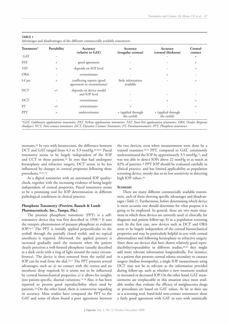
There are many different commercially available tonom-eters, each of them showing specific advantages and disadvan-tages (Table 1). Furthermore, before determining which device is more accurate one should determine for what purpose it is going to be employed. In general, there are two main situa-tions in which these devices are currently used: a) clinically, for diagnosis and patient follow-up; b) as a population screening tool. In the first case, new devices such as DCT and ORA seem to be largely independent of the corneal biomechanical properties and may be particularly helpful in eyes with corneal abnormalities and following keratoplasty or refractive surgery. Since these are devices that have shown relatively good repro-ducibility/repeatability in different studies,49,56 they might add more relevant information longitudinally. For instance, in a patient that presents corneal edema secondary to cataract surgery (bullous keratopathy), a single IOP measurement using DCT may not be as relevant as the information provided during follow-up, such as whether a new treatment resulted in increased or decreased IOP. On the other hand, GAT meas-urements are irreplaceable in this situation since most avail-able studies that evaluate the efficacy of antiglaucoma drugs or procedures are based on GAT values. As far as their use as a screening tool, hand-held non-contact tonometers show a fairly good agreement with GAT in eyes with statistically
TABLE 1 Advantages and disadvantages of the different commercially available tonometers Tonometer* Portability Accuracy Accuracy Accuracy Corneal (relative to GAT) (irregular corneas) (corneal thickness) contact GAT - - - +
PAT + good agreement - - +
TAT + depends on IOP level + - +
ORA - overestimates - + -
I-Care + conflicting reports (good little information - + agreement or overestimates) available
NCT +/- depends on device model and IOP level - - -
DCT - overestimates - + +
PT +/- overestimates + - +
PPT + underestimates + (applied through + (applied through - the eyelid) the eyelid)
*GAT, Goldmann applanation tonometer; PAT, Perkins applanation tonometer; TAT, Tono-Pen applanation tonometer; ORA, Ocular Response Analyzer; NCT, Non-contact tonometer; DCT, Dynamic Contour Tonometer; PT, Pneumatonometer; PPT, Phosphene tonometer.
J Optom, Vol. 1, No. 2, October-December 2008
48 Tonometry and Cornea: De Moraes CG et al.
normal IOP (15 – 22 mmHg) and CCT (525- 555). Except for PPT, most of the devices described above could provide reproducible and reliable IOP measurements even when used by non-trained personnel in screening projects. Moreover, portable tonometers with reduced applanation area (such as Tono-Pen) are especially helpful in non-collaborative patients and when corneal irregularities may be affecting the accuracy of the measurement.
Although new non-invasive technologies seem to be get-ting closer to a precise estimation of the true IOP, most of the available literature and large clinical trials in glaucoma (e.g., OHTS, EMGT, AGIS)1-5 were based on GAT readings. We believe that even if a new technology one day proves itself to provide a very precise estimation of the true IOP regardless of corneal properties, such information would be at first of limited applicability for the clinician. New definitions of “statistically normal” or “target” IOP will need to be refor-mulated before the practitioner can actually start making decisions based on a new tonometer.
On the other hand, these new devices provide not only information about IOP, but also new ocular parameters, such as corneal hysteresis (ORA) and ocular pulse amplitude (DCT). As some studies have reported differences in these parameters between glaucomatous and non-glaucomatous patients,20,31,39,45,46,71,72 we believe that in the future these parameters might prove useful to better understand the patho-physiology in different eye conditions.
It is important to emphasize that the IOP readings from these devices are not interchangeable, and even in patients with regular and corneal thickness in the average range, some of them seem to constantly disagree with GAT values. Rather then using multiple devices in the same patient, the clinician should choose one that better fits each clinical indication and use it consistently.
Among all the available tonometers, there is still no device capable of providing IOP readings with high accuracy regard-less of the value of the corneal thickness, the surface irregu-larities, or the particular conditions the patient may have. A customized application seems more reasonable. Fifty years after its invention, Goldman tonometry has helped build most of the knowledge available regarding aqueous humor dynamics and IOP monitoring whereas it remains as the gold standard all new technologies should be compared with.
REFERENCES
1. Leske MC, Heijl A, Hussein M, et al. Early Manifest Glaucoma Trial Group. Factors for glaucoma progression and the effect of treatment: the Early Manifest Glaucoma Trial. Arch Ophthalmol. 2003;121:48-56.
2. Kass MA, Heuer DK, Higginbotham EJ, et al. The Ocular Hypertension Treatment Study: a randomized trial determines that topical ocular hypotensive medication delays or prevents the onset of primary open-angle glaucoma. Arch Ophthalmol. 2002;120:701-713.
3. Lichter PR, Musch DC, Gillespie BW, et al. CIGTS Study Group. Interim clinical outcomes in the Collaborative Initial Glaucoma Treatment Study comparing initial treatment randomized to medica-tions or surgery. Ophthalmology. 2001;108:1943-1953.
4. The Advanced Glaucoma Intervention Study (AGIS): 7. The relation-ship between control of intraocular pressure and visual field deteriora-tion.The AGIS Investigators. Am J Ophthalmol. 2000;130:429-440.
5. Collaborative Normal-Tension Glaucoma Study Group. The effective-ness of intraocular pressure reduction in the treatment of normal-ten-sion glaucoma. Am J Ophthalmol. 1998;126:498-505.
6. Quigley HA, Broman AT. The number of people with glaucoma world-wide in 2010 and 2020. Br J Ophthalmol. 2006;90:262-267.
7. Liu JH. Diurnal measurement of intra-ocular pressure. J. Glaucoma. 2001;10:39-41.
8. Asrani S, Zeimer R, Wilensky J, et al. Large diurnal fluctuations in intraocular pressure are an independent risk factor in patients with glaucoma. J Glaucoma 2000;9:134-142.
9. Drance SM. Diurnal variation of intra-ocular pressure in treated glau-coma. Significance in patients with chronic simple glaucoma. Arch Ophthalmol. 1963;70:302-311.
10. Doughty MJ, Zaman ML. Human corneal thickness and its impact on intraocular pressure measures: a review and meta-analysis approach. Surv Ophthalmol. 2000;44:367-408.
11. Shimmyo M, Ross AJ, Moy A, et al. Intraocular pressure, Goldmann applanation tension, corneal thickness, and corneal curvature in Caucasians, Asians, Hispanics, and African Americans. Am J Ophthalmol. 2003;136:603-613.
12. Liu J, Roberts CJ. Influence of corneal biomechanical properties on intraocular pressure measurement: quantitative analysis. J Cataract Refract Surg. 2005;31:146-155.
13. Medeiros FA, Weinreb RN. Evaluation of the influence of corneal bio-mechanical properties on intraocular pressure measurements using the ocular response analyzer. J Glaucoma 2006;15:364-370.
14. Goldmann H, Schmidt T. Applanation tonometry. Ophthalmologica 1957;134:221-242.
15. Ceruti P, Morbio R, Marraffa M, Marchini G. Comparison of Goldmann applanation tonometry and dynamic contour tonometry in healthy and glaucomatous eyes. Eye 2008;25. (Epub ahead of print)
16. Schneider E, Grehn F. Intraocular pressure measurement-comparison of dynamic contour tonometry and Goldmann applanation tonometry. J Glaucoma 2006;15:2-6.
17. Bohm A, Kohlhaas M, Lerche RC, et al. Measuring intraocular pressure in keratoconus. Effect of the changed biomechanics. Ophthalmologe. 1997;94:771-774.
18. Brooks AM, Robertson IF, Mahoney AM. Ocular rigidity and intraocu-lar pressure in keratoconus. Aust J Ophthalmol. 1984;12:317-324.
19. Ismail AR, Lamont M, Perera S, et al. Comparison of IOP measure-ment using GAT and DCT in patients with penetrating keratoplasties. Br J Ophthalmol. 2007;91:980-981.
20. Papastergiou GI, Kozobolis V, Siganos DS. Assessment of the pascal dynamic contour tonometer in measuring intraocular pressure in kera-toconic eyes. J Glaucoma 2008;17:484-488.
21. Meyenberg A, Iliev ME, Eschmann R, Frueh BE. Dynamic con-tour tonometry in keratoconus and postkeratoplasty eyes. Cornea 2008;27:305-310.
22. Ceruti P, Morbio R, Marraffa M, Marchini G. Comparison of dynamic contour tonometry and goldmann applanation tonometry in deep lamellar and penetrating keratoplasties. Am J Ophthalmol. 2008;145:215-221.
23. Rosa N, Cennamo G, Breve MA, et al. Goldmann applanation tonom-etry after myopic photorefractive keratectomy. Acta Ophthalmol Scand 1998;76: 550-554.
24. Svedberg H, Chen E, Hamberg-Nyström H. Changes in corneal thick-ness and curvature after different excimer laser photorefractive proce-dures and their impact on intraocular pressure measurements. Graefes Arch Clin Exp Ophthalmol. 2005;243:1218-1220.
25. Pepose JS, Feigenbaum SK, Qazi MA, et al. Changes in corneal biome-chanics and intraocular pressure following LASIK using static, dynamic, and noncontact tonometry. Am J Ophthalmol. 2007;143:39-47.
26. Kirwan C, O’Keefe M. Measurement of intraocular pressure in LASIK and LASEK patients using the Reichert Ocular Response Analyzer and Goldmann applanation tonometry. J Refract Surg. 2008;24:366-70.
27. Hjortdal JØ, Møller-Pedersen T, Ivarsen A, et al. Corneal power, thick-ness, and stiffness: results of a prospective randomized controlled trial of PRK and LASIK for myopia. J Cataract Refract Surg. 2005;31:21-29.
28. Gunvant P, O’Leary DJ, Baskaran M, et al. Evaluation of tonometric correction factors. J Glaucoma 2005;14:337-343.
29. Azuara-Blanco A, Bhojani TK, Sarhan AR, et al. Tono-Pen determina-tion of intraocular pressure in patients with band keratopathy or glued cornea. Br J Ophthalmol. 1998;82:634-636.
30. Salvetat ML, Zeppieri M, Tosoni C, et al. Comparisons between Pascal dynamic contour tonometry, the TonoPen, and Goldmann applana-tion tonometry in patients with glaucoma. Acta Ophthalmol Scand. 2007;85:272-279.
31. Broman AT, Congdon NG, Bandeen-Roche K, et al. Influence of corneal structure, corneal responsiveness, and other ocular parameters on tonometric measurement of intraocular pressure. J Glaucoma. 2007;16:581-588.
Tonometry and Cornea: De Moraes CG et al. 49
J Optom, Vol. 1, No. 2, October-December 2008
32. Mollan SP, Wolffsohn JS, Nessim M, et al. Accuracy of Goldmann, Ocular Response Analyser, Pascal and TonoPen XL Tonometry in Keratoconic and Normal Eyes. Br J Ophthalmol. 2008;29 (Epub ahead of print).
33. Rao VJ, Gnanaraj L, Mitchell KW, et al. Clinical comparison of ocular blood flow tonometer, Tonopen, and Goldmann applanation tonom-eter for measuring intraocular pressure in postkeratoplasty eyes. Cornea. 2001;20:834-838.
34. Wozniak K, Köller AU, Spörl E, et al. Intraocular pressure measurement during the day and night for glaucoma patients and normal controls using Goldmann and Perkins applanation tonometry. Ophthalmologe 2006;103:1027-1031.
35. Baskett JS, Goen TM, Terry JE. A comparison of Perkins and Goldmann applanation tonometry. J Am Optom Assoc. 1986;57:832-834.
36. dos Santos MG, Makk S, Berghold A, et al. Intraocular pressure difference in Goldmann applanation tonometry versus Perkins hand-held applanation tonometry in overweight patients. Ophthalmology 1998;105:2260-2263.
37. Feltgen N, Leifert D, Funk J. Correlation between central corneal thickness, applanation tonometry, and direct intracameral IOP read-ings. Br J Ophthalmol 2001;85:85-87.
38. Sullivan-Mee M, Billingsley SC, Patel AD, et al. Ocular Response Analyzer in subjects with and without glaucoma. Optom Vis Sci. 2008;85:463-470.
39. Laiquzzaman M, Bhojwani R, Cunliffe, et al. Diurnal variation of ocular hysteresis in normal subjects: relevance in clinical context. Clin Experiment Ophthalmol. 2006;34:114-118.
40. Touboul D, Roberts C, Kérautret J, et al. Correlations between cor-neal hysteresis, intraocular pressure, and corneal central pachymetry. J Cataract Refract Surg. 2008;34:616-622.
41. Lim LS, Gazzard G, Chan YH, et al. Cornea biomechanical character-istics and their correlates with refractive error in Singapore children. Invest Ophthalmol Vis Sci. 2008;49:3852-3857.
42. Kynigopoulos M, Schlote T, Kotecha A, et al. Repeatability of intraocular pressure and corneal biomechanical properties measurements by the ocu-lar response analyzer. Klin Monatsbl Augenheilkd. 2008;225:357-360.
43. Hager A, Schroeder B, Sadeghi M, et al. The influence of corneal hys-teresis and corneal resistance factor on the measurement of intraocular pressure. Ophthalmologe 2007;104:484-489.
44. Annette H, Kristina L, Bernd S, et al. Effect of central corneal thickness and corneal hysteresis on tonometry as measured by dynamic contour tonometry, ocular response analyzer, and Goldmann tonometry in glaucomatous eyes. J Glaucoma 2008;17:361-365.
45. Congdon NG, Broman AT, Bandeen-Roche K, et al. Central corneal thickness and corneal hysteresis associated with glaucoma damage. Am J Ophthalmol. 2006;141:868-875.
46. Bochmann F, Ang GS, Azuara-Blanco A. Lower corneal hysteresis in glaucoma patients with acquired pits of the optic nerve (APON). Graefes Arch Clin Exp Ophthalmol. 2008;246:735-738.
47. Ortiz D, Piñero D, Shabayek MH, et al. Corneal biomechanical prop-erties in normal, post-laser in situ keratomileusis, and keratoconic eyes. J Cataract Refract Surg. 2007;33:1371-1375.
48. González-Méijome JM, Queirós A, Jorge J, et al. Intraoffice variability of corneal biomechanical parameters and intraocular pressure (IOP). Optom Vis Sci. 2008;85:457-462.
49. Moreno-Montañés J, Maldonado MJ, García N, et al. Reproducibility and clinical relevance of the ocular response analyzer in nonoperated eyes: corneal biomechanical and tonometric implications. Invest Ophthalmol Vis Sci. 2008;49:968-974.
50. Moseley MJ, Evans NM, Fielder AR. Comparison of a new non-contact tonometer with Goldmann applanation. Eye. 1989;3:332-337.
51. Gupta V, Sony P, Agarwal HC, et al. Inter-instrument agreement and influence of central corneal thickness on measurements with Goldmann, pneumotonometer and noncontact tonometer in glauco-matous eyes. Indian J Ophthalmol. 2006;54:261-265.
52. Parker VA, Herrtage J, Sarkies NJ. Clinical comparison of the Keeler Pulsair 3000 with Goldmann applanation tonometry. Br J Ophthalmol. 2001;85:1303-1304.
53. Jorge J, Díaz-Rey JA, González-Méijome JM, et al. Clinical perform-ance of the Reichert AT550: a new non-contact tonometer. Ophthalmic Physiol Opt. 2002;22:560-564.
54. Domke N, Hager A, Wiegand W. Intraocular pressure and corneal thickness. A comparison between non-contact tonometry and applana-tion tonometry. Ophthalmologe. 2006;103:583-587.
55. Scibilia GD, Ehlers WH, Donshik PC. The effects of therapeu-
tic contact lenses on intraocular pressure measurement. CLAO J. 1996;22:262-265.
56. Tonnu PA, Ho T, Sharma K, et al. A comparison of four methods of tonometry: method agreement and interobserver variability. Br J Ophthalmol. 2005;89:847-850.
57. Tonnu PA, Ho T, Newson T, et al. The influence of central corneal thickness and age on intraocular pressure measured by pneumotonom-etry, non-contact tonometry, the Tono-Pen XL, and Goldmann appla-nation tonometry. Br J Ophthalmol. 2005;89:851-854.
58. Chui WS, Lam A, Chen D, Chiu R. The influence of corneal properties on rebound tonometry. Ophthalmology 2008;115:80-84.
59. Diaz A, Yebra-Pimentel E, Resua CG, et al. Accuracy of the ICare rebound tonometer in glaucomatous eyes with topical ocular hypoten-sive medication. Ophthalmic Physiol Opt. 2008;28:29-34.
60. Van der Jagt LH, Jansonius NM. Three portable tonometers, the TGDc-01, the ICARE and the Tonopen XL, compared with each other and with Goldmann applanation tonometry. Ophthalmic Physiol Opt. 2005;25:429-435.
61. Nakamura M, Darhad U, Tatsumi Y, et al. Agreement of rebound tonometer in measuring intraocular pressure with three types of appla-nation tonometers. Am J Ophthalmol. 2006;142:332-334.
62. Chui WS, Lam A, Chen D, et al. The influence of corneal properties on rebound tonometry. Ophthalmology 2008;115:80-84.
63. Jóhannesson G, Hallberg P, Eklund A, et al. Pascal, ICare and Goldmann applanation tonometry- a comparative study. Acta Ophthalmol. 2008;86:614-621.
64. Brusini P, Salvetat ML, Zeppieri M, et al. Comparison of ICare tonom-eter with Goldmann applanation tonometer in glaucoma patients. J Glaucoma. 2006;15:213-217.
65. Fernandes P, Díaz-Rey JA, Queirós A, et al. Comparison of the ICare rebound tonometer with the Goldmann tonometer in a normal popula-tion. Ophthalmic Physiol Opt. 2005;25:436-440.
66. Boehm AG, Weber A, Pillunat LE, et al. Dynamic contour tonometry in comparison to intracameral IOP measurements. Invest Ophthalmol Vis Sci. 2008;49:2472-2477.
67. Barreto J Jr, Babic M, Vessani RM, et al. Dynamic contour tonometry and Goldman applanation tonometry in eyes with keratoconus. Clinics. 2006;61:511-514.
68. Hamilton KE, Pye DC, Kao L, et al. The effect of corneal edema on dynamic contour and goldmann tonometry. Optom Vis Sci. 2008; 85:451-456.
69. Viestenz A, Langenbucher A, Seitz B, et al. Evaluation of dynamic contour tonometry in penetrating keratoplasties. Ophthalmologe 2006; 103:773-776.
70. Kaufman C, Bachmann LM, Thiel MA. Intraocular pressure measure-ments using dynamic contour tonometry after laser in situ keratomileu-sis. Invest Ophthalmol Vis Sci. 2003;44:3790-3794.
71. Schmidt KG, von Rückmann A, Mittag TW. Ocular pulse amplitude in ocular hypertension and open-angle glaucoma. Ophthalmologica 1998;212:5-10.
72. Schmidt KG, von Rückmann A, Kemkes-Matthes B, et al. Ocular pulse amplitude in diabetes mellitus. Br J Ophthalmol. 2000;84:1282-1284.
73. Milla E, Duch S, Buchacra O, Masuet C. Poor agreement between Goldmann and Pascal tonometry in eyes with extreme pachymetry. Eye 2008;28 (ahead of print).
74. Siganos DS, Papastergiou GI, Moedas C. Assessment of the Pascal dynamic contour tonometer in monitoring intraocular pressure in unoperated eyes and eyes after LASIK. J Cataract Refract Surg. 2004; 30:746-751.
75. Fresco BB. A new tonometer--the pressure phosphene tonome-ter: clinical comparison with Goldman tonometry. Ophthalmology 1998;105:2123-2126.
76. Danesh-Meyer HV, Niederer R, Gaskin BJ, Gamble G. Comparison of the Proview pressure phosphene tonometer performed by the patient and examiner with the Goldmann applanation tonometer. Clin Experiment Ophthalmol. 2004;32:29-32.
77. Lam DS, Leung DY, Chiu TY, et al. Pressure phosphene self-tonom-etry: a comparison with Goldmann tonometry in glaucoma patients. Invest Ophthalmol Vis Sci. 2004;45:3131-136.
78. Alvarez TL, Gollance SA, Thomas GA, et al. The Proview phosphene tonometer fails to measure ocular pressure accurately in clinical prac-tice. Ophthalmology. 2004;111:1077-1085.
79. Rai S, Moster MR, Kesen M, et al. Level of disagreement between Proview phosphene tonometer and Goldmann applanation tonometer intraocular pressure readings. J Glaucoma. 2005;14:120-123.